Article

Is Bioelectrical Impedance Vector Analysis (BIVA) a Useful Exploratory Tool to Assess Exercise-Induced Metabolic and Mechanical Responses in Endurance-Trained Male Trail Runners?

by
Fabrizio Gravina-Cognetti
1,2,
Javier Espasa-Labrador
1,
Álex Cebrián-Ponce
1,
Marta Carrasco-Marginet
1,
Silvia Puigarnau
1,
Diego Chaverri
1,
Xavier Iglesias
1 and
Alfredo Irurtia
1,*
1
INEFC-Barcelona Sport Sciences Research Group (GRCEIB), National Institute of Physical Education of Catalonia (INEFC), University of Barcelona, 08038 Barcelona, Spain
2
Faculty of Health Sciences, University of Valladolid (UVa), 47002 Valladolid, Spain
*
Author to whom correspondence should be addressed.
Appl. Sci. 2025, 15(19), 10768; https://doi.org/10.3390/app151910768
Submission received: 26 August 2025 / Revised: 25 September 2025 / Accepted: 1 October 2025 / Published: 7 October 2025
(This article belongs to the Special Issue Advances in Sports Science and Biomechanics)

Abstract

This study tested whether classic and specific bioelectrical impedance vector analysis (BIVA) parameters could explain metabolic and mechanical performance in endurance-trained trail runners. Fifteen males ( V ˙ O2max 61.04 ± 6.91 mL·kg−1·min−1) completed a 60-min treadmill protocol at 70% V ˙ O2max across randomized slopes (−7% to +7%), with continuous gas-exchange, heart-rate, and running-power recording; whole-body BIVA was obtained immediately pre- and post-exercise. Post-test, impedance and resistance increased (+2.73%, +2.84%), while reactance (Xc) and phase angle decreased (−2.36%, −4.91%); all were significant and mirrored by both classic and specific indices, consistent with acute fluid loss and altered cellular status. After Benjamini–Hochberg adjustment, baseline Xc/height correlated inversely with V ˙ CO2peak and V ˙ CO2mean, whereas exercise-induced changes in ΔXc/height and ΔXcspecific correlated positively with both metabolic variables and mean power. Stepwise regression retained ΔXc/h or ΔXcspecific as the only BIVA predictors for V ˙ CO2peak, V ˙ CO2mean, and mean power output, explaining ~31–36% and ~22–23% of the variance, respectively; classic and specific approaches performed similarly. No bioelectrical variable predicted V ˙ O2max. These preliminary findings suggest that acute reactance shifts may provide a modest yet sensitive, non-invasive index of exercise-induced physiological responses, warranting confirmation in larger and more diverse cohorts.

1. Introduction

Bioelectrical impedance vector analysis (BIVA), introduced by Piccoli in the 1990s, interprets raw resistance (R), reactance (Xc), and phase angle (PhA) on the RXc graph to infer hydration and cellular properties without prediction equations [1]. Its non-invasive, rapid, and repeatable nature has led to increasing use in exercise and sports science, where acute fluid shifts and membrane-related changes are central to performance and recovery [2]. Rather than directly measuring cellular properties, BIVA provides inferential information on membrane capacitance and cellular status through its resistive (R) and reactive (Xc) components. In particular, PhA geometrical measured derived from R and Xc, has been consistently associated in the literature with indices of membrane integrity, intracellular hydration, and mitochondrial function, although these relationships remain indirect rather than direct measurements of the cellular health [3,4]. In athletes, two complementary whole-body approaches are used. Classic BIVA normalizes R and Xc by height (R/h, Xc/h) and is particularly sensitive to whole-body fluid redistribution, whereas specific BIVA additionally incorporates the cross-sectional geometry of the arm, trunk, and leg (Rsp, Xcsp, Zsp), thereby improving sensitivity to athletes’ morphology [5].
Beyond classic or specific BIVA, metabolically, PhA has emerged as a robust biomarker of cellular health—characterizing membrane integrity, mitochondrial density, and intracellular hydration [6]—and may capture tissue capacity to maintain ionic gradients and buffer pH under exercise-induced metabolic stress [7], processes fundamental to oxygen utilization efficiency and, ultimately, fitness performance [8]. Because mitochondrial function underpins athletic performance and adapts via biogenesis and fusion–fission dynamics—to which PhA appears sensitive—higher PhA plausibly reflects greater metabolic efficiency and resilience under training and competitive workloads [9].
In sports medicine and exercise science, converging evidence links PhA with aerobic capacity and power. In professional futsal players, PhA was identified as a valid predictor of V ˙ O2max, showing a positive correlation (r = 0.493, p < 0.001), which remained significant after adjusting for age and body mass [10]. Similarly, in elite combat athletes, regional PhA (leg-to-leg) emerged as a significant predictor of V ˙ O2max in regression models (β = 0.233–0.249, p < 0.05), with male athletes displaying both higher PhA values (7.02 ± 0.65°) and greater V ˙ O2max (56.04 ± 9.43 mL·kg−1·min−1) compared to females (PhA = 6.38 ± 0.57°; V ˙ O2max = 46.32 ± 7.39 mL·kg−1·min−1) [11]. Beyond aerobic fitness, higher PhA has also been associated with superior repeat-sprint performance, as noted in the futsal literature, while whole-body bioelectrical impedance vector analysis (BIVA) profiles successfully discriminated athletes according to V ˙ O2max terciles (p < 0.001) [10]. Moreover, in handball players, PhA correlates positively with grip strength and jump performance: whole-body PhA was significantly associated with squat jump (r = 0.376, p < 0.013), countermovement jump (r = 0.419, p < 0.013), and dominant-hand grip strength (r = 0.448, p < 0.013), while segmental PhA of the upper limbs showed even stronger correlations with grip strength (r = 0.630, p = 0.001) [12]. Complementing these cross-sectional findings, longitudinal field data across a competitive season demonstrated that BIVA vector length (Z/H) and PhA tracked criterion-measured shifts in body fluids: reductions in vector length were correlated with increases in total (r = −0.718, p < 0.01) and intracellular body water (r = −0.630, p < 0.01), whereas increases in PhA correlated positively with total (r = 0.458, p < 0.01) and intracellular body water (r = 0.564, p < 0.01) [13]. In endurance modalities—and specifically for acute exercise-induced changes—RXc-vector displacement reflects sweat loss and intra-/extracellular water redistribution, as shown in ultra-endurance triathlons where immediate post-race assessments revealed vector lengthening with body mass loss (≈−5%, p = 0.0001) consistent with dehydration, while a significant decline in PhA was observed only during recovery at 48 h (−8.7%, p < 0.017), suggesting extracellular dehydration and metabolic strain over time [14].
In trail running, whole-body vectors and PhA kinetics delineate distance-dependent dehydration profiles and sex-specific responses, underscoring BIVA’s value for monitoring systemic hydration and informing recovery [15]. However, how these bioelectrical signals map onto aerobic–mechanical performance remains insufficiently defined. Emerging evidence suggests that pre-exercise vector position may index lower-limb fluid distribution and relate to downhill running economy, while acute post-exercise vector shortening aligns with reduced stride frequency, consistent with neuromuscular coordination costs of intracellular dehydration. Moreover, athlete data indicate that specific BIVA is more informative for adiposity/morphology whereas classic BIVA better indexes total body water, and that PhA reflects ECW/ICW balance—supporting the concurrent use of both approaches when probing systemic fluid shifts alongside performance-relevant cellular properties [3]. Given the heterogeneity of terrain and workload in field conditions, controlled testing is needed to isolate exercise-induced vector shifts and examine their association with metabolic and mechanical outputs; accordingly, the primary objective of this study was to characterize the acute exercise-induced shift in whole-body BIVA—between pre- and post-exercise—during a standardized 60-min uphill–downhill treadmill protocol, using both classic and specific indices. Secondary objectives were: (i) to determine whether baseline bioelectrical profiles and their acute deltas (Δ) were associated with aerobic fitness and with in-test metabolic and mechanical performance, and (ii) to identify possible independent BIVA predictors of these outcomes using stepwise multiple linear regression.

2. Materials and Methods

2.1. Experimental Design

This investigation employed a descriptive, cross-sectional, correlational, and multivariate pre–post exercise design to examine the relationship between BIVA parameters and performance outcomes in endurance-trained trail runners during a standardized 60-min treadmill protocol. Predictors (independent): (i) baseline whole-body bioelectrical parameters—R, Xc, impedance (Z), and PhA; (ii) classic BIVA indices normalized by height (R/h, Xc/h, Z/h); (iii) specific BIVA parameters adjusted for cross-sectional areas (Rsp, Xcsp, Zsp); and (iv) pre- to post-exercise acute changes (Δ) in all BIVA variables. Outcomes (dependent): (i) metabolic performance—maximal oxygen uptake (VO2max), mean and peak values of oxygen uptake ( V ˙ O2mean, V ˙ O2peak), carbon dioxide production ( V ˙ CO2mean, VCO2peak), respiratory quotient (RQmean, RQpeak), heart rate (HRmean, HRpeak), energy expenditure minute (EEMmean; EEMpeak); and (ii) mechanical performance—mean and peak power output (POmean, POpeak) measured during uphill and downhill phases.

2.2. Participants

A convenience sample of 15 endurance-trained male trail runners was recruited through local trail running clubs and training groups in Catalonia (Spain). Athletes were contacted via club coaches and personal networks, following the same recruitment protocol recently detailed [16]. Sample size was consistent with previous studies involving trail runners [17] and met inclusion criteria. Participants were classified according to the established Participant Classification Framework as Tier 3 (Highly Trained/National Level) endurance athletes [18].
Inclusion criteria required: (i) a minimum of 10 h of weekly structured training for mountain running disciplines; (ii) at least three years of competitive mountain running experience with documented participation in regional or national-level competitions; (iii) absence of acute or chronic musculoskeletal injury for the preceding three months; (iv) being 18 years of age or older; (v) male sex to eliminate potential confounding effects of hormonal fluctuations on bioimpedance measurements; and (vi) participants maintaining their normal hydration and dietary habits while avoiding caffeine and alcohol consumption for 12 h prior to testing [19]. Exclusion criteria comprised: (i) current use of medications known to affect fluid balance or cardiovascular responses; (ii) presence of implanted electronic devices; (iii) history of cardiovascular, metabolic, or renal disorders; and (iv) inability to complete the full experimental protocol.
All participants were fully informed about the study objectives, experimental procedures, potential risks, and benefits through comprehensive information sheets and verbal briefings conducted by qualified researchers. Written informed consent was obtained from all participants prior to any data collection procedures. The study protocol adhered strictly to the principles outlined in the Declaration of Helsinki [20] regarding ethical conduct of human research and received formal ethical approval from the Ethics Committee for Clinical Research of the Catalan Sports Council (protocol code 020-CEICGC-2022). Participant characteristics are presented in Table 1.

2.3. Procedures

VO2max was determined for each participant using an incremental treadmill protocol one week prior to the experimental session. Individual speeds corresponding to 70% VO2max were calculated from this assessment and used for the experimental protocol. The experimental session consisted of a standardized 60-min treadmill protocol designed to simulate trail running demands across varying gradients. Following a 5-min warm-up at 2.77 m·s−1 with varied slopes (0%, 2%, and −2%), participants performed five consecutive 5-min running intervals at their predetermined speed (70% VO2max) across different inclines: −7%, −5%, 0%, +5%, and +7%. Slope order was randomized for each participant to minimize systematic bias. Between each interval, participants underwent a standardized 5-min seated recovery period to ensure physiological return to baseline conditions. Data extraction focused on minutes 2–4 of each interval, representing steady-state conditions and excluding potential transitional artifacts from interval initiation and termination phases.
Cardiorespiratory parameters were continuously monitored using a portable metabolic system (K5®, Cosmed SRL, Rome, Italy) for breath-by-breath gas exchange measurements. The system was calibrated before each testing session using standard gas mixtures (16% O2, 5% CO2) and a 3-L calibration syringe (Cosmed Srl, Rome, Italy) according to manufacturer specifications. Heart rate was simultaneously recorded using a Polar H10® chest strap (firmware v3.2.0, software v7.15, Polar Electro Oy, Kempele, Finland). Energy expenditure per minute was calculated using the integrated Cosmed Omnia® software (version 2.1, Cosmed Srl, Rome, Italy), which applied standard equations incorporating oxygen consumption and carbon dioxide production rates. Peak metabolic values ( V ˙ O2peak, V ˙ CO2peak, RQpeak, HRpeak, Eempeak) were determined as the highest instantaneous values recorded across all intervals regardless of gradient condition, while mean values ( V ˙ O2mean, V ˙ CO2mean, RQmean, HRmean, Eemmean) represented the average of all measurements obtained during minutes 2–4 of each 5-min interval.
Running power output was measured using the Stryd Footpod® (Stryd, Boulder, CO, USA), a validated triaxial accelerometer-based device incorporating gyroscope and barometric sensors [21]. The Stryd Footpod® does not require additional calibration. Following manufacturer guidelines, the device was updated (firmware v2.1.15, software v7.8), securely mounted on the dorsal surface of the participants’ preferred running shoe, and the slope was manually entered before each effort, as previously reported [16]. Power output data were continuously recorded and exported in Flexible and Interoperable Data Transfer (FIT) format via the Stryd application: http://www.stryd.com/powercenter (accessed on 13 October 2023). Data processing involved conversion of .FIT files to .csv format using Golden Cheetah software (version 3.4, open-source software) for subsequent analysis in Microsoft Excel® (version 2016, Microsoft Corp., Redmond, WA, USA). POpeak was defined as the maximum instantaneous power output recorded across all testing intervals, while mean power output (POmean) represented the average power during steady-state periods (minutes 2–4) of each interval.
Whole-body bioimpedance measurements were obtained immediately before the exercise protocol and immediately after, using a phase-sensitive single-frequency impedance plethysmograph (BIA 101 BIVA® PRO, Akern Srl, Florence, Italy). Post-exercise measurements were performed following a 10-min cold shower (coldest tolerable water) to minimize artifacts related to elevated skin temperature, cutaneous blood flow, and residual sweat/electrolytes, in line with stabilization procedures reported in the literature [22]. The device emitted a 400 μA alternating sinusoidal current at 50 kHz and was calibrated prior to each session using a known impedance circuit provided by the manufacturer (R = 383 ± 10 Ω, Xc = 45 ± 5 Ω). Pre-measurement preparation included skin cleansing with alcohol, hair removal when necessary, and application of conductive gel at electrode sites. Participants assumed a supine position with limbs abducted at 30° and separated from the body using non-conductive foam supports to prevent electrical interference. Injector electrodes (BIVATRODE, Akern Srl, Florence, Italy) were positioned on the dorsal surface of the right hand (proximal to the third metacarpal-phalangeal joint) and foot (proximal to the third metatarsal-phalangeal joint). Following a mandatory 5-min stabilization period for optimal body fluid distribution, three consecutive measurements were obtained at 60-s intervals, with the mean value used for subsequent calculations. Raw bioimpedance parameters were acquired directly from the device (R, Xc, PhA), and impedance modulus (Z) was calculated using the standard formula: Z = √(R2 + Xc2). For classic BIVA analysis, bioimpedance values were normalized by height (R/h, Xc/h, Z/h), while specific BIVA incorporated additional adjustments for cross-sectional areas of arm, trunk, and leg segments (Rsp, Xcsp, Zsp). All procedures were conducted in a climate-controlled laboratory environment (temperature 21 ± 2 °C, relative humidity 45–55%).

2.4. Statistical Analysis

Descriptive statistics were computed for all study variables (mean, standard deviation and distribution range). Data normality was assessed using the Shapiro–Wilk test for each variable. Homogeneity of variances was evaluated using Levene’s test where applicable. Changes in individual bioelectrical impedance vector analysis (BIVA) parameters were analyzed using paired-samples t-tests for normally distributed data or Wilcoxon signed-rank tests for non-normally distributed data. Associations between bioelectrical variables (both baseline values and exercise-induced changes expressed by delta values: %Δ) and performance outcomes were examined using Pearson’s correlation coefficient for normally distributed data (r) and Spearman’s rank-order correlation coefficient for non-parametric data. Correlation strength was interpreted according to Hopkins’ established thresholds [23]: trivial (0.00–0.09), small (0.10–0.29), moderate (0.30–0.49), large (0.50–0.69), very large (0.70–0.89), nearly perfect (0.90–0.99), and perfect (1.00). However, we also report the corresponding coefficients of determination (R2) to provide a more accurate estimate of variance explained and to avoid overinterpretation of statistical significance.
To control for the multiple comparisons problem and reduce the risk of type I error inflation, the Benjamini–Hochberg false discovery rate (FDR) correction was applied to all correlation analyses. The FDR threshold was set at q = 0.05, with only correlations surviving this correction considered statistically significant.
Finally, stepwise multiple linear regression was employed to identify independent BIVA predictors of metabolic and mechanical performance outcomes. The forward selection method was utilized with entry criterion p < 0.05 and removal criterion p > 0.10. Prior to regression analysis, standard assumptions were verified: multicollinearity was assessed, linearity was evaluated through scatterplot examination, homoscedasticity was confirmed, independence of residuals was verified, and normality of residuals was assessed. To reduce the risk of overfitting, we carefully limited the number of predictors considered by first addressing multicollinearity and retaining only one variable per correlated pair. In addition, only those predictors that remained significant after FDR correction were introduced into the regression models. Adjusted R2 values were reported to indicate the proportion of explained variance in each model, accounting for the number of predictors included. Standardized beta coefficients (β) with their associated significance levels were reported to indicate the relative importance and direction of each predictor variable in the final models. All statistical analyses were conducted using IBM SPSS Statistics version 26.0 (IBM Corp., Armonk, NY, USA).

3. Results

All metabolic and mechanical outcomes from the treadmill protocol are presented in Table 2.
Acute exercise induced consistent and significant alterations in body mass (pre-test: 70.89 ± 7.00 kg vs. post-test: 69.97 ± 6.94 kg (Δ%: −1.35 ± 0.45; p = 0.001) and whole-body bioimpedance parameters (Table 3). Body mass decreased by an average of 1.3% following exercise (p = 0.001), indicating measurable fluid loss. Both whole-body Z and R increased significantly post-exercise (+2.7% and +2.8%, respectively; both p = 0.001), consistent with reductions in total body water. Conversely, significant declines were observed in Xc (Δ%: −2.4%; p = 0.02) and PhA (Δ%: −4.9%; p = 0.01), suggesting compromised cellular membrane integrity and hydration status acutely after effort. These trends were consistently mirrored across both classic (height-normalized) and specific (segmental-adjusted) BIVA indices, with all Z and R markers increasing and Xc indices (Xc/h, Xcsp) decreasing after exercise. The robust and directionally uniform changes across BIVA parameters reinforce their sensitivity to acute shifts in fluid distribution and cell function secondary to endurance exercise. Full descriptive statistics, including ranges and confidence intervals, are reported in Table 3.
After Benjamini–Hochberg correction (q = 0.05), significant correlations emerged between bioelectrical variables and both metabolic and mechanical performance metrics (Figure 1). Baseline Xc/h showed inverse associations with carbon dioxide production, for both VCO2peak (r = −0.55, R2 = 0.30, p = 0.03) and VCO2mean (r = −0.52, R2 = 0.27, p = 0.05). In contrast, acute changes in Xc, computed as both height-normalized (ΔXc/h) and segmental-specific (ΔXcsp), correlated positively with VCO2peak (r = 0.64 for both, R2 = 0.41, p = 0.01) and VCO2mean (r = 0.60 for both, R2 = 0.36, p = 0.01). Mechanical performance (POmean) followed similar patterns, with negative correlations observed with baseline Xc/h (r = −0.52, R2 = 0.27, p = 0.05), and positive associations documented for ΔXc/h (r = 0.53, R2 = 0.28, p = 0.04), ΔXcsp (r = 0.53, R2 = 0.28, p = 0.04), and changes in PhA (r = 0.55, R2 = 0.30, p = 0.05). These R2 values indicate that the explained variance was modest across all associations.
In stepwise multiple linear regression, acute changes in Xc were the only bioelectrical predictors retained for V ˙ CO2peak, V ˙ CO2mean, and POmean. For V ˙ CO2peak, ΔXc/h (classic) and ΔXcsp (specific) yielded nearly identical one-predictor models (ΔXc/h: F1,13 = 8.79, p = 0.01, adjusted R2 = 0.36; ΔXcsp: F1,13 = 8.78, p = 0.01, adjusted R2 = 0.36), indicating that greater exercise-induced decreases in Xc were associated with higher peak CO2 output. The same pattern was observed for V ˙ CO2mean (ΔXc/h: F1,13 = 7.27, p = 0.02, adjusted R2 = 0.31; ΔXcsp: F1,13 = 7.26, p = 0.02, adjusted R2 = 0.31). For POmean, ΔXc/h (F1,13 = 5.10, p = 0.04, adjusted R2 = 0.23) and ΔXcsp (F1,13 = 5.00, p = 0.04, adjusted R2 = 0.22) explained ~22–23% of the variance. Collectively, these models indicate that acute reductions in Xc, whether height-normalized or specific, explained ~31–36% of the variance in CO2 kinetics and ~22–23% in sustained mechanical output, with classic and specific BIVA offering essentially equivalent predictive capacity. Although statistically significant, these proportions of explained variance are modest and should be interpreted with caution.

4. Discussion

Despite increasing use of bioelectrical impedance vector analysis (BIVA) in sports science, few studies have investigated its capacity to capture acute physiological responses during exercise in endurance-trained athletes. This gap limits our understanding of whether BIVA can provide meaningful, non-invasive insights into exercise-induced metabolic and mechanical demands. Addressing this gap is important, as the ability to monitor cellular-level perturbations during exercise could support individualized training and recovery strategies.
Against this background, the present study examined whether whole-body BIVA predicts metabolic and mechanical performance in endurance-trained trail runners. Our findings showed that acute, exercise-induced changes in reactance (Xc) were significant predictors of V ˙ CO2 kinetics and sustained mechanical output, explaining ~22–36% of the variance. Although statistically significant, these proportions of explained variance are modest and their clinical significance is limited; conclusions should therefore be considered preliminary and exploratory. These results provide initial evidence of BIVA’s sensitivity to exercise-related cellular perturbations and suggest its potential as a non-invasive biomarker for assessing physiological adaptations in endurance-trained athletes. Nevertheless, the small sample size and the specificity of the cohort warrant cautious interpretation and may limit generalizability to other athletic populations. It should also be noted that these interpretations are indirect, as BIVA does not directly measure membrane integrity or mitochondrial density but provides inferential information through its resistive and reactive components.
The observed post-exercise increases in Z (+2.73%) and R (+2.84%), together with decreases in Xc (−2.36%) and PhA (−4.91%), are indicative of acute fluid redistribution and membrane-level perturbations consistent with the physiological demands of prolonged physical exercise stress [24,25,26]; mechanistically, the coordinated rise in R and fall in Xc reflect complex fluid shifts within skeletal muscle during sustained exercise, with the increase in R signaling reduced total body water and intracellular-to-extracellular fluid shifts, while the concomitant Xc decline points to changes in cell-membrane capacitance (e.g., surface area, ionic milieu, structural integrity), a picture consistent with imaging evidence of exercise-/dehydration-related intramuscular volume reductions and with training-induced modulation of cell volume (via glycogen-bound water) that alters both resistive and capacitive properties [27]. This response mirrors the predominant pattern described in endurance contexts—elevations in R and Z accompanied by reductions in Xc and PhA after high-intensity or long-duration exercise—classically interpreted as vector lengthening along the major axis with concomitant total body water loss and diminished membrane capacitance [28,29]. However, BIVA kinetics are protocol-dependent: under shorter or lower-load bouts, with ad libitum fluid intake or marked skin-temperature effects, vectors may shorten and R or Z can even decline [5]; in this regard, small post-training decreases in R and Z were observed in university athletes, attributed to the specific session characteristics rather than dehydration per se [25], whereas endurance protocols imposing greater dehydration stress consistently report the same direction of change seen here [14,15,28,29]. In our cohort, body mass decreased significantly (−1.35 ± 0.45%; mean −0.92 kg; t = 11.02; p = 0.001; 95% CI: 0.74–1.10 kg), a validated field marker of acute dehydration that converges with the BIVA-derived vector lengthening and PhA reduction, reinforcing both the adequacy of our protocol and the physiological plausibility of our findings; moreover, the magnitude of the Xc decline (−2.36% ± 3.48) lies within the range reported in trail runners across race distances (−2.60% to +14.5%) [15], supporting the ecological validity of our 60-min simulated trail-running protocol for reproducing field-relevant cellular stress.
Regarding the inverse correlation between baseline Xc/h and V ˙ CO2peak (significant after multiple-comparison adjustment), this finding supports a mechanistic link between membrane capacitance and metabolic output: Xc, as an index of cell-membrane charge-storage capacity, reflects cellular integrity, and higher baseline Xc/h associated with lower peak CO2 suggests greater metabolic efficiency—plausibly via superior mitochondrial function and reduced reliance on anaerobic pathways. However, this association explained only about 30% of the variance (R2 ≈ 0.30), which limits its clinical applicability and highlights the exploratory nature of the finding. This interpretation is consonant with evidence positioning PhA as a proxy of mitochondrial density and cellular quality [30,31]: in professional futsal players higher PhA correlated with higher VO2max (r = 0.49, p < 0.001) and greater repeated-sprint power [11], in recreational runners each 1° increase in PhA related to a 0.52–0.57 km·h−1 faster running speed, independently of demographics [26], and in healthy young adults PhA correlated with maximal running speed and, more modestly, with critical speed [32]. Endurance-sport data further indicate that bioelectrical metrics track metabolic stress and adaptation: during the Giro d’Italia, Xc and PhA captured acute and cumulative perturbations in hydration and muscle integrity, with PhA/Xc reductions paralleling fatigue and performance changes, and longitudinal segmental measures—especially in lower limbs—evidencing muscle-water imbalance and strain [7]. Cross-sectional comparisons show professional/elite cyclists display higher Xc and PhA than amateurs, consistent with greater cell mass and metabolic efficiency, a pattern mirrored by vector profiles indicative of more favorable hydration–muscle status [24]. Beyond performance, PhA also predicts resting energy expenditure and aerobic power, linking BIVA metrics to energetic capacity and training status [33]. Across these reports, correlations typically explained less than 25% of the variance, underscoring that statistical significance does not necessarily translate into strong predictive or clinical value. Taken together, these lines of evidence reinforce that BIVA—particularly Xc and PhA—can serve as surrogate markers of cellular metabolic capacity in endurance athletes and provide a plausible physiological context for the inverse Xc/h– V ˙ CO2peak association observed here.
The observation that acute decreases in Xc predicted higher CO2 output and sustained power production provides a novel, albeit seemingly counterintuitive, insight into exercise physiology. Larger reductions in the membrane-capacitive component are observed in athletes capable of sustaining greater metabolic flux and mechanical output [34]. This phenomenon may be explained by the greater transient stress imposed on sarcolemmal and intracellular membranes due to increased external work during exercise [35,36,37]. Such stress is consistent with calcium-triggered membrane perturbation and transient mitochondrial strain during intense exercise [38,39,40], and is reflected in short-term decreases in Xc—bioelectrical indices of membrane capacitance—reported by BIVA under muscular stress or damage [41]. Accordingly, acute Xc reductions may be viewed as bioelectrical signatures of short-lived cellular remodeling compatible with heightened oxidative metabolism [42]. Nonetheless, and consistent with our earlier statements, the predictive models in our study explained only ~22–36% of the variance, which should be regarded as a modest effect and interpreted with caution rather than as a definitive marker of performance.
Translating these physiological insights into measurement practice, the virtually identical predictive capacity of classic (height-normalized) and specific (segment-adjusted) BIVA observed in this cohort suggests that height normalization alone may suffice to detect acute, whole-body bioelectrical responses in relatively homogeneous endurance-trained athletes. This is relevant for field-based assessments, where classic BIVA reduces anthropometric and computational demands [2,3].
Several limitations should be acknowledged when interpreting our findings. First, the exclusive inclusion of male athletes limits generalizability to female endurance runners, who may exhibit different BIVA response patterns due to hormonal influences on fluid balance and cellular membrane properties. Second, our laboratory-based protocol, while standardized, may not fully replicate the variable terrain and environmental conditions encountered in actual trail running competition. The relatively small sample size (n = 15) also warrants cautious interpretation, particularly for correlation analyses where individual outliers could disproportionately influence the results. We sought to mitigate the influence of multiple comparisons by applying Benjamini–Hochberg false discovery rate correction; nevertheless, the modest R2 values combined with the limited sample size emphasize that these conclusions must remain exploratory. Future studies with larger, more diverse cohorts are needed to confirm these preliminary findings and establish more robust predictive models. Additionally, the cross-sectional design precludes assessment of training-induced adaptations in BIVA parameters, which could provide valuable insights into long-term cellular remodeling processes.

Practical Applications and Future Directions

Our preliminary findings indicate that acute changes in Xc may help anticipate metabolic and mechanical outputs, positioning BIVA as a potential field tool to monitor training responses and guide recovery. If confirmed in larger, more diverse cohorts, these exploratory associations could support the use of pre-/post-exercise BIVA to gauge session load, flag athletes at risk of excessive cellular stress, and support individualized recovery strategies. To enable implementation, sport-specific reference values and externally validated predictive models are needed. Future work should prioritize longitudinal designs aligned with periodization, relate chronic BIVA adaptations to performance change, and include mechanistic studies (e.g., muscle biology, metabolomics) to clarify the cellular processes underpinning BIVA dynamics.

5. Conclusions

In a cohort of 15 endurance-trained trail runners completing a controlled uphill–downhill treadmill run, whole-body BIVA showed a clear pre–post shift—vector lengthening with higher R and Z and lower Xc and PhA—replicated by both classic and specific analyses. Beyond this descriptive change, a simple, actionable signal emerged: acute reductions in Xc tracked higher CO2 kinetics and greater sustained mechanical power (POmean), whereas no bioimpedance metric explained V ˙ O2max. Altogether, acute Xc dynamics explained only ~22–36% of the variance in metabolic and mechanical outputs, indicating a modest effect that should be interpreted with caution. These insights remain exploratory and preliminary, and confirmation in larger, more diverse cohorts and under competitive conditions is required before BIVA can be deployed widely as a decision-support tool in endurance sports.

Author Contributions

Conceptualization, A.I., X.I. and F.G.-C.; methodology, A.I., X.I., D.C., J.E.-L. and M.C.-M.; software, D.C. and J.E.-L.; validation, A.I. and X.I.; formal analysis, J.E.-L. and F.G.-C.; investigation, S.P., M.C.-M., F.G.-C., Á.C.-P. and D.C.; resources, S.P., M.C.-M. and X.I.; data curation, J.E.-L. and Á.C.-P.; writing—original draft preparation, F.G.-C. and J.E.-L.; writing—review and editing, A.I., X.I., Á.C.-P., M.C.-M., S.P., D.C., J.E.-L. and F.G.-C.; visualization, X.I. and J.E.-L.; supervision, A.I., X.I. and M.C.-M.; project administration, A.I. All authors have read and agreed to the published version of the manuscript.

Funding

This research received no external funding.

Institutional Review Board Statement

The study was conducted in accordance with the Declaration of Helsinki, and approved by the Committee for Clinical Investigations of the Sports Administration of Catalonia (020-CEICGC-2022), on 10 June 2022.

Informed Consent Statement

Informed consent was obtained from all subjects involved in the study.

Data Availability Statement

The data supporting the findings of this study are available from the corresponding author upon reasonable request, due to the sensitive nature of the individual level performance metrics and the need to protect participant confidentiality.

Acknowledgments

We would like to express our deepest gratitude to all the volunteer participants whose commitment and enthusiasm made this study possible. We also sincerely thank the laboratory technicians for their professionalism and support throughout the data collection process. Special recognition goes to all members of the INEFC Barcelona Sports Sciences Research Group (GRCEIB) for their invaluable collaboration, especially Zea Noriega.

Conflicts of Interest

The authors declare no conflicts of interest.

Abbreviations

The following abbreviations are used in this manuscript:
BIABioelectrical impedance analysis
BIVABioelectrical impedance vector analysis
RResistance
XcReactance
ZImpedance modulus
PhAPhase angle
R/h, Xc/h, Z/hHeight-normalized resistance, reactance, impedance
Rsp, Xcsp, ZspSpecific BIVA indices adjusted for segment cross-sectional areas
RXcResistance–reactance graph
V ˙ O2Oxygen uptake
V ˙ CO2Carbon dioxide production
V ˙ O2maxMaximal oxygen uptake
RQRespiratory quotient
HRHeart rate
EEMEnergy expenditure per minute
POPower output
POmean, POpeakMean, peak power output
SDStandard deviation
CIConfidence interval
FDRFalse discovery rate
BHBenjamini–Hochberg (procedure)
Change (pre- to post-exercise)
EIMElectrical impedance myography (muscle-localized bioimpedance)

References

  1. Piccoli, A.; Rossi, B.; Pillon, L.; Bucciante, G. A new method for monitoring body fluid variation by bioimpedance analysis: The RXc graph. Kidney Int. 1994, 46, 534–539. [Google Scholar] [CrossRef] [PubMed]
  2. Campa, F.; Toselli, S.; Mazzilli, M.; Gobbo, L.A.; Coratella, G. Assessment of body composition in athletes: A narrative review of available methods with special reference to quantitative and qualitative bioimpedance analysis. Nutrients 2021, 13, 1620. [Google Scholar] [CrossRef] [PubMed]
  3. Marini, E.; Campa, F.; Buffa, R.; Stagi, S.; Matias, C.N.; Toselli, S.; Sardinha, L.B.; Silva, A.M. Phase angle and bioelectrical impedance vector analysis in the evaluation of body composition in athletes. Clin. Nutr. 2020, 39, 447–454. [Google Scholar] [CrossRef] [PubMed]
  4. Lukaski, H.C.; Talluri, A. Phase angle as an index of physiological status: Validating bioelectrical assessments of hydration and cell mass in health and disease. Rev. Endocr. Metab. Disord. 2023, 24, 371–379. [Google Scholar] [CrossRef]
  5. Castizo-Olier, J.; Irurtia, A.; Jemni, M.; Carrasco-Marginet, M.; Fernández-García, R.; Rodríguez, F.A. Bioelectrical impedance vector analysis (BIVA) in sport and exercise: Systematic review and future perspectives. PLoS ONE 2018, 13, e0197957. [Google Scholar] [CrossRef]
  6. Ward, L.C.; Brantlov, S. Bioimpedance basics and phase angle fundamentals. Rev. Endocr. Metab. Disord. 2023, 24, 381–391. [Google Scholar] [CrossRef]
  7. Cebrián-Ponce, Á.; Irurtia, A.; Castizo-Olier, J.; Garnacho-Castaño, M.V.; Espasa-Labrador, J.; Noriega, Z.; Carrasco-Marginet, M. Bioelectrical, anthropometric, and hematological analysis to assess body fluids and muscle changes in elite cyclists during the Giro d’Italia. Biology 2023, 12, 450, Erratum in Biology 2023, 12, 863. [Google Scholar] [CrossRef]
  8. Short, T.; Yamada, P.M. Exploring the mechanistic trail connecting cellular function, health, and athletic performance with phase angle: A review on the physiology of phase angle and exercise-based interventions. Top. Exerc. Sci. Kinesiol. 2024, 5, 7. [Google Scholar]
  9. Annunziata, G.; Paoli, A.; Frias-Toral, E.; Marra, S.; Campa, F.; Verde, L.; Colao, A.; Lukaski, H.; Simancas-Racines, D.; Muscogiuri, G.; et al. Use of phase angle as an indicator of overtraining in sport and physical training. J. Transl. Med. 2024, 22, 1084. [Google Scholar] [CrossRef]
  10. Tortu, E.; Deliceoglu, G.; Nefes, A.; Kaya, S. Body composition and regional phase angle as indicators of VO2max in elite male and female combat athletes. Int. J. Sport Cult. Sci. 2024, 12, 1–11. [Google Scholar] [CrossRef]
  11. Matias, C.N.; Campa, F.; Cerullo, G.; D’antona, G.; Giro, R.; Faleiro, J.; Reis, J.F.; Monteiro, C.P.; Valamatos, M.J.; Teixeira, F.J. Bioelectrical impedance vector analysis discriminates aerobic power in futsal players: The role of body composition. Biology 2022, 11, 505. [Google Scholar] [CrossRef]
  12. Silvino, V.O.; Barros, K.R.B.; Brito, F.M.; Magalhães, F.M.D.; Carioca, A.A.F.; Loureiro, A.C.C.; Veras-Silva, A.S.; Drummond, M.D.M.; dos Santos, M.A.P. Phase angle as an indicator of body composition and physical performance in handball players. BMC Sports Sci. Med. Rehabil. 2024, 16, 114. [Google Scholar]
  13. Campa, F.; Matias, C.N.; Marini, E.; Heymsfield, S.B.; Toselli, S.; Sardinha, L.B.; Silva, A.M. Identifying athlete body fluid changes during a competitive season with bioelectrical impedance vector analysis. Int. J. Sports Physiol. Perform. 2020, 15, 361–367. [Google Scholar] [CrossRef] [PubMed]
  14. Castizo-Olier, J.; Carrasco-Marginet, M.; Roy, A.; Chaverri, D.; Iglesias, X.; Pérez-Chirinos, C.; Rodríguez, F.; Irurtia, A. Bioelectrical impedance vector analysis (BIVA) and body-mass changes in an ultra-endurance triathlon event. J. Sports Sci Med. 2018, 17, 571–579. [Google Scholar] [PubMed]
  15. Cebrián-Ponce, Á.; Marini, E.; Stagi, S.; Castizo-Olier, J.; Carrasco-Marginet, M.; Garnacho-Castaño, M.V.; Noriega, Z.; Espasa-Labrador, J.; Irurtia, A. Body fluids and muscle changes in trail runners of various distances. PeerJ 2023, 11, e16563. [Google Scholar] [CrossRef] [PubMed]
  16. Gravina-Cognetti, F.; Chaverri, D.; Planas, A.; Montraveta, J.; Carrasco-Marginet, M.; Puigarnau, S.; Espasa-Labrador, J.; Iglesias, X. Mechanical running power and energy expenditure in uphill and downhill running. Sports 2025, 13, 294. [Google Scholar] [CrossRef]
  17. Björklund, G.; Swarén, M.; Born, D.P.; Stöggl, T. Biomechanical adaptations and performance indicators in short trail running. Front. Physiol. 2019, 10, 506. [Google Scholar] [CrossRef]
  18. McKay, A.K.A.; Stellingwerff, T.; Smith, E.S.; Martin, D.T.; Mujika, I.; Goosey-Tolfrey, V.L.; Sheppard, J.; Burke, L.M. Defining training and performance caliber: A participant classification framework. Int. J. Sports Physiol. Perform. 2022, 17, 317–331. [Google Scholar] [CrossRef]
  19. Williamson, C.M.; Nickerson, B.S.; Bechke, E.E.; McLester, C.N.; Kliszczewicz, B.M. Influence of acute consumption of caffeine vs. placebo over Bia-derived measurements of body composition: A randomized, double-blind, crossover design. J. Int. Soc. Sports Nutr. 2018, 15, 7. [Google Scholar] [CrossRef]
  20. World Medical Association. World Medical Association Declaration of Helsinki: Ethical Principles for Medical Research Involving Human Subjects. JAMA 2013, 310, 2191–2194. [Google Scholar] [CrossRef]
  21. Cerezuela-Espejo, V.; Hernández-Belmonte, A.; Courel-Ibáñez, J.; Conesa-Ros, E.; Mora-Rodríguez, R.; Pallarés, J.G. Are we ready to measure running power? repeatability and concurrent validity of five commercial technologies. Eur. J. Sport Sci. 2021, 21, 341–350. [Google Scholar] [CrossRef]
  22. Campa, F.; Gatterer, H.; Lukaski, H.; Toselli, S. Stabilizing bioimpedance-vector-analysis measures with a 10-min cold shower after running exercise to enable assessment of body hydration. Int. J. Sports Physiol. Perform. 2019, 14, 1006–1009. [Google Scholar] [CrossRef]
  23. Hopkins, W.G.; Marshall, S.W.; Batterham, A.M.; Hanin, J. Progressive statistics for studies in sports medicine and exercise science. Med. Sci. Sports Exerc. 2009, 41, 3–13. [Google Scholar] [CrossRef]
  24. Giorgi, A.; Vicini, M.; Pollastri, L.; Lombardi, E.; Magni, E.; Andreazzoli, A.; Gatterer, H. Bioimpedance patterns and bioelectrical impedance vector analysis (BIVA) of road cyclists. J. Sports Sci. 2018, 36, 2608–2613. [Google Scholar] [CrossRef]
  25. Abdelnour, M.; Berkachy, R.; Nasreddine, L.; Fares, E.J. Bioelectrical impedance vector analysis (BIVA) for assessment of hydration status: A comparison between endurance and strength university athletes. Sensors 2024, 24, 6024. [Google Scholar] [CrossRef]
  26. Genton, L.; Mareschal, J.; Norman, K.; Karsegard, V.L.; Delsoglio, M.; Pichard, C.; Graf, C.; Herrmann, F.R. Association of phase angle and running performance. Clin. Nutr. ESPEN 2020, 37, 65–68. [Google Scholar] [CrossRef]
  27. Sardinha, L.B.; Rosa, G.B. Phase angle, muscle tissue, and resistance training. Rev. Endocr. Metab. Disord. 2023, 24, 393–414. [Google Scholar] [CrossRef] [PubMed]
  28. Carrasco-Marginet, M.; Castizo-Olier, J.; Rodríguez-Zamora, L.; Iglesias, X.; Rodríguez, F.A.; Chaverri, D.; Brotons, D.; Irurtia, A. Bioelectrical impedance vector analysis (BIVA) for measuring the hydration status in young elite synchronized swimmers. PLoS ONE 2017, 12, e0178819. [Google Scholar] [CrossRef] [PubMed]
  29. Nescolarde, L.; Roca, E.; Bogónez-Franco, P.; Hernández-Hermoso, J.; Bayes-Genis, A.; Ara, J. Relationship between bioimpedance vector displacement and renal function after a marathon in non-elite runners. Front. Physiol. 2020, 11, 352. [Google Scholar] [CrossRef] [PubMed]
  30. Costa Pereira, J.P.D.; Rebouças, A.S.; Prado, C.M.; Gonzalez, M.C.; Cabral, P.C.; Diniz, A.D.S.; Trussardi Fayh, A.P.; Silva, F.M. Phase angle as a marker of muscle quality: A systematic review and meta-analysis. Clin. Nutr. 2024, 43, 308–326. [Google Scholar] [CrossRef]
  31. Bellido, D.; García-García, C.; Talluri, A.; Lukaski, H.C.; García-Almeida, J.M. Future lines of research on phase angle: Strengths and limitations. Rev. Endocr. Metab. Disord. 2023, 24, 563–583. [Google Scholar] [CrossRef]
  32. Short, T.; Yamada, P.M. Maximal running speed and critical speed are positively related to phase angle in healthy young adults. Int. J. Exerc. Sci. 2024, 17, 405–417. [Google Scholar] [CrossRef] [PubMed]
  33. Marra, M.; Di Vincenzo, O.; Cioffi, I.; Sammarco, R.; Morlino, D.; Scalfi, L. Resting energy expenditure in elite athletes: Development of new predictive equations based on anthropometric variables and bioelectrical impedance analysis derived phase angle. J. Int. Soc. Sports Nutr. 2021, 18, 68. [Google Scholar] [CrossRef] [PubMed]
  34. Nescolarde, L.; Yanguas, J.; Hernández-Hermoso, J.A. Localized hamstring bioimpedance in marathon runners is related to muscle high-energy enzyme serum levels and predicts race time. Front. Physiol. 2024, 15, 1413949. [Google Scholar] [CrossRef] [PubMed]
  35. Yamada, Y.; Hirata, K.; Iida, N.; Kanda, A.; Shoji, M.; Yoshida, T.; Myachi, M.; Akagi, R. Membrane capacitance and characteristic frequency are associated with contractile properties of skeletal muscle. Med. Eng. Phys. 2022, 106, 103832. [Google Scholar] [CrossRef]
  36. Renaud, J.M.; Ørtenblad, N.; McKenna, M.J.; Overgaard, K. Exercise and fatigue: Integrating the role of K+, Na+ and Cl in the regulation of sarcolemmal excitability of skeletal muscle. Eur. J. Appl. Physiol. 2023, 123, 2345–2378. [Google Scholar] [CrossRef]
  37. McKenna, M.J.; Renaud, J.M.; Ørtenblad, N.; Overgaard, K. A century of exercise physiology: Effects of muscle contraction and exercise on skeletal muscle Na+, K+-ATPase, Na+ and K+ ions, and on plasma K+ concentration-historical developments. Eur. J. Appl. Physiol. 2024, 124, 681–751. [Google Scholar] [CrossRef]
  38. Bishop, D.J.; Granata, C.; Eynon, N. Can we optimise the exercise training prescription to maximise improvements in mitochondria function and content? Biochim. Biophys. Acta (BBA)-Gen. Subj. 2014, 1840, 1266–1275. [Google Scholar] [CrossRef]
  39. Cooper, S.T.; McNeil, P.L. Membrane repair: Mechanisms and pathophysiology. Physiol. Rev. 2015, 95, 1205–1240. [Google Scholar] [CrossRef]
  40. Memme, J.M.; Erlich, A.T.; Phukan, G.; Hood, D.A. Exercise and mitochondrial health. J. Physiol. 2021, 599, 803–817. [Google Scholar] [CrossRef]
  41. Cebrián-Ponce, Á.; Irurtia, A.; Carrasco-Marginet, M.; Saco-Ledo, G.; Girabent-Farrés, M.; Castizo-Olier, J. Electrical impedance myography in health and physical exercise: A systematic review and future perspectives. Front. Physiol. 2021, 12, 740877. [Google Scholar] [CrossRef]
  42. da Silva, B.R.; Gonzalez, M.C.; Cereda, E.; Prado, C.M. Exploring the potential role of phase angle as a marker of oxidative stress: A narrative review. Nutrition 2022, 93, 111493. [Google Scholar] [CrossRef]
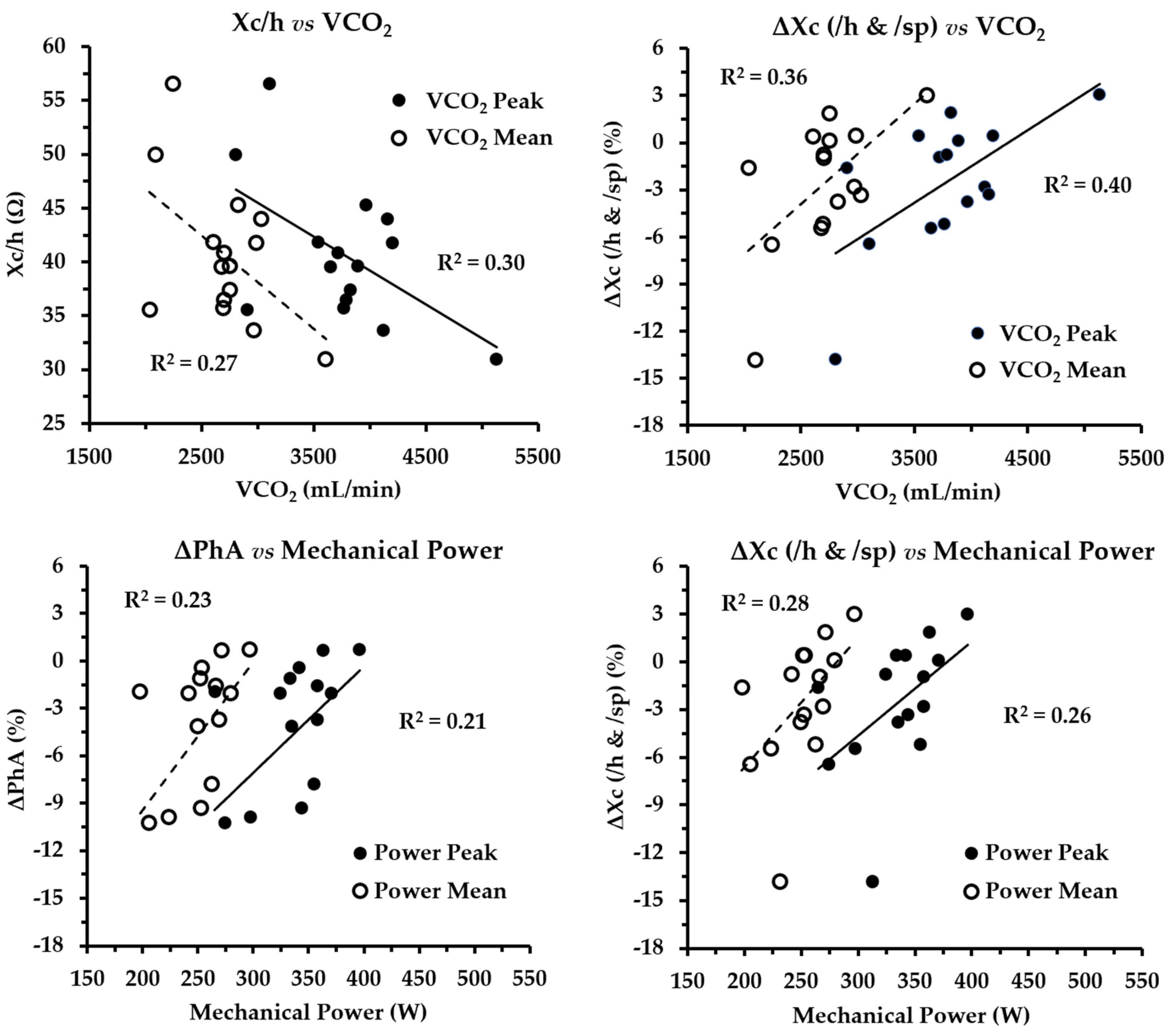
Figure 1. Scatterplots of statistically significant correlations between bioimpedance variables and metabolic/mechanical outcomes.
Applsci 15 10768 g001
Table 1. General characteristics of the 15 endurance-trained trail runners (mean ± SD; range [min–max]).
Mean ± SD
(n = 15)
Range
(Min–Max)
Age (years)37.27 ± 6.5527.32–48.25
Body mass (kg)70.89 ± 7.0561.80–81.80
Stature (cm)176.06 ± 5.96167.60–187.00
BMI (kg/m2)22.85 ± 1.6320.19–25.03
BMI: body mass index.
Table 2. Metabolic and mechanical performance metrics during the treadmill protocol (mean ± SD; range) in the 15 endurance-trained trail runners.
Mean ± SDRange (Min–Max)
VO2max (mL·min−1·kg−1)61.04 ± 6.9151.01–76.42
VO2mean (mL·min−1)3387.89 ± 532.432258.04–4114.46
VO2peak (mL·min−1)4401.4 ± 700.182919.10–5294.99
VCO2mean (mL·min−1)2712.37 ± 390.082037.35–3608.51
VCO2peak (mL·min−1)3772.23 ± 569.952806.70–5130.74
RQmean0.80 ± 0.080.67–0.92
RQpeak0.87 ± 0.110.71–1.10
HRmean (bpm)138.26 ± 9.50115.00–157.99
HRpeak (bpm)158.61 ± 9.33138.00–176.44
EEMmean (kcal·min−1)16.2 ± 2.4111.05–19.31
EEMpeak (kcal·min−1)21.27 ± 3.1414.59–25.00
POmean (W)250.21 ± 26.95197.74–296.90
POpeak (W)335.21 ± 35.75265.00–396.17
VO2max: maximal oxygen uptake; VO2: oxygen uptake; VCO2: carbon dioxide production; RQ: respiratory quotient; HR: heart rate; EEM: energy expenditure minute; PO: mechanical power output.
Table 3. Pre- to post-exercise changes in bioelectrical impedance vector analysis (BIVA) variables in the 15 endurance-trained trail runners.
Pre-TestPost-TestΔ%Statistical Differences
Mean ± SD (Range)Mean ± SD (Range)(Delta Value Differences)
Impedance (Ω)512.03 ± 47.87 (432.01–593.31)525.71 ± 46.78 (435.98–602.27)2.73 ± 2.11t = −5.03; p = 0.001; CI 95% = −19.52, −7.85
Resistance (Ω)506.97 ± 48.06 (428.29–588.50)521.05 ± 46.81 (432.49–597.5)2.84 ± 2.20t = −5.02; p = 0.001; CI 95% = −20.09, −08.06
Reactance (Ω)71.08 ± 9.58 (56.4–96.1)69.32 ± 8.87 (55–89.9)−2.36 ± 3.48t = 2.70; p = 0.02; CI 95% = 0.36, 3.16
Impedance (Ω/h)291.30 ± 27.21 (256.84–344.38)298.76 ± 26.80 (259.2–345.55)2.60 ± 1.96t = −5.18; p = 0.001; CI 95% = −10.55, −4.37
Resistance (Ω/h)288.41 ± 27.23 (254.63–341.38)298.43 ± 25.88 (257.13–342.79)2.71 ± 2.05t = −5.16; p = 0.001; CI 95% = −10.89, −4.50
Reactance (Ω/h)40.50 ± 5.97 (30.97–56.53)39.43 ± 5.38 (31.91–52.88)−2.46 ± 3.74t = 2.66; p = 0.02; CI 95% = 0.21, 1.94
Impedance (Ω/sp)350.07 ± 26.76 (295.33–397.09)358.49 ± 26.50 (307.37–405.91)2.44 ± 1.92t = −5.04; p = 0.001; CI 95% = −12.00, −4.84
Resistance (Ω/sp)346.62 ± 27.09 (289.13–393.83)355.31 ± 26.59 (302.16–402.71)2.55 ± 2.01t = −5.07; p = 0.001; CI 95% = −12.36, −5.02
Reactance (Ω/sp)48.56 ± 5.60 (36.78–60.21)47.29 ± 5.58 (36.19–56.61)−2.60 ± 4.13t = 2.63; p = 0.02; CI 95% = 0.24, 2.32
Phase angle (°)8.02 ± 1.22 (6.88–11.76)7.57 ± 0.95 (6.19–10.56)−4.91 ± 4.78W = 115; p = 0.01; CI 95% = 0.13, 0.66
Disclaimer/Publisher’s Note: The statements, opinions and data contained in all publications are solely those of the individual author(s) and contributor(s) and not of MDPI and/or the editor(s). MDPI and/or the editor(s) disclaim responsibility for any injury to people or property resulting from any ideas, methods, instructions or products referred to in the content.