Article

Effect of Denture Adhesives on the Surface Roughness and Hardness of Denture Base Resins—A Preliminary Study

by
Guilherme Bezerra Alves
1,2,
Maria Margarida Sampaio-Fernandes
1,3,*,
Carlos Fernandes
3,4,*,
Francisco Góis
1,
Bruno Graça
5,
Estevam Bonfante
2 and
Maria Helena Figueiral
1,3
1
Faculdade de Medicina Dentária da Universidade do Porto (FMDUP), 4200-393 Oporto, Portugal
2
Faculdade de Odontologia da Universidade de São Paulo (USP), São Paulo 05508-000, Brazil
3
Instituto de Ciência e Inovação em Engenharia Mecânica e Engenharia Industrial (INEGI), 4200-465 Oporto, Portugal
4
Faculdade de Engenharia, Universidade do Porto (FEUP), 4200-465 Oporto, Portugal
5
Faculdade de Medicina Dentária, Universidade de Lisboa (FMDUL), 1600-277 Lisbon, Portugal
*
Authors to whom correspondence should be addressed.
Appl. Sci. 2025, 15(19), 10749; https://doi.org/10.3390/app151910749
Submission received: 1 July 2025 / Revised: 30 September 2025 / Accepted: 2 October 2025 / Published: 6 October 2025

Abstract

This study aimed to evaluate the impact of different adhesive solutions on the surface roughness and hardness of denture base materials. Twenty specimens (20 × 20 × 5 mm) were produced for each material group: heat-cured ProBase Hot®, 3D-printed NextDent Denture 3D+®, and PMMA-milled Exaktus®. They were then divided into five solution subgroups (n = 4): control (T0), distilled water, Corega PowerMax®, Elgydium Fix®, and Kukident Pro Ultimate®. Specimens were immersed in the solution at 37 °C daily for 28 days, simulating continuous use. Profilometry and Shore D hardness tests were performed at baseline and after 28 days of the immersion protocol. Data analysis was done using IBM SPSS Statistics 30.0, considering a confidence level of 0.05. At baseline, the materials differed in surface roughness and Shore D hardness, with the 3D-printed group showing the highest median values for the Rz parameter (p = 0.023) and the lowest for hardness (p = 0.023). Elgydium Fix had a significant effect on the heat-cured resin, with increased Rz and decreased hardness. Kukident caused higher roughness and lower hardness in the 3D-printed and milled resins (not significant). Corega showed minor effects in all tested materials. In conclusion, the denture base material and the adhesive formulation influence the physical and mechanical properties of denture base resins.

1. Introduction

The adhesives for removable dental prostheses play an important role in enhancing the retention, stability and function of this type of rehabilitation [1,2]. Although a considerable number of denture wearers use them, they are seldom recommended by health professionals as they are often associated with facilitating the continued use of ill-fitting dentures [3,4]. In turn, current evidence indicates that the use of adhesives in well-adapted dentures can provide benefits, including greater stability, increased incisal bite force, reduced discomfort and improved quality of life [2,5,6]. In addition, the use of these products can contribute to the protection of the oral mucosa by maintaining an adequate blood supply [7]. These advantages are particularly relevant in patients with xerostomia or limited muscle control, in whom adhesives can substantially improve the denture experience [8].
Denture adhesives are available in various forms, such as creams, powders, and adhesive pads, associated with different application properties, viscosities, retention times, and patient comfort [2,6,9]. The typical formulation of cream adhesives includes copolymers of maleic acid and polyvinylmethylether (PVM/MA), which confer adhesive properties, as well as sodium carboxymethylcellulose, liquid paraffin, petroleum jelly, and natural gums, which act as thickeners and texture modifiers [10]. Certain formulations also include zinc salts, such as zinc sulfate, to promote greater fixation; however, their chronic and cumulative ingestion has been associated with neurological adverse effects, leading to the development of zinc-free options [11]. The clinical choice between formulations with or without zinc must take into account the frequency of use, the extent of the prosthetic base, and the patient’s individual characteristics [12,13].
The surface properties of denture base materials are an important issue, as several studies have shown a direct correlation between their roughness, the accumulation of microbial biofilm and the adhesion of Candida albicans (C. albicans) [13,14,15,16]. The growing presence of microorganisms can be associated with the development of pathological conditions, such as denture-related stomatitis, gingival inflammation, and caries in the supporting teeth [17,18].
Denture-related stomatitis is an inflammation that affects the supporting mucosa in contact with removable prostheses, occurring most frequently on the palate. Studies show a high prevalence of this condition, reaching around 50% among denture wearers [13,19]. Although its causes are multifactorial, the genus Candida, especially the species C. albicans, stands out as the main etiological factor [20,21]. This yeast’s ability to adhere to surfaces is an important virulence factor [14]. After this initial adhesion, cell proliferation and the formation of an extracellular matrix occur [21,22], resulting in complex biofilms that attach to the internal surfaces of dentures, serving as reservoirs for pathogens. Compared to isolated microorganisms, these biofilms show greater resistance to both antifungal therapy and immunological defenses, thereby increasing the risk of systemic infections, especially in immunocompromised individuals [21].
Several studies indicate that the formation of dental biofilms is directly associated with the roughness of polymethylmethacrylate (PMMA) surfaces, a material widely used in denture bases, but also with the roughness of new materials for 3D-printed and milled denture bases [16,22,23,24,25]. Rougher surfaces favor the adhesion of microorganisms such as Candida albicans and Streptococcus spp. since the microscopic irregularities offer niches that are difficult to clean [19,20,21,26,27]. The surface roughness can be quantified by parameters such as Ra (average roughness) and Rz (maximum profile height). Ra represents the arithmetic mean of the absolute variations in relation to the midline of the profile, while Rz expresses the mean of the sum of the five largest peaks and the five largest valleys of the surface. Higher Ra and Rz values indicate surfaces with greater biofilm retention potential, which contributes not only to increased adhesion but also to greater resistance of the biofilms formed [14,28,29]. Beyond texture, the surface wettability is very important in the performance of denture adhesives, as well as in biofilm formation and bacterial adhesion on denture base resins [22,30]. Higher wettability (smaller contact angle, higher hydrophilicity) has been associated with smoother surfaces as observed on milled base materials with digital production and less tendency to candida adhesion [23,25,31]. Hardness is another important mechanical property for prosthetic base materials, as it reflects the surface’s resistance to deformation and penetration. Low hardness values can compromise resistance to wear during chewing and brushing, leading to the progressive degradation of the material. Recent studies have shown that factors such as the type of resin used, manufacturing processes, and aging in aqueous media, artificial saliva or denture adhesives can significantly influence the hardness and other characteristics of acrylic materials used in removable prostheses [24,32,33,34,35,36,37]. Evaluating these properties is, therefore, essential to understanding the clinical impact of continued adhesive use on the performance and longevity of dentures.
Although denture adhesives are widely used in clinical practice, there are still few studies investigating their direct effects on the physical–mechanical properties of denture base materials. Preliminary results suggest that certain adhesives can cause measurable changes in surface roughness, potentially impacting microbial adhesion and material integrity [36,37]. In addition, there are indications that prolonged adhesive use may also affect the surface hardness of acrylic resins used in prosthetic bases; however, this relationship has yet to be systematically validated. In this context, it is crucial to investigate, in a controlled and comparative way, the influence of different commercial adhesives on the roughness and hardness of prosthetic base materials obtained by different technologies, thus contributing to the more conscious and evidence-based use of these products in the field of oral rehabilitation [32,33,34,36,37].
The main objective of this study is to evaluate the effect of various dental adhesives on the surface roughness and Shore D hardness of three denture base materials obtained by different techniques: heat-curing, 3D printing, and milling. The secondary objective is to compare the surface roughness and hardness of the materials.
The following null hypotheses were formulated:
H01. 
There are no significant differences in initial surface roughness and Shore D hardness between the denture base materials studied.
H02. 
Exposure to adhesives does not significantly alter the surface roughness or the Shore D hardness of the materials studied.

2. Materials and Methods

2.1. Preparation of Resin Specimens for Denture Base

The specimens used in this research were made from a digital model (.stl file) with dimensions of 20 × 20 × 5 mm, created using Meshmixer 3.5 software (Autodesk, San Francisco, CA, USA). Three groups of 20 specimens per denture base material (a total of 60 specimens) were made: (H) heat-cured PMMA ProBase Hot® (IVOCLAR, Schaan, Liechtenstein), obtained using the conventional lost wax technique, considered the gold standard; (P) light-curing resin for 3D printing NextDent Denture 3D+® (3D Systems, Rock Hill, SC, USA), printed on the NextDent 5100® (3D Systems, Rock Hill, SC, USA) with 0° orientation, in Premium mode, with a 100-µm layer thickness; and (M) PMMA for CAD-CAM milling ref. AROSE 30 (Exaktus, Vila Nova de Gaia, Portugal), using the Zirkhonzan M2® milling machine (Zirkhonzan, Gais, South Tyrol, Italy). Finally, all the specimens were polished with 600- and 1000-grit water sandpaper Dexter (Leroy Merlin, Lisboa, Portugal), followed by finishing with a pumice stone and the polishing paste Dentaurum® (Dentaurum, Ispringen, Germany). Figure 1 shows one specimen of each material group.

2.2. Dental Adhesives

Three commercial denture adhesives with different compositions (Table 1) were tested in cream formulation: Corega PowerMax® (Haleon, Dungarvan, County Waterford, Irland), Elgydium Fix® (Pierre Fabre, Castres, France) and Kukident Pro Ultimate® (Procter & Gamble, Schwalbach, Germany).
Table 2 presents the information on the products used in this investigation.

2.3. Experimental Protocol

Prior to immersion in the solutions, the surface roughness (Ra and Rz) and Shore D hardness of four randomly selected specimens from each experimental group were assessed, constituting the control subgroups (initial—T0). These specimens did not undergo the immersion protocol, remaining in a dry environment throughout the study. Thus, they served as a negative control group, representing the initial condition of the materials before exposure to the adhesive solutions or the aqueous environment.
In each group (heat-cured, 3D-printed, and milled resins), the rest of the specimens were randomly distributed into four subgroups (n = 4), according to the solution used: distilled water W5 (Lidl & Cia., Sintra, Portugal), Corega, Elgydium, and Kukident.
During the immersion experiments, ambient conditions varied between 23–25 °C and 40–50% relative humidity.
In the water subgroups, the specimens were immersed only in distilled water for the independent assessment of the aqueous medium’s effects. The remaining subgroups consisted of specimens immersed in Corega, Elgydium, and Kukident adhesive solutions, respectively, all prepared in a ratio of 1 g of adhesive to 10 mL of distilled water for each specimen.
The specimens were immersed in sealed plastic containers containing the respective solutions and, after being closed, they were placed in a stove for 23 h a day, for 28 days, at a constant temperature of 37 °C. Between immersion cycles, the specimens were washed under tap water (municipal supply, moderate water hardness) and air-dried for about 1 h. The adhesive and water solutions were renewed daily. In total, each specimen was immersed in its adhesive solution for 644 h. After the 28-day immersion protocol in the solutions (T28), the surface roughness and hardness of the specimens were assessed again under the same initial conditions.
The pH of the solutions was controlled by a Cumbur3 Test® (Roche, Mannheim, Germany) at T0 and after 23 h of immersion, to control the potential chemical changes.
Finally, the sample mass of each specimen of all subgroups was recorded using an analytical balance Radwag AS 220.R2 (Radwag, Radom, Poland).

2.3.1. Surface Roughness

The parameters Ra (average roughness) and Rz (maximum profile height) were measured using the contact profilometer HommelWerke LV-50 (Hommelwerke GmbH, Mannheim, Germany). The measurements were taken using a diamond tip with a radius of 5 µm over a travel length of 4.8 mm. Three longitudinal measurements were taken per specimen, and their average was considered for statistical analysis.

2.3.2. Shore D Hardness

Shore D hardness was assessed using the analog durometer Sauter® (Sauter Ibéria, Oeiras, Portugal) equipped with a standard indenter, which exerts a force perpendicularly to the surface of the material. The reading was taken after stabilizing the penetrator in accordance with the ISO 868 criteria [41]. A reading system converts the vertical displacement of the penetrator into a Shore D hardness value on an appropriate scale ranging from 0 to 100. In this work, each specimen was tested three times, and then the average was calculated for each one.

2.4. Statistical Analysis

The data obtained were initially recorded and organized in Excel - Microsoft 365 (Microsoft, Redmond, WA, USA) and then analyzed statistically using IBM SPSS Statistics® 30.0 (IBM Corp., Armonk, NY, USA). The descriptive analysis included calculating the median and interquartile range (IQR) of each group and subgroup.
The normality of the distributions was checked using the Shapiro–Wilk test. Considering the small sample size of each subgroup (n = 4), the non-parametric Kruskal–Wallis test was used to compare independent groups—used to compare the three parameters assessed both between the different materials at T0 and the subgroups of each material. The significance level adopted was 5% (α = 0.05).

3. Results

Table 3, Table 4 and Table 5 present the median and IQR of the three parameters assessed for all material groups and solution subgroups (T0 and T28).

3.1. Baseline Evaluation (T0)—Comparison Between Materials

At T0, the medians of the roughness parameters (Ra and Rz) were higher in the 3D-printed group (0.11 µm; 1.53 µm) compared to the heat-cured (0.04 µm; 0.33 µm) and milled (0.05 µm; 0.45 µm) groups—Figure 2a,b. The distribution of results of both roughness parameters showed greater variability (higher IQR) in the printed resin. Kruskal–Wallis tests confirmed statistically significant differences between the material groups for the Rz parameter (p = 0.023) but not for the Ra parameter (p = 0.051).
The heat-cured and milled materials had a higher similar median of Shore D hardness (92.74 and 92.77, respectively). In contrast, the printed material had a significantly lower Shore D hardness (86.67), accompanied by a higher IQR—Figure 2c. The differences in hardness were statistically significant (p = 0.023), specifically between heat-cured and 3D-printed materials (p = 0.027) and between 3D-printed and milled materials (p = 0.012).
Regarding the sample mass, heat cured resin showed the lowest median (2.24 g [2.20–2.31]), followed by milled group (2.35 g [2.32–2.36]) and 3D printed (2.38 g [2.35–2.40]). The differences at T0 were significant (p = 0.017), specifically between heat-cured and printed resins (p = 0.004). In each material group, independent comparisons were carried out, and no statistically significant differences were found between T0 and solutions subgroups (p ˃ 0.05).

3.2. Effect of Adhesive Solutions (T28)—Comparison Between Subgroups

After 28 days of the immersion protocol (T28), the Ra roughness parameter showed a tendency to increase, especially in the 3D-printed resin. In turn, the hardness tended to decrease in all the groups, particularly in the 3D-printed group.
Distilled water and Kukident solutions presented pH of 5, both initially at T0 and after immersion period of the three materials tested. Following the same pattern, Corega and Elgydium solutions had pH of 6 which remains unchanged after 23 h of immersion.

3.2.1. Heat-Cured Group

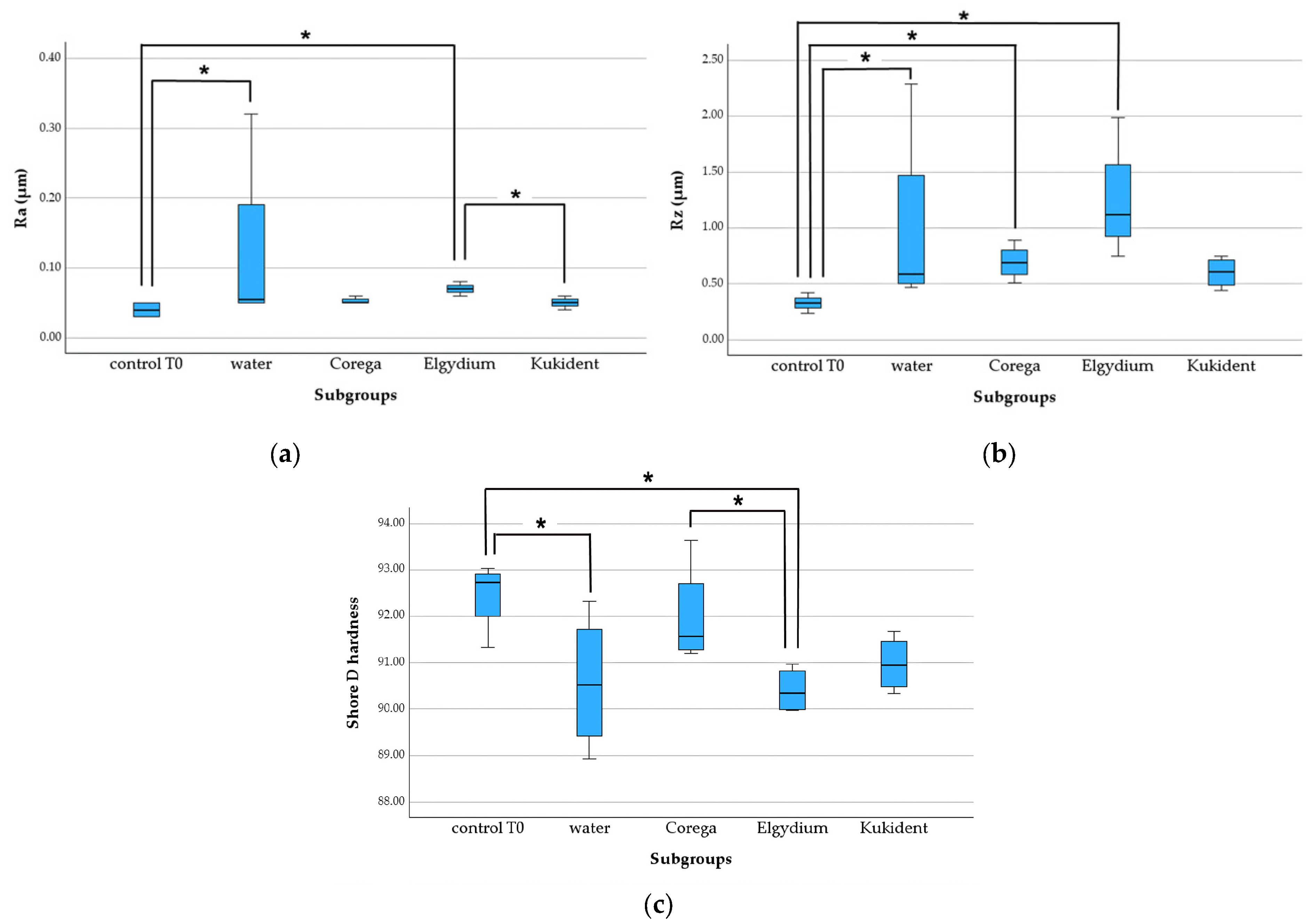
In the heat-cured group, Ra and Rz tended to increase in the solution subgroups compared to the control T0, and the highest median was observed in the Elgydium one (0.07 [0.06–0.08]) (Figure 3a,b). The hardness observed after 28 days of immersion in the solutions (water or denture adhesives) was lower than that observed in the control—Figure 3c.
Kruskal–Wallis tests showed statistically significant differences (p < 0.05) between the subgroups for the roughness parameters (Ra: p = 0.026; Rz: p = 0.014) and the Shore D hardness (p = 0.033), specifically between the following subgroups:
  • Ra: control—water (p = 0.047), control—Elgydium (p = 0.02), and Elgydium—Kukident (p = 0.028);
  • Rz: control—water (p = 0.042), control—Corega (p = 0.027), and control—Elgydium (p < 0.001);
  • Shore D hardness: control—water (p = 0.023), control—Elgydium (p = 0.009), and Corega—Elgydium (p = 0.031).
Thus, the roughness parameters of the heat-cured specimens increased after the 28-day immersion protocol in water and denture adhesive solutions. The adhesive solutions also caused reductions in hardness values, especially the Elgydium adhesive, in contrast with the Corega one.

3.2.2. 3D-Printed Group

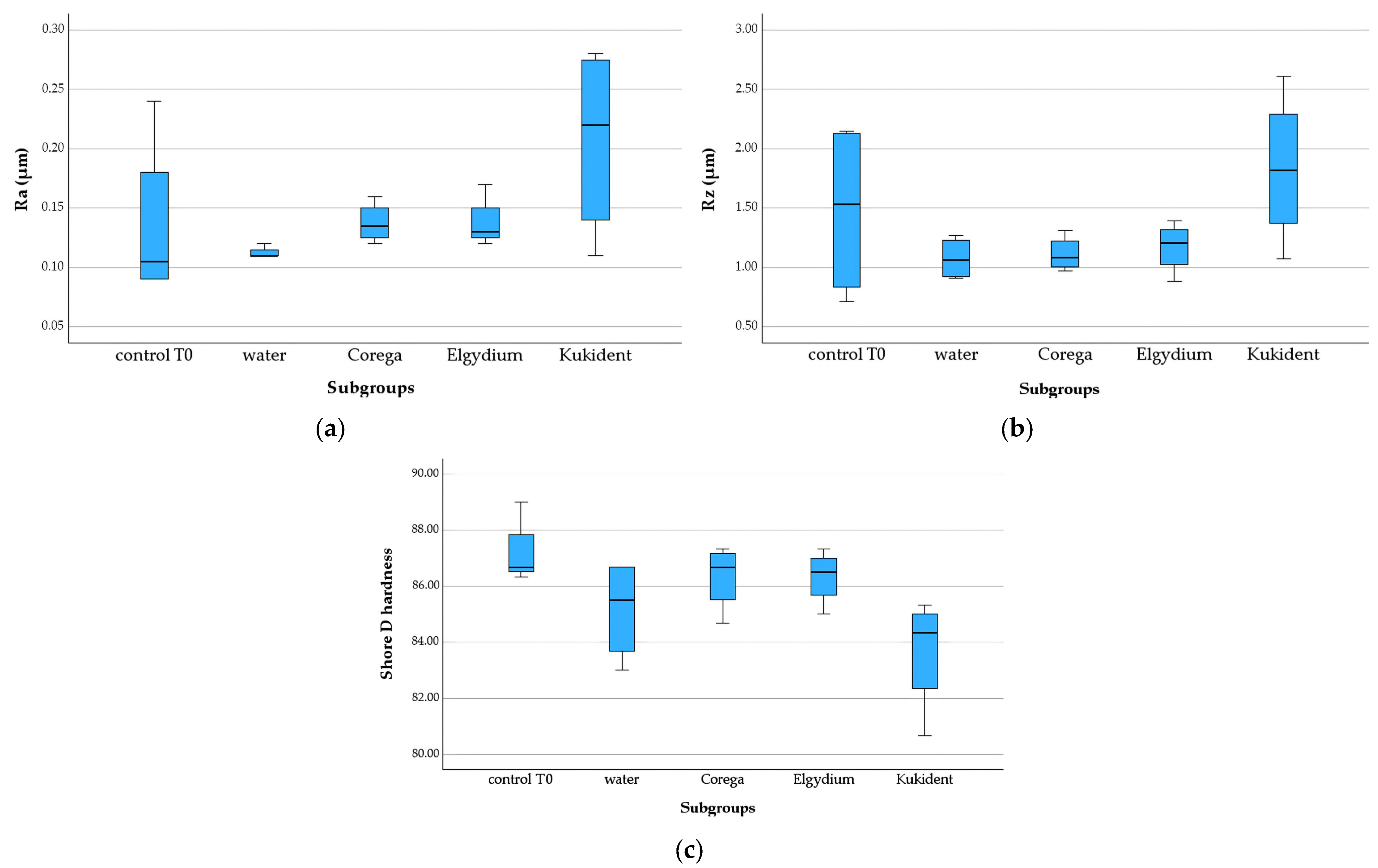
The median Ra values of this material’s subgroups at T28 were consistently higher than those of the control group (0.11 µm), with the Kukident subgroup (0.22 µm) showing the highest median and the greatest IQR (Figure 4a). In contrast, the median Rz value of the Kukident subgroup (1.88 µm) was similar to that of the control (1.53 µm), and both subgroups showed higher IQR (Figure 4b). In the other subgroups (water, Corega, and Elgydium), the Rz value decreased, indicating less distance between peaks and valleys on the surface of the specimens. However, there are no significant differences between the solution subgroups and the initial control (p ˃ 0.05) for both roughness parameters (Ra: p = 0.127; Rz: p = 0.438).
After the 28-day immersion protocol, there was a reduction in the median hardness, more evident in the water and Kukident subgroups, which also showed a wider IQR (Figure 4c). Although there were no significant differences between the subgroups (p = 0.103), there was a clear gap between the median hardness of the Kukident subgroup and the control, Corega, and Elgydium subgroups.

3.2.3. Milled Group

In this group, the median Ra was higher in the Corega subgroup, followed by Kukident, water, and Elgydium (T28 subgroups), compared to the control (Figure 5a). Regarding the Rz parameter, Figure 5b shows that the median is only lower than the control (0.45 µm) in the Elgydium subgroup (0.40 µm). In the other subgroups, the Rz median was always higher than the control T0, with greater expression in the Corega subgroup (0.82 µm, compared to 0.49 µm in Kukident and water). Despite the tendency for the Elgydium adhesive to have a lesser effect on the roughness of the milled specimens, there were no significant differences between the solutions and the baseline control for both parameters assessed (Ra: p = 0.167; Rz: p = 0.531).
Figure 5c shows that the variations in hardness were more marked in the Corega subgroup (greater reduction compared to the control) and that the Kukident subgroup was the only one that showed a slightly increased median. However, the Kruskal–Wallis test revealed no statistically significant differences (p = 0.107).

4. Discussion

In this study, the surface roughness parameters Ra and Rz and the Shore D hardness were used to characterize and compare three denture base materials at baseline (T0) and after a 28-day immersion protocol in water and adhesive solutions (T28).
The comparison of T0 results for the tested materials revealed significant differences (p < 0.05) in roughness (only for the Rz parameter) and hardness. The 3D-printed group showed the highest median value for Rz and the lowest median value for Shore D hardness compared to the milled and heat-cured groups. Therefore, the null hypothesis H01 was rejected. In general, the data obtained indicate that the 3D-printed material initially has a rougher surface, which may have clinical implications in terms of increased bacterial adhesion and risk of denture-related stomatitis.
The discrepancy in the statistical results between Ra and Rz may be attributed to the different nature of these two parameters. While Ra represents the arithmetic mean of surface irregularities, smoothing out local variations, Rz reflects the extremes of the profile, being more sensitive to sharp peaks and valleys. Thus, it is possible that the differences between the materials were mainly manifested by particular irregularities, which were sufficiently marked to have an impact on the Rz parameter but not on the overall average expressed by Ra. Furthermore, the p-value for Ra (p = 0.051) indicates a tendency toward significance, which may have been attenuated by the variability of the data or the limitations imposed by the sample size.
Shore D hardness was assessed as an indicator of the surface resistance of denture base materials to prolonged exposure to adhesive solutions. This parameter is fundamental because harder surfaces tend to show less plastic deformation, greater resistance to mechanical wear, and less susceptibility to physical–chemical alterations in the oral environment [32].
The additive manufacturing technique used in the 3D-printed group is often related to discrete layers and residual porosities, which affect surface uniformity, and less dense and more heterogeneous polymer structures, with a higher residual monomer content and a lower degree of conversion [33], which may explain the lower hardness values observed in this group. Despite the greater variability of results observed in the group of printed specimens, the printing and post-processing parameters (print orientation, layer thickness, or post-curing) were similar and comparable within the group, since the resin was thoroughly stirred beforehand, the manufacturer’s instructions were followed, and all samples were printed at once (a full printer tray), trying to achieve the best possible uniformity. On the other hand, milled PMMA (subtractive manufacturing) is produced from pre-polymerized blocks under high pressure and temperature, resulting in greater structural homogeneity and dimensional stability [34]. Moreover, the milled and heat-cured groups showed greater and more homogeneous values of hardness, which may reflect the greater cohesion of the acrylic matrix inherent to the density and homogeneity of the milled industrial blocks [29], or to the flask compression for heat-curing of the conventional PMMA.
These results obtained at T0 are consistent with findings from other studies which compare the surface characteristics and hardness between conventional PMMA denture resins and other base materials produced by digital technologies [13,15,16,42].
Regarding the effect of exposure to adhesives on the properties studied, at least one adhesive influenced surface roughness and hardness in at least one of the materials tested. Thus, the null hypothesis H02 was partially rejected.
After immersion (T28), the heat-cured group showed significant surface changes: the Elgydium and water subgroups presented higher Ra values than the control (T0), while the water, Corega, and Elgydium subgroups showed a higher Rz median than the control, with even more evident changes. In this conventional material, a significant reduction in hardness was also detected in all T28 subgroups, especially in the water and Elgydium subgroups. These findings suggest that prolonged exposure to adhesive solutions can promote relevant topographical and mechanical modifications in heat-cured materials, possibly due to the absorption of water and adhesive components, which act as matrix plasticizers, thereby promoting relaxation of the polymer network and consequent loss of surface rigidity or removal of surface polymer [32,33,36].
In the 3D-printed material, there were no significant differences between adhesives, but some tendencies are worth noting. In the water, Corega, and Elgydium subgroups (in contrast to Kukident), Ra tended to be lower than in the control subgroup. Rz tended to increase with immersion only with the Kukident solution. This finding suggests that immersion may have had a slight surface smoothing effect and that Kukident adhesive seems to promote a significant change in the surface of the printed material, unlike other adhesives, possibly due to its specific chemical composition (denture adhesive containing zinc). This hypothesis reinforces the importance of considering the compatibility between the constituents of the adhesive solutions and the polymeric matrix of the prosthetic materials tested [34]. Regarding the hardness, the lowest median observed (greater decrease) in the Kukident subgroup reveals its greater surface degradation. The IQR also raises the hypothesis of individual variability between specimens, a characteristic recognized in 3D-printed materials [43].
Finally, the milled group showed greater stability in the immersion protocol, with no significant differences in Ra, Rz, or Shore D hardness observed between the solution subgroups and the control. This behavior is in line with the high physical–mechanical properties of CAD-CAM materials, which tend to have a less permeable structure and greater resistance to surface alterations [44]. This industrially polymerized block, produced under controlled pressure and temperature [29], shows greater resistance to liquid absorption and less change in its properties after artificial aging [29,36].
In the simulated conditions of this in vitro study, the heat-cured material appears to be the most susceptible to changes after prolonged exposure to adhesives, while the milled material seems more stable and resistant to the adhesive immersion protocol.
Recognizing the importance of sample mass for test protocols (such as immersion, thermocycling, or chemical aging), this parameter was analysed. At T0, the median value of heat-cured resin was significantly lower in comparison with the other resins. However, under the conditions of this in vitro study, no statistically significant differences were found between T0 and the solution subgroups. Moreover, in all groups, the pH of the solutions did not change over the immersion period (23 h).
From the perspective of the denture adhesive, the considerations below should be highlighted.
Elgydium Fix® showed the greatest impact on the heat-cured resin (compared to 3D-printed or milled resins), causing a significant increase in roughness parameters and a significant reduction in Shore D hardness. This behavior suggests a potentially more intense chemical interaction between the adhesive components and the heat-cured polymer matrix [45]. Potential interaction types include plasticization that occurs when hydrophilic components (PVM/MA, cellulose gum) absorb water and swell. In addition, surface softening may happen if solvents or mineral oil (PVP, isopropyl palmitate and myristate in the specific formulation of Elgydium) are present, as lipophilic compounds facilitate penetration into the acrylic matrix and contribute to the disorganization of its internal structure. Also, there may be a “smoothing” effect on the surface of the PMMA, especially if the surface has been poorly polished or has microretentions (pores, microcracks) [43].
The immersion in Kukident Pro Ultimate® adhesive caused the greatest reduction in hardness and the largest increase in surface roughness of the 3D-printed material (no statistical significance). These observations may reflect more penetration or plasticization of the matrix and surface degradation by Kukident components, such as petrolatum or liquid paraffin [43]. The calcium/zinc partial salt is used to modulate the adhesive properties of the formulation, potentially influencing setting time, bonding strength, and retention. Zinc may also impart mild antimicrobial activity and affect the copolymer’s behavior through ionic interactions. Although some studies indicate that silica nanoparticles or fillers improve hardness or mechanical properties when incorporated into PMMA [46], there is no clear recent evidence that hydrated silica used as a thickener or abrasive in this formulation causes measurable mechanical wear of PMMA surfaces under typical adhesive use. Apart from the compounds, a recent work tested aging in artificial saliva at different pH levels and suggested that surface roughness increased with pH-induced aging, peaking at acidic pH 3. Therefore, since the Kukident solution had pH of 5 (lower than the other adhesives), this parameter should be considered in the interaction denture base resin—denture adhesive [47].
In turn, Corega PowerMax® was the adhesive that caused the largest roughness increase and hardness reduction in the milled material (not significant), while the changes observed in the printed and heat-cured materials were minimal or absent with this specific solution. This fact may be related to the presence of effervescent and surfactant agents (sodium PVM/MA copolymer, cellulose gum, liquid paraffin) in the composition of this denture adhesive, which promotes physicochemical changes at the material interface. The lack of statistical significance in the hardness of the milled material is in accordance with the recognized physical–mechanical stability of CAD-CAM materials, which results in high density and low porosity, thereby restricting the absorption of liquids even in contact with potentially aggressive solutions such as adhesives [48].
All proposed mechanisms of interaction denture material-denture adhesive are presented as hypotheses or areas for future research, not as established effects evidenced on this work.
Another important aspect to consider is the translation of findings into clinical practice. The experimental protocol employed in this study involved immersing the specimens in adhesive solutions for 28 days, with 23 h of daily exposure and one hour dedicated to washing and air-drying. This approach aimed to simulate, in an accelerated manner, the cumulative effects of prolonged adhesive use on the base materials of removable prostheses.
When considering actual clinical use, several studies suggest that the average daily contact time of a denture with an intraoral adhesive is approximately 16 h, followed by a rest period of around 8 h in a humid environment (simulating the action of saliva during non-use). Based on this model, the protocol adopted in this investigation would correspond to approximately 40 days of continuous clinical use of the denture, lending validity to the proposal to analyze the chronic effects of the solutions tested.
Recent studies have tested resin aging protocols in isolation (without immersion in adhesive solutions), and the results after aging pointed to an increase in roughness and reduction in hardness in all materials, especially in 3D-printed ones [25,35], and to an increase in wettability [22,23,30,31] and biofilm adhesion [13], supporting the need to replace dentures every five years [16].
Direct comparison with other studies is still limited because the existing literature shows significant methodological variations, especially regarding exposure time, type of acrylic material, and adhesive formulation. Previous studies [49,50] have generally focused on evaluating a single physical–mechanical parameter (such as hardness or roughness) or just one type of material (conventional), not including different manufacturing technologies (heat-curing, CAD-CAM milling and 3D printing) and multiple commercial adhesive formulations. This gap highlights the originality of this study and points to the need for future research that replicates clinical conditions with greater precision, associating periods of intermittent use, pH variations, salivary simulation, and aging (thermal or chemical) in order to more accurately reflect the intraoral reality of patients wearing removable prostheses.
Although this investigation provides relevant data on the effects of adhesive solutions on the surface roughness and Shore D hardness of denture base materials, some methodological limitations should be considered when interpreting the results. Firstly, the period of exposure to the adhesive solutions was limited to 28 days, which was sufficient to simulate continuous and standardized use but does not represent the entire time of clinical use of a denture, which can extend over years. Therefore, there is a need for long-term experimental protocols that can reproduce chronic use and assess the cumulative impact of adhesives on the structure and integrity of the materials. Additionally, even though the specimens were manufactured in a standardized manner, they can still show intrinsic variability between them, especially in the printed group, whose additive technology depends on multiple critical steps. Discrete differences in finishing, surface homogeneity or even residual porosity can influence the roughness and hardness values observed, contributing to the variability of the results, as shown in some subgroups.
Furthermore, the number of specimens per group was limited, which could reduce the statistical power of the analysis and restrict the generalizability of the findings. It is a preliminary work, and future studies with larger samples could make the results more robust and allow for more accurate stratified analyses.
More extensive in vitro studies should be conducted with accelerated aging protocols that include, for example, thermal cycles (thermocycling), simulated brushing, pH variations, artificial salivary simulation, and intermittent immersion protocols to better reflect the fluctuations of the oral environment over time. The inclusion of these factors will enable not only the assessment of chemical degradation but also the mechanical and abrasive effects that occur simultaneously in the oral cavity. It would also be relevant to explore the response of different commercial adhesive formulations (including those with and without zinc) on additional properties of acrylic materials, such as water absorption, solubility, bacterial adhesion, and color changes. Microstructural characterization using techniques such as scanning electron microscopy (SEM), Fourier-transform infrared spectroscopy (FTIR), or three-dimensional profilometry analysis could also provide more robust data on interactions at the adhesive-material interface. Moreover, other mechanical properties of base materials, such as flexural or compressive strength, and the material’s plasticity, could be affected by the use of denture adhesives.
In addition, it is essential to acknowledge that this study was conducted in a controlled laboratory environment under in vitro conditions. Although this experimental design allows for strict control of variables and greater reproducibility of procedures, it does not reflect the complexity of the intraoral environment. In the clinical context, denture bases are continually exposed to multiple factors that interact simultaneously, such as the constant presence of saliva (with variable composition and pH), thermal cycles related to food and drink intake, intermittent masticatory forces, abrasion from brushing, and the presence of microbial biofilm [29]. All these elements can significantly modify the way adhesives interact with the surface of materials over time. Therefore, while in vitro data are essential to support hypotheses and elucidate basic degradation mechanisms, clinical validation through in vivo studies—with patients using removable prostheses in real-word settings—is crucial. These studies should include longitudinal monitoring, repeated measurements, and evaluation of the functional and microbiological performance of the dentures, allowing the real clinical relevance of the physical–mechanical changes observed in the laboratory to be verified.
Future research should be carried out to gain a deeper understanding of the degradation mechanisms of prosthetic materials exposed to adhesive solutions.

5. Conclusions

Considering the results and limitations presented, this study reveals that prolonged exposure to cream adhesives can significantly influence the surface roughness (Ra and Rz) and Shore D hardness of the denture base resins (heat-cured, printed, and milled).
Greater roughness and lower hardness are the trend effects of denture adhesives, for all materials tested. The heat-cured resin was the material most affected by adhesive solutions and the milled resin the least. Elgydium seems to be less suitable for heat-cured resin; Kukident should be avoided for 3D-printed bases and Corega appears to be safer than others for clinical recommendation.
Considering the preliminary nature of this work and the inherent limitations, further studies are recommended to investigate and sustain the long-term effects of different adhesive formulations on denture base resins as well as to develop safer and more personalized clinical recommendations for the rational use of adhesives in removable dental prostheses.

Author Contributions

Conceptualization, M.H.F., M.M.S.-F. and E.B.; methodology, C.F.; investigation, G.B.A.; resources, B.G. and F.G.; writing—original draft preparation, G.B.A. and M.M.S.-F.; writing—review and editing, M.M.S.-F., C.F. and M.H.F.; supervision, M.H.F. and E.B. All authors have read and agreed to the published version of the manuscript.

Funding

This research received no external funding.

Institutional Review Board Statement

Not applicable.

Informed Consent Statement

Not applicable.

Data Availability Statement

Data is contained within the article.

Acknowledgments

The authors acknowledge the support given by Pierre Fabre, Haleon and Procter & Gamble companies (donation of denture adhesives used for experiments).

Conflicts of Interest

The authors declare no conflicts of interest.

References

  1. Grasso, J.E. Denture adhesives: Changing attitudes. J. Am. Dent. Assoc. 1996, 127, 90–96. [Google Scholar] [CrossRef]
  2. Lemos, C.A.A.; da Fonte Porto Carreiro, A.; Rosa, C.D.D.R.D.; Luna Gomes, J.M.; de Oliveira Limirio, J.P.J.; Mendonça, G.; Pellizzer, E.P. Does the use of an adhesive improve conventional complete dentures? A systematic review of randomized controlled trials. J. Prosthet. Dent. 2022, 128, 150–157. [Google Scholar] [CrossRef] [PubMed]
  3. Florêncio Costa, R.T.; Leite Vila-Nova, T.E.; Barbosa de França, A.J.; Gustavo da Silva Casado, B.; de Souza Leão, R.; Dantas de Moraes, S.L. Masticatory performance of denture wearers with the use of denture adhesives: A systematic review. J. Prosthet. Dent. 2022, 127, 233–238. [Google Scholar] [CrossRef] [PubMed]
  4. Gad, M.M.; Almusallam, M.O.; Almarar, F.H.; Al khaldi, H.O.; Aldossary, A.K.; Almutairi, W.M.; Alghamdi, N.A.; Alssaialiy, Y.S.; Alghamdi, N.I.; Nazir, M.A. Denture Adhesive Utilization and Associated Factors among Dental Practitioners in the Eastern Province, Saudi Arabia. Medicina 2023, 59, 974. [Google Scholar] [CrossRef]
  5. Felton, D.; Cooper, L.; Duqum, I.; Minsley, G.; Guckes, A.; Haug, S.; Meredith, P.; Solie, C.; Avery, D.; Deal Chandler, N. Evidence-based guidelines for the care and maintenance of complete dentures: A publication of the American College of Prosthodontists. J. Prosthodont. 2011, 20, S1–S12. [Google Scholar] [CrossRef]
  6. Ereifej, N.S.; Oweis, Y.G.; Abu-Awwad, M. The effect of using denture adhesives on patient satisfaction with complete dentures; a randomized clinical trial. BMC Oral Health 2023, 23, 1027. [Google Scholar] [CrossRef]
  7. Kelsey, C.C.; Lang, B.R.; Wang, R.F. Examining patients’ responses about the effectiveness of five denture adhesive pastes. J. Am. Dent. Assoc. 1997, 128, 1532–1538. [Google Scholar] [CrossRef]
  8. Pradíes, G.; Sanz, I.; Evans, O.; Martínez, F.; Sanz, M. Clinical study comparing the efficacy of two denture adhesives in complete denture patients. Int. J. Prosthodont. 2009, 22, 361–367. [Google Scholar]
  9. Pinto, J.R.; Bonifácio, C.C.; Campos, J.C. Avaliação das propriedades físico-químicas de adesivos para próteses totais. Rev. Odontol. UNESP 2020, 49, e20200056. [Google Scholar]
  10. Pisani, M.X.; Malheiros-Segundo, P.A.; Paranhos, H.F. Uso de adesivos para prótese total: Revisão e recomendações clínicas. Rev. Odontol. Bras. Cent. 2015, 24, 34–39. [Google Scholar]
  11. Shay, K. Denture adhesives: Choosing wisely. Compend. Contin. Educ. Dent. 2003, 24, 18–26. [Google Scholar]
  12. Polyzois, G.L.; Lagouvardos, P.E.; Vahidi, F. Effects of zinc-containing denture adhesives on oral and systemic health: A review. J. Prosthodont. 2011, 20, 294. [Google Scholar]
  13. Osman, R.B.; Khoder, G.; Fayed, B.; Kedia, R.A.; Elkareimi, Y.; Alharbi, N. Influence of Fabrication Technique on Adhesion and Biofilm Formation of Candida albicans to Conventional, Milled, and 3D-Printed Denture Base Resin Materials: A Comparative In Vitro Study. Polymers 2023, 15, 1836. [Google Scholar] [CrossRef] [PubMed]
  14. Radford, D.R.; Sweet, S.P.; Challacombe, S.J.; Walter, J.D. Adherence of Candida albicans to denture-base materials with different surface finishes. J. Dent. 1998, 26, 577–583. [Google Scholar] [CrossRef]
  15. Arzani, S.; Khorasani, E.; Mokhlesi, A.; Azadian, S.; Ghodsi, S.; Mosaddad, S.A. Do 3D-Printed and Milled Denture Bases Differ in Microbial Activity and Adhesion? A Systematic Review and Meta-Analysis. Int. Dent. J. 2025, 75, 100857. [Google Scholar] [CrossRef]
  16. Perić, M.; Petrović, S.; Čairović, A.; Vlajić Tovilović, T.; Racić, A.; Panajotović, R.; Živković, R.; Miličić, B.; Radunović, M. Influence of ageing on surface properties and biofilm adhesion on denture base resin material fabricated by different manufacturing techniques. Clin. Oral Investig. 2025, 29, 423. [Google Scholar] [CrossRef]
  17. Gendreau, L.; Loewy, Z.G. Epidemiology and etiology of denture stomatitis. J. Prosthodont. 2011, 20, 251–260. [Google Scholar] [CrossRef]
  18. Webb, B.C.; Thomas, C.J.; Willcox, M.D.P.; Harty, D.W.S.; Knox, K.W. Candida-associated denture stomatitis. Aetiology and management: A review. Part 1. Factors influencing distribution of Candida species in the oral cavity. Aust. Dent. J. 1998, 43, 45–50. [Google Scholar] [CrossRef]
  19. Figueiral, M.H.; Azul, A.; Pinto, E.; Fonseca, P.A.; Branco, F.M.; Scully, C. Denture-related stomatitis: Identification of aetiological and predisposing factors—A large cohort. J. Oral Rehabil. 2007, 34, 448–455. [Google Scholar] [CrossRef]
  20. Sanitá, P.V.; Pavarina, A.C.; Giampaolo, E.T.; Silva, M.M.; Mima, E.G.; Ribeiro, D.G.; Vergani, C.E. Candida spp. prevalence in well-controlled type 2 diabetic patients with denture stomatitis. Oral Surg. Oral Med. Oral Pathol. Oral Radiol. Endod. 2011, 111, 726–733. [Google Scholar] [CrossRef]
  21. Ramage, G.; Martínez, J.P.; López-Ribot, J.L. Candida biofilms on implanted biomaterials: A clinically significant problem. FEMS Yeast Res. 2006, 6, 979–986. [Google Scholar] [CrossRef]
  22. Hanno, K.I.; Metwally, N.A. The wettability of complete denture base materials constructed by conventional versus digital techniques: An in-vitro study. BMC Oral Health 2024, 24, 1081. [Google Scholar] [CrossRef] [PubMed]
  23. Sri, H.; Maiti, S.; Rajaraman, V.; Ganapathy, D. Comparative evaluation of Wettability of Conventional vs CAD/CAM denture base resins. J. Coast. Life Med. 2022, 10, 270–279. [Google Scholar]
  24. Srinivasan, M.; Kalberer, N.; Kamnoedboon, P.; Mekki, M.; Durual, S.; Özcan, M.; Müller, F. CAD-CAM complete denture resins: An evaluation of biocompatibility, mechanical properties, and surface characteristics. J. Dent. 2021, 114, 103785. [Google Scholar] [CrossRef] [PubMed]
  25. Atalay, S.; Çakmak, G.; Fonseca, M.; Schimmel, M.; Yilmaz, B. Effect of thermocycling on the surface properties of CAD-CAM denture base materials after different surface treatments. J. Mech. Behav. Biomed. Mater. 2021, 121, 104646. [Google Scholar] [CrossRef]
  26. Chandra, J.; Kuhn, D.M.; Mukherjee, P.K.; Hoyer, L.L.; McCormick, T.; Ghannoum, M.A. Biofilm formation by the fungal pathogen Candida albicans: Development, architecture, and drug resistance. J. Bacteriol. 2001, 183, 5385–5394. [Google Scholar] [CrossRef]
  27. Shahal, Y.; Steinberg, D.; Hirschfeld, Z.; Bronshteyn, M.; Kopolovic, K. In vitro bacterial adherence onto pellicle-coated aesthetic restorative materials. J. Oral Rehabil. 1998, 25, 52–58. [Google Scholar] [CrossRef]
  28. Verran, J.; Maryan, C.J. Retention of Candida albicans on acrylic resin and silicone of different surface topography. J. Prosthet. Dent. 1997, 77, 535–539. [Google Scholar] [CrossRef]
  29. Gomes, A.S.; Sampaio-Maia, B.; Vasconcelos, M.; Fonseca, P.A.; Figueiral, H. In situ evaluation of the microbial adhesion on a hard acrylic resin and a soft liner used in removable prostheses. Int. J. Prosthodont. 2015, 28, 65–71. [Google Scholar] [CrossRef]
  30. Poker, B.C.; Oliveira, V.C.; Macedo, A.P.; Gonçalves, M.; Ramos, A.P.; Silva-Lovato, C.H. Evaluation of surface roughness, wettability and adhesion of multispecies biofilm on 3D-printed resins for the base and teeth of complete dentures. J. Appl. Oral Sci. 2024, 32, e20230326. [Google Scholar] [CrossRef]
  31. Singh, B.; Jain, S.; Bhasin, N.; Kaur, J.; Borse, P.; Longkumer, P. Comparative Evaluation of Surface Roughness, Wettability, and Hardness of Conventional, Heat-Polymerized, Computer-Aided Designed and Milled, and Three-Dimensionally Printed Polymethyl Methacrylate Denture Base Resins: An In Vitro Study. Cureus 2025, 17, e85008. [Google Scholar] [CrossRef]
  32. Ghahramanzadeh, S.; Givshad, F.G.; Allahbakhshi, H. Effect of accelerated aging on surface roughness and hardness of conventional and CAD-CAM denture base materials. J. Prosthet. Dent. 2022, 127, 138–143. [Google Scholar]
  33. Gultekin, P.; Akin, H.; Akin, G.E. Effect of denture adhesives on the surface hardness of denture base materials. Eur. Oral Res. 2021, 55, 128–135. [Google Scholar]
  34. Sharma, P.; Arora, A.; Madan, R.; Singhal, S. Effect of denture adhesive on surface roughness and bond strength of denture base materials: An in vitro study. J. Indian Prosthodont. Soc. 2017, 17, 379–386. [Google Scholar]
  35. Gad, M.M.; Fouda, S.M.; Abualsaud, R.; Alshahrani, F.A.; Al-Thobity, A.M.; Khan, S.Q.; Akhtar, S.; Ateeq, I.S.; Helal, M.A.; Al-Harbi, F.A. Strength and Surface Properties of a 3D-Printed Denture Base Polymer. J. Prosthodont. 2022, 31, 412–418. [Google Scholar] [CrossRef]
  36. Darwish, M.; Nassani, M.Z. Evaluation of the effect of denture adhesives on surface roughness of two chemically different denture base resins. Eur. J. Dent. 2016, 10, 321–326. [Google Scholar] [CrossRef]
  37. Ibraheem, E.M.A.; Hammad, H.G.H. Effect of commercially available denture adhesives on microhardness of a flexible denture base material. Open Access Maced. J. Med. Sci. 2019, 7, 862–868. [Google Scholar] [CrossRef]
  38. Haleon. Corega Power Max. Available online: https://www.haleonhealthpartner.com/pt-br/oral-health/brands/corega/overview/ (accessed on 16 May 2025).
  39. Pierre Fabre Oral Care. Elgydium Fix. Available online: https://www.pierrefabre-oralcare.com/pt-pt (accessed on 16 May 2025).
  40. Procter & Gamble. Kukident Pro Ultimate. Available online: https://kukident.es/es-es (accessed on 16 May 2025).
  41. ISO 868:2003(E); Plastics and Ebonite—Determination of Indentation Hardness by Means of a Durometer (Shore Hardness). International Organization for Standardization: Geneva, Switzerland, 2003.
  42. Al-Dwairi, Z.N.; Tahboub, K.Y.; Baba, N.Z.; Goodacre, C.J. A comparison of the surface properties of CAD/CAM and conventional denture base materials. J. Prosthodont. 2019, 28, 452–457. [Google Scholar] [CrossRef]
  43. Fujimoto, K.; Minami, N.; Goto, T.; Ishida, Y.; Watanabe, M.; Nagao, K.; Ichikawa, T. Hardness, cohesiveness, and adhesiveness of oral moisturizers and denture adhesives: Selection criteria for denture wearers. Dent. J. 2016, 4, 34. [Google Scholar] [CrossRef]
  44. Ahmed, K.E.; McCord, J.F. Assessment of Shore D hardness of acrylic resin denture base materials in dry and wet conditions. J. Prosthodont. 2017, 26, 65–71. [Google Scholar]
  45. Kattadiyil, M.T.; AlHelal, A. An update on computer-engineered complete dentures: A systematic review on clinical outcomes. J. Prosthet. Dent. 2017, 117, 478–485. [Google Scholar] [CrossRef]
  46. Hata, K.; Ikeda, H.; Nagamatsu, Y.; Masaki, C.; Hosokawa, R.; Shimizu, H. Dental Poly(methyl methacrylate)-Based Resin Containing a Nanoporous Silica Filler. J. Funct. Biomater. 2022, 13, 32. [Google Scholar] [CrossRef]
  47. Arslan, E.; Akay, C.; Erdönmez, D.; Avukat, E.N. Evaluation of the effect of new generation denture base materials aged in artificial saliva at different pH levels on surface roughness and Candida albicans adhesion. BMC Oral Health 2025, 25, 356. [Google Scholar] [CrossRef]
  48. Garcia, R.C.M.R.; Souza, J.A.J., Jr.; Rached, R.N.; Del Bel Cury, A.A. Effect of denture cleansers on the surface roughness and hardness of a microwave-cured acrylic resin and dental alloys. J. Prosthodont. 2004, 13, 173–178. [Google Scholar] [CrossRef]
  49. Davi, L.R.; Felipucci, D.N.B.; Souza, R.F.; Bezzon, O.L.; Silva, C.H.L. Effect of denture cleansers on metal ion release and surface roughness of denture base materials. Braz. Dent. J. 2012, 23, 387–393. [Google Scholar] [CrossRef]
  50. Oliveira, M.C.; Oliveira, V.M.; Vieira, A.C.; Rambob, I. In vivo assessment of the effect of an adhesive for complete dentures on colonisation of Candida species. Gerodontology 2010, 27, 303–307. [Google Scholar] [CrossRef]
Figure 1. Example of a specimen produced with each denture base material: (A) heat-cured; (B) 3D-printed; (C) milled resin.
Applsci 15 10749 g001
Figure 2. Distribution of roughness parameters at T0 in the three groups of materials: (a) Ra; (b) Rz; (c) Shore D hardness. Box plots represent median (line inside the box), interquartile range (box limits), and minimum and maximum values (whiskers). * Statistical significance.
Applsci 15 10749 g002
Figure 3. Distribution of the three parameters in the heat-cured material subgroups: (a) Ra; (b) Rz; (c) Shore D hardness. Box plots represent median (line inside the box), interquartile range (box limits), and minimum and maximum values (whiskers). * Statistical significance.
Applsci 15 10749 g003
Figure 4. Distribution of the three parameters in the 3D-printed material subgroups: (a) Ra; (b) Rz; (c) Shore D. Box plots represent median (line inside the box), interquartile range (box limits), and minimum and maximum values (whiskers).
Applsci 15 10749 g004
Figure 5. Distribution of the three parameters in the milled material subgroups: (a) Ra; (b) Rz; (c) Shore D. Box plots represent median (line inside the box), interquartile range (box limits), and minimum and maximum values (whiskers).
Applsci 15 10749 g005
Table 1. Denture adhesives’ composition.
Denture Adhesive Composition
Corega Power Max®
Haleon [38]
Sodium PVM/MA copolymer, petrolatum, cellulose gum, liquid paraffin.
Elgydium Fix®
Pierre Fabre [39]
Calcium/sodium copolymer PVM/MA, petrolatum, cellulose gum, liquid paraffin, PVP, isopropyl palmitate; isopropyl myristate.
Kukident Pro Ultimate®
Procter & Gamble [40]
Calcium/zinc copolymer PVM/MA (35%), petrolatum, cellulose gum (20%), liquid paraffin, hydrated silica, BHT, tocopherol, CI 45410, CI 15985 (colorants).
PVM/MA—Copolymer of methyl methacrylate and maleic acid; PVP—Polyvinylpyrrolidone; BHT—Butylated hydroxytoluene; CI—Color index, for cosmetic colorants.
Table 2. Product information: lot and expiry date.
Denture Adhesive/Denture Base MaterialLot NumberExpiry Date
Corega PowerMax®A 58F05/2027
Elgydium Fix®T 7202/2028
Kukident Pro Ultimate®41002889003/2026
ProBase Hot®, IVOCLAR20190524NA *
NextDent Denture 3D+®WU495N0307/12/2025
Exaktus® AROSE 3020190524NA *
* Not applicable.
Table 3. Median and respective interquartile range (Median [IQR]) of the roughness parameter Ra (µm) at T0 and T28 for each subgroup of each material group.
Material GroupsT0T28T28T28T28
ControlWaterCoregaElgydiumKukident
Heat-cured resin0.04 [0.03–0.05]0.06 [0.05–0.25]0.05 [0.05–0.06]0.07 [0.06–0.08]0.05 [0.04–0.06]
3D-printed resin0.11 [0.09–0.21]0.11 [0.11–0.12]0.14 [0.12–0.16]0.13 [0.12–0.16]0.22 [0.13–0.28]
Milled resin0.04 [0.03–0.09]0.06 [0.04–0.07]0.07 [0.45–0.08]0.04 [0.03–0.05]0.06 [0.05–0.11]
Table 4. Median and respective interquartile range (Median [IQR]) of the roughness parameter Rz (µm) at T0 and T28 for each subgroup of each material group.
Material GroupsT0T28T28T28T28
ControlWaterCoregaElgydiumKukident
Heat-cured resin0.33 [0.25–0.40]0.59 [0.49–1.88]0.69 [0.55–0.85]1.12 [0.84–1.78]0.61 [0.47–0.73]
3D-printed resin1.53 [0.77–2.14]1.07 [0.92–1.25]1.09 [0.99–1.27]1.21 [0.95–1.35]1.88 [1.22–2.45]
Milled resin0.45 [0.27–0.66]0.49 [0.44–0.72]0.82 [0.44–0.97]0.40 [0.29–0.76]0.49 [0.43–0.84]
Table 5. Median and respective interquartile range (Median [IQR]) of Shore D hardness at T0 and T28 for each subgroup of each material group.
Material GroupsT0T28T28T28T28
ControlWaterCoregaElgydiumKukident
Heat-cured resin92.74 [91.67–92.97]90.52 [89.17–92.03]91.57 [91.24–93.17]90.34 [89.98–90.90]90.95 [90.41–91.57]
3D-printed resin86.67 [86.42–88.42]85.50 [83.33–86.67]86.67 [85.09–87.25]86.55 [85.33–87.17]84.34 [81.50–85.17]
Milled resin92.77 [92.28–93.51]91.17 [90.75–91.59]91.50 [90.58–92.77]92.37 [92.33–92.90]92.94 [91.47–93.25]
Disclaimer/Publisher’s Note: The statements, opinions and data contained in all publications are solely those of the individual author(s) and contributor(s) and not of MDPI and/or the editor(s). MDPI and/or the editor(s) disclaim responsibility for any injury to people or property resulting from any ideas, methods, instructions or products referred to in the content.