Article

Biocompatibility of Biomedical Materials: Reliability of Cell Viability Tests in the Context of Retinal Prostheses

by
Anna Cieślik
1,2 and
Joanna Raczkowska
2,*
1
Doctoral School of Exact and Natural Sciences, Jagiellonian University, Łojasiewicza 11, 30-348 Kraków, Poland
2
Smoluchowski Institute of Physics, Faculty of Physics, Astronomy and Applied Computer Science, Jagiellonian University, Łojasiewicza 11, 30-348 Kraków, Poland
*
Author to whom correspondence should be addressed.
Appl. Sci. 2025, 15(19), 10684; https://doi.org/10.3390/app151910684
Submission received: 3 September 2025 / Revised: 29 September 2025 / Accepted: 30 September 2025 / Published: 2 October 2025

Abstract

The biocompatibility of materials used in biomedical applications, especially those in direct contact with human tissue, is crucial to ensuring their safety. Ensuring material biocompatibility requires a wide range of in vitro and in vivo tests, with in vitro tests using cell culture systems being the first step in biomaterial characterization. Among the commonly used methods for assessing cell viability are colorimetric tests, such as MTT and LDH assays. While these assays provide valuable information about cell viability, their results can be affected by biochemical substances. This study focused on evaluating the reliability of MTT and LDH assays in nicotinamide-supplemented medium, which optimized culture conditions for the differentiation of ARPE-19 cells. The results were compared with a live/dead viability test based on fluorescence staining, providing insight into the effectiveness of different cell viability assessment methods in this specific context. This research is important in developing biomaterials for retinal prostheses, where maintaining high biocompatibility is essential for successful implantation.

1. Introduction

All materials designed for biomedical application, especially in direct contact with human tissue, must meet numerous criteria, specified for the selected application, and ensuring the safety of their use. The most important of these is biocompatibility, which means not causing toxic or injurious effects to biological systems [1,2]. Due to the complex interactions between materials and the biological environment, affected by various factors, the evaluation of biocompatibility requires a broad spectrum of specific in vitro and in vivo tests. In vitro tests using cell culture systems are the first step in the characterization of biomaterials, providing basic data on their cytocompatibility. The most widely used methods to assess cell viability in response to a given condition are based on colorimetric tests, such as MTT or LDH assays [3,4,5]. The MTT assay is based on the reduction of the tetrazolium salt MTT (3-[4,5-dimethylthiazol-2-yl]-2,5-diphenyl tetrazolium bromide) by living cells in mitochondrial processes, manifested as a change of yellow to blue color, with intensity proportional to cell density [6,7,8]. The LDH assay is based on the detection of lactate dehydrogenase (LDH), the cytosolic enzyme, in extracellular medium, caused by irreversible cell death due to cell membrane damage. The release of LDH into culture medium can be quantified by a coupled enzymatic reaction, leading to the reduction of a tetrazolium salt to a red formazan product that can be measured spectrophotometrically at 490 nm [6,9,10,11]. Although both assays provide information on cell viability, their results may be disturbed by the specific properties of the investigated cells, especially when treated with biochemical substances [4,5]. This statement might seem obvious to experts in biochemistry, but it is not that evident to material engineers, who focus on the physicochemical properties of biomaterials and only cursorily determine their cytotoxicity, without delving into the biochemical basis of viability assays. In fact, the cell viability provided by the LDH assay may be affected by the composition of the medium or the presence of reactive chemicals or bacteria [12,13,14,15]. It should also be noted that although LDH assay is sensitive to low-level damage to the cell membrane, it is less specific for apoptosis compared to other methods [13,16,17,18]. In turn, mitochondria are one of the most sensitive organelles to various factors; therefore, their metabolic activity, recognized by the MTT assay, can be easily affected [19] either negatively, when they are exposed to environmental toxicants such as metals, chemicals, pesticides, and nanoparticles, or positively, especially in the presence of mitotropic substances, including electron and proton carriers, vitamins, minerals, and micronutrients. Moreover, comparative studies of cell viability determined using different assays have indicated that their reliability depends strongly on analyzed cells and investigated processes [5,6,9,11].
This situation takes place in the case of studies on new biomaterials that could be applied in retinal prostheses, which are frequently the only solution that can restore vision lost due to degeneration of the retina [20,21,22,23,24,25]. For intraocular implantation of a biomedical device, a higher level of biocompatibility is required compared to other organs because inflammatory responses and fibrous encapsulation of a foreign structure could lead to irreversible destruction of the retina [26]. Most research on various aspects of cell growth and differentiation of retinal pigment epithelium (RPE) cells is driven for the ARPE-19 cell line, whose properties originally strongly resembled the characteristics of well-differentiated RPE cells, such as melanin pigmentation and hexagonal shape. However, after multiple passages, ARPE-19 cells lost their specialized properties and need to undergo a differentiation process to regain the RPE characteristics [27,28,29,30]. Samuel et al. observed epithelial phenotype differentiation in ARPE-19 cells after 4 months of culture, manifested by dark pigmentation of the cells and the expression of genes and proteins preferentially expressed in RPE [29]; similar results were obtained by Abmado et al. [30]. Unfortunately, such a long culture time has some serious disadvantages: it significantly prolongs the time of experiments, hampers the maintenance of identical, reproducible experimental conditions, and increases the risk of contamination of the culture. Therefore, an improved method to culture ARPE-19 cells was proposed, involving medium supplementation with nicotinamide, resulting in a differentiation time reduced to 2 weeks [27,28] and functioning effectively also for induced pluripotent stem-derived RPE [31], embryonic stem cells [32], and primary cells [33].
Nicotinamide, a form of vitamin B3, is a biosynthetic precursor to nicotinamide adenine dinucleotide (NAD+), a crucial cofactor for mitochondrial bioenergetics, and increasing cellular NAD+ through vitamin B3 supplementation is a widely used strategy to restore mitochondrial homeostasis [34,35,36,37]. Although the exact pathways that link mitochondrial metabolism with RPE cellular functions are not fully resolved, mitochondrial respiration was identified as a key factor in RPE cell differentiation [27,38]. Unfortunately, this strong impact of nicotinamide on RPE metabolism of RPE [27] may limit the reliability of cell viability assessed by the MTT assay, which is based on the evaluation of mitochondrial activity. On the other hand, nicotinamide has been shown to increase the ratio of two forms of nicotinamide adenine dinucleotide (NAD+/NADH) [39]. Therefore, LDH viability tests, which are based on the conversion of lactate to pyruvate via reduction of NAD+ to NADH, may also be disturbed by medium supplementation.
The main objective of this research is to optimize culture conditions to promote differentiation and to test the reliability of MTT and LDH assays for the determination of cell viability when cultured in nicotinamide-supplemented medium. For this purpose, cells were cultured in different media, and changes in their shape and color, indicating differentiation, were recorded using phase contrast microscopy. ARPE-19 cells were also cultured in different concentrations of nicotinamide in the medium most promoting differentiation, chosen on the basis of long-term culture. The viability of cells in nicotinamide-supplemented media was then examined using MTT and LDH colorimetric tests. The results were additionally verified with a live/dead viability test using fluorescence staining based on cell membrane continuity, which is a different type of mechanism for assessing cell viability from that used in MTT and LDH tests.

2. Materials and Methods

2.1. Cell Culture

ARPE-19 cells were obtained from the American Tissue Culture Collection (ATCC, CRL-2302) and maintained at 37 °C in a humidified atmosphere with 5% CO2 in T75 cm2 cell culture flasks. After thawing, cells were initially maintained in Dulbecco′s Modified Eagle′s Medium/Nutrient Mixture F-12 Ham (DMEM/F12, Sigma-Aldrich, Darmstadt, Germany, D8437) supplemented with 10% Fetal Bovine Serum (FBS, Sigma-Aldrich, Darmstadt, Germany, F9665) and 2% penicillin–streptomycin stock solution (5000 units/mL penicillin, 5 mg/mL streptomycin, Sigma-Aldrich, Darmstadt, Germany, P4458). For the experiments, cells at passages 2–7 were used. The cell passage of non-differentiated ARPE-19 was performed every week before a monolayer was formed to prevent spontaneous differentiation. For this purpose, culture medium was discarded and cells were washed twice with Dulbecco’s Phosphate Buffered Saline (DPBS, Sigma-Aldrich, Darmstadt, Germany, D8537). The cells were then incubated for 3–5 min in 2 mL of 0.05% trypsin-EDTA solution (0.25% solution diluted in DPBS, Sigma-Aldrich, Darmstadt, Germany, T4049) at 37 °C in a humidified atmosphere with 5% CO2 until they were detached from the surface of the flask. Trypsin was neutralized with 6 mL of full medium, and then the solution was collected and centrifuged (LMC-4200R, Biosan, Jozefow, Poland) for 7 min at 120 g. The pellet was suspended in fresh medium, and cells were seeded for the following culture at a ratio 1:4 or for experiments with the appropriate density. To optimize differentiation conditions, cells were seeded in T25 cm2 culture flasks, and after three days. When confluence was reached, the medium was changed to DMEM with 4.5 g/L glucose, L-glutamine, and pyruvate with 10% or 1% FBS (DMEM HG 10%, DMEM HG 1%) or DMEM/F12 with 10% or 1% (DMEM/F12 10%, DMEM/F12 1%) FBS, both supplemented with 2% penicillin–streptomycin. Throughout the experiment, cells cultured in T25 cm2 flasks were maintained in a 5 mL volume with media changed twice a week. For long-term incubation with 10 mM nicotinamide, cells were maintained in T75 cm2 flasks in DMEM HG 10% supplemented with penicillin–streptomycin solution. The 8 mL with freshly added nicotinamide solution was changed twice a week. The nicotinamide solution was prepared by dissolving nicotinamide crystals (Sigma-Aldrich, Darmstadt, Germany, 72340) in sterile water to reach a concentration of 50 mg/mL. The solution was kept at −20 °C between usage.

2.2. MTT and LDH Tests

For both the MTT and LDH assays, ARPE-19 cells were seeded in triplicate in a 96-well plate at a density of 1.5 × 10⁴ cells/cm2. The cells were maintained in 100 μL of DMEM HG medium containing 1% FBS throughout the experiment. After 24 h, the medium was changed to one containing nicotinamide at concentrations of 10, 20, and 50 mM. After 1, 3, and 7 days, MTT and LDH tests were performed. For the MTT test, Cell Proliferation Kit I (Sigma-Aldrich, Darmstadt, Germany, 11465007001) was used, and the CyQUANTTM LDH Cytotoxicity Assay (Thermo Fisher Scientific, Waltham, MA, USA, C20300) was used for the LDH assay. Both assays were performed according to the manufacturer’s protocols. Finally, the absorbance of the resulting color solutions was quantified by a scanning multi-well spectrophotometer (SPECTROstar Nano, BMG Labtech, Ortenberg, Germany). The absorbance was measured at 560 nm for MTT and 490 nm and 680 nm for LDH, with the latter as the reference wavelength. To obtain correct results, the absorbance measured at 680 nm was then subtracted from the absorbance at 490 nm.

2.3. Fluorescent Staining

For fluorescent staining of the cell nuclei and actin cytoskeleton, ARPE-19 cells were seeded on 15 mm × 15 mm glass coverslips. Firstly, coverslips were sterilized in 96% ethanol for 5 min and then placed in the wells of a 12-well flat bottom plate. Cells were passaged and seeded in plate wells with coverslips in a density of 2.4 × 10⁴ cells/cm2 in DMEM HG medium with 1% FBS. After 24 h, the medium was changed to nicotinamide supplemented with nicotinamide at concentrations of 10, 20, or 50 mM. Cells were fixed after 6 days of incubation with the compound. For this purpose, the coverslips were washed with DPBS, incubated in a solution of 3.7% paraformaldehyde in PBS (Thermo Fisher Scientific, Waltham, MA, USA, 169650010) for 15 min at room temperature and washed with DPBS again. Subsequently, the fixed cells were incubated for 8 min with a 0.1% Triton-X100 solution (Sigma-Aldrich, Darmstadt, Germany, T8787) for membrane permeabilization and washed with DPBS. To dye the actin skeleton, phalloidin conjugated with Alexa Fluor 488 was used (Alexa Fluor 488 Phalloidin, Thermo Fisher Scientific, Waltham, MA, USA, A12379) at 1:500 dilution. To visualize the cell nuclei, 1 µg/mL solution of Hoechst 34,580 dye (Thermo Fisher Scientific, Waltham, MA, USA, H21486) was used. A mixture of phalloidin and Hoechst was prepared in a 1% bovine serum albumin solution in DPBS (Sigma-Aldrich, Darmstadt, Germany, 840653). Fixed cells were incubated with fluorescent dye solution for 1 h, then washed with H2O (5 times for 5 min). The cells on the coverslips were mounted with fluorescence mounting medium (Dako Omnis, Agilent, Santa Clara, CA, USA, S3023), placed on microscope slides, and left to dry. Fluorescent images were collected using an Olympus IX51 microscope equipped with a 100 W mercury light source (Olympus, U-LH100HG), a U-MWIG2 filter (λexit = 530–550 nm, λemit = 590 nm), and a U-MNB2 filter (λexit = 470–490 nm, λemit = 520 nm). The images were recorded with an XC30 digital camera (Olympus) equipped with CellSense Dimensions 1.14 software (Olympus).

2.4. Live and Dead Staining

The viability of cells incubated with nicotinamide was tested with the use of the Live/Dead Cell Double Staining Kit (Sigma-Aldrich, Darmstadt, Germany, 04511). For this purpose, cells were seeded in a 12-well plate at a density of 1.5 × 10⁴ cells/cm2 and maintained in DMEM HG medium supplemented with 1% FBS and 2% penicillin–streptomycin. After incubation for 1, 3, or 7 days, the medium was collected from each well into separate centrifuge tubes to make sure apoptotic, non-adherent cells would be included in the experiment. Then, DPBS was used to wash the well bottoms and collected into the corresponding tubes. The cells were then incubated with trypsin solution (0.05%), and after 3–5 min of incubation at 37 °C, after cell detachment, the solution was collected in the corresponding tubes. The samples were centrifuged for 7 min at 120 g, and the pellet was suspended in 100 μL DPBS. From this point on, samples were kept on ice to prevent cell death not caused by incubation with nicotinamide. A staining solution was prepared with reagents provided by the kit according to the manufacturer’s protocol and contained calcein-AM and propidium iodide to stain living and dead cells, respectively. Living cells showed green fluorescence, while dead cells were stained red. To stain the cells, 10 µL of their suspension was mixed on a microscope slide with 10 µL of staining solution and covered with a coverslip. Images of dyed cells were acquired at a 490 nm wavelength to show living and dead cells simultaneously with an Olympus IX51 microscope, as stated in Section 2.3. For data analysis, cells were counted, and the ratio between living cells and the sum of all cells was calculated.

2.5. Statistical Analysis

Data are presented as mean ± standard deviation (SD) for the indicated number of biological replicates from at least three independent experiments. Statistical analysis was performed by multivariate ANOVA, using Origin (OriginLab, Northampton, MA, USA). Statistical significance between the groups was determined by performing Bonferroni’s post hoc analysis. Statistical significance was achieved for p < 0.005.

3. Results

3.1. Cell Differentiation

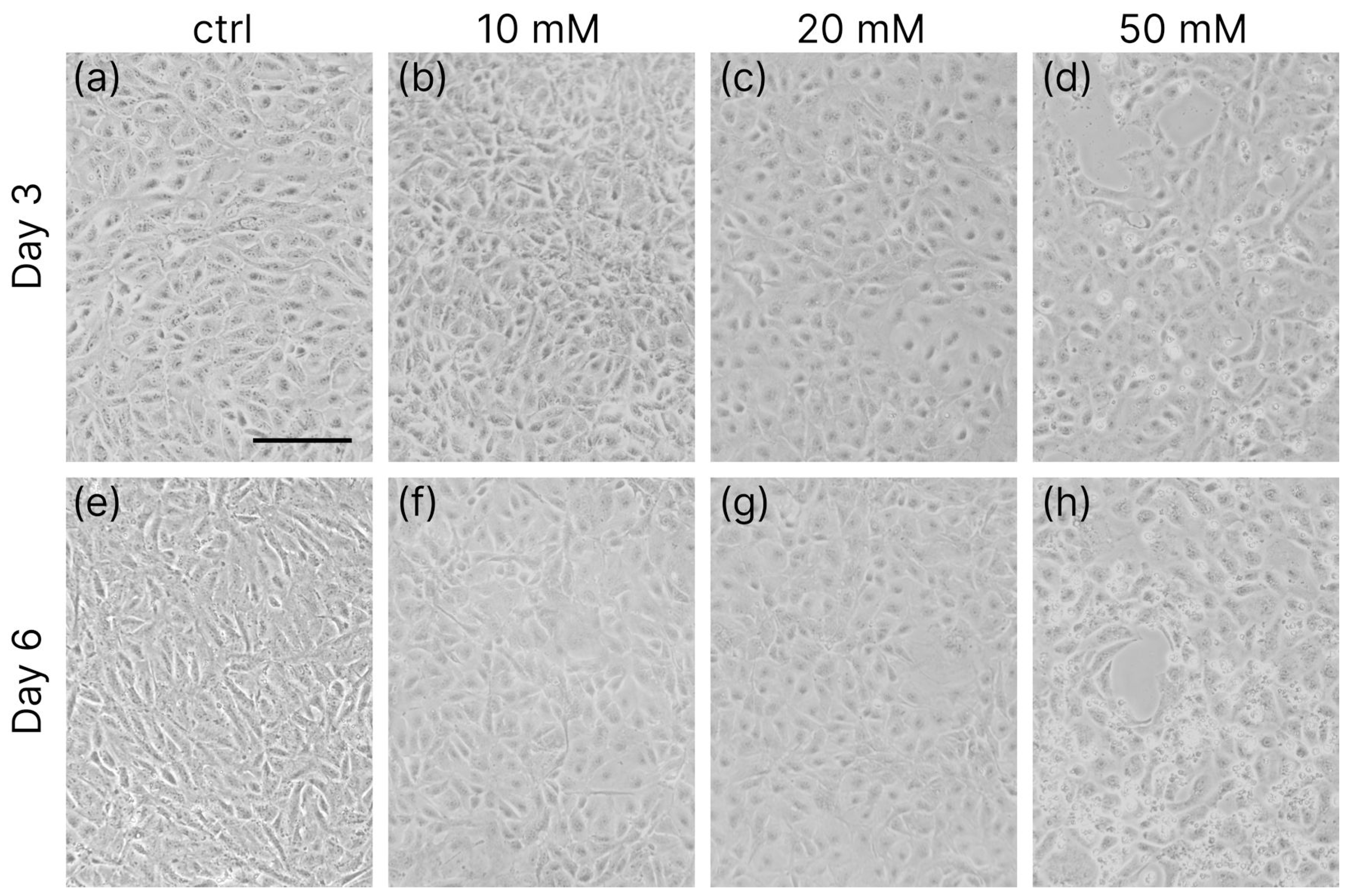
The growth and differentiation of ARPE-19 cells in long-term culture in four different media were traced using phase contrast microscopy (Figure 1).
Two types of medium were used, along with two different concentrations of fetal bovine serum (FBS). Four different combinations used were as follows: Dulbecco′s Modified Eagle′s Medium with high glucose containing 1% or 10% FBS (DMEM HG 10% or DMEM HG 1%) and Dulbecco′s Modified Eagle′s Medium/Nutrient Mixture F-12 Ham with 1% or 10% FBS (DMEM/F12 1% or DMEM/F12 10%). For all examined media, a monolayer of cells formed within 3 days of culture. Hexagonally shaped cells could first be observed after 11 weeks of culture in DMEM HG with both 1% and 10% FBS (Figure 1g–h). At this time, only cells maintained in DMEM HG 1% were pigmented, but their shape was more round compared to densely packed cells incubated in DMEM HG 10%. Melanin pigmentation in cells cultured in DMEM HG 10% was observed a month later (Figure 1l). For longer culture times, the differentiation process continued, resulting in an increasing number of cells with characteristic epithelial shape and increased pigmentation only in DMEM HG medium (Figure 1k,o,p). After 5 months of culture, hexagonal shape and melanin pigmentation could be observed in a larger fraction of the population maintained in DMEM HG 10% than for the cells cultured in DMEM HG 1%. No differentiated cells were observed in any of the DMEM/F12 media.
Then, DMEM HG 1% culture medium was additionally supplemented with nicotinamide at concentrations equal to 10, 20, and 50 mM, and cell growth and differentiation were visualized using phase contrast and fluorescence microcopy.
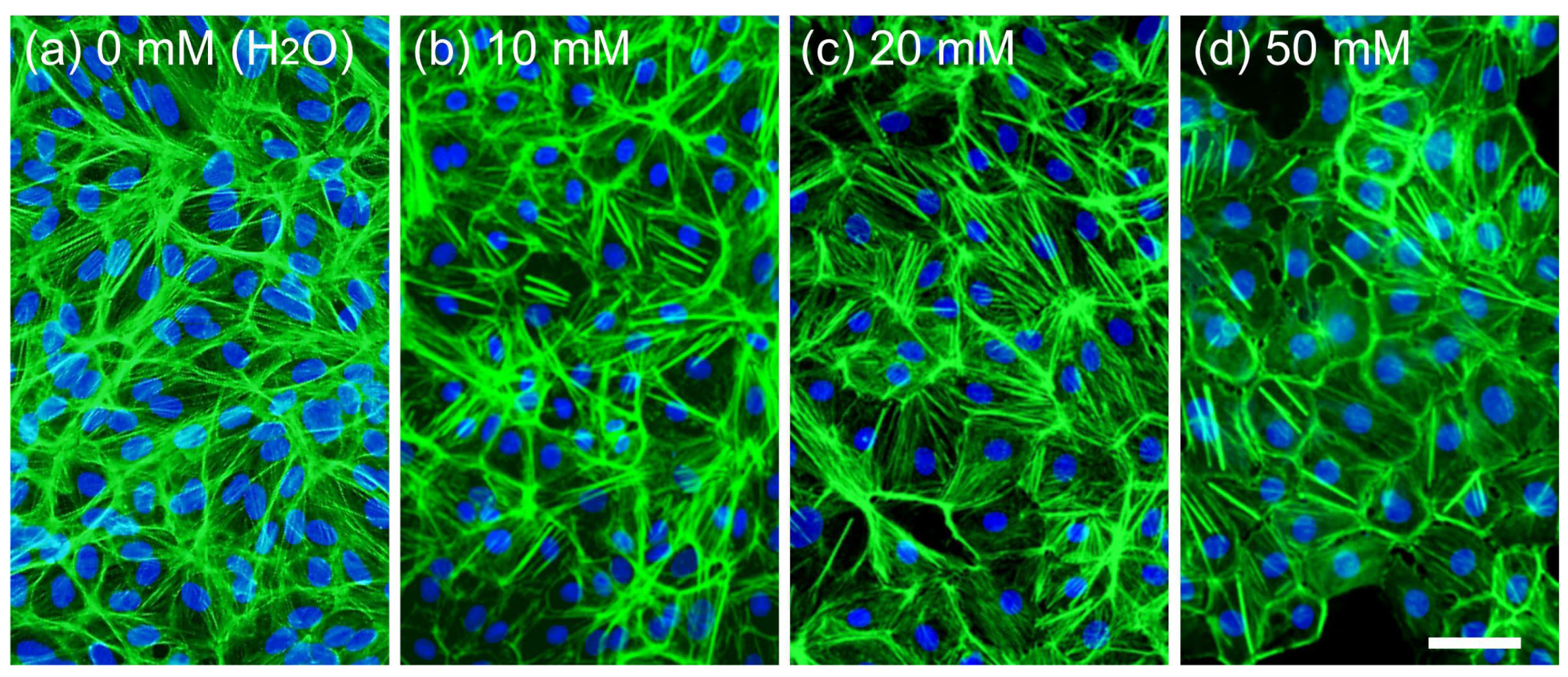
The recorded microscopic images show that within 3 days of culture, the cell morphology did not resemble the fibroblast-like, elongated forms, and cells took more rounded shapes compared to the culture without additives (cf. Figure 2a–d). In turn, after 6 days of culture, cells started to form islands of hexagonally arranged epithelial-like cells, typical for RPE (Figure 2f–h). This effect is also visible in fluorescence micrographs recorded after 6 days of culture (Figure 3), showing the epithelial-like morphology of the cells. However, in cultures incubated with 50 mM nicotinamide, multiple apoptotic cells were visible within 3 days of incubation, as well as after 6 days, which could contribute to the fact that the cells did not form a monolayer. Furthermore, the morphology of the cells was not homogenous; multiple elongated, fibroblast-like cells were found (Figure 2d,h).
In addition to information about the impact of nicotinamide on cell differentiation, the recorded images also provide information about its effect on cell growth. For a low concentration of nicotinamide (10 mM, Figure 2b,f), only viable cells that formed a confluent monolayer within 3 days of culture were visible. A similar situation was observed for cells cultured in medium supplemented with 20 mM nicotinamide (Figure 2c,g). In turn, for the highest concentration of nicotinamide of 50 mM (Figure 2d,h), microscopic images show a large number of small, rounded cells not flattened at the substrate, suggesting their limited viability or death. Furthermore, even after 6 days of culture, the confluent monolayer of the cells does not form, indicating an adverse effect of nicotinamide at high concentrations on cells.
Fluorescence microscopy was used to visualize the impact of different concentrations of nicotinamide on the ARPE-19 actin cytoskeleton (Figure 3). The images show that actin stress fibers were present in every condition. However, in higher concentrations of nicotinamides, more actin filaments accumulated near the cell–cell barrier as a consequence of cytoskeleton rearrangement. After closer examination of stained cells cultured in medium with 50 mM nicotinamide (Figure 3d), gaps between differentiated cells can be visible. There were no such gaps in lower concentrations of nicotinamide (Figure 3b,c), nor in standard medium (Figure 3a), where the monolayer was intact.
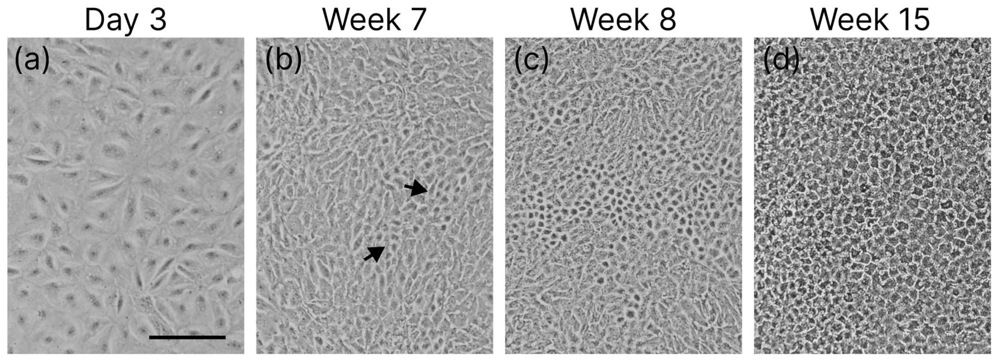
The long-term culture of ARPE-19 cells in the presence of 10 mM nicotinamide (Figure 4) also points to their more effective differentiation. First, islands of cells with modified shapes were observed within 7 weeks of culture, and after 15 weeks, an entire monolayer was formed by cells with a characteristic epithelial shape and increased pigmentation, although not as intense as in DMEM HG 10%.

3.2. Enzymatic Viability Tests

To assess quantitative information about the impact of nicotinamide on cell viability, MTT and LDH tests were performed for cells cultured in medium with nicotinamide concentrations equal to 10, 20, and 50 mM, as well as standard culture medium for 1 and 7 days of culture.
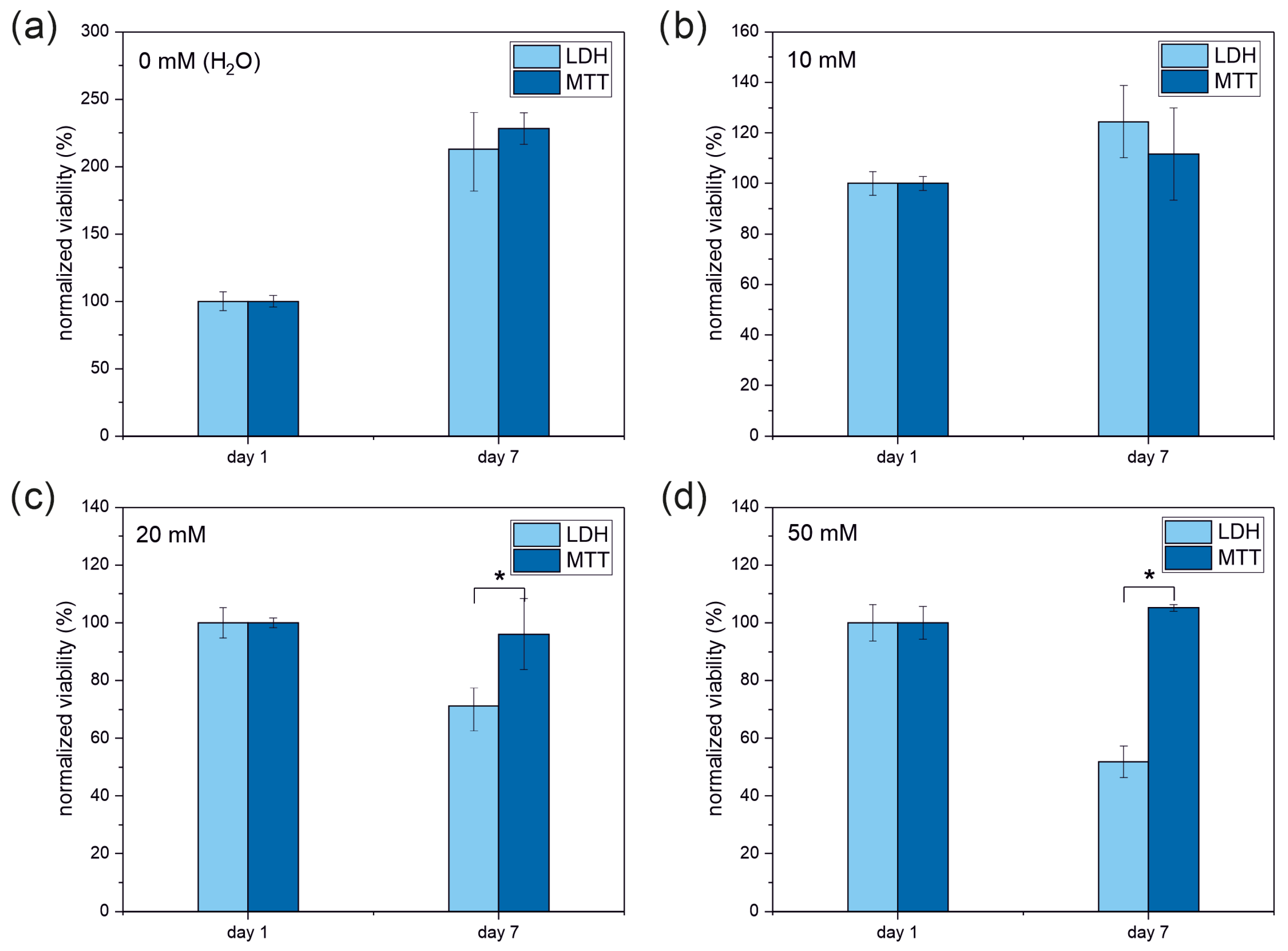
The results determined for the standard culture conditions (Figure 5a), without nicotinamide additives, show a significant increase in cell viability, which doubled after 7 days of culture, and the values are comparable for both applied tests. Similarly, for cells supplemented with 10 mM nicotinamide (Figure 5b), both viability assays showed similar results. However, the observed growth in viability was significantly lower and reached 110–120% of the initial value for the MTT and LDH tests, respectively. In turn, for cells cultured in medium with 20 and 50 mM nicotinamide added (Figure 5c and Figure 5d, respectively), the MTT test did not show any increase in cell viability with increasing culture time, while the LDH assay indicated a reduction of cell viability, to 70% for 20 mM nicotinamide. Cell viability was further reduced for 50 mM nicotinamide, where it dropped down to 50% of the initial value. The results of the LDH tests are in agreement with the images presented in Figure 2e–h and Figure 3a–d, depicting almost no changes in cell morphology for 10 mM nicotinamide, and a slight loss of cell monolayer continuity for 20 mM nicotinamide, which intensified with the highest concentration of nicotinamide. Furthermore, cultures supplemented with 50 mM nicotinamide showed cells with apoptotic morphology. These results indicate that the LDH test is more reliable for assessing the viability of cells in response to an external factor.

3.3. Live and Dead Cell Staining

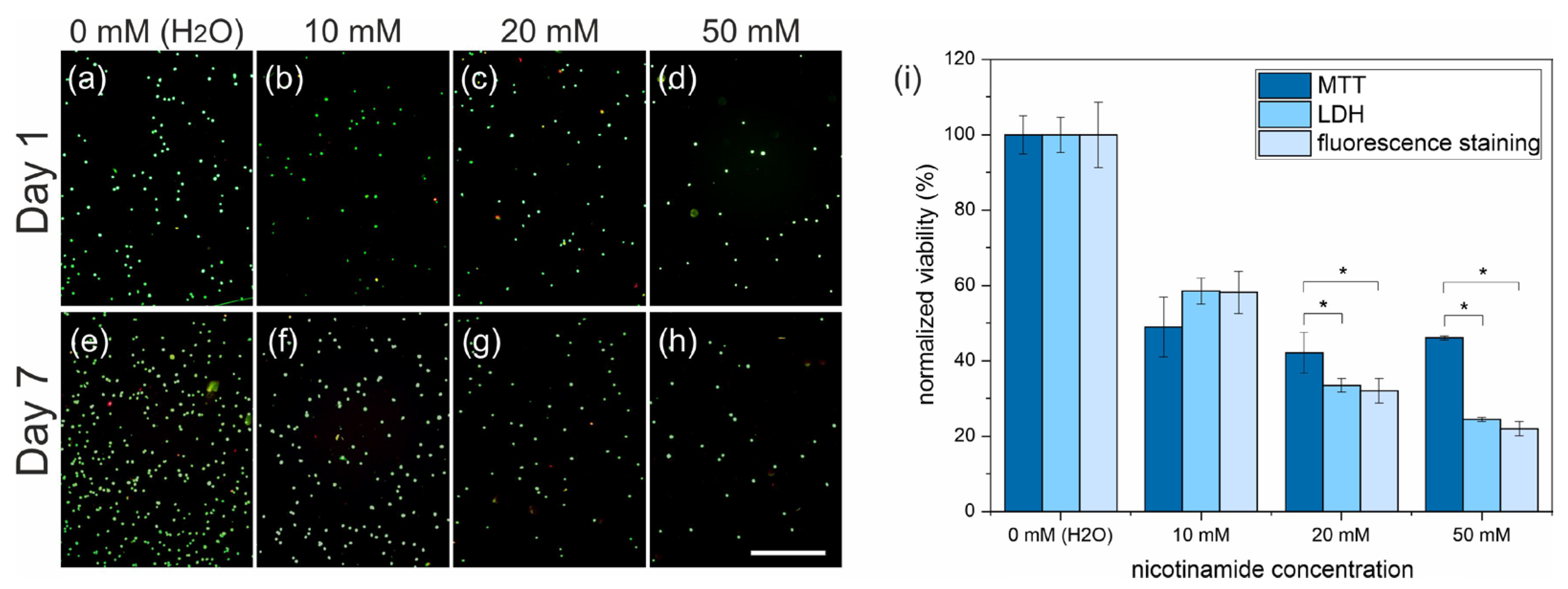
To verify which of the performed tests provided more reliable data, live/dead staining was applied. This assay relies on dyeing with calcein-AM and propidium iodine, resulting in the green and red labeling of live and dead cells, respectively, and its results are not falsified by undesirable biochemical interactions caused by nicotinamide. The stained cells were visualized using fluorescence microscopy. Representative fluorescence micrographs, presented in Figure 6, show that nicotinamide affected cell growth, and their numbers were significantly reduced compared to culture in a standard medium.
These observations were quantitatively verified by estimating the number of live cells as a function of nicotinamide concentration and used for comparative analysis with the LDH and MTT results after 7 days of incubation with nicotinamide (Figure 6i). The number of cells determined in the fluorescence images decreased monotonically with the abundance of nicotinamide, reaching 60, 30, and 20% for 10, 20, and 50 mM of nicotinamide, respectively. These results closely resemble the LDH data, showing almost the same values of cell viability. On the contrary, the MTT assay indicated a significant decrease in cell viability for cell culture in medium containing nicotinamide, which remained constant independently with respect to its concentration.
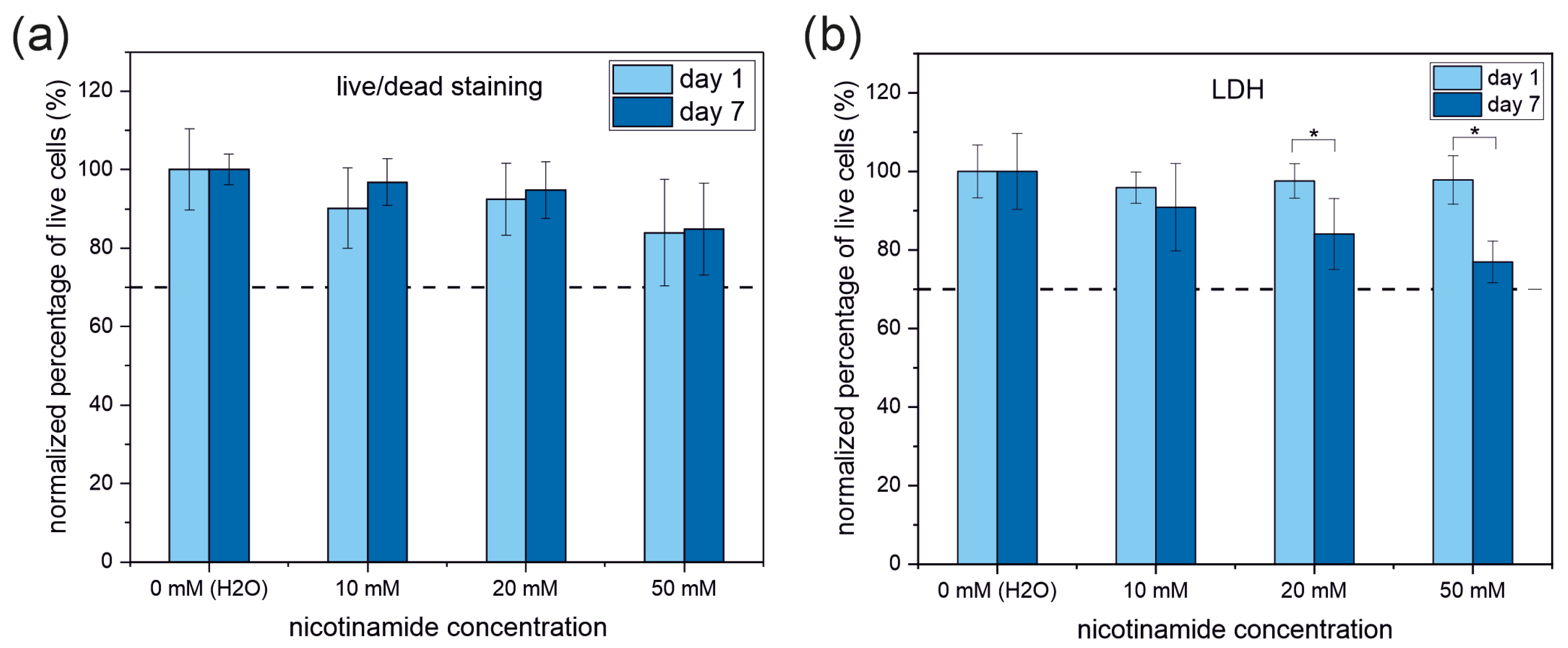
Additionally, the percentage of live cells was calculated as the ratio between live and all (live and dead) cells (Figure 7a), showing that approximately 90% of cells were alive for 10 and 20 mM of nicotinamide. This number was slightly reduced to approximately 80% for the highest concentration of nicotinamide. However, it should be noted that this result is not statistically relevant. Moreover, this situation did not change with culture time.
These results were compared with the percentage of live cells calculated based on LDH data (Figure 7b). In this method, the number of live cells is proportional to the difference between the maximum LDH activity—determined for lysed cells and reflecting both live and dead cells—and the spontaneous LDH activity found in the culture media and related to dead cells. After 1 day of culture, the contribution of live cells was constant for all experimental conditions and varied between 95 and 97% compared to the culture in standard medium. In turn, for cells cultured for 7 days, the percentage of live cells decreased with increasing nicotinamide concentration, and dropped to 90, 85 and 75% for the 10, 20, and 50 mM nicotinamide concentrations, respectively.

4. Discussion

The retina is a highly organized stratified neural tissue composed of pigment epithelium (RPE) and neural retina (NR). It consists of interconnecting layers of specialized cells organized in strict localizations, maintaining interaction with other cells in the 3D retinal space [40]. Retinal degeneration, mainly caused by diabetic and age-related dysfunctions, is the leading cause of blindness. Until now, there has been no treatment capable of reversing retinal degeneration, and only the use of retinal prostheses provides a chance to restore some vision in blind eyes. As the successful realization of a retinal prosthesis depends on the survival and function of remaining retinal cells, complex interactions between the cell and the substrate are critical. Efficiency in the research on this subject requires a cell line that resembles the properties of human RPE cells, which are in direct contact with retinal prostheses and a reliable characterization method, providing information about cell viability first. The most suitable candidates for retinal-orientated studies are primary cells harvested from donors or of animal origin. Another choice is the commercially available ARPE-19 cell line, which, however, cultured in standard medium, proliferates in an undifferentiated form not resembling RPE cells. Therefore, in the first step, a series of experiments aimed at finding culture conditions suitable to trigger the differentiation process was carried out. Based on the data from the literature, [29,30,41] ARPE-19 cells were cultured in four enriched variants of DMEM medium, namely DMEM/F12 supplemented with 1% or 10% FBS, and high-glucose DMEM HG, also supplemented with 1% or 10% of FBS. Different concentrations of FBS were used due to the fact that its presence and concentration can influence cell behavior and thus the outcome of the experiment [42,43]. The results show that FBS concentration influences the rate at which ARPE-19 cells differentiate and produce melanin in DMEM HG medium. Although this approach led to the differentiation of ARPE-19 cells in both variants of DMEM HG medium, the cells developed epithelial-like morphology with changed shapes and increased pigmentation after 11 weeks. Even after 3 months, the culture was heterogeneous, and not all cells were pigmented or phenotypically transformed (Figure 1). Such long-term culture does not provide the appropriate conditions for effective research on cell–substrate interactions; therefore, another medium modification was proposed, based on nicotinamide supplementation [27,28].
The choice of nicotinamide abundance was motivated by the data from the literature, pointing to the effective differentiation of RPE cells already after 2 weeks for medium supplemented with 10 mM nicotinamide [27,28,31,32,33]. Although the reported reduction in differentiation time is impressive, it is still not sufficient for some experiments, e.g., testing cell sheet engineering platforms based on thermoresponsive polymer brushes, which enable the spontaneous detachment of RPE cell sheets due to the cell–substrate interactions modified by a rapid reduction of temperature. In this case, the impact of the thermoresponsive platform may be reduced or even completely eliminated by the formation of a well-developed extracellular matrix during prolonged culture [44,45] and effectively preventing cell detachment [46]. Therefore, the possibility of further acceleration of RPE cell differentiation with increases in nicotinamide was verified. For this purpose, cells were cultured in DMEM medium with 10, 20, and 50 mM nicotinamide, and visualized using phase and fluorescence microscopy.
This strategy significantly improved the efficiency of restoration of properties resembling physiological RPE cells, and already after 6 days of culture, cells began to take hexagonal shapes (Figure 2 and Figure 3). Unfortunately, high concentrations of nicotinamide turned out to have adverse effects on cultured cells, resulting in a reduced number of cells and loss of monolayer integrity (Figure 2 and Figure 3), so the proposed approach may not be used to prompt RPE cell differentiation. In order to bypass this limitation, e.g., in an effort to study detachment of differentiated RPE cells from the thermoresponsive platforms, the culture time may be shortened by using pre-differentiated for the experiment. Although the proliferation rate of differentiated cells generally decreases, RPE cells have the intrinsic capacity to re-enter the cell cycle, especially in injury contexts, and regain their proliferation potential [46,47,48].
The experiments also showed that supplementation of DMEM HG 10% medium, chosen according to the long-term culture experiment, with 10 mM nicotinamide reduced the time required to obtain morphologically differentiated cells by a few weeks compared to culture without nicotinamide. This duration, significantly longer than reported elsewhere, could probably be further reduced by using permeable membranes for cell culture, which have been shown to promote cell differentiation [28,49,50].
The mentioned observations imposed the need for a detailed and quantitative analysis of the dose-dependent impact of nicotinamide on the growth and viability of ARPE-19 cells. Cellular viability is usually determined using colorimetric tests based on enzymatic activity. However, nicotinamide was shown to enhance the mitochondrial activity of ARPE-19 cells and influence the NAD+/NADH ratio [27,28], which could affect the reliability of tests based on mitochondrial activity, such as the MTT test. Moreover, differentiated ARPE-19 cells undergo a metabolic switch, from glycolysis-dependent to oxidative phosphorylation-dependent, which strongly affects the entire metabolic equilibrium; thus, also other enzyme-based viability tests, such as LDH, may be adulterated. Therefore, cell viability was determined by both colorimetric tests, and their results were compared with each other, showing good agreement for short incubation times and significant differences for long periods of incubation, especially for the highest nicotinamide concentration. The MTT-derived viability did not indicate any significant difference between cells cultured in medium supplemented with various amounts of nicotinamide, whereas the LDH assay showed a monotonic decrease in cell viability with increasing nicotinamide concentration. To verify which test provided the most adequate results, the number of live cells, proportional to their viability, was determined using an independent method based on membrane integrity. The results obtained, summarized in Table 1, show that the values provided by the LDH assay are more consistent with the number determined using live/dead staining. These results indicate that the presence of nicotinamide significantly influenced the results of the MTT test due to the growth of mitochondrial activity caused by nicotinamide supplementation, with the effect being more pronounced for longer incubation times. In turn, for the LDH test, no disturbances caused by nicotinamide were observed, indicating that possible changes in the NAD+/NADH ratio did not impact the conversion of lactate to the pyruvate path.
In addition to viability, the percentage of living cells was also calculated based on the LDH assay, showing a constant ratio of live cells for short culture times and a slight reduction in their contribution for longer culture times, with the strength of this effect increasing with the growing concentration of nicotinamide, thus indicating the long-term, dose-dependent negative effect of nicotinamide on ARPE-19 cells. However, even for the highest fraction of nicotinamide, supplementation of live cells does not decrease below 70%, thus not crossing the border line defining the cytocompatible material [2]. Similar results were also provided by live/dead staining, which showed a very high and almost constant ratio of living cells, slightly decreasing for the highest nicotinamide concentration. However, in this case, reductions were observed for both short and long incubation times.

5. Conclusions

In this work, the significant impact of the composition of the medium on the capability of ARPE-19 cells to differentiate was shown. Supplementation of the medium with nicotinamide leads to the most effective phenotypic transformation of ARPE-19 cells. However, the concentration of nicotinamide must be optimized to provide differentiation and avoid adverse effects that occur predominantly for high concentrations of nicotinamide. Furthermore, the applicability of MTT and LDH enzymatic assays for determining ARPE-19 cell viability was verified, indicating that LDH provides more reliable information. Therefore, it can be used for effective research on the biocompatibility of different materials for retinal prostheses, significantly increasing the probability of the construction of novel and effective devices that restore vision. However, it should be noted that the reliability of MTT and LDH assays depends strongly on the analyzed cell type and the investigated process. Therefore, the viability of cells determined using LDH and MTT assays should be carefully interpreted, considering the potential impact of external factors. Additionally, conclusions on the cytotoxicity and biocompatibility of materials, pharmaceuticals, or medical procedures should be drawn with awareness of the limitations of both tests, which may lead to over- or underestimated impact of the examined factor on cell viability. Regardless of the method selected by the researcher—MTT, LDH, live/dead assays, or any other technique—the results should always be independently verified. At a minimum, qualitative verification should be conducted by direct observation using optical microscopy.
Together, all the assays performed provide a simple viability metrics, sufficient for the basic evaluation of material safety and biological performance. However, to assess a comprehensive understanding of cellular responses to external factors, more specific analysis is required, e.g., with assays targeting apoptosis and cell cycle dynamics. Such assays provide insights into programmed cell death mechanisms triggered by external factors and reveal disruptions in cell proliferation and progression through specific phases, indicating potential sub-lethal stress or genotoxic effects. This provides a holistic view and enables detailed interpretation of the results.

Author Contributions

Conceptualization, A.C. and J.R.; methodology, A.C. and J.R.; validation, J.R.; formal analysis, A.C. and J.R.; investigation, A.C.; resources, A.C. and J.R.; writing—original draft preparation, A.C. and J.R.; writing—review and editing, A.C. and J.R.; visualization, A.C. and J.R.; supervision, J.R.; project administration, A.C. and J.R.; funding acquisition, A.C. and J.R. All authors have read and agreed to the published version of the manuscript.

Funding

The purchase of the experimental equipment used in this research was funded by Priority Research Areas DigiWorld (absorbance microplate reader) and SciMat (incubator) under the program Excellence Initiative—Research University at Jagiellonian University in Kraków. The study was funded by the “Research support module” as part of the “Excellence Initiative—Research University” program at Jagiellonian University in Kraków (RSM/43/CA).

Institutional Review Board Statement

Not applicable.

Informed Consent Statement

Not applicable.

Data Availability Statement

Data will be made available upon request.

Conflicts of Interest

The authors declare no conflicts of interest.

Abbreviations

The following abbreviations are used in this manuscript:
MTT3-[4,5-dimethylthiazol-2-yl]-2,5-diphenyl tetrazolium bromide
LDHLactate dehydrogenase
RPERetinal pigment epithelium
NAD+/NADHNicotinamide adenine dinucleotide
DMEMDulbecco′s Modified Eagle′s Medium
FBSFetal Bovine Serum
DPBSDulbecco’s Phosphate Buffered Saline
HGHigh glucose

References

  1. Sousa, A.; Neves, S.C.; Gonçalves, I.C.; Barrias, C.C. 12—In Vitro Interaction of Polymeric Biomaterials with Cells; Tanzi, M.C., Farè, S., Eds.; Woodhead Publishing: Cambridge, UK, 2017; pp. 285–315. ISBN 978-0-08-100737-2. [Google Scholar]
  2. Huzum, B.; Puha, B.; Necoara, R.; Gheorghevici, S.; Puha, G.; Filip, A.; Sirbu, P.; Alexa, O. Biocompatibility assessment of biomaterials used in orthopedic devices: An overview (Review). Exp. Ther. Med. 2021, 22, 1315. [Google Scholar] [CrossRef]
  3. Barmshuri, M.; Kholdebarin, B.; Sadeghi, S. Applications of comet and MTT assays in studying Dunaliella algae species. Algal Res. 2023, 70, 103018. [Google Scholar] [CrossRef]
  4. Abel, S.D.A.; Baird, S.K. Honey is cytotoxic towards prostate cancer cells but interacts with the MTT reagent: Considerations for the choice of cell viability assay. Food Chem. 2018, 241, 70–78. [Google Scholar] [CrossRef]
  5. de la Fuente-Jiménez, J.L.; Rodríguez-Rivas, C.I.; Mitre-Aguilar, I.B.; Torres-Copado, A.; García-López, E.A.; Herrera-Celis, J.; Arvizu-Espinosa, M.G.; Garza-Navarro, M.A.; Arriaga, L.G.; García, J.L.; et al. A Comparative and Critical Analysis for In Vitro Cytotoxic Evaluation of Magneto-Crystalline Zinc Ferrite Nanoparticles Using MTT, Crystal Violet, LDH, and Apoptosis Assay. Int. J. Mol. Sci. 2023, 24, 12860. [Google Scholar] [CrossRef] [PubMed]
  6. Bopp, S.K.; Lettieri, T. Comparison of four different colorimetric and fluorometric cytotoxicity assays in a zebrafish liver cell line. BMC Pharmacol. 2008, 8, 8. [Google Scholar] [CrossRef] [PubMed]
  7. Mosmann, T. Rapid colorimetric assay for cellular growth and survival: Application to proliferation and cytotoxicity assays. J. Immunol. Methods 1983, 65, 55–63. [Google Scholar] [CrossRef]
  8. Edmondson, J.M.; Armstrong, L.S.; Martinez, A.O. A rapid and simple MTT-based spectrophotometric assay for determining drug sensitivity in monolayer cultures. J. Tissue Cult. Methods 1988, 11, 15–17. [Google Scholar] [CrossRef]
  9. Fotakis, G.; Timbrell, J.A. In vitro cytotoxicity assays: Comparison of LDH, neutral red, MTT and protein assay in hepatoma cell lines following exposure to cadmium chloride. Toxicol. Lett. 2006, 160, 171–177. [Google Scholar] [CrossRef]
  10. Smith, S.M.; Wunder, M.B.; Norris, D.A.; Shellman, Y.G. A simple protocol for using a LDH-Based cytotoxicity assay to assess the effects of death and growth inhibition at the same time. PLoS ONE 2011, 6, e26908. [Google Scholar] [CrossRef]
  11. Lobner, D. Comparison of the LDH and MTT assays for quantifying cell death: Validity for neuronal apoptosis? J. Neurosci. Methods 2000, 96, 147–152. [Google Scholar] [CrossRef]
  12. Kendig, D.M.; Tarloff, J.B. Inactivation of lactate dehydrogenase by several chemicals: Implications for in vitro toxicology studies. Toxicol. Vitr. 2007, 21, 125–132. [Google Scholar] [CrossRef]
  13. Singhal, M.; Shaha, S.; Katsikogianni, M. Comparative Analysis of Cytotoxicity Assays, from Traditional to Modern Approaches. In Cytotoxicity—A Crucial Toxicity Test for In Vitro Experiments; Erkekoğlu, P., Ed.; IntechOpen: London, UK, 2024; ISBN 978-1-83634-031-7. [Google Scholar]
  14. Aslantürk, Ö.S. In Vitro Cytotoxicity and Cell Viability Assays: Principles, Advantages, and Disadvantages. In Genotoxicity—A Predictable Risk to Our Actual World; Larramendy, M.L., Soloneski, S., Eds.; IntechOpen: London, UK, 2017; ISBN 978-1-78923-419-0. [Google Scholar]
  15. Van den Bossche, S.; Vandeplassche, E.; Ostyn, L.; Coenye, T.; Crabbé, A. Bacterial Interference With Lactate Dehydrogenase Assay Leads to an Underestimation of Cytotoxicity. Front. Cell. Infect. Microbiol. 2020, 10, 494. [Google Scholar] [CrossRef] [PubMed]
  16. Parhamifar, L.; Andersen, H.; Moghimi, S.M. Lactate dehydrogenase assay for assessment of polycation cytotoxicity. Methods Mol. Biol. 2019, 1943, 291–299. [Google Scholar] [CrossRef] [PubMed]
  17. Khalef, L.; Lydia, R.; Filicia, K.; Moussa, B. Cell viability and cytotoxicity assays: Biochemical elements and cellular compartments. Cell Biochem. Funct. 2024, 42, e4007. [Google Scholar] [CrossRef] [PubMed]
  18. Madorran, E.; Ambrož, M.; Knez, J.; Sobočan, M. An Overview of the Current State of Cell Viability Assessment Methods Using OECD Classification. Int. J. Mol. Sci. 2025, 26, 220. [Google Scholar] [CrossRef]
  19. Han, Q.; Zhang, W.; Guo, J.; Zhu, Q.; Chen, H.; Xia, Y.; Zhu, G. Mitochondrion: A sensitive target for Pb exposure. J. Toxicol. Sci. 2021, 46, 345–358. [Google Scholar] [CrossRef]
  20. Friedlander, M. Fibrosis and diseases of the eye. J. Clin. Investig. 2007, 117, 576–586. [Google Scholar] [CrossRef]
  21. Tan, W.; Zou, J.; Yoshida, S.; Jiang, B.; Zhou, Y. The role of inflammation in age-related macular degeneration. Int. J. Biol. Sci. 2020, 16, 2989–3001. [Google Scholar] [CrossRef]
  22. Tenbrock, L.; Wolf, J.; Boneva, S.; Schlecht, A.; Agostini, H.; Wieghofer, P.; Schlunck, G.; Lange, C. Subretinal fibrosis in neovascular age-related macular degeneration: Current concepts, therapeutic avenues, and future perspectives. Cell Tissue Res. 2022, 387, 361–375. [Google Scholar] [CrossRef]
  23. Higashijima, F.; Hasegawa, M.; Yoshimoto, T.; Kobayashi, Y.; Wakuta, M.; Kimura, K. Molecular mechanisms of TGFβ-mediated EMT of retinal pigment epithelium in subretinal fi brosis of age-related macular degeneration. Front. Ophthalmol. 2023, 2, 1060087. [Google Scholar] [CrossRef]
  24. Bellapianta, A.; Cetkovic, A.; Bolz, M.; Salti, A. Retinal Organoids and Retinal Prostheses: An Overview. Int. J. Mol. Sci. 2022, 23, 2922. [Google Scholar] [CrossRef]
  25. Singh, M.S.; Issa, P.C.; Butler, R.; Martin, C.; Lipinski, D.M.; Sekaran, S.; Barnard, A.R.; MacLaren, R.E. Reversal of end-stage retinal degeneration and restoration of visual function by photoreceptor transplantation. Proc. Natl. Acad. Sci. USA 2013, 110, 1101–1106. [Google Scholar] [CrossRef] [PubMed]
  26. Bae, S.H.; Che, J.H.; Seo, J.M.; Jeong, J.; Kim, E.T.; Lee, S.W.; Koo, K.I.; Suaning, G.J.; Lovell, N.H.; Dan Cho, D., II; et al. In vitro biocompatibility of various polymer-based microelectrode arrays for retinal prosthesis. Investig. Ophthalmol. Vis. Sci. 2012, 53, 2653–2657. [Google Scholar] [CrossRef] [PubMed]
  27. Hazim, R.A.; Paniagua, A.E.; Tang, L.; Yang, K.; Kim, K.K.O.; Stiles, L.; Divakaruni, A.S.; Williams, D.S. Vitamin B3, nicotinamide, enhances mitochondrial metabolism to promote differentiation of the retinal pigment epithelium. J. Biol. Chem. 2022, 298, 102286. [Google Scholar] [CrossRef] [PubMed]
  28. Hazim, R.A.; Volland, S.; Yen, A.; Burgess, B.L.; Williams, D.S.; Angeles, L.; Angeles, L. Rapid differentiation of the human RPE cell line, ARPE-19, induced by nicotinamide. Exp Eye Res. 2019, 179, 18–24. [Google Scholar] [CrossRef]
  29. Samuel, W.; Jaworski, C.; Postnikova, O.A.; Kutty, R.K.; Duncan, T.; Tan, L.X.; Poliakov, E.; Lakkaraju, A.; Redmond, T.M. Appropriately differentiated ARPE-19 cells regain phenotype and gene expression profiles similar to those of native RPE cells. Mol. Vis. 2017, 23, 60–89. [Google Scholar]
  30. Ahmado, A.; Carr, A.J.; Vugler, A.A.; Semo, M.; Gias, C.; Lawrence, J.M.; Chen, L.L.; Chen, F.K.; Turowski, P.; da Cruz, L.; et al. Induction of differentiation by pyruvate and DMEM in the human retinal pigment epithelium cell line ARPE-19. Investig. Ophthalmol. Vis. Sci. 2011, 52, 7148–7159. [Google Scholar] [CrossRef]
  31. D’Antonio-Chronowska, A.; D’Antonio, M.; Frazer, K.A. In vitro Differentiation of Human iPSC-derived Retinal Pigment Epithelium Cells (iPSC-RPE). Bio Protoc. 2019, 9, e3469. [Google Scholar] [CrossRef]
  32. Idelson, M.; Alper, R.; Obolensky, A.; Ben-Shushan, E.; Hemo, I.; Yachimovich-Cohen, N.; Khaner, H.; Smith, Y.; Wiser, O.; Gropp, M.; et al. Directed Differentiation of Human Embryonic Stem Cells into Functional Retinal Pigment Epithelium Cells. Cell Stem Cell 2009, 5, 396–408. [Google Scholar] [CrossRef]
  33. Hazim, R.A. Enhanced mitochondrial metabolism promotes differentiation of the retinal pigment epithelium following treatment with nicotinamide. Investig. Ophthalmol. Vis. Sci. 2023, 64, 953. [Google Scholar]
  34. Makarov, M.V.; Trammell, S.A.J.; Migaud, M.E. The chemistry of the vitamin B3 metabolome. Biochem. Soc. Trans. 2018, 47, 131–147. [Google Scholar] [CrossRef]
  35. Yusri, K.; Jose, S.; Vermeulen, K.S.; Tan, T.C.M.; Sorrentino, V. The role of NAD+ metabolism and its modulation of mitochondria in aging and disease. NPJ Metab. Health Dis. 2025, 3, 26. [Google Scholar] [CrossRef] [PubMed]
  36. Yu, F.; Zhao, H.; Luo, L.; Wu, W. Nicotinamide Adenine Dinucleotide Supplementation to Alleviate Heart Failure: A Mitochondrial Dysfunction Perspective. Nutrients 2025, 17, 1855. [Google Scholar] [CrossRef]
  37. Xie, N.; Zhang, L.; Gao, W.; Huang, C.; Huber, P.E.; Zhou, X.; Li, C.; Shen, G.; Zou, B. NAD+ metabolism: Pathophysiologic mechanisms and therapeutic potential. Signal Transduct. Target. Ther. 2020, 5, 227. [Google Scholar] [CrossRef]
  38. Rizwan, S.; Toothman, B.; Li, B.; Engel, A.J.; Lim, R.R.; Niernberger, S.; Lu, J.; Ratliff, C.; Xiang, Y.; Eminhizer, M.; et al. Metabolic Phenotyping of Healthy and Diseased Human RPE Cells. Investig. Ophthalmol. Vis. Sci. 2024, 65, 5. [Google Scholar] [CrossRef] [PubMed]
  39. Boo, Y.C. Mechanistic Basis and Clinical Evidence for the Applications of Nicotinamide (Niacinamide) to Control Skin Aging and Pigmentation. Antioxidants 2021, 10, 1315. [Google Scholar] [CrossRef] [PubMed]
  40. Grigoryan, E.N. Self-Organization of the Retina during Eye Development, Retinal Regeneration In Vivo, and in Retinal 3D Organoids In Vitro. Biomedicines 2022, 10, 1458. [Google Scholar] [CrossRef]
  41. Villarroel, M.; García-Ramírez, M.; Corraliza, L.; Hernández, C.; Simó, R. Effects of high glucose concentration on the barrier function and the expression of tight junction proteins in human retinal pigment epithelial cells. Exp. Eye Res. 2009, 89, 913–920. [Google Scholar] [CrossRef]
  42. Luo, Y.; Zhuo, Y.; Fukuhara, M.; Rizzolo, L.J. Effects of culture conditions on heterogeneity and the apical junctional complex of the ARPE-19 cell line. Investig. Ophthalmol. Vis. Sci. 2006, 47, 3644–3655. [Google Scholar] [CrossRef]
  43. Oka, M.S.; Landers, R.A.; Bridges, C.D.B. A serum-free defined medium for retinal pigment epithelial cells. Exp. Cell Res. 1984, 154, 537–547. [Google Scholar] [CrossRef]
  44. Kumar, P.; Satyam, A.; Fan, X.; Collin, E.; Rochev, Y.; Rodriguez, B.J.; Gorelov, A.; Dillon, S.; Joshi, L.; Raghunath, M.; et al. Macromolecularly crowded in vitro microenvironments accelerate the production of extracellular matrix-rich supramolecular assemblies. Sci. Rep. 2015, 5, 8729. [Google Scholar] [CrossRef]
  45. Torres, Y.; Gluais, M.; Da Silva, N.; Rey, S.; Grémare, A.; Magnan, L.; Kawecki, F.; L’Heureux, N. Cell-assembled extracellular matrix (CAM) sheet production: Translation from using human to large animal cells. J. Tissue Eng. 2021, 12, 2041731420978327. [Google Scholar] [CrossRef]
  46. Cieślik, A.; Shymborska, Y.; Tymetska, S.; Stetsyshyn, Y.; Bernasik, A.; Brzychczy-Włoch, M.; Drożdż, K.; Szajna, K.; Krok, F.; Budkowski, A.; et al. Cell sheet engineering platforms integrating antibacterial and thermo-responsive functionalities: Cu-nanoparticle-loaded P4VP brushes for retinal cell sheet harvesting. Chem. Eng. J. 2025, 513, 162985. [Google Scholar] [CrossRef]
  47. Cooper, G.M. The Cell: A Molecular Approach, 2nd ed.; Sinauer Associates: Sunderland, MA, USA, 2000; ISBN 0-87893-106-6. [Google Scholar]
  48. Ho, A.; Dowdy, S.F. Regulation of G1 cell-cycle progression by oncogenes and tumor suppressor genes. Curr. Opin. Genet. Dev. 2002, 12, 47–52. [Google Scholar] [CrossRef]
  49. Michelet, F.; Balasankar, A.; Teo, N.; Stanton, L.W.; Singhal, S. Rapid generation of purified human RPE from pluripotent stem cells using 2D cultures and lipoprotein uptake-based sorting. Stem Cell Res. Ther. 2020, 11, 47. [Google Scholar] [CrossRef]
  50. Liu, H.; Huang, S.S.; Lingam, G.; Kai, D.; Su, X.; Liu, Z. Advances in retinal pigment epithelial cell transplantation for retinal degenerative diseases. Stem Cell Res. Ther. 2024, 15, 390. [Google Scholar] [CrossRef]
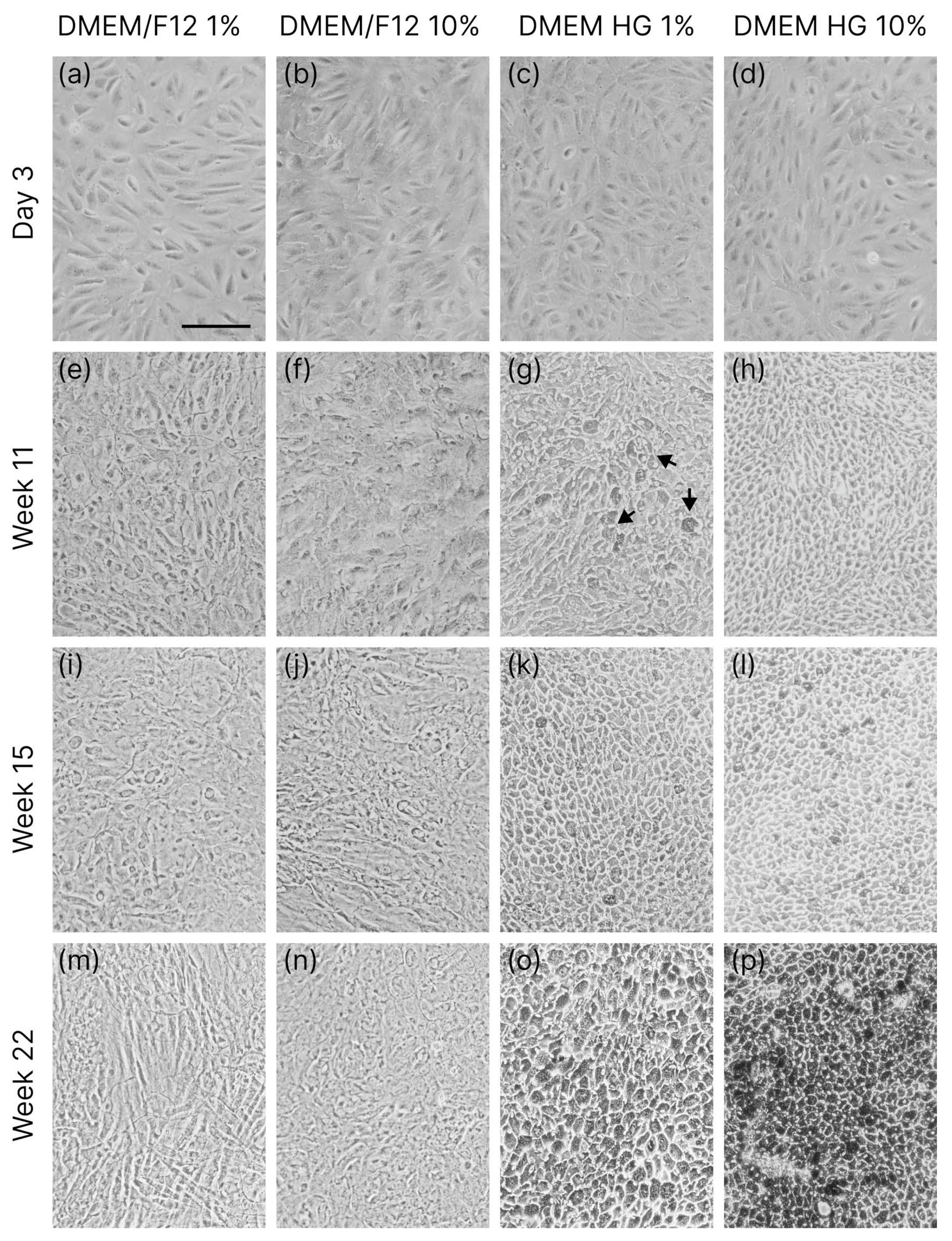
Figure 1. Growth and differentiation of ARPE-19 cells cultured in DMEM with 1% F12 (a,e,i,m), 10% F12 (b,f,j,n), 1% HG (c,g,k,o), and 10% HG (d,h,l,p) in long-term culture traced using phase-contrast microscopy. Arrows point to cells with melanin expression. The scale bar corresponds to 100 µm.
Applsci 15 10684 g001
Figure 2. The growth and differentiation of ARPE-19 cells in DMEM HG 1% medium (a,e) and nicotinamide supplemented with concentrations of 10 mM (b,f), 20 mM (c,g), and 50 mM (d,h), traced using phase contrast microscopy. The scale bar corresponds to 100 µm.
Applsci 15 10684 g002
Figure 3. Differentiation of ARPE-19 cells in DMEM HG 1% (a) and 10 mM (b), 20 mM (c), and 50 mM (d) nicotinamide-supplemented medium visualized using fluorescence microscopy after 6 days of culture (actin filaments—green; nuclei—blue). The scale bar corresponds to 50 μm.
Applsci 15 10684 g003
Figure 4. Growth and differentiation of ARPE-19 cells in long-term culture maintained in DMEM HG 1% medium with 10 mM nicotinamide, traced using phase contrast microscopy after 3 days (a), and 7 (b), 8 (c), and 15 weeks (d). The arrows point to the islands of epithelial-like cells. The scale bar corresponds to 100 µm.
Applsci 15 10684 g004
Figure 5. Results of the MTT and LDH viability assay after 1 and 7 days of incubation with nicotinamide-supplemented medium with nicotinamide concentrations equal to 0 (a), 10 (b), 20 (c), and 50 mM (d). Results normalized to appropriate controls. Data are presented as mean ± SD; * corresponds to statistical significance, p < 0.005.
Applsci 15 10684 g005
Figure 6. Live (green) and dead (red) ARPE-19 cells visualized using fluorescence microscopy (ah) with quantitative analysis (i). The scale bar corresponds to 500 μm. Data are presented as mean ± SD; * corresponds to statistical significance, p < 0.005.
Applsci 15 10684 g006
Figure 7. Percentage of live cells determined using live/dead staining (a) and LDH assay (b). Data are presented as mean ± SD; * corresponds to statistical significance, p < 0.005.
Applsci 15 10684 g007
Table 1. Impact of different concentrations of nicotinamide on the results of MTT, LDH, and live/dead viability tests. Data are presented as mean ± SD; * corresponds to statistical significance, p < 0.005.
MTTLDHLive/Dead
10 mM49 ± 7 58 ± 358 ± 5
20 mM42 ± 4 (*)33 ± 131 ± 3
50 mM46 ± 1 (*)24 ± 121 ± 2
Disclaimer/Publisher’s Note: The statements, opinions and data contained in all publications are solely those of the individual author(s) and contributor(s) and not of MDPI and/or the editor(s). MDPI and/or the editor(s) disclaim responsibility for any injury to people or property resulting from any ideas, methods, instructions or products referred to in the content.