Article

Phytochemical Profile and Biological Activities of Biscutella laevigata: A Comparative Study of Leaves, Seeds, and Microshoot Cultures

by
Marta Klimek-Szczykutowicz
1,*,
Magdalena Anna Malinowska
2,
Anna Śliwa
2,
Ivica Blažević
3,
Azra Ðulović
3,
Karolina Wiśniewska
4,
Renata Piwowarczyk
4,
Paulina Paprocka
5,
Małgorzata Wrzosek
1,6 and
Agnieszka Szopa
7,*
1
Department of Pharmaceutical Sciences, Medical College, Jan Kochanowski University of Kielce, IX Wieków Kielc 19a, 25-516 Kielce, Poland
2
Department of Organic Chemistry and Technology, Faculty of Chemical Engineering and Technology, Cracow University of Technology, Warszawska 24, 31-155 Kraków, Poland
3
Department of Organic Chemistry, Faculty of Chemistry and Technology, University of Split, Ruđera Boškovića 35, 21000 Split, Croatia
4
Center for Research and Conservation of Biodiversity, Department of Environmental Biology, Institute of Biology, Jan Kochanowski University of Kielce, Uniwersytecka 7, 25-406 Kielce, Poland
5
Department of Microbiology and Immunology, Institute of Medical Science, Medical College, Jan Kochanowski University of Kielce, IX Wieków Kielc 19a, 25-516 Kielce, Poland
6
Department of Biochemistry and Pharmacogenomics, Medical University of Warsaw, Banacha 1, 02-097 Warsaw, Poland
7
Department of Medicinal Plant and Mushroom Biotechnology, Faculty of Pharmacy, Jagiellonian University Medical College, Medyczna 9, 30-688 Kraków, Poland
*
Authors to whom correspondence should be addressed.
Appl. Sci. 2025, 15(19), 10462; https://doi.org/10.3390/app151910462
Submission received: 8 July 2025 / Revised: 22 September 2025 / Accepted: 22 September 2025 / Published: 26 September 2025

Abstract

Biscutella laevigata (Brassicaceae) is an endemic species confined to European mountain regions, with a distribution range extending from the Iberian Peninsula through the Carpathians to the Balkans. The objective of this study was to investigate the phytochemical composition and biological properties of extracts obtained from leaves, seeds, and in vitro-derived microshoot cultures. UHPLC-DAD-MS/MS profiling of glucosinolates (GSLs) revealed six compounds exclusively present in seed extracts, with glucohirsutin identified as the predominant constituent (15.06 mg/100 g DW). No glucosinolates were detected in either leaf or microshoot extracts. Notably, 8-(methylsulfonyl)octyl GSL was reported in B. laevigata for the first time. The seed extract exhibited the highest total polyphenol content (TPC, 25,701.00 mg GAE/100 g DW), while leaf and microshoot extracts contained similar amounts (16,244.00 and 16,552.00 mg GAE/100 g DW, respectively). Among phenolic compounds, rutin was the most abundant, reaching up to 1609.21 mg/100 g DW in leaf extracts. Antioxidant capacity, assessed by ABTS and DPPH assays, was strongest in the seed extract (90.56% and 69.24% inhibition, respectively). The same extract demonstrated the greatest anti-elastase activity (12.68%), whereas the microshoot extract displayed a considerable Fe2+-chelating ability (12.48%). All tested extracts showed antimicrobial potential against Staphylococcus aureus, Escherichia coli, Cutibacterium acnes, and the fungus Candida albicans.

1. Introduction

Species belonging to the Brassicaceae family are extensively utilized across the food, cosmetic, and pharmaceutical sectors owing to their richness in bioactive constituents, including glucosinolates (GSLs), polyphenols (such as anthocyanins), isothiocyanates, and vitamins A, C, E, and K. Many representatives of this family have long been incorporated into culinary traditions as well as folk medicine. Owing to their diverse phytochemical profile, Brassicaceae species are regarded as a valuable source of functional foods with well-documented biological properties, particularly antibacterial, anticancer, and antiviral activities [1].
Within this family, the genus Biscutella comprises more than 50 species, distributed mainly across southern Europe, northern Africa, and southwestern Asia. Most Biscutella species are perennials, with a smaller number being annuals. Many of them occupy narrow ecological niches and have restricted geographic ranges, often being endemic to specific regions [2].
One representative of this genus is Biscutella laevigata L., a glacial relict occurring in mountainous areas of Europe, from the Iberian Peninsula to the Carpathians and the Balkans. It is the only Biscutella species found in Poland, where it inhabits primarily limestone rocks and screes in high mountain regions, but can also be found sporadically in meadow communities and on metalliferous waste heaps (mainly zinc and lead) in lowland areas [2]. In Poland, B. laevigata is classified as vulnerable due to its isolated localities outside the main distribution range. On the Małopolska Upland it is regarded as regionally endangered and listed as Critically Endangered (CR) [3] Although it was previously included in the national red list of vascular plants as Vulnerable (VU) [4], it is no longer present in the most recent edition [5].
Ecologically, B. laevigata is considered a pseudometallophyte, as it can grow both in soils rich and poor in heavy metals such as zinc, lead, cadmium, nickel, chromium, magnesium, and thallium. These habitats are typically stony, alkaline, and nutrient-poor. The species has developed tolerance to toxic elements, accumulating them mainly in the roots (zinc, lead, cadmium) and stems (lead, thallium) [2,6,7,8].
Despite its ecological adaptations, B. laevigata has not been studied for potential applications in cosmetics or pharmaceuticals. Currently, no commercial products contain its extracts, and its chemical composition and biological activities remain unexplored. Only one related species, B. raphanifolia, has been examined, showing promising antioxidant, antibacterial, and anticholinesterase properties. Extracts from this species inhibited acetylcholinesterase and butyrylcholinesterase activity, enzymes involved in the degradation of acetylcholine, suggesting potential in delaying the progression of Alzheimer’s disease [9].
Plant biotechnology offers tools to investigate such rare and endangered plants. Controlled in vitro cultures can serve as a sustainable source of valuable secondary metabolites, providing an alternative to conventional harvesting and supporting the conservation of species with declining natural populations [10]. Previous studies on B. laevigata have been limited mainly to micropropagation and its ability to accumulate heavy metals [11,12].
Therefore, the aim of this study was to evaluate the cosmetic potential of B. laevigata and to compare the biological activity of extracts obtained from plants grown in natural habitats and under laboratory conditions. The research objectives included analyzing the chemical composition and biological activities of B. laevigata extracts—such as antioxidant activity, metal chelation capacity, elastase inhibition, and antimicrobial properties—and assessing the potential of in vitro cultures as a source of bioactive compounds.

2. Materials and Methods

2.1. Parent Plant Material

Leaves and seeds of Biscutella laevigata L. (Figure 1) were collected in September 2021 from the Botanical Garden in Kielce. The seed material was subsequently deposited at the Center for Research and Conservation of Biodiversity, Institute of Biology, Jan Kochanowski University (Uniwersytecka 7, PL-25-406 Kielce, Poland; 50.882032, 20.657719).
Initially, B. laevigata seedlings were introduced into the collection of rare and endangered species at the aforementioned garden from a locality in the Małopolska Upland—Zagórzyce near Kozubów in the Pińczów administrative district—where the species grows on xerothermic grasslands [3].
Before analyses were carried out, the plant material was subjected to lyophilization (Labconco Corporation, Kansas City, MO, USA). Voucher specimens were deposited in the Herbarium of Jan Kochanowski University in Kielce (KTC), Poland. Although individual accession numbers were not assigned, the species are systematically organized in alphabetically arranged boxes labeled with their names.

2.2. Initiation of In Vitro Cultures

Seeds of Biscutella laevigata L., obtained from the Botanical Garden in Kielce, were employed to initiate in vitro cultures. The seeds were surface-sterilized by immersion in a 3% (v/v) chloramine T solution for 15 min with continuous agitation, followed by three rinses in sterile distilled water. Aseptically, the sterilized seeds were transferred onto Murashige and Skoog (MS) basal medium [13] containing 0.8% (w/v) plant agar (Duchefa, Haarlem, The Netherlands), 3% (w/v) sucrose, 1 mg/L 6-benzyladenine (BA), and 1 mg/L 1-naphthaleneacetic acid (NAA). After two weeks, viable microshoot cultures displaying normal green pigmentation were successfully established. The cultures were maintained in vitro at 25 ± 2 °C under continuous artificial illumination (15.52 W/m2; L 36 W/77 lamp, OSRAM, Munich, Germany). Before the experiment, the cultures were subcultured at 20-day intervals to increase the biomass. A total of five passages were performed.

2.3. Experimental In Vitro Cultures

B. laevigata microshoots were cultivated in agar media for this study. The cultures were maintained on MS basal medium supplemented with 3% (w/v) sucrose, 1 mg/L 6-benzyladenine (BA), and 1 mg/L 1-naphthaleneacetic acid (NAA). The microshoots were grown under continuous white light (15.52 W/m2) at 25 ± 2 °C over 20-day intervals.

2.4. Extraction

Two extraction methods were selected in this study to address different analytical and functional objectives. Methanol was used for phytochemical and antimicrobial analyses because it efficiently extracts a broad range of polar secondary metabolites, including glucosinolates and phenolic acids, providing a comprehensive profile of bioactive compounds. Ethanol (70% v/v) was applied for antioxidant and anti-elastase assays, as ethanolic extracts are more relevant for potential cosmetic applications due to their safety, compatibility with topical formulations, and ability to extract compounds that retain biological activity in skin-relevant conditions. Using these two solvents allowed us to both thoroughly characterize the chemical composition and evaluate the functional bioactivity of B. laevigata extracts in contexts relevant to future applied studies.
For phytochemical and antimicrobial analyses, lyophilized biomass (Labconco Corporation, Kansas City, MO, USA) from in vitro cultures was pulverized and accurately weighed (0.7 g). The sample was extracted twice with 15 mL of methanol (STANLAB, Lublin, Poland) using an ultrasound-assisted extraction method for 20 min in an ultrasonic bath (POLSONIC 2, Warsaw, Poland). The extracts were centrifuged for 7 min at 2000× g (MPW-223E; MPW, Warsaw, Poland) and subsequently filtered through 0.22 μm syringe filters (Millex® GP; Merck Millipore, Burlington, MA, USA).
For assessment of antioxidant and anti-elastase potential, ethanolic extracts were prepared. Extracts from microshoot cultures, leaves, and seeds were prepared by ultrasound-assisted extraction in 70% ethanol (v/v) (Chempur, Poland) for 30 min using an ultrasonic bath (IS-3, UltraSonic, Haverhill, MA, USA). Following extraction, the samples were centrifuged at 35,000 rpm for 10 min (EBA 20, HETTICH, Kirchlengern, Germany) to separate the supernatant from the sediment. Then, the extracts were then filtered with a filter syringe. The obtained extract had a concentration of 20 mg/mL.

2.5. Analysis of GSL

Isolation of desulfoglucosinolates (dGSLs) from dried plant material was carried out according to previously established protocols [14,15]. The plant material was initially extracted for 5 min at 90 °C using 2 × 1 mL of methanol/water (70:30 v/v; Gram-Mol d.o.o, Zagreb, Croatia). The resulting supernatant was applied to mini-columns packed with DEAE-Sephadex A-25 anion-exchange resin (Sigma-Aldrich, St. Louis, MO, USA), after which the columns were washed to remove residual non-polar compounds. To establish optimal conditions for the sulfatase reaction, mini columns were initially rinsed with 20 mM sodium acetate buffer (Merck, Darmstadt, Germany), followed by the addition of sulfatase (type H-1 from Helix pomatia; Sigma-Aldrich, St. Louis, MO, USA). The reaction was allowed to proceed overnight, and the resulting desulfoglucosinolates (dGSLs) were eluted the following day with ultrapure water (Merck Millipore, Burlington, MA, USA). Qualitative and quantitative analyses of dGSLs were carried out using UHPLC-DAD-MS/MS (Ultimate 3000 RS coupled to a TSQ Quantis MS/MS detector, Thermo Fisher Scientific, Waltham, MA, USA) on a Hypersil GOLD column (3 µm, 3 mm× 100 mm, Thermo Fisher Scientific, Waltham, MA, USA). Detailed chromatographic and mass spectrometric conditions have been described previously [14,15]. Quantification of dGSLs was performed using an external calibration curve (y = 0.0516x + 0.2371, R2 = 0.9992, LOD = 0.55 µM, LOQ = 2.82 µM) of pure desulfosinigrin solution (concentrations used: 13.63, 27.50, 54.50, 163.50, 272.50, and 545.00 µM) with recovery rates in the range 87.5–119%. For each individual dGSL, response factors (RPF) were taken in accordance with the literature: RPF 0.28 for 4-hydroxyglucobrassicin, 0.29 for glucobrassicin, 0.95 for 8-(methylsulfonyl)octyl GSL, 1.1 for glucohirsutin, arbitrary 1.0 for 7-(methylsulfinyl)heptyl GSL, 8-(methylsulfanyl)octyl GSL and glucoarabin [16,17]. Sinigrin was obtained from Sigma Aldrich, while glucohirsutin, glucoarabin, glucobrassicin and 4-methoxyglucobrassicin were obtained from Phytoplan (Heidelberg, Germany). All other chemicals and reagents were of analytical grade.

2.6. Total Phenolic Assay

Total polyphenol content (TPC) was determined in plant extracts using the Folin–Ciocalteu assay according to the protocol of Singleton and Rossi [18]. A 10 µL aliquot of 20 mg/mL ethanolic extract was combined with 10 µL 7.5% NaHCO3, 200 µL deionized water, and 20 µL FC reagent, then incubated at room temperature for 2 h. Absorbance was recorded at 765 nm on an Infinite M Nano multiwell plate reader (Tecan, Männedorf, Switzerland). Gallic acid solutions (0.0625, 0.125, 0.25, 0.5, 1, and 2 mg/mL) were used to prepare a calibration curve, and sample TPC values were calculated using the equation y = 0.3064x + 0.0572.

2.7. Polyphenol Profiling by HPLC-DAD

Methanolic extracts, prepared as outlined in Section 2.4, were analyzed with an HPLC-DAD system (Merck-Hitachi, Merck KGaA, Darmstadt, Germany) equipped with a Purospher RP-18e column (4 × 250 mm, 5 µm; Merck, Boston, MA, USA). Detailed chromatographic and mass spectrometric conditions have been described previously [19,20]. Identification and quantification were based on comparison with 29 phenolic acid standards (caffeic (y = 59,811,859.92x − 145,632.2687, R2 = 0.9996), 4-O-caffeoylquinic, caftaric, chlorogenic, m-, o-, p-coumaric, cryptochlorogenic, 1,5-dicaffeoylquinic, 3,4-dicaffeoylquinic, 3,5-dicaffeoylquinic, 3,4-dihydroxyphenylacetic, ellagic, ferulic (y = 60,949,159.57x + 44,967.3333, R2 = 0.9983), 4-feruloylquinic, gallic, gentisic, hydrocaffeic, p-hydroxybenzoic, isochlorogenic, isoferulic, neochlorogenic, phenylacetic, protocatechuic (y = 135,776,068.2 − 259,871.0199, R2 = 0.9991), rosmarinic, salicylic, sinapic, syringic, vanillic (y = 127,687,413.9 − 169,231.5174, R2 = 0.9998), precursors (benzoic and cinnamic acids), and 24 flavonoids standards (apigenin, apigenin-7-glucuronide, astragalin (y = 85,889,594.47 − 418,744.4583, R2 = 0.9999), avicularin, cymaroside, chrysin, guaiaverin, hyperoside, isoquercetin, kaempferol (y = 59,797,159.34 − 3,146,083.292, R2 = 0.9996), kaempferol-4-glucoside, kaempferol-7-rhamnoside, quercetin, quercimetrin, quercitrin, luteolin, myricetin, naringin, populin, rhamnetin, robinin, rutin (y = 59,420,774.19 + 66,560, R2 = 0.9998), trifolin, vitexin). All compounds were obtained from Sigma-Aldrich (Sigma-Aldrich, Saint Louis, MO, USA). Quantification used external calibration curves. According to the validated protocol [19], limits of detection (LOD) ranged from 0.007 to 0.024 mg/mL, and limits of quantification (LOQ) from 0.023 to 0.072 mg/mL.

2.8. Assessment of Biological Activity

2.8.1. Total Antioxidant Potential Assay by ABTS

The antioxidant potential of the extracts (Section 2.4) was assessed using the ABTS (2,2′-azino-bis(3-ethylbenzothiazoline-6-sulfonic acid)) assay [21]. The ABTS solution was prepared a day in advance by mixing 7 mM ABTS with 2.45 mM potassium persulfate and incubating in the dark for 12 h. Before the assay, the solution was diluted with ethanol and deionized water and mixed thoroughly. For testing, 10 µL of a 20 mg/mL extract was added to 190 µL of the ABTS solution in a 96-well plate and incubated for 15 min at room temperature. Absorbance was measured at 630 nm using a multiplate reader (Tecan Infinite 200 Pro, TK Biotech, Warszawa, Poland), and antioxidant activity was calculated as the percentage of inhibition, was calculated according the formula (Equation (1)).
% inhibition = A c A s A c × 100,
where: Ac—absorbance of control sample and As—absorbance of tested sample.

2.8.2. Total Antioxidant Potential by DPPH

The free radical–scavenging activity of the extracts (Section 2.4) was measured using the DPPH (2,2-diphenyl-1-picrylhydrazyl) radical [22]. In 96-well plates, 10 µL of a 20 mg/mL extract was mixed with 190 µL of 60 µM DPPH solution and incubated at room temperature for 15 min. Absorbance was recorded at 517 nm using a multiplate reader (Tecan Infinite 200 Pro, TK Biotech, Warszawa, Poland). Antioxidant potential was expressed as % inhibition (Equation (2)):
% inhibition   =   A c A s A c × 100,
where: Ac is the control absorbance and As is the sample absorbance.

2.8.3. The Chelating Capacity of Iron Ions Fe2+

The metal-chelating capacity of the extracts was determined using a colorimetric method [23]. In 96-well plates, 200 µL of a 20 mg/mL extract was mixed with 100 µL of methanol, 10 µL of 2 mM FeCl3, and 20 µL of 5 mM ferrozine. Samples and blanks were prepared in triplicate. After 10 min of incubation at room temperature, absorbance was measured at 562 nm using an Infinite multiplate reader (Tecan Infinite 200 Pro, TK Biotech, Warszawa, Poland), and chelating activity was calculated relative to the blank.

2.8.4. Elastase Inhibition Ability Test

Elastase inhibition was tested in 96-well plates following Wittenauer’s method [24]. Each well contained 25 µL of a 20 mg/mL extract, 25 µL of porcine elastase, and 25 µL of 0.1 M Tris-HCl buffer (pH 8.0) (Tris = tris(hydroxymethyl)aminomethane). After incubation at room temperature for 15 min in an multiplate reader (Tecan Infinite 200 Pro, TK Biotech, Warszawa, Poland), 100 µL of N-Succinyl-Ala-Ala-p-nitroanilide substrate was added, followed by another 15 min incubation. The substrate was prepared by dissolving 13.5 mg in 2 mL Tris-HCl buffer and mixing with 19.6 mL of buffer containing elastase. Oleanolic acid (200 µg/mL, Merck Millipore, Burlington, MA, USA) was used as a reference inhibitor. Inhibition was calculated as (Equation (3)):
% inhibition   =   A c A s A c × 100,
where: Ac—absorbance of control sample and As—absorbance of tested sample.

2.8.5. Anti-Microbial Assay

The antimicrobial activity of the tested extracts of B. laevigata seeds, leaves and microshoot cultures after a 20-day growth period and classical antibiotics (tetracycline or fluconazole) was assessed against Staphylococcus epidermidis ATCC 12228, Staphylococcus aureus ATCC 29213, Escherichia coli ATCC 25922, Cutibacterium acnes ATCC 6919, Cutibacterium acnes ATCC 11827 and Candida albicans ATCC 14053. Antimicrobial activity was determined by evaluating the minimum inhibitory concentration (MIC) and minimum bactericidal/fungicidal concentration (MBC/MFC, in the case of C. albicans) according to EUCAST guidelines [25]. Dried extracts were dissolved in DMSO, and broth microdilution assays were performed in 96-well plates. Serial two-fold dilutions (0.25–256 µg/mL) were prepared in Mueller–Hinton broth (Thermofisher Scientific, Waltham, MA, USA) for S. epidermidis, S. aureus, and E. coli, Brucella broth (Thermofisher Scientific, Waltham, MA, USA) for C. acnes, and RPMI broth (Sigma-Aldrich, Saint Louis, MO, USA) for C. albicans. Bacterial inocula were prepared from 18–24 h cultures and adjusted to ~5 × 10⁵ CFU (colony-forming unit)/mL; for C. albicans, the inoculum was ~2 × 10⁵ CFU/mL. Incubation was carried out at 37 °C for bacteria (aerobically for S. epidermidis, S. aureus, and E. coli; anaerobically for up to 5 days for C. acnes) and at 35 °C for 24–48 h for C. albicans. MIC values were read macroscopically. To determine MBC/MFC, aliquots from wells without visible growth were plated on appropriate agar media: LB agar (Thermofisher Scientific, Waltham, MA, USA) for S. aureus, S. epidermidis, and E. coli (aerobic, 37 °C, 18 ± 2 h), Schaedler agar for C. acnes (anaerobic, 37 °C, 5 days), and Sabouraud agar (Thermofisher Scientific, Waltham, MA, USA) for C. albicans (35 °C, 24–48 h). MBC/MFC was defined as the lowest concentration showing no growth on solid medium. EUCAST Definitive Document E.Def 7.4 Method for the determination of broth dilution MIC of antifungal agents for yeasts, recommends incubation for 24–48 h at 35 °C to obtain reliable susceptibility test results. Growth begins to be visible within 24 h, but more accurate results are usually obtained within 48 h.

2.9. Study on Cosmetic Formulation

2.9.1. Preparation of Emulsion

A facial emulsion containing the experimental seed extract of B. laevigata was prepared. A control formulation without the extract was also produced. The detailed composition of both formulations is presented in Table 1.
The oil and water phases were heated separately to 70 °C in a water bath and stirred with a mechanical mixer (uniSTIRRER OH2, LLG Labware, Meckenheim, Germany) at 250 rpm until complete dissolution of all components. The water phase was then gradually added to the oil phase in small portions under continuous stirring at 400 rpm. The combined mixture was stirred for approximately 10 min and then removed from the water bath. Once the emulsion temperature reached 40 °C, the previously prepared B. laevigata seed extract, sodium hyaluronate, D-panthenol, niacinamide, retinyl palmitate, and tocopherol acetate were incorporated. The emulsion was then mixed for an additional 10 min at 300 rpm before being transferred to sterile storage containers.

2.9.2. Physiochemical Tests

Stability Tests and pH Measurement
The stability of the prepared emulsions was evaluated through centrifugation and temperature stress tests. For the centrifugation assay, samples were spun at 3500 rpm for 20 min using a HETTICH EBA 20 centrifuge. Temperature stress testing involved placing 5 mL emulsion samples in containers and subjecting them to alternating 24 h cycles at 40 °C (Schülke incubator, New Delhi, India), −20 °C (freezer), and room temperature (22 °C). After each cycle, the samples were visually inspected for changes in appearance, odor, and consistency. Samples showing no alterations were considered stable. The pH of all emulsions was measured using a Seven Multi pH meter (Mettler Toledo, Columbus, OH, USA).
Rheological Properties Testing
The rheological properties of the emulsions were assessed using a BROOKFIELD R/S Plus (BROOKFIELD, Middleboro, MA, USA) rheometer equipped with a cone-plate geometry (cone and RCTO-25-2 plate). Measurements were conducted at room temperature over a shear rate range of 1–1000 s−1, with 60 data points collected during a 60 s run. Parameters including viscosity, shear rate, and shear stress were analyzed using the Rheo3000 software (https://store.brookfieldengineering.com/rheo3000-software-standard-edition/, accessed on 9 February 2025). All measurements were conducted in triplicate.
Evaluation of Antioxidant Properties Using the DPPH Method
The antioxidant properties of the emulsions were determined using the DPPH assay. For each sample, 1 g of emulsion was mixed with 5 mL of 70% (v/v) ethanol in a Falcon tube and vortexed for 2 min (IKA VORTEX 3, IKA, Warsaw, Poland). The mixture was then centrifuged at 3500 rpm for 10 min (Hettich centrifuge, Andreas Hettich GmbH, Tuttlingen, Germany) and subsequently filtered through a syringe filter. Subsequently, 2 mL of the emulsion extract was combined with 3 mL of DPPH solution in a cuvette. After 30 min of incubation, absorbance was measured at 517 nm using a Nanocolor UV/VIS spectrophotometer (Macherey-Nagel GmbH & Co. KG, Düren, Germany). Each test was performed in triplicate. The total antioxidant capacity was calculated using the formula (2).

2.10. Statistical Analysis

All measurements were performed in triplicate, and results are expressed as mean ± SD (standard deviation). Statistical analyses were conducted using Statistica 13 software (StatSoft, Kraków, Poland), and Tukey’s test was applied to compare pairs of samples (p < 0.05).

3. Results

3.1. Microshoot Appearance

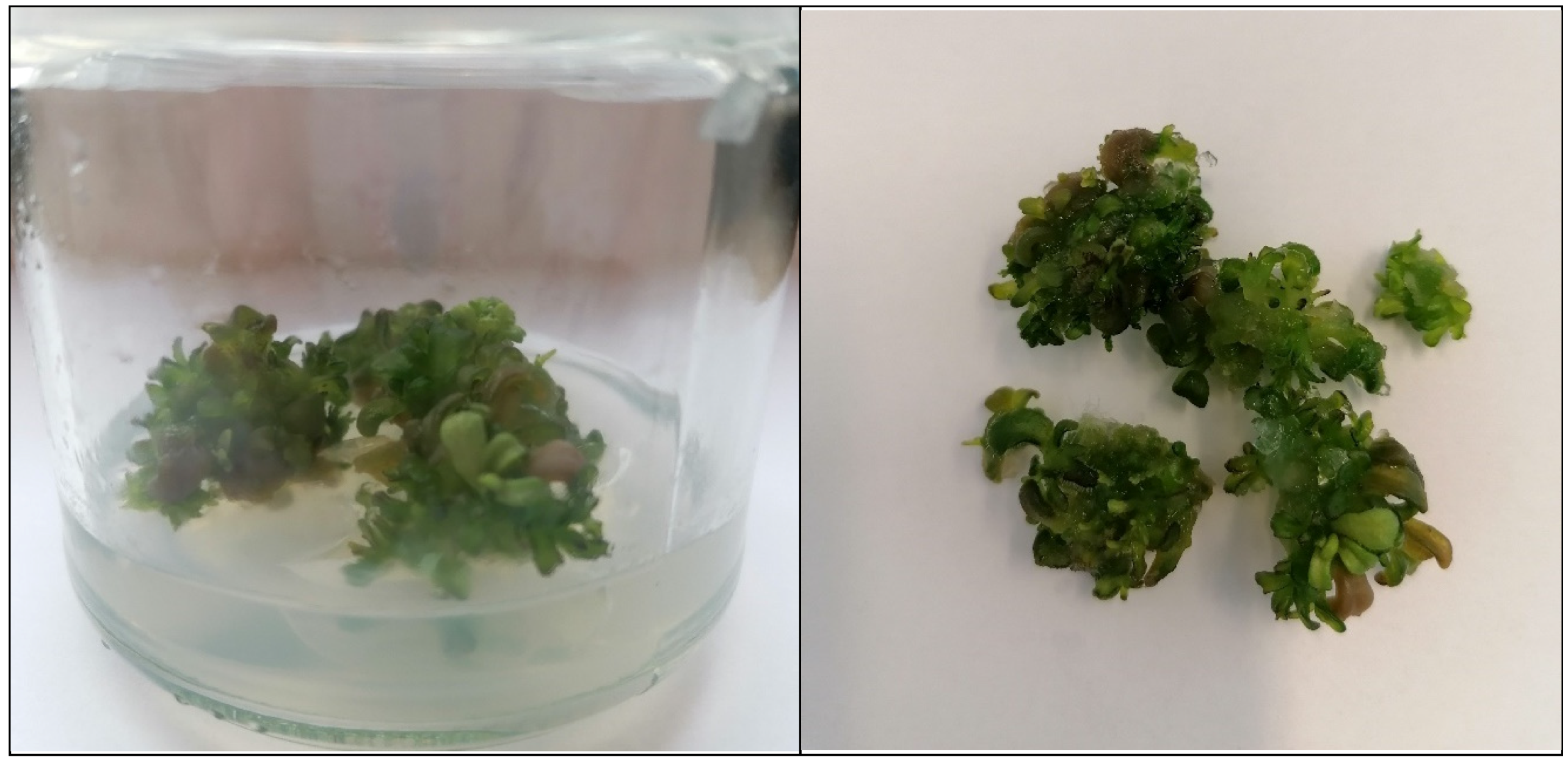
Germination of B. laevigata seeds occurred approximately 7 days after surface sterilization. The cultures were maintained on MS medium supplemented with 1 mg/L BA and 1 mg/L NAA for initiation and subsequent growth. After 20 days, the cultures were transferred to fresh medium. At this stage, they exhibited vigorous growth, numerous microshoots, and a dark green coloration. Biomass was collected after 20 days, and the microshoots demonstrated a dense, healthy appearance (Figure 2).

3.2. GSLs Profiling and Individual Contents

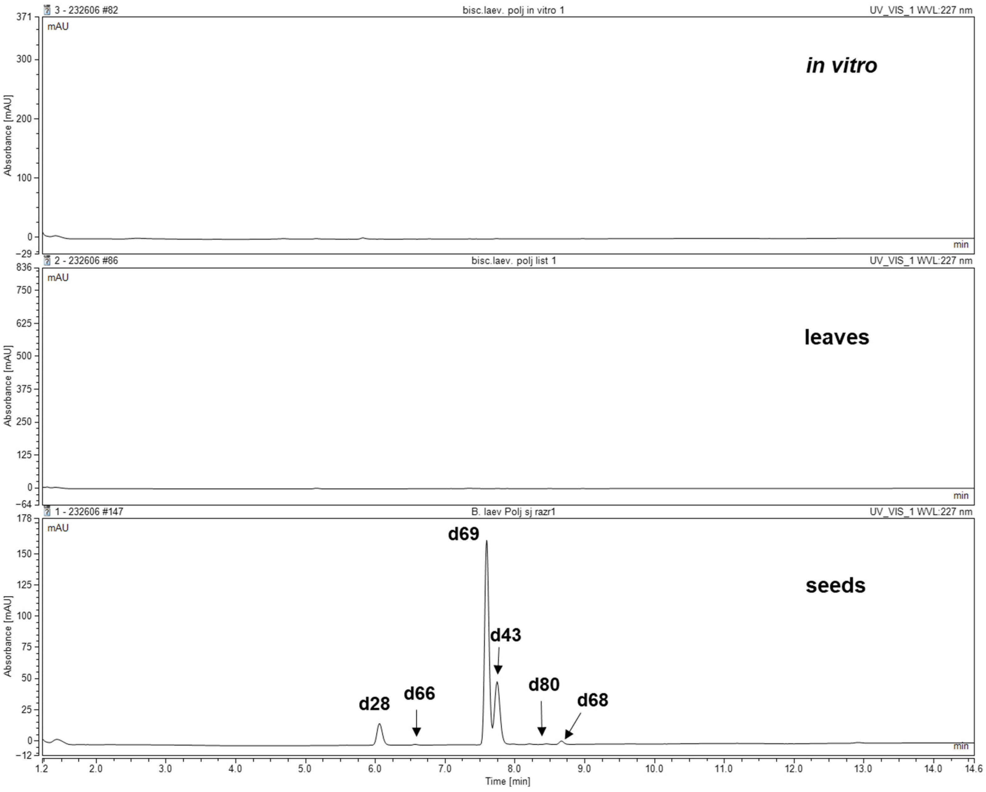
Qualitative and quantitative analysis using UHPLC-DAD-MS/MS showed that glucosinolates (GSLs) were present only in seed extracts of B. laevigata. In leaf and microshoot culture extracts, the presence of GSLs was not detected (Figure 3).
In the seed extracts, the presence of four methionine-derived GSLs was confirmed: 7-(methylsulfinyl)heptyl GSL (66), 8-(methylsulfinyl)octyl GSL (glucohirsutin) (69), 8-(methylsulfonyl)octyl GSL (80), and 9-(methylsulfinyl)nonyl GSL (glucoarabin) (68); as well as two tryptophan-derived GSLs: 4-hydroxyindol-3-ylmethyl GSL (4-hydroxyglucobrassicin) (28) and indol-3-ylmethyl GSL (glucobrassicin) (43) (Table 2, Figure S1).
The total GSL content in the seed extracts ranged from 0.21 to 15.06 mg/100 g DW. The dominant compound was glucohirsutin (15.06 mg/100 g DW), while the lowest content was recorded for glucoarabin (0.21 mg/100 g DW). Trace amounts (<0.1 μmol/g DW) were detected for 7-(methylsulfinyl)heptyl GSL and 8-(methylsulfonyl)octyl GSL.

3.3. Total and Individual Polyphenol Profiling

The TPC in extracts obtained from seeds, leaves, and microshoot cultures of B. laevigata was measured using the Folin–Ciocalteu method (Table 3). Seed extracts exhibited the highest polyphenol levels (25,701.00 mg GAE/100 g DW), while leaf and microshoot culture extracts showed similar values of 16,244.00 mg GAE/100 g DW and 16,552.00 mg GAE/100 g DW, respectively.
HPLC-DAD analysis of 53 compounds in seed and leaf extracts identified four phenolic acids (caffeic, ferulic, protocatechuic, and vanillic acids) and three flavonoids (astragalin, kaempferol, and rutin). Microshoot culture extracts contained the same four phenolic acids but only two flavonoids (astragalin and kaempferol) (Table 3).
Quantitative analysis revealed that rutin was the most abundant flavonoid across all extracts, with the highest concentration found in leaf extracts (1609.21 mg/100 g DW). In leaves, phenolic acids ranged from 0.88 to 14.23 mg/100 g DW, with ferulic acid being dominant and vanillic acid the least abundant. Flavonoid levels varied from 38.18 to 1609.21 mg/100 g DW (Table 3).
Seed extracts contained phenolic acids from 5.60 to 20.85 mg/100 g DW and flavonoids from 26.96 to 943.92 mg/100 g DW. Ferulic acid was the predominant phenolic acid (20.85 mg/100 g DW), while caffeic acid was present at the lowest level (5.60 mg/100 g DW) (Table 3).
Microshoot culture extracts had the lowest polyphenol content compared to seeds and leaves. Phenolic acids ranged from 2.22 to 6.67 mg/100 g DW, with caffeic acid being the most abundant and vanillic acid the least. Flavonoid concentrations varied from 0.78 to 11.66 mg/100 g DW, with rutin as the main flavonoid and astragalin present in the lowest amount (Table 3).

3.4. Assessment of Biological Activity

3.4.1. The Antioxidant Potential Assessed by ABTS and DPPH

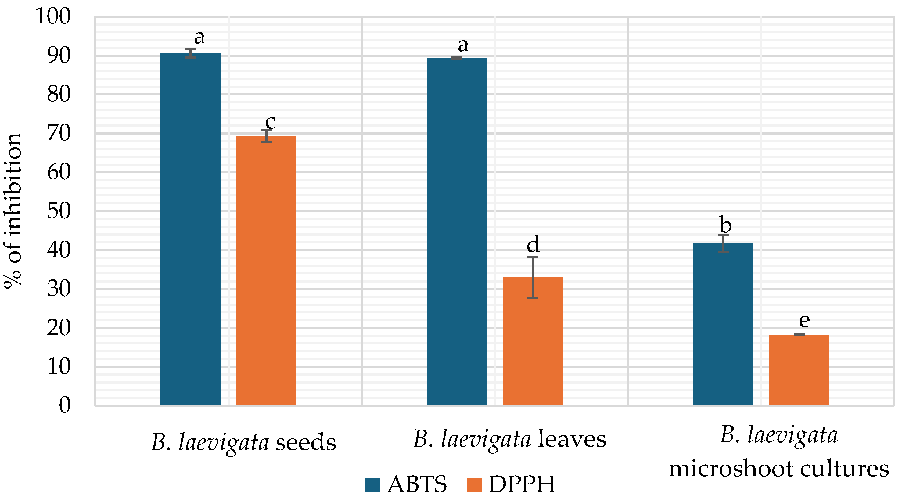
The antioxidant potential of B. laevigata extracts from seeds, leaves, and microshoot cultures was assessed using two assays, ABTS and DPPH (Figure 4). The antioxidant potential of the extracts assessed using the ABTS method ranged from 41.76% to 90.56%. The highest inhibition was observed in the seed extract (90.56%), followed closely by the leaf extract (89.33%). The lowest activity was recorded for the microshoot culture extract (41.76%) (Table 3).
In the DPPH assay, the antioxidant potential of the extracts ranged from 1.27% to 69.24% (Figure 4). The seed extract exhibited the highest inhibition, reaching 69.24%. The leaf extract showed a moderate inhibition of 33.01%, while the microshoot culture extract exhibited the lowest activity, with only 18.27% inhibition (Figure 4).

3.4.2. The Chelating Capacity of Iron Ions Fe2+

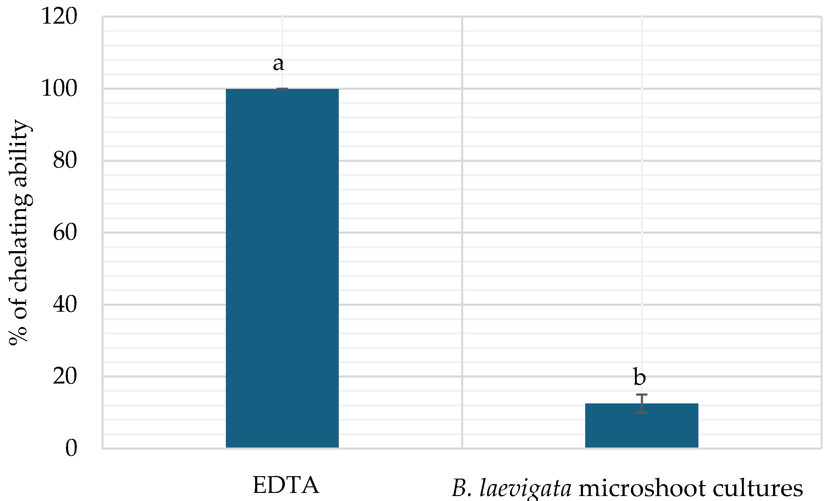
B. laevigata is a pseudometallophyte, meaning it can accumulate metals. This study evaluated the metal-chelating capacity of extracts derived from various plant parts as well as from in vitro cultures. Analyses of extracts from B. laevigata seeds, leaves, and microshoot cultures revealed that only the microshoot culture extract exhibited metal-chelating activity. The chelation capacity of the microshoot extract was 12.48% (Figure 5).

3.4.3. Elastase Inhibition Ability Assay

The study showed that seed extracts exhibited the strongest elastase-inhibiting properties (Figure 6). The percentage of inhibition for the seed extract was 12.68%, while the leaf and microshoot culture extracts showed inhibition levels of 5.17% and 5.86%, respectively. For comparison, an inhibition test was performed for a reference sample of oleanolic acid at a concentration of 200 μg/mL. The results for this compound were 53.45 ± 3.67%. This inhibitory activity is notably stronger compared to the seed extracts and significantly more potent than the leaf and petiole extracts tested in our study. It can be attributed to its standardized concentration and absence of potential antagonistic matrix effects that may interfere with enzyme binding in complex plant extracts.

3.4.4. Anti-Microbial Assay

The effects of different B. laevigata extracts were tested against Gram-positive bacteria (S. epidermidis, S. aureus, C. acnes), Gram-negative bacteria (E. coli), and the yeast C. albicans. MIC values varied depending on the type of extract and microbial strain, ranging from 625 to 5000 µg/mL (Table 4).
For Gram-positive bacteria, the lowest MIC was observed for S. epidermidis, with all extracts showing an MIC of 1250 µg/mL. For S. aureus, MICs ranged from 1250 to 2500 µg/mL, with microshoot culture extracts exhibiting the lowest value (1250 µg/mL), while seed and leaf extracts showed higher MICs (2500 µg/mL). For C. acnes ATCC 11827, all extracts had an MIC of 1250 µg/mL; however, for ATCC 6919, the leaf extract showed a higher MIC of 2500 µg/mL.
All tested extracts demonstrated an MIC of 1250 µg/mL against C. albicans. Notably, leaf and microshoot culture extracts showed the strongest activity against E. coli with an MIC of 625 µg/mL, compared to 1250 µg/mL for the seed extract (Table 4).

3.5. Study on Cosmetic Formulation

3.5.1. Physicochemical Tests

Stability Tests and pH Measurement
In the centrifuge test, no phase separation or precipitation of any components was observed. The study confirmed that both preparations were stable.
After the temperature shock test, the cream without the extract did not exhibit any changes in color or smell. In contrast, the sample containing the B. laevigata extract became darker, although its smell remained unchanged. The consistency of both preparations was affected by the test, and they became thicker.
The measured pH of the emulsions ranged from 5.01 to 5.21, falling within the recommended slightly acidic range for cosmetic formulations (pH 4–6) [28]. The emulsion containing the plant extract exhibited a slightly lower pH (5.01) than the control (5.21).
Rheological Study of Emulsions
The resulting data were analyzed to construct viscosity and flow curves, facilitating a detailed characterization of the samples’ rheological behavior. Analysis of the graphs confirmed that the preparations exhibited pseudoplastic, non-Newtonian fluid behavior. The addition of B. laevigata extract caused a decrease in both shear stress and viscosity of the face cream. The viscosity of the tested emulsions decreased with increasing shear rate. The shear stress values obtained for the same shear rates differed slightly between the samples. The flow curves of the emulsions also showed minor differences. Interestingly, the addition of the extract to the emulsion slightly increased its viscosity at rest, which positively influenced the organoleptic properties (e.g., emulsion consistency). Table 5 provides a comparative analysis of viscosity values for the formulations at specified shear rates.
Evaluation of Antioxidant Properties of Emulsions Using the DPPH Method
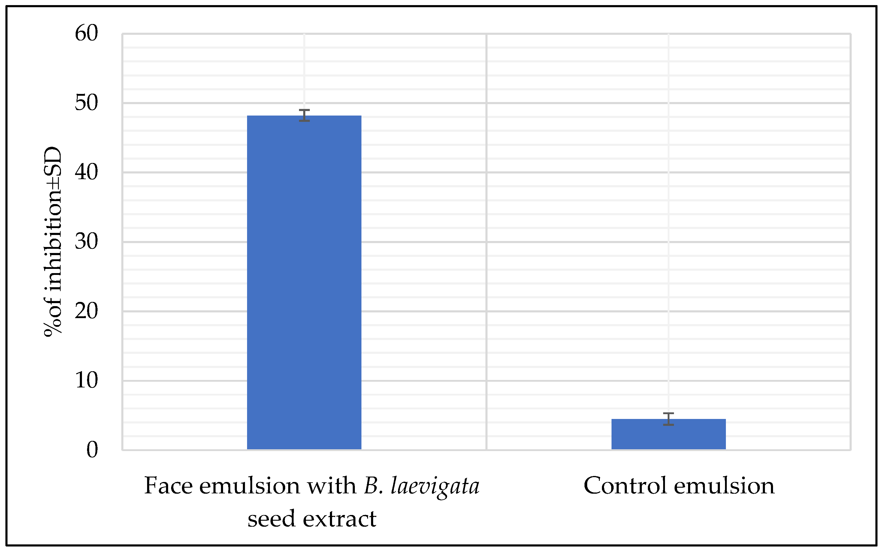
The DPPH assay showed that the cream with B. laevigata extract had a much higher inhibition of oxidation processes (48.21%) compared to the cream without the extract (4.48%) (Figure 7). The incorporation of the extract markedly augmented the antioxidant capacity of the cosmetic formulation. The findings revealed pronounced disparities in antioxidant potential between emulsions containing B. laevigata seed extract and the control formulations. Specifically, the emulsion enriched with 2% B. laevigata seed extract demonstrated a significantly elevated total antioxidant capacity, averaging 48.21%, in contrast to 4.48% recorded for the control emulsion. This strong free radical-scavenging ability is especially important for protecting the skin from reactive oxygen species (ROS), which contribute to accelerated aging. Oxidation of protein and lipid structures in the skin contributes to tissue degradation and significantly impairs the function of intercellular cement.

4. Discussion

This work constitutes the first detailed study of the phytochemical profile and biological activities of Biscutella laevigata. The production of glucosinolates (GSLs) and polyphenols was analyzed, alongside evaluations of antioxidant, anti-elastase, and antimicrobial properties. Extracts from seeds, leaves, and microshoot cultures of B. laevigata were examined. Furthermore, physicochemical and antioxidant assessments were carried out on an emulsion containing B. laevigata seed extract, which demonstrated the highest levels of active compounds and biological activity.
The initiation of in vitro cultures of B. laevigata was successfully achieved. Shoot germination from seeds occurred within 7 days. The initiation and subsequent culture medium was Murashige and Skoog (MS) medium supplemented with 1 mg/L BA and 1 mg/L NAA. Hanus-Fajerska et al. [12] also reported the successful initiation of microshoot cultures from B. laevigata seeds, although in their study MS medium without plant growth regulators (PGRs) was used for culture initiation. Both our findings and those of Hanus-Fajerska et al. demonstrate that B. laevigata seeds possess a high capacity for rapid germination in vitro and effectively develop green microshoots. The medium composition in the present study was selected based on our earlier in vitro investigations of another Brassicaceae species, Nasturtium officinale R.Br., which exhibited a high growth index (Gi), elevated glucosinolate and phenolic compound content, and strong total antioxidant potential [29]. Additionally, studies on endangered species, including Sorbus redliana, Origanum dictamnus, and Magnolia sirindhorniae, demonstrate that the establishment of in vitro cultures is critical for the ex situ conservation of threatened taxa [30,31,32]. Such cultures function as a repository of genetic resources and, under controlled conditions, enable the consistent production of valuable secondary metabolites. Studies on various species have shown that in vitro cultivation also allows the selection of lines resistant to abiotic and biotic stresses, including drought, salinity, pathogens, and heavy metals [33,34,35,36]. Similarly, in vitro cultures of B. laevigata provide opportunities for further studies, including the investigation of elicitor effects on secondary metabolite production and potential phytoremediation applications. Moreover, as B. laevigata is a pseudometallophyte, in vitro cultivation allows the growth and use of microshoots free from heavy metal contamination by enabling precise control over substrate composition.
A characteristic group of compounds in the Brassicaceae family is glucosinolates (GSLs). This is the first study to investigate GSL profiles present in seeds, leaves and microshoot cultures of B. laevigata using LC-MS/MS with available reference standards and/or MS2 spectra. The results clearly demonstrated that GSLs were present exclusively in the seeds; no GSLs were detected in the extracts from leaves or microshoot cultures (Table 2). Early studies on GSLs in B. laevigata by Cole (1976) and Daxenbichler (1991) identified 2-phenylethyl GSL, 8-(methylsulfinyl)octyl GSL (glucohirsutin), and 9-(methylsulfinyl)nonyl GSL (glucoarabin) through their breakdown products using GC-MS [37,38]. Subsequently, Bennett et al. (2003) screened intact GSLs in seeds using LC-MS (without standards), reporting aliphatic glucohirsutin as the major GSL, and glucoarabin and 7-(methylsulfinyl)heptyl GSL as minor components as well as two indolic GSLs—4-hydroxyindol-3-ylmethyl GSL (4-hydroxyglucobrassicin) and indol-3-ylmethyl GSL (glucobrassicin) [39]. For comparison, Lockwood and Belkhiri (1991) [40] analyzed B. didyma root extracts, detecting 2-phenylethyl GSL, benzyl GSL, 4-(methylsulphinyl)butyl GSL, and 3-(methylsulfanyl)propyl GSL, with the major compound being 4-(methylsulphinyl)butyl GSL (9 mg/100 g DW). These findings highlight qualitative differences in GSL profiles between B. laevigata and B. didyma. The more recent study by Metz et al. [41] on B. didyma leaf extracts identified five aliphatic GSLs—7-(methylsulfinyl)heptyl GSL, glucohirsutin, 8-(methylsulfanyl)octyl GSL, two minor unidentified aliphatic GSLs—and two indole GSLs, namely indol-3-ylmethyl GSL and 4-methoxyindol-3-ylmethyl GSL. Moreover, their study suggested that GSL content can vary with climatic conditions, such as dry versus humid environments.
The absence of detectable GSLs in leaves and in vitro cultures was unexpected, as Brassicaceae are generally characterized by GSL accumulation in vegetative tissues. This finding suggests the need for further investigation of GSL biosynthesis in different plant parts of B. laevigata as well as under in vitro conditions. Czerniawski et al. [42] analyzed the composition of GSLs using LC/MS in leaves, siliques of mature plants, roots of mature plants, and inflorescences of mature plants of selected species representing Capsella, Camelina, and Neslia genera belonging to the Brassicaceae family. The studies showed the presence of GSLs in all organs except leaves. Most of the analyzed GSLs belonged to the aliphatic and indole GSL groups. Czerniawski et al. [42] suggest that glucosinolates preferentially accumulate in siliques and roots rather than in leaves due to evolutionary shifts in their defensive role, ensuring protection of reproductive and survival organs while limiting their presence in short-lived leaf tissues. Additionally, Zhang et al. [43] confirmed that glucosinolate levels in different parts of Isatis indigotica were highest in reproductive organs, particularly in seeds, whereas mature vegetative organs contained the lowest levels. The dynamic changes observed during seed germination, with an initial decline in total glucosinolates followed by a partial increase at later developmental stages, further support the tight regulation of glucosinolate metabolism across plant growth phases. Indole-type glucosinolates exhibited a distinct accumulation pattern, rapidly increasing after germination and fluctuating throughout seedling development, suggesting their potential role in early defense. Moreover, elicitor treatments, including mechanical damage and MeJA, markedly enhanced glucosinolate accumulation in vegetative organs, demonstrating the responsiveness of this metabolic pathway to stress signals.
Notably, in vitro cultures provide controlled humidity, temperature, and light conditions, which could support the production of bioactive compounds with more consistent profiles. However, the observed interspecies differences in GSL composition within the Biscutella genus underscore the need for further investigations into their biosynthetic pathways and environmental influences.
For the first time, the total polyphenol content (TPC) of different parts of Biscutella laevigata was comprehensively compared. Our study revealed that seed extracts exhibited the highest polyphenol content, while extracts from leaves and microshoot cultures contained approximately 1.5 times lower levels.
In comparison, TPC analysis of Eruca sativa Mill. leaf extracts showed a content of 8.13 mg GAE/100 g DW, and Brassica oleracea var. italica (broccolo nero) extracts had 8.32 mg/100 g DW—both values notably lower than those observed in B. laevigata seed extracts [44]. Other studies reported that total polyphenol content in aerial parts of Lepidium sativum L. was 10.96 mg GAE/g DW, approximately 6.2 times lower than in B. laevigata leaf and microshoot extracts [45]. By contrast, Arena et al. [46] reported TPC values of 64.06 mg GAE/g in extracts from Sinapis pubescens subsp. pubescens leaves, which are comparable to the levels detected in B. laevigata leaf and microshoot extracts.
To date, there are no published qualitative or quantitative studies on the phenolic acid and flavonoid content in B. laevigata extracts. However, one study on Biscutella raphanifolia reported the presence of four flavonoids: quercetin-3-O-β-D-glucoside, quercetin-3-O-[β-D-glucosyl(1→2)-O-β-D-glucoside], and kaempferol-3-O-[β-D-glucosyl(1→2)-[(6‴p-coumaroyl)-β-D-glucoside] [9]. These findings suggest that species within the Biscutella genus may exhibit distinct and diverse phytochemical profiles, warranting further comparative analyses.
The antioxidant activity of plant extracts can vary considerably depending on the solvent used, plant organ, and cultivation conditions. In our study, the percentage of oxidation inhibition in B. laevigata extracts ranged from 14.07 to 90.17%, with leaf extracts exhibiting particularly high activity (89.33%). Boudouda et al. [9] reported inhibition values ranging from 4.03 to 80.59% in extracts of the aerial part of B. raphanipholia, highlighting the influence of extraction methods and plant material on antioxidant potential. Arena et al. [46] demonstrated that different parts of Sinapis pubescens subsp. pubescens exhibit varying antioxidant activity, with leaves showing the highest activity using the DPPH method. Similarly, Bose et al. [47] investigated Malaxis acuminata, comparing plant material and in vitro cultures, and confirmed high antioxidant activity in both, although extracts from parent plants consistently showed higher activity than those from in vitro cultures. Notably, our previous studies on microshoot cultures of N. officinale also demonstrated antioxidant activity that could be enhanced through the application of elicitors, precursors, and optimized in vitro cultivation strategies [14,29,48,49]. These findings collectively indicate that in vitro cultures of B. laevigata and other species provide a versatile platform not only for investigating antioxidant properties, but also for stimulating bioactive compound production and exploring potential applications in pharmaceuticals, nutraceuticals, and functional foods.
In our studies, the ability to chelate metals was demonstrated only by microshoot cultures of B. laevigata and was about 12.48%. Studies have shown that species of the genus Brassicaceae have the ability to chelate metals. Arena et al. [46] in their studies showed that hydroalcoholic extracts from flowers, leaves and stems have the ability to chelate metals, depending on the type of plant part used. The highest ability was demonstrated for flower extracts. Bose et al. [47] demonstrated that extracts from in vitro cultures of M. acuminata exhibited a high metal-chelating capacity of 83.8%. These results were higher than those obtained for microshoot cultures of B. leavigata, but in both studies higher metal chelating capacities of extracts from in vitro cultures were observed. Abbasi et al. [50] also demonstrated the metal-chelating ability of extracts from callus cultures of Isodon rugosus.
Our studies showed that seed extracts exhibited elastase-inhibitory properties. Bose et al. [47] reported that extracts from the leaves, stems, and in vitro cultures of Malaxis acuminata demonstrated potent elastase-inhibitory activity across all tested extracts. Consistent with these findings, the present study indicated that extracts obtained from in vitro cultures displayed reduced inhibitory effects compared to those from parent plant material. These results support previous suggestions that species within the Brassicaceae family may hold potential for application in anti-aging cosmetic formulations. Szewczyk et al. [51] investigated the elastase-inhibitory properties of Eutrema japonicum (Miq.) Koidz., reporting the highest activity in root extracts (90.18%). Furthermore, microshoot cultures of Nasturtium officinale (Brassicaceae) grown in PantForm bioreactors also exhibited high elastase-inhibitory capacity (79.78%) [52].
Extracts from B. laevigata seeds, leaves, and microshoot cultures demonstrated antibacterial activity against Cutibacterium acnes, Staphylococcus aureus, and Staphylococcus epidermidis, as well as antifungal activity against Candida albicans. In our study, B. laevigata extracts inhibited the growth of C. acnes, consistent with previous findings for Eutrema japonicum root extracts [51]. Notably, B. laevigata extracts also suppressed S. aureus and S. epidermidis, effects not reported for E. japonicum. In vitro cultures of other Brassicaceae species, such as N. officinale, similarly exhibited inhibitory activity against C. acnes [48,52]. Additionally, both B. laevigata and N. officinale extracts were effective against E. coli and C. albicans, although MIC values for B. laevigata extracts were generally lower compared to N. officinale microshoots and herb [14]. Comparable antimicrobial activity has been reported for in vitro cultures of Eleutherococcus senticosus, Codonopsis pilosula, Platanthera bifolia, Saposhnikovia divaricata [53], Plectranthus bourneae [54], and Capparis spinosa [55]. The above-mentioned studies confirm the potential use of in vitro cultures in the inhibition of skin pathogens.
Although there is a clear difference between the MIC values obtained for the tested compounds (625–2500 µg/mL) and classic antibiotics (0.125–125 µg/mL), it is worth noting that these compounds exhibit antimicrobial activity. Considering their potential low toxicity, multidirectional mechanism of action, and possibility of synergy with conventional drugs, they are worth closer examination as sources of potential antimicrobial agents. High MIC values are typical for many natural substances and do not preclude their potential therapeutic usefulness [56].
The obtained results concerning the evaluation of the compounds’ content, as well as potential biological activity (antioxidant, anti-elastase and antimicrobial properties) contributed to the development of a face emulsion with the addition of B. leavigata seed extract. This extract was selected due to its high content of compounds, as well as higher antioxidant potential and elastase inhibition capacity compared to other tested extracts.
The study included analysis of technical parameters of emulsions, such as stability, rheological properties, and pH of the formulation. A base emulsion without extract was used as a control. The prepared emulsions demonstrated stability and exhibited pH values within the optimal range for skin compatibility (5.01 to 5.21). The appropriate pH value of cosmetic preparations is very important for maintaining the proper condition of the skin [57].
The viscosity profiles of the emulsions exhibited minimal variation between formulations, indicating consistent rheological behavior. Minor differences in shear stress and viscosity were observed among the samples. The control emulsion obtained higher values, but the changes were not small. It can be stated that the rheological profiles of the samples were similar. These parameters affect the texture, consistency and sensory properties of the emulsion [58,59]. The results indicated that the incorporation of the extract did not adversely impact the stability of these parameters.
The facial emulsion enriched with B. laevigata seed extract exhibited an approximately 10.8-fold increase in antioxidant activity compared to the control formulation (Figure 5). These results confirm that the seed extract effectively enhances the antioxidant properties of skincare products, supporting its potential use in formulations aimed at skin protection, anti-aging, and rejuvenation. Furthermore, the findings indicate that the extract may help protect the skin against environmental stressors.

5. Conclusions

This work provides the first detailed phytochemical and biological evaluation of Biscutella laevigata extracts derived from seeds, leaves, and in vitro microshoot cultures. Seed extracts were found to contain the highest levels of bioactive compounds, including glucosinolates—particularly glucohirsutin—and polyphenols, and demonstrated the strongest antioxidant, elastase-inhibitory, and antimicrobial activities. Rutin and ferulic acid were identified as key constituents.
Microshoot cultures, although lacking glucosinolates, accumulated polyphenols and demonstrated Fe2+-chelating and antimicrobial properties, supporting their potential as a sustainable and controlled source of active compounds. Incorporation of seed extracts into a cosmetic emulsion significantly enhanced its antioxidant capacity, indicating possible applications in skincare.
The results are highly encouraging and scientifically valuable. Further toxicological in vivo and formulation stability studies are needed to confirm safety and efficacy. Nonetheless, the findings provide a strong foundation for future research and product development. Upcoming work will focus on refining in vitro culture conditions and exploring scalable production strategies to support the sustainable use of B. laevigata in dermatological and cosmetic applications, while contributing to the conservation of this rare and regionally endangered species.

Supplementary Materials

The following supporting information can be downloaded at: https://www.mdpi.com/article/10.3390/app151910462/s1, Figure S1: MS2 spectra of detected desulfoglucosinolates (dGSL): d66—7-(methylsulfinyl)heptyl dGSL; d69—8-(methylsulfinyl)octyl dGSL (deulfoglucohirsutin); d80—8-(methylsulfonyl)octyl dGSL; d68—9-(methylsulfinyl)octyl dGSL (desulfoglucoarabin); d28—4-hydroxyindol-3-ylmethyl dGSL (desulfo-4-hydroxyglucobrassicin); d43—indol-3-ylmethyl dGSL (desulfoglucobrassicin).

Author Contributions

Conceptualization was carried out by M.K.-S.; methodology and formal analysis were performed by M.K.-S., A.S., M.A.M., I.B., A.Ð. and P.P.; investigation was conducted by M.K.-S., A.S., A.Ś., M.A.M., I.B., A.Ð. and P.P.; data curation was handled by M.K.-S. and M.A.M.; the original draft was prepared by M.K.-S., A.S. and M.A.M.; visualization was performed by M.K.-S. and A.S.; funding acquisition was managed by M.K.-S. and I.B.; critical review and editing of the manuscript were provided by A.S., M.A.M., I.B., A.Ð., K.W., R.P. and M.W.; the final version of the manuscript was written and formatted collaboratively by M.K.-S., A.S., M.A.M., I.B., A.Ð., K.W., R.P., P.P. and M.W. All authors contributed to the experiments, manuscript preparation, and approved the final version. All authors have read and agreed to the published version of the manuscript.

Funding

This research was also supported by grants from Jan Kochanowski University of Kielce (no. SUPB.RN.24.001 and SUPB.RN.25.055).

Institutional Review Board Statement

Not applicable.

Informed Consent Statement

Not applicable.

Data Availability Statement

The original contributions presented in this study are included in the article. Further inquiries can be directed to the corresponding author.

Acknowledgments

The scientific research equipment was funded by an EU program titled “Functional integration of the University of Split, PMF-ST, PFST, and KTFST through the development of scientific and research infrastructure,” and I.B. gratefully acknowledges this support.

Conflicts of Interest

The authors declare no conflicts of interest. The funders had no role in the design of the study; in the collection, analyses, or interpretation of data; in the writing of the manuscript, or in the decision to publish the results.

References

  1. Alloggia, F.P.; Bafumo, R.F.; Ramirez, D.A.; Maza, M.A.; Camargo, A.B. Brassicaceae Microgreens: A Novel and Promissory Source of Sustainable Bioactive Compounds. Curr. Res. Food Sci. 2023, 6, 100480. [Google Scholar] [CrossRef]
  2. Rostanski, A.; Szarek-Łukaszewska, G.; Jędrzejczyk-Korycińska, M. The Buckler Mustard (Biscutella laevigata L.)—Species Description. In Buckler Mustard (Biscutella laevigata L.) an Extraordinary Plant on Ordinary Mine Heaps Near Olkusz; Szarek-Łukaszewska, G., Ed.; W. Szafer Institute of Botany, Polish Academy of Sciences: Krakow, Poland, 2020; pp. 79–93. [Google Scholar]
  3. Bróż, E.; Przemyski, A. The Red List of Vascular Plants in the Wyżyna Małopolska Upland (S Poland). In Rare, Relict and Endangered Plants and Fungi in Poland; Mirek, Z., Nikel, A., Eds.; W. Szafer Institute of Botany, Polish Academy of Sciences: Krakow, Poland, 2009; pp. 123–136. [Google Scholar]
  4. Zarzycki, K.; Szeląg, Z. Red List of the Vascular Plants in Poland. In Red List of Plants and Fungi in Poland; Mirek, Z., Zarzycki, K., Wojewoda, W., Szeląg, Z., Eds.; W. Szafer Institute of Botany, Polish Academy of Sciences: Krakow, Poland, 2006; pp. 9–20. [Google Scholar]
  5. Kaźmierczakowa, R.; Bloch-Orłowska, J.; Celka, Z.; Cwener, A.; Dajdok, Z.; Michalska-Hejduk, D.; Pawlikowski, P.; Szczęśniak, E.; Ziarnek, K. Polish Red List of Pteridophytes and Flowering Plants; Instytut Ochrony Przyrody PAN: Krakow, Poland, 2016. [Google Scholar]
  6. Ciarkowska, K.; Hanus-Fajerska, E.; Gambuś, F.; Muszyńska, E.; Czech, T. Phytostabilization of Zn-Pb Ore Flotation Tailings with Dianthus Carthusianorum and Biscutella laevigata after Amending with Mineral Fertilizers or Sewage Sludge. J. Environ. Manag. 2017, 189, 75–83. [Google Scholar] [CrossRef] [PubMed]
  7. LaCoste, C.; Robinson, B.; Brooks, R.; Anderson, C.; Chiarucci, A.; Leblanc, M. The Phytoremediation Potential of Thallium-Contaminated Soils Using Iberis and Biscutella Species. Int. J. Phytoremediation 1999, 1, 327–338. [Google Scholar] [CrossRef]
  8. Remigio, A.; Poscic, F.; Nkrumah, P.; Edraki, M.; Spiers, K.; Brueckner, D.; van der Ent, A. Comprehensive Insights in Thallium Ecophysiology in the Hyperaccumulator Biscutella laevigata. Sci. Total Environ. 2022, 838, 155899. [Google Scholar] [CrossRef] [PubMed]
  9. Boudouda, H.B.; Zeghib, A.; Karioti, A.; Bilia, A.R.; Öztürk, M.; Aouni, M.; Kabouche, A.; Kabouche, Z. Antibacterial, Antioxidant, Anti-Cholinesterase Potential and Flavonol Glycosides of Biscutella raphanifolia (Brassicaceae). Pak. J. Pharm. Sci. 2015, 28, 153–158. [Google Scholar]
  10. Karuppusamy, S. A Review on Trends in Production of Secondary Metabolites from Higher Plants by in Vitro Tissue, Organ and Cell Cultures. J. Med. Plants Res. 2009, 3, 1222–1239. [Google Scholar]
  11. Muszyńska, E.; Hanus-Fajerska, E.; Piwowarczyk, B.; Augustynowicz, J.; Ciarkowska, K.; Czech, T. From Laboratory to Field Studies—The Assessment of Biscutella laevigata Suitability to Biological Reclamation of Areas Contaminated with Lead and Cadmium. Ecotoxicol. Environ. Saf. 2017, 142, 266–273. [Google Scholar] [CrossRef]
  12. Hanus-Fajerska, E.; Wiszniewska, A.; Muszynska, E. In Vitro Multiplication and Acclimatization of Biscutella laevigata (Brassicaceae) to Cultivation in Greenhouse Conditions. Biotechnologia 2012, 93, 97–101. [Google Scholar] [CrossRef]
  13. Murashige, T.; Skoog, F. A Revised Medium for Rapid Growth and Bioassays with Tobacco Tissue Cultures. Physiol. Plant. 1962, 15, 473–497. [Google Scholar] [CrossRef]
  14. Klimek-Szczykutowicz, M.; Dziurka, M.; Blažević, I.; Đulović, A.; Granica, S.; Korona-Glowniak, I.; Ekiert, H.; Szopa, A. Phytochemical and Biological Activity Studies on Nasturtium officinale (Watercress) Microshoot Cultures Grown in RITA® Temporary Immersion Systems. Molecules 2020, 25, 5257. [Google Scholar] [CrossRef]
  15. Grosser, K.; van Dam, N.M. A Straightforward Method for Glucosinolate Extraction and Analysis with High-Pressure Liquid Chromatography (HPLC). J. Vis. Exp. 2017, 2017, 55425. [Google Scholar] [CrossRef]
  16. Wathelet, J.P.; Iori, R.; Leoni, O.; Quinsac, O.; Palmieri, S. Guidelines for Glucosinolate Analysis in Green Tissues Used for Biofumigation. Agroindustria 2004, 3, 257–266. [Google Scholar]
  17. Brown, P.D.; Tokuhisa, J.G.; Reichelt, M.; Gershenzon, J. Variation of Glucosinolate Accumulation among Different Organs and Developmental Stages of Arabidopsis thaliana. Phytochemistry 2003, 62, 471–481. [Google Scholar] [CrossRef] [PubMed]
  18. Singleton, V.; Rossi, J. Colorimetry of Total Phenolics with Phosphomolybdic-Phosphotungstic Acid Reagents. Am. J. Enol. Vitic. 1965, 16, 144–158. [Google Scholar] [CrossRef]
  19. Sułkowska-Ziaja, K.; Maślanka, A.; Szewczyk, A.; Muszyńska, B. Physiologically Active Compounds in Four Species of Genus Phellinus. Nat. Prod. Commun. 2017, 12, 363–366. [Google Scholar] [CrossRef] [PubMed]
  20. Ellnain-Wojtaszek, M.; Zgórka, G. High-Performance Liquid Chromatography and Thin-Layer Chromatography of Phenolic Acids from Gingko biloba L. Leaves Collected within Vegetative Period. J. Liq. Chromatogr. Relat. Technol. 1999, 22, 1457–1471. [Google Scholar] [CrossRef]
  21. Re, R.; Pellegrini, N.; Proteggente, A.; Pannala, A.; Yang, M.; Rice-Evans, C. Antioxidant Activity Applying an Improved ABTS Radical Cation Decolorization Assay. Free Radic. Biol. Med. 1999, 26, 1231–1237. [Google Scholar] [CrossRef]
  22. Brand-Williams, W.; Cuvelier, M.; Berset, C. Use of a Free Radical Method to Evaluate Antioxidant Activity. LWT—Food Sci. Technol. 1995, 28, 25–30. [Google Scholar] [CrossRef]
  23. Kubica, P.; Kokotkiewicz, A.; Malinowska, M.A.; Synowiec, A.; Gniewosz, M.; Hussain, S.; Yaqoob, M.; Bonn, G.K.; Jakschitz, T.; Mahmoud, E.A.; et al. Phenylpropanoid Glycoside and Phenolic Acid Profiles and Biological Activities of Biomass Extracts from Different Types of Verbena Officinalis Microshoot Cultures and Soil-Grown Plant. Antioxidants 2022, 11, 409. [Google Scholar] [CrossRef] [PubMed]
  24. Wittenauer, J.; Sonja, M.; Sußmann, D.; Schweiggert-Weisz, U.; Carle, R. Inhibitory Effects of Polyphenols from Grape Pomace Extract on Collagenase and Elastase Activity. Fitoterapia 2015, 101, 179–187. [Google Scholar] [CrossRef] [PubMed]
  25. EUCAST. Available online: https://www.eucast.org (accessed on 22 May 2025).
  26. Agerbirk, N.; Hansen, C.C.; Olsen, C.E.; Kiefer, C.; Hauser, T.P.; Christensen, S.; Jensen, K.R.; Ørgaard, M.; Pattison, D.I.; Lange, C.B.A.; et al. Glucosinolate Profiles and Phylogeny in Barbarea Compared to Other Tribe Cardamineae (Brassicaceae) and Reseda (Resedaceae), Based on a Library of Ion Trap HPLC-MS/MS Data of Reference Desulfoglucosinolates. Phytochemistry 2021, 185, 112658. [Google Scholar] [CrossRef]
  27. Blažević, I.; Montaut, S.; Burčul, F.; Olsen, C.E.; Burow, M.; Rollin, P.; Agerbirk, N. Glucosinolate Structural Diversity, Identification, Chemical Synthesis and Metabolism in Plants. Phytochemistry 2020, 169, 112100. [Google Scholar] [CrossRef] [PubMed]
  28. Lukić, M.; Pantelić, I.; Savić, S.D. Towards Optimal pH of the Skin and Topical Formulations: From the Current State of the Art to Tailored Products. Cosmetics 2021, 8, 69. [Google Scholar] [CrossRef]
  29. Klimek-Szczykutowicz, M.; Szopa, A.; Dziurka, M.; Komsta, Ł.; Tomczyk, M.; Ekiert, H. The Influence of Nasturtium officinale R. Br. Agar and Agitated Microshoot Culture Media on Glucosinolate and Phenolic Acid Production, and Antioxidant Activity. Biomolecules 2020, 10, 1216. [Google Scholar] [CrossRef] [PubMed]
  30. Sarropoulou, V.; Maloupa, E.; Grigoriadou, K. Cretan Dittany (Origanum dictamnus L.), a Valuable Local Endemic Plant: In Vitro Regeneration Potential of Different Type of Explants for Conservation and Sustainable Exploitation. Plants 2023, 12, 182. [Google Scholar] [CrossRef]
  31. Mendler-Drienyovszki, N.; Magyar-Tábori, K. Response of Rowan Berry (Sorbus redliana) Shoot Culture to Slow Growth Storage Conditions. Plants 2023, 12, 1287. [Google Scholar] [CrossRef] [PubMed]
  32. Cui, Y.; Deng, Y.; Zheng, K.; Hu, X.; Zhu, M.; Deng, X.; Xi, R. An Efficient Micropropagation Protocol for an Endangered Ornamental Tree Species (Magnolia sirindhorniae Noot. & Chalermglin) and Assessment of Genetic Uniformity through DNA Markers. Sci. Rep. 2019, 9, 9634. [Google Scholar] [CrossRef]
  33. Park, C.H.; Sathasivam, R.; Nguyen, B.V.; Baek, S.A.; Yeo, H.J.; Park, Y.E.; Kim, H.H.; Kim, J.K.; Park, S.U. Metabolic Profiling of Primary Metabolites and Galantamine Biosynthesis in Wounded Lycoris radiata Callus. Plants 2020, 9, 1616. [Google Scholar] [CrossRef] [PubMed]
  34. Mishra, N.; Tripathi, M.K.; Tiwari, S.; Tripathi, N.; Sapre, S.; Ahuja, A.; Tiwari, S. Cell Suspension Culture and in Vitro Screening for Drought Tolerance in Soybean Using Poly-Ethylene Glycol. Plants 2021, 10, 517. [Google Scholar] [CrossRef]
  35. Dogan, M. Effect of Salt Stress on in Vitro Organogenesis from Nodal Explant of Limnophila aromatica (Lamk.) Merr. and Bacopa monnieri (L.) Wettst. and Their Physio-Morphological and Biochemical Responses. Physiol. Mol. Biol. Plants 2020, 26, 803–816. [Google Scholar] [CrossRef]
  36. Marzilli, M.; Di Santo, P.; Palumbo, G.; Maiuro, L.; Paura, B.; Tognetti, R.; Cocozza, C. Cd and Cu Accumulation, Translocation and Tolerance in Populus alba Clone (Villafranca) in Autotrophic in Vitro Screening. Environ. Sci. Pollut. Res. 2018, 25, 10058–10068. [Google Scholar] [CrossRef]
  37. Cole, R. Isothiocyanates, Nitriles and Thiocyanates as Products of Autolysis of Glucosinolates in Cruciferae. Phytochemistry 1976, 15, 759–762. [Google Scholar] [CrossRef]
  38. Daxenbichler, M.E.; Spencer, G.F.; Carlson, D.G.; Rose, G.B.; Brinker, A.M.; Powell, R.G. Glucosinolate Composition of Seeds from 297 Species of Wild Plants. Phytochemistry 1991, 30, 2623–2638. [Google Scholar] [CrossRef]
  39. Bennett, R.N.; Mellon, F.A.; Foidl, N.; Pratt, J.H.; Dupont, M.S.; Perkins, L.; Kroon, P.A. Profiling Glucosinolates and Phenolics in Vegetative and Reproductive Tissues of the Multi-Purpose Trees Moringa oleifera L. (Horseradish Tree) and Moringa stenopetala L. J. Agric. Food Chem. 2003, 51, 3546–3553. [Google Scholar] [CrossRef]
  40. Lockwood, G.B.; Belkhiri, A. Glucosinolate Spectrum of Some Algerian Cruciferae. Plant Syst. Evol. 1991, 176, 11–20. [Google Scholar] [CrossRef]
  41. Metz, J.; Ribbers, K.; Tielborger, K.; Muller, C. Long- and Medium-Term Effects of Aridity on the Chemical Defence of a Widespread Brassicaceae in the Mediterranean. Environ. Exp. Bot. 2014, 105, 39–45. [Google Scholar] [CrossRef]
  42. Czerniawski, P.; Piasecka, A.; Bednarek, P. Evolutionary Changes in the Glucosinolate Biosynthetic Capacity in Species Representing Capsella, Camelina and Neslia genera. Phytochemistry 2021, 181, 112571. [Google Scholar] [CrossRef] [PubMed]
  43. Zhang, T.; Liu, R.; Zheng, J.; Wang, Z.; Gao, T.; Qin, M.; Hu, X.; Wang, Y.; Yang, S.; Li, T. Insights into Glucosinolate Accumulation and Metabolic Pathways in Isatis indigotica Fort. BMC Plant Biol. 2022, 22, 78. [Google Scholar] [CrossRef]
  44. Arena, D.; Ben Ammar, H.; Major, N.; Kovačević, T.K.; Goreta Ban, S.; Treccarichi, S.; Lo Scalzo, R.; Branca, F. Light Use Efficiency of Broccoli (Brassica oleracea var. italica Plenck) and Rocket (Eruca sativa L.) during the Initial Plant Growth Stages. Sci. Hortic. 2024, 336, 113408. [Google Scholar] [CrossRef]
  45. Al-Ogaili, N. The Evaluation of Total Flavonoids, Total Phenolic Content and Biological Activity of Iraqi Lipedium sativum L. Crude Extract Obtained by Optimized Ultrasound Assisted Extraction Conditions. Plant Sci. Today 2024, 11, 214–220. [Google Scholar] [CrossRef]
  46. Arena, P.; Miceli, N.; Marino, A.; Davì, F.; Cavò, E.; Spadaro, V.; Raimondo, F.M.; Cacciola, F.; Laganà Vinci, R.; Mondello, L.; et al. Comparative Study on Phenolic Profile and Biological Activities of the Aerial Parts of Sinapis pubescens L. Subsp. Pubescens (Brassicaceae) Wild from Sicily (Italy). Chem. Biodivers. 2023, 20, e202300309. [Google Scholar] [CrossRef]
  47. Bose, B.; Choudhury, H.; Tandon, P.; Kumaria, S. Studies on Secondary Metabolite Profiling, Anti-Inflammatory Potential, in Vitro Photoprotective and Skin-Aging Related Enzyme Inhibitory Activities of Malaxis acuminata, a Threatened Orchid of Nutraceutical Importance. J. Photochem. Photobiol. B Biol. 2017, 173, 686–695. [Google Scholar] [CrossRef]
  48. Klimek-Szczykutowicz, M.; Dziurka, M.; Blažević, I.; Đulović, A.; Miazga-Karska, M.; Klimek, K.; Ekiert, H.; Szopa, A. Precursor-Boosted Production of Metabolites in Nasturtium officinale Microshoots Grown in Plantform Bioreactors, and Antioxidant and Antimicrobial Activities of Biomass Extracts. Molecules 2021, 26, 4660. [Google Scholar] [CrossRef] [PubMed]
  49. Klimek-Szczykutowicz, M.; Dziurka, M.; Blažević, I.; Đulović, A.; Apola, A.; Ekiert, H.; Szopa, A. Impacts of Elicitors on Metabolite Production and on Antioxidant Potential and Tyrosinase Inhibition in Watercress Microshoot Cultures. Appl. Microbiol. Biotechnol. 2022, 106, 619–633. [Google Scholar] [CrossRef]
  50. Abbasi, B.H.; Siddiquah, A.; Tungmunnithum, D.; Bose, S.; Younas, M.; Garros, L.; Drouet, S.; Giglioli-Guivarc’h, N.; Hano, C. Isodon rugosus (Wall. Ex Benth.) Codd in Vitro Cultures: Establishment, Phytochemical Characterization and In Vitro Antioxidant and Anti-Aging Activities. Int. J. Mol. Sci. 2019, 20, 452. [Google Scholar] [CrossRef]
  51. Szewczyk, K.; Pietrzak, W.; Klimek, K.; Miazga-Karska, M.; Firlej, A.; Flisiński, M.; Grzywa-Celińska, A. Flavonoid and Phenolic Acids Content and in Vitro Study of the Potential Anti-Aging Properties of Eutrema japonicum (Miq.) Koidz Cultivated in Wasabi Farm Poland. Int. J. Mol. Sci. 2021, 22, 6219. [Google Scholar] [CrossRef]
  52. Klimek-Szczykutowicz, M.; Malinowska, M.A.; Gałka, A.; Blažević, I.; Ðulović, A.; Paprocka, P.; Wrzosek, M.; Szopa, A. Nasturtium officinale Microshoot Culture Multiplied in PlantForm Bioreactor—Phytochemical Profiling and Biological Activity. Molecules 2025, 30, 936. [Google Scholar] [CrossRef]
  53. Milentyeva, I.S.; Le, V.M.; Kozlova, O.V.; Velichkovich, N.S.; Fedorova, A.M.; Loseva, A.I.; Yustratov, V.P. Secondary Metabolites in in Vitro Cultures of Siberian Medicinal Plants: Content, Antioxidant Properties, and Antimicrobial Characteristics. Foods Raw Mater. 2021, 9, 153–163. [Google Scholar] [CrossRef]
  54. Rajaram, K.; Sriramji, P.; Sureshkumar, P. Micropropagation, Antioxidant and Antimicrobial Effect of Plectranthus bourneae Gamble: An Endangered Medicinal Plant. J. Chem. Pharm. Sci. 2015, 8, 142–147. [Google Scholar]
  55. Shahrour, W.G.; Shatnawi, M.A.; Al-Alawi, M.; Shibli, R.A.; Alqudah, T.S.; Majdalawi, M.M.; Al-Tawaha, A.R.; Aljammal, A. In Vitro Multiplication, Antimicrobial, and Insecticidal Activity of Capparis spinosa L. Not. Bot. Horti Agrobot. Cluj-Napoca 2024, 52, 13609. [Google Scholar] [CrossRef]
  56. Jo, D.M.; Tabassum, N.; Oh, D.K.; Ko, S.C.; Kim, K.W.; Yang, D.; Kim, J.Y.; Oh, G.W.; Choi, G.; Lee, D.S.; et al. Green Medicine: Advancing Antimicrobial Solutions with Diverse Terrestrial and Marine Plant-Derived Compounds. Processes 2024, 12, 2316. [Google Scholar] [CrossRef]
  57. Akaza, N.; Takasaki, K.; Matsudaira, T.; Usui, A.; Iijima, A.; Miura, S.; Yashiro, Y. Relationship between Skin Fungal and Bacterial Microbiomes and Skin PH. Int. J. Cosmet. Sci. 2023, 45, 362–372. [Google Scholar] [CrossRef] [PubMed]
  58. Ordoñez-Toro, A.; Montero-Vilchez, T.; Muñoz-Baeza, J.; Sanabria-De-la-Torre, R.; Buendia-Eisman, A.; Arias-Santiago, S. The Assessment of Skin Homeostasis Changes after Using Different Types of Excipients in Healthy Individuals. Int. J. Environ. Res. Public Health 2022, 19, 16678. [Google Scholar] [CrossRef] [PubMed]
  59. Pinto, D.; Lameirão, F.; Delerue-Matos, C.; Rodrigues, F.; Costa, P. Characterization and Stability of a Formulation Containing Antioxidants-Enriched Castanea sativa Shells Extract. Cosmetics 2021, 8, 49. [Google Scholar] [CrossRef]
Figure 1. Morphological appearance of Biscutella laevigata grown in Botanical Garden in Kielce (Poland): (A) herb, (B) leaves, (C) flat-sided fruits (siliques) with two flattened seeds.
Applsci 15 10462 g001
Figure 2. Microshoot cultures of B. laevigata (20 days of growth period) cultivated on MS medium with 1 mg/L BA and 1 mg/L NAA.
Applsci 15 10462 g002
Figure 3. Chromatogram of desulfoglucosinolates (dGSL): d66—7-(methylsulfinyl)heptyl dGSL; d69—8-(methylsulfinyl)octyl dGSL (deulfoglucohirsutin); d80—8-(methylsulfonyl)octyl dGSL; d68 9-(methylsulfinyl)octyl dGSL (desulfoglucoarabin); d28—4-hydroxyindol-3-ylmethyl dGSL (desulfo-4-hydroxyglucobrassicin); d43—indol-3-ylmethyl dGSL (desulfoglucobrassicin).
Applsci 15 10462 g003
Figure 4. Antioxidant potential of B. laevigata extracts from seeds, leaves, and microshoot cultures, determined by ABTS and DPPH assays (% inhibition ± SD); n = 3; different letters indicate significant differences between the tested extracts; α = 0.05.
Applsci 15 10462 g004
Figure 5. Chelating capacity of EDTA and extract from B. laevigata microshoot cultures; n = 3; different letters indicate significant differences between the tested extracts; α = 0.05.
Applsci 15 10462 g005
Figure 6. Elastase inhibitory activity of B. laevigata extracts from seeds, leaves, and microshoot cultures (% inhibition ± SD); n = 3; different letters indicate significant differences between the tested extracts; α = 0.05.
Applsci 15 10462 g006
Figure 7. Antioxidant potential, DPPH assay (% inhibition ± SD) of tested emulsions.
Applsci 15 10462 g007
Table 1. Composition and quantitative proportions of ingredients utilized in the preparation of the experimental facial emulsions.
PhaseControl EmulsionFace Emulsion with B. laevigata Seed Extract
IngredientConcentration %IngredientConcentration %
Oil phasePersea gratissima
(Avocado) Oil
7.5Persea gratissima
(Avocado) Oil
7.5
Cocos nucifera Oil7.5Cocos nucifera Oil7.5
Cetearyl Olivate,
Sorbitan Olivate
5.0Cetearyl Olivate,
Sorbitan Olivate
5.0
Butyrospermum parkii Butter3.0Butyrospermum parkii Butter3.0
Cera Flava2.0Cera Flava2.0
Cetyl Alcohol1.0Cetyl Alcohol1.0
Retinyl Palmitate0.5Retinyl Palmitate0.5
Tocopherol Acetate0.5Tocopherol Acetate0.5
Water phaseAqua68.0Aqua66.0
Biscutella laevigata Seed Extract-Biscutella laevigata Seed Extract2.0
Sodium Hyaluronate1.0Sodium Hyaluronate1.0
D-panthenol1.0Panthenol1.0
Niacinamide0.5Niacinamide0.5
Sodium Benzoate0.5Sodium Benzoate0.5
Table 2. The presence and concentrations (mg/100 g DW ± SD) of glucosinolate compounds in Biscutella laevigata seed extracts were determined using UHPLC-DAD-MS/MS analysis.
No.Glucosinolate (GSL)
(Trivial Name)
tR
(min)
[M + Na]+Glucosinolate Content
(mg/100 g DW)
Methionine-derived
667-(Methylsulfinyl)heptyl GSL a6.58422tr
698-(Methylsulfinyl)octyl GSL (Glucohirsutin) a,b7.5943615.06 ± 1.99
808-(Methylsulfonyl)octyl GSL a7.99452tr
689-(Methylsulfinyl)octyl GSL (Glucoarabin) a,b8.454500.21 ± 0.01
Tryptophan-derived
284-Hydroxyindol-3-ylmethyl GSL (4-Hydroxyglucobrassicin) a,b6.094070.48 ± 0.09
43Indol-3-ylmethyl GSL (Glucobrassicin) a,b7.753911.08 ± 0.09
a MS2 spectra compared to available literature [26]; b comparison with used standard; No.—numbers are related to the glucosinolate number given in review paper by Blažević et al. [27]. [M + Na]+, sodium adduct of desulfoglucosinolate; DW, dry weight; tr < 0.1 μmol/g DW. Data are expressed as the mean value ± standard error (n = 3).
Table 3. Total polyphenol content (TPC) and concentrations of individual phenolic compounds in B. laevigata extracts from seeds, leaves, and microshoot cultures (mg/100 g DW ± SD).
PolyphenolsB. laevigata
SeedsLeavesMicroshoot Cultures
TPC25,701.00 ± 41.23 a16,244.00 ± 72.09 b16,552.00 ± 110.36 b
Phenolic acidCaffeic acid5.60 ± 0.37 c5.35 ± 0.46 c6.67 ± 0.35 d
Ferulic acid20.85 ± 0.55 e14.23 ± 0.25 f5.78 ± 0.25 g
Protocatechuic acid6.68 ± 0.18 h7.18 ± 0.16 i5.28 ± 0.12 j
Vanillic acid7.79 ± 0.23 k0.88 ± 0.01 l2.22 ± 0.20 m
FlavonoidAstragalin (kaempferol-3-glucoside)26.96 ± 2.30 n106.29 ± 3.70 o0.78 ± 0.02 p
Kaempferol45.31 ± 2.60 r38.18 ± 3.89 rnd *
Rutin (quercetin 3-rutinoside)943.92 ± 21.97 s1609.21 ± 15.44 t11.66 ± 0.24 u
* nd—not detected, n = 3; different letters indicate significant differences between the tested extracts; α = 0.05.
Table 4. Anti-microbial activity of B. laevigata extracts from seeds, leaves and microshoot cultures.
MicroorganismExtractAntibiotic/Antifungal DrugMIC (µg/mL)MBC or MFC (µg/mL)
B. laevigata SeedsB. laevigata LeavesB. laevigata Microshoot Cultures
MIC (µg/mL)MBC or MFC (µg/mL)MIC (µg/mL)MBC or MFC (µg/mL)MIC (µg/mL)MBC or MFC (µg/mL)
BacteriaStaphylococcus epidermidis125012501250125012501250Tetracycline128256
ATCC 12228
Staphylococcus aureus250050002500500012505000Tetracycline10.5
ATCC 29213
Escherichia coli1250250062512506252500Tetracycline0.250.25
ATCC 25922
Cutibacterium acnes125025002500500012502500Tetracycline0.516
ATCC 6919
Cutibacterium acnes125025001250125012502500Tetracycline0.516
ATCC 11827
FungiCandida albicans125012501250125012501250Fluconazole10.5
ATCC 14053
Applsci 15 10462 i001Applsci 15 10462 i002
Table 5. Emulsion tangential stress and viscosity value.
EmulsionTangential Stress (Pa)Viscosity (Pa·s)
Face emulsion with B. laevigata seed extract100.71 ± 0.351.42 ± 0.01
Control emulsion109.57 ± 0.971.55 ± 0.02
Disclaimer/Publisher’s Note: The statements, opinions and data contained in all publications are solely those of the individual author(s) and contributor(s) and not of MDPI and/or the editor(s). MDPI and/or the editor(s) disclaim responsibility for any injury to people or property resulting from any ideas, methods, instructions or products referred to in the content.