Abstract
Sex estimation is a cornerstone of biological profiling in forensic anthropology. However, in cases involving badly decomposed, burnt, or fragmented remains, traditional assessments may be unfeasible, requiring alternative approaches, such as radiological and medical imaging. The cranium is a valuable indicator of sex, yet its dimorphic traits may be fragmented or altered, hindering interpretation. In such scenarios, radiological techniques targeting protected cranial structures—such as the paranasal sinuses—offer a promising alternative. Although these sinuses exhibit sexual dimorphism, their full potential for sex estimation remains partially underexplored. This study aimed to develop a logistic regression model for sex estimation in a contemporary Italian population based on volumetric measurements of the frontal, maxillary, and sphenoid sinuses, combined with selected cranial linear dimensions (biorbital breadth, upper facial height, and nasal spine length). CT scans from 222 individuals were analyzed. Volumetric measurements were obtained from 3D sinus models individually segmented from the CT scans, while linear cranial dimensions were measured on volume-rendered 3D skull reconstructions. Two predictive models were developed on a training subset and subsequently validated on an independent validation subset, both achieving an overall accuracy of approximately 80% in both phases. The most predictive variables were the volumes of the right frontal and maxillary sinuses, upper facial height, and nasal spine length, which showed the most significant sexual dimorphism. These findings are consistent with the literature on sexual dimorphism of paranasal sinuses and reflect the anatomical variability of structures like the sphenoid sinus. This study demonstrates that volumetric assessment of paranasal sinuses combined with selected cranial dimensions can provide more reliable sex estimation in forensic contexts. The integration of radiological imaging with statistical modelling offers a practical framework for situations where conventional skeletal analysis is compromised, reinforcing the role of advanced radiology in expanding the methodological toolkit of forensic anthropology.
1. Introduction
The technological advancements in medical imaging and the use of three-dimensional (3D) models allow for non-invasive analyses, providing support and answers to clinical, surgical, and medicolegal questions. For example, when examining human remains in forensic contexts, imaging tools aid in the interpretation and timing of traumatic injuries [1,2,3] and the detection of foreign bodies, implants, or pathological conditions that may contribute to establishing the cause of death and the personal identification of the subject [4,5]. Over time, due to their invaluable contribution, the application of radiologic tools has intensified both in day-to-day postmortem examinations, Disaster Victim Identification (DVI) operations, and clinical forensic medicine [6,7]. Common modalities include X-rays, computed tomography (CT), and magnetic resonance imaging (MRI), with CT scans increasingly enabling detailed, non-invasive, three-dimensional reconstructions of skeletal and soft tissue structures [8]. Indeed, forensic radiology has grown into a well-established specialized branch of medical imaging that applies radiological techniques to medicolegal investigations, including the creation of a biological profile of unidentified remains through virtual autopsies. Preliminary to personal identification is, in fact, the creation of a biological profile, which includes four main pillars: sex, population affinity, age at death, and stature. The biological profile enables narrowing down the pool of people potentially representing the remains, and sex estimation holds a unique role as it can immediately reduce the number of potential matches by approximately 50% [9,10]. Furthermore, sex has a significant impact on the evaluation of the other components of the biological profile [11,12,13,14], making reliable sex determination a fundamental first step in the analysis of human remains.
In well-preserved bodies, sex can often be determined by examining the external and internal genitalia. However, in cases involving decomposing, severely decomposed, burned, or dismembered remains, soft tissues are typically no longer informative, and sex estimation must rely on the underlying skeletal structures. In forensic anthropology, the pelvis, the postcranial bones, and the cranium are used to estimate sex with accuracy rates ranging from 88 to 100% [15,16,17]. While research has shown that the pelvis and long bones are the skeletal regions that offer higher accuracy [18,19], the cranium remains a valuable indicator, particularly when other skeletal elements are incomplete or damaged. Indeed, morphological sexually dimorphic features can be directly assessed and include the mastoid process, the nuchal crest, the supraorbital margin, the glabella, and the mental eminence [20,21]. In addition, linear measurements and, more recently, advanced metrics such as geometric morphometrics are widely used and investigated tools for assessing sexual dimorphism in the human crania [22,23,24,25]. However, the diagnostic value of these features and tools can be compromised by postmortem damage or hidden by soft tissues in decomposing remains that would require time-consuming “cleaning” procedures to obtain the skeletal elements on which to perform the assessments directly. Moreover, cleaning skeletal remains from decomposing corpses requires not only a significant amount of time but also specialized expertise and suitable facilities. This opens new avenues for research aimed at improving sex estimation methods in cases where traditional skeletal indicators are absent, compromised, or still covered by soft tissues to be removed. Therefore, attention has turned to more internally located and structurally persistent anatomical features that may be preserved [26,27] and that can be investigated by implementing indirect radiological analysis.
One such group of structures is the paranasal sinuses, air-filled cavities within the maxillofacial and neurocranial bones. Frontal sinuses, pneumatized chambers located in the frontal bone and anterior to the ethmoid bone, are usually bilateral—although agenesis of one or both sinuses can occur [28,29]—and separated by one main or several minor septa into additional recesses that communicate with each other. The pneumatization process of frontal sinuses starts around the first year of postnatal life and completes full growth around 18–20 years of age [30]. Sphenoid sinuses consist of two air-filled cavities encased by the body of the sphenoid bone and divided by the intersphenoidal septum. Their pneumatization begins early, sometimes visible at birth, and initially expands in a downward and backward direction around 15 to 18 months of age. The sinuses continue to grow until they reach their full size around age 12, with complete development typically occurring during puberty [31]. Maxillary sinuses, the largest of the paranasal sinuses, are located within the maxillary bones, positioned to the sides of the nasal cavities and beneath the orbits. Their formation starts as early as 10 fetal weeks and continues until 20 years of age when they reach their full configuration [32,33].
While paranasal sinuses have already been demonstrated as valuable tools for personal identification [34,35,36,37,38,39,40,41], their potential as indicators of sex remains relatively underexplored and, at the same time, debated [27]. In the past, the size and shape of paranasal sinuses were examined through planar X-rays, but the introduction of CT scans allowed for a more comprehensive assessment of these structures [42]. The general consensus is that the size of the sinuses is larger in males than in females [43,44,45] and that sexual dimorphism of these structures is influenced by geographic affinity variability [46,47]. Indeed, relationships between shape and dimensions, and sex of the individual were demonstrated, although the sex prediction power was generally low, but increased in specific population groups [47]. Linear measurements (e.g., height, width, and length) produced variable results for the discriminative power between males and females, with moderate accuracy rates between 50% and 75% [48,49,50,51]. Although paranasal sinus morphology has shown potential in sex estimation, the overall accuracy remains inferior to that achieved by methods relying on conventional osteological approaches. As a result, the diagnostic contribution of paranasal sinus metrics, particularly when integrated together or with complementary radiological parameters, requires further systematic investigation and validation. Additionally, volumes are increasingly investigated for their diagnostic potential [52,53]. At present, the role of the volume of paranasal sinuses in sex estimation has been limitedly investigated, again with variable results. Overall, in most studies, accuracy rates of paranasal sinuses as sex indicators are between 57% and 88% [27,50,54,55,56,57], with some notable exceptions [58,59] recording higher accuracy rates (92–97%). So far, volumes seem to reach higher levels of accuracy in discriminating males and females, although their potential is still to be fully explored.
This paper presents a volumetric analysis of the frontal, maxillary, and sphenoid sinuses segmented from cranial CT scans of an Italian population. A metric analysis involving three linear cranial measurements taken from the same volume-rendered CT scans was also performed in combination with the volumetric assessment. The aim was to investigate the potential of sinus volume as a predictor of sex through the development of regression equations based on all these parameters. By evaluating whether these internal cranial structures exhibit measurable sexual dimorphism, the study aims to assess their applicability in forensic radiological contexts, particularly in those cases where traditional skeletal traits are not applicable (because absent, covered by soft tissues, fragmented, or otherwise unsuitable for traditional analysis) and so where radiological imaging can offer a valid alternative contribution.
2. Materials and Methods
The study sample included paranasal sinuses CT-scans obtained from the database of the Fatebenefratelli Hospital in Milan (Italy), which comprised 700 CT-scans. To constitute the sample, the CT-scans in DICOM (Digital Imaging and Communications in Medicine) file format were anonymized and retrospectively searched according to the following inclusion criteria: age over 18 years, European population affinity, absence of craniofacial deformities, previous or recent traumatic events involving the paranasal sinuses, sinusitis, edentulism of the posterior maxillary arches, and monolateral or bilateral agenesia of at least one sinus. Finally, a total sample of 222 Italians representing European affinity, and of known sex (114 females; 108 males) and age (mean: 51.8 ± 22.1 years; range: 18 to 96 years) was collected.
The CT acquisitions were performed using a second-generation dual-source scanner, the Somatom Definition Flash (Siemens, Forchheim, Germany), with the following settings: tube voltage of 120 kV, tube current of 320 mAs, collimation of 40 × 0.6 mm, and a tube rotation time of 1 s. The images were reconstructed with a slice thickness of 1 mm, using two reconstruction filters: H21s (smooth) for soft tissues and H60 (sharp) for bone structures. The study was conducted in accordance with the Declaration of Helsinki [60] and received approval from the local ethical committee of the Comitato Etico Milano Area 1 (protocol: 7331/2019, date: 18 February 2019). The patient’s informed consent was waived due to the retrospective nature of the study and the use of anonymized CT images.
2.1. Volumetric and Linear Measurements
Semi-automatically segmentation of left and right frontal, sphenoid, and maxillary sinuses was performed using the open-source software ITK-SNAP (version 4.0.0) [61] employing a region-growing algorithm. Segmentation was performed by first determining the region of interest and setting a threshold value to define the gray-level range corresponding to the anatomical structure of interest [61]. One or more seed points were then placed within the target region, and the software automatically propagated the segmentation based on the intensity homogeneity relative to the defined threshold. Only voxels exhibiting gray-level values comparable to those at the seed points were included. Upon completion, the software generated the 3D reproductions of the segmented structures (Figure 1), which were then exported as stereolithographic (.stl) files. The 3D models were imported into the Vectra Analysis Module© software (VAM© version 7.4.6; Canfield Scientific Inc., Parsippany, NJ, USA) to calculate the volume of each sinus individually: right and left frontal sinuses (RFS and LFS), right and left maxillary sinuses (RMS and LMS), and right and left sphenoid sinuses (RSS and LSS). In addition to single sinus volumes, paired sinus volumes (e.g., RFS and LFS) and total volumes (i.e., combined FS, MS, and SS volumes) were calculated.
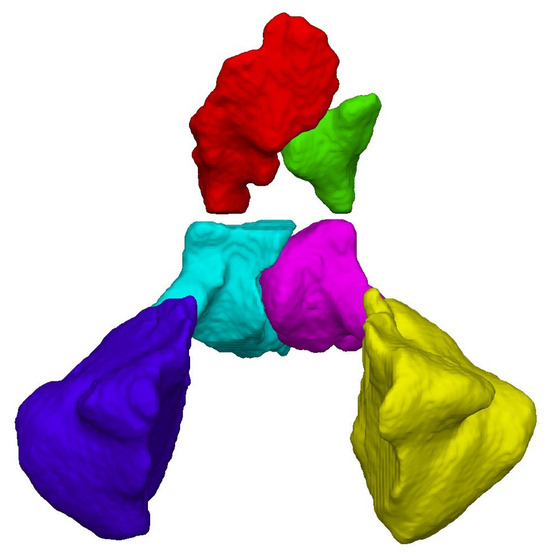
Figure 1.
Frontal view of the segmented paranasal sinuses: right frontal sinus (RFS) in red; left frontal sinus (LFS) in green; right maxillary sinus (RMS) in dark blue; left maxillary sinus (LMS) in yellow; right sphenoid sinus (RSS) in light blue; and left sphenoid sinus (LSS) in purple.
In addition to volumetric measurements, three cranial linear distances were measured as displayed in Figure 2: Biorbital Breadth (BOB; the maximum distance between the left and right ectoconchion points); Upper Facial Height (UFH; the distance between nasion and prosthion); and Nasal Spine Length (NSL; the distance between the anterior (ANS) and posterior (PNS) nasal spine. The CT scans were originally acquired as part of routine clinical screenings performed for a variety of diagnostic purposes. Consequently, the resulting images did not consistently capture the entire skull. For this reason, three specific linear measurements were selected, as the anatomical region of interest was reliably represented across all scans, regardless of cranial completeness. Moreover, these measurements were chosen for their documented or potential expression of sexual dimorphism, as well as for their direct relevance to midfacial morphology and the anatomical positioning of the paranasal sinuses. Their inclusion was intended to complement volumetric data and potentially enhance the performance of the classification models. Linear distances were computed on the 3D volume renderings of each cranium, derived from CT DICOM data imported into the open-source platform 3D Slicer (https://www.slicer.org/) [62].
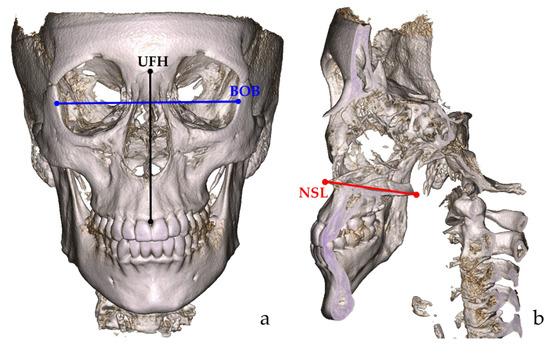
Figure 2.
Frontal view (a) and sagittal section (b) of the three cranial linear distances measured on a volume-rendered cranium obtained from a CT DICOM file imported in 3D Slicer: Biorbital breadth (BOB) in blue; Upper facial height (UFH) in black; and Nasal spine length (NSL) in red.
2.2. Statistical Analysis
The reliability of the segmentation procedure has been verified in previous studies [42] as well as for the cranial measurements [63,64], and was therefore not evaluated in the present analysis.
The normality of the data distribution and the homogeneity of variances were assessed using the Kolmogorov–Smirnov and Levene’s tests, respectively. Statistically significant differences between males and females according to age, paranasal volumes, and cranial measurements were tested through Student’s t-test for independent samples when the normality assumption was met; otherwise, non-parametric tests (i.e., Mann–Whitney U test) were applied. To account for multiple comparisons, a Bonferroni correction was implemented, adjusting the significance threshold to α = 0.004 (0.05/14). Additionally, descriptive statistics, including mean, standard deviation, minimum, and maximum, were calculated separately for males and females for each paranasal volume along with their pair and total sums, and for linear measurements as well.
Two binary logistic regression analyses were performed to develop sex classification models: the first was based on the volume of the frontal, maxillary, and sphenoid sinuses, whereas the second combined these volumes with the three cranial measurements. Binary logistic regression is a statistical approach used to model the relationship between a dichotomous dependent variable (in this case, sex) and one or more independent variables (predictors), which may be either categorical or continuous in nature. As sinus volumes and cranial dimensions are anatomically and developmentally correlated, we tested the possible existence of multicollinearity among the predictor variables through the Variance Inflation Factor (VIF), for which values greater than 10 are indicative of highly correlated variables [65].
The study sample was randomly divided into two subsets: a training set comprising 80% of the sample (178 subjects: 91 females; 87 males), and a validation set including the remaining 20% (44 subjects: 23 females; 21 males). To avoid overestimation or underestimation of the models’ performance when applied to the validation set, we evaluated the consistency of the sex ratio and age distribution between the training and validation sets. Specifically, we calculated the female-to-male ratio by dividing the number of females by the number of males, while the consistency in the age distribution was tested through a Mann–Whitney U test for the two sexes separately.
The regression models were built using the training set, with “female” defined as the reference category. Model fit was evaluated using the Hosmer–Lemeshow goodness-of-fit test, with non-significant p-values indicating an adequate model fit. The regression output included the estimated coefficient for each predictor variable, along with their corresponding odds ratios with 95% confidence intervals, and p-values indicating statistical significance. The coefficients reflect the direction and magnitude of the association between each predictor and the probability of classification as male (the non-reference category). Odds ratios greater than 1 indicate an increased likelihood of male classification with a one-unit increase in each predictor, whereas values lower than 1 suggest a decreased likelihood. The estimated coefficients were subsequently used to formulate the logistic equations for predicting the probability p of a subject being classified as male. A cut-off value of 0.5 was adopted for binary classification: probabilities below 0.5 indicate a higher likelihood of the subject being female, while probabilities over 0.5 suggest classification as a male subject. The predictive performance of the model was tested on the validation set. For both the training and validation subsets, key performance metrics were computed, including the overall accuracy (i.e., proportion of correctly classified subjects), sensitivity (i.e., proportion of correctly identified females), specificity (i.e., proportion of correctly identified males), and sex bias (i.e., the absolute difference between female and male classification rates).
All statistical tests were performed in SPSS version 29.0.1 (IBM Corp., Armonk, NY, USA), and the significance was set to α = 0.05.
3. Results
Age distribution of males deviated from normality according to the Kolmogorov–Smirnov test (p = 0.044). Consequently, age differences between sexes were evaluated using the Mann–Whitney U test, which revealed that females were significantly older than males (51.8 ± 21.2 and 44.9 ± 21.0 years, respectively; p < 0.001).
All volumetric (left and right frontal, maxillary, and sphenoid sinuses, and paired and total sums) and linear cranial measurements (biorbital breadth; UFH: upper facial height; NSL: nasal spine length) met the assumption of normality and homogeneity of variance across sex groups. Accordingly, Student’s t-test was used to assess sex-related differences. Descriptive and inferential statistics are reported in Table 1. Except for the right sphenoid sinus (RSS), all variables were significantly larger in males compared to females, even after applying the Bonferroni correction (α = 0.004).

Table 1.
Descriptive statistics of the paranasal volumes and cranial linear measurements in the two sexes and related p-values.
Two binary logistic regression models were developed to estimate the probability p of an individual being classified as either male (p > 0.5) or female (p < 0.5), and both models were trained on 80% of the sample represented by the same subjects and subsequently tested on the validation subset. The female-to-male sex ratio was consistent between the two samples, as they were 1.046:1 and 1.095:1 for the training and validation subsets, respectively. Additionally, the Mann–Whitney U test revealed no significant age differences between the validation and the training subsets for both males (p = 0.505) and females (p = 0.706).
The first model included only paranasal sinus volumes, and the second combined the volumes with the three cranial linear measurements.
The first model proved an acceptable fit to the data, as indicated by a non-significant Hosmer–Lemeshow test (p = 0.082), even though this value approached the conventional α level of 0.05. Additionally, the Akaike information criterion (AIC) was 206.01. The logistic regression estimates, odds ratios with 95% confidence intervals, the significance of the included predictors (namely, paranasal sinuses volumes), and the related variance inflation factors (VIF) that disproved multicollinearity are reported in Table 2.
Table 2.
Regression model based only on paranasal sinus volumes: logistic regression estimates, p-values, odds ratios with 95% confidence intervals, and variance inflation factors (VIF).
In this model, significant predictors positively associated with male classification were the volumes of right frontal and maxillary sinuses (RFS and RMS, respectively), and the probability (p) of sex classification was estimated using the coefficient of all predictors, according to the following equation:
where e (Euler’s number).
In the training subset, the model achieved an overall accuracy of 72.5%, with 74.7% sensitivity, 70.1% specificity, and a sex classification bias of 4.6%. Performance improved in the validation subset, with accuracy of 79.5%, and sensitivity of 73.9%, specificity of 85.7%, and sex classification bias of −11.8%. Confusion matrices for the training and validation subsets are reported in Table 3.
Table 3.
Confusion matrix for sex prediction in the training and validation samples using the regression model based on paranasal sinus volumes.
Following these results, the second model was designed to improve the classification performance of the first one. This model exhibited a better fit to the data, as indicated by a higher Hosmer–Lemeshow test p-value (p = 0.239) and a lower AIC (173.34), suggesting a more efficient and better-performing model. Additionally, the predictors proved to be non-multicollinear as the VIF values are smaller than 10. The estimates, odds ratios with 95% confidence intervals, the significance of the included predictors, and the related VIF values are reported in Table 4.
Table 4.
Regression model based on the paranasal sinus volume and cranial linear measurements: Logistic regression estimates, p-values, odds ratios with 95% confidence intervals, and variance inflation factors (VIF).
In the second model, the right frontal sinus volume (RFS), the upper facial height (UFH), and the nasal spine length (NSL) emerged as the more significant predictors of sex, all positively associated with male classification. Notably, RFS and UFH showed the strongest predictive power. The logistic regression equation estimating the probability p of an individual being classified either as male (p > 0.5) or female (p < 0.5) was developed using all coefficients and resulted in the following:
where e (Euler’s number).
The model achieved an overall accuracy of 78.7% in the training subset, with a sensitivity of 80.2%, a specificity of 77.0%, and a sex classification bias of 3.2%. By applying the model to the validation subset, the overall accuracy was 79.5%, with a sensitivity of 78.3% and a specificity of 81.0%, and a sex classification bias of −2.7%. The confusion matrices for both subsets are provided in Table 5.
Table 5.
Confusion matrix for sex prediction in the training and validation samples using the regression model based on paranasal sinus volumes and cranial linear measurements.
A preset Excel sheet for practical use by experts is provided in the Supplementary Material for calculating the estimated sex probability according to the two proposed models.
4. Discussion
Sex estimation is a fundamental component in the reconstruction of a reliable biological profile in both archeological and forensic contexts. Traditionally, it relies on the external genitalia in well-preserved cadavers, or on morphological and metric analysis of sexually dimorphic skeletal portions, such as the pelvis, long bones, and cranium. However, in real-case scenarios, human remains are often recovered in fragmentary, incomplete, or taphonomically altered conditions, frequently with varying degrees of soft tissue preservation. These challenges limit the applicability of classical methods and have prompted researchers to explore alternative anatomical structures and diagnostic methods for sex estimation.
Among these, the paranasal sinuses, already recognized for their value in personal identification, have attracted increasing interest for this purpose, due to their deep anatomical location within the skull, which offers protection against fragmentation, taphonomic alteration, environmental degradation, and thermal phenomena (e.g., incineration). Moreover, paranasal sinuses have been shown to exhibit sexual dimorphism in both their linear dimensions and volumetric features. When combined with the widespread use of advanced medical imaging modalities such as CT and CBCT, which enable accurate, non-invasive three-dimensional reconstruction, these anatomical structures emerge as promising candidates for forensic sex estimation. Accordingly, the present study aimed to develop population-specific logistic regression models to predict sex in a contemporary Italian sample of European affinity, based on a comprehensive set of measurements including the volumes of the frontal, maxillary, and sphenoid sinuses, as well as three cranial linear measurements. The measurements were selected for their documented sexual dimorphism and their relevance to midfacial morphology, which is known to differ significantly between sexes. The inclusion of linear cranial parameters relies on the assumption that combining volumetric and linear features can enhance the classification performance of predictive models, as demonstrated by previous studies, such as those incorporating foramen magnum dimensions [59].
The average measurements of both paranasal sinus volumes and cranial linear measurements are in accordance with those reported in the literature [54,56,63,64], as well as for their standard deviation, whose broad ranges reflect the high inter-individual variability of the measured structures, in part explaining the limited accuracy of our models. Indeed, the logistic regression models developed in this study achieved an overall accuracy of approximately 75% and 80% in both the training and validation subsets and, although both models showed comparable performance in terms of overall accuracy on the validation subset (79.5% for both models), the model incorporating the three cranial measurements, which resulted to be not correlated with the paranasal sinus volumes, demonstrated an overall superior performance and an unaffected predictive reliability. These findings are supported by a lower Akaike Information Criterion (AIC), a better fit to the data (higher p-value in the Hosmer–Lemeshow test), and a more balanced classification of the sexes, reducing the sex bias from −11.8% to −2.7%. Thus, the model including the cranial linear measurements proved greater generalizability and robustness.
Notably, although the strongest predictors included upper facial height and nasal spine length, the volumes of the right frontal and maxillary sinuses were also of importance in the sex probability estimation. These findings are consistent with the broader literature on the sexual dimorphism of the paranasal sinuses, which is well documented for both linear and volumetric parameters [54], with males consistently displaying greater measurements than females. In line with these findings, our study revealed significant sex-related differences in the volumes of the bilateral frontal and maxillary sinuses, as well as in the left sphenoid sinus. Interestingly, the volume of the right sphenoid sinus, although larger in males, did not reach statistical significance. This observation closely reflects the outcomes reported by Hekimoğlu et al. [54], who similarly found no significant sexual dimorphism in this specific structure. Overall, while frontal sinuses are consistently shown to exhibit sexual dimorphism in both linear and volumetric dimensions [45,55,58,59,66,67,68,69], the findings concerning the maxillary and sphenoid sinuses are more heterogeneous in the literature. Specifically, several studies report significantly larger maxillary sinuses in males [45,56,58,59,69,70,71], while others do not find significant sex-related differences [72,73,74]. As for sphenoid sinuses, the literature is particularly inconsistent: while some authors confirm the presence of sexual dimorphism [45,75,76,77], others fail to identify any significant difference between sexes [66,69,78,79]. One possible explanation for the lack of dimorphism in the right sphenoid sinus may lie in the intrinsic anatomical variability and asymmetry of this structure, which shows considerable heterogeneity in pneumatization patterns and volume across individuals [80,81]. Moreover, the developmental onset and trajectory of the sphenoid sinus are relatively late, and we can speculate that it may be less directly influenced by changes to the skeletal structures driven by sex hormones compared to other paranasal sinuses. Additionally, methodological differences, such as variations in segmentation techniques, threshold settings in volumetric analysis, and sample characteristics, may contribute to inconsistencies in detecting dimorphic features in this anatomical structure across studies.
Despite the sexual dimorphism of the volume of the paranasal sinuses being recognized, the studies that have investigated the accuracy of sex prediction combining all these structures are limited in the literature and summarized in Table 6.
Table 6.
Comparison of the performance of different sex-prediction models in diverse populations.
Through a linear discriminant function analysis (LDA) applied to a Turkish sample (50 males, 50 females), Hekimoğlu et al. [54] reported a classification accuracy of 68.0%, while Wanzeler et al. [59], using the same approach to a population of 163 Brazilians reached a much higher accuracy (94.48%) when using only the volumes of the sinuses, and a 100% accuracy when these were combined with dimensions of the foramen magnum. Other authors explored the volumes of single or two pairs of sinuses. In an Egyptian group, Ibrahim et al. [58] developed two separate LDA models based on both volumetric and linear measurements of frontal and maxillary sinuses, obtaining an accuracy of 94% and 92%, respectively. Two additional studies involved limited samples from France. Radulesco et al. [56] investigated the maxillary sinus volume in 50 females and 50 males, reporting a 68.0% accuracy. Michel et al. [55] analyzed the frontal sinus volumes in 35 females and 34 males, developing a discriminative model with an accuracy of 72.5%. However, the equations by Michel et al. [55] reached a lower accuracy (59.6%) when tested on Western Australians (47 males; 52 females) by Thottungal et al. [57]. Moreover, in the same population, Thottungal et al. [57] attempted to develop their own population-specific models with logistic regression (LR) equations based on unilateral and bilateral volumes of the frontal sinuses. The models’ accuracy, however, ranged from 57.2% to 62.5%. Similarly, other studies have preferred LR to LDA, again mostly limited to a single sinus type, and only occasionally in combination with other cranial measurements. Choi et al. [68] developed an LR model based on several parameters of the frontal sinuses (volume, occupied area, roundness, width, perimeter, circularity, aspect ratio, Feret’s diameter, and major axis of the ellipse) in a Brazilian population (65 males; 65 females). In this case, the accuracy reached was 80.0%. Concerning maxillary sinuses, while Salim et al. [83] proposed an LR model based on their height, width, and length in addition to the volume with an accuracy of 68.2% developed on a Turkish population, Wu et al. [82] developed two separate models for the left and right structures with accuracies of 74.3% and 78.6%, respectively.
Overall, regardless of the type of sinuses examined, which is one of the main sources of variation, the differences in accuracy rates reported by previous studies and by ours can depend on several factors. First among these is the presence of population-specific variations, which are also well-documented in the morphology and dimensions of the cranium [21,71,84].
Sexual dimorphism is not a strict dichotomy but a continuum. Typical or more nuanced male or female skeletal traits can coexist and exhibit a considerable overlap. We can therefore consider our models a fair compromise in case others based on different cranial parameters are unapplicable. Concerning population affinity, previous studies have investigated the Italian population, but only proposing predictive models based on cranial measurements and cranial morphological traits, with accuracy percentages up to 88.6% and 91.7%, respectively [21,84], testifying that in the case of complete crania, the traditional methods still represent the elective ones.
The approach used for building the predictor models is another important aspect to consider. We decided to implement LR as it offers several methodological advantages, especially in forensic contexts, a practice where uncertainty is inherent and where data often violate underlying assumptions [85,86]. LR can, in fact, produce more robust results when dealing with real-world biological data that frequently deviate from ideal statistics. In addition to this, LR provides outputs that can be more easily interpreted, such as odds ratios and predicted probabilities, which can facilitate the practical application of sex estimation models in practice [85]. Given the critical role of sex estimation in forensic investigations—and the significant consequences that can arise from potential misclassification—practitioners generally rely on well-established methods whose reliability has been extensively tested and validated. Methods that achieve high accuracy rates are generally regarded as more reliable; however, there is no universally accepted benchmark that definitively establishes the superiority of one method over another. As a result, certain approaches are regarded as more trustworthy than others. For example, morphological or metric traits of the innominate and long bones yield more satisfactory rates in terms of accuracy [18,87,88], and the cranium is generally considered to be less reliable than the abovementioned elements. However, reliability is often context-dependent, shaped by factors such as the reference population, which can significantly influence the accuracy of sex estimation [21,89,90], the quality and preservation of the remains, and the specific circumstances of the forensic investigation. The completeness and preservation state of human remains is inherently unpredictable due to the stochastic nature of taphonomic processes. Isolated crania are, however, a common finding in forensic investigations [21,91], hence the need to devise alternative tools that contribute to estimating sex when more dimorphic elements are unavailable. The accuracy rates produced by the combination of linear and volumetric measurements (around 80%) in this study may not be high enough to deem this method as a decisive one, in contrast to the pelvis, which is considered the first choice and line of evidence [92]. However, when the preservation state of the cranium does not allow a direct and reliable assessment of traditional traits, but portions of the maxillofacial skeleton and the sinuses are intact, the models presented here could provide an auxiliary tool for sex estimation. Consequently, the choice of method should be guided not only by reported accuracy but also by its applicability and robustness under the conditions at hand. For this reason, it remains essential to explore and assess new tools and techniques, ensuring that forensic anthropology continues to expand its methodological repertoire and maintains its effectiveness even under challenging preservation conditions.
An additional strength of logistic regression analysis is the combined approach, including the unilateral volumes of the frontal, maxillary, and sphenoid sinuses, and the craniometric measurements. This approach can provide a more general and robust prediction model for sex estimation, as confirmed by the low sex bias of the correct sex classifications obtained within the validation subset and the increase in the accuracy percentage. Moreover, the validation subset represents an additional advantage that supports the value of this study, compared to previous studies in which this aspect was not investigated.
Furthermore, other discrepancies among all the above-mentioned studies may be attributable to heterogeneous methodologies, including sample size and composition, 3D imaging typology (such as CT, CBCT, or MRI) and characteristics (resolution/slice thickness), segmentation protocols, measurements calculation, and number and typology of sinuses included [71]. All these factors can, to some extent, influence and shape the outcomes of the above-mentioned studies, thereby hindering a proper and reliable comparative evaluation. Given the continuous advancements in medical imaging and their growing application in forensic practice, the standardization of procedures and protocols has become a relevant issue. In recent years, this topic has gained increasing attention, as it directly impacts the validity of imaging-based analyses and their integration into routine forensic workflows [63,93,94]. This includes establishing consistent methods for the acquisition and segmentation of virtual anatomical models, as well as for measurements and overall data collection from imaging studies. Automatic or semi-automatic procedures to acquire volumes can help reduce the variability among operators, although further studies are needed to thoroughly evaluate potential biases. However, several factors may limit the creation of univocal procedures, including access to resources, more expensive and elaborate software, or the expertise and background of the researchers devising the methods. Nevertheless, standardization is essential to ensure meaningful cross-study comparisons, reproducibility, and reliability of results across different research groups and forensic cases; thus, it should be deeply investigated and pursued.
On a final note, although our sample was larger than those used in most of the previous studies, it presented an age imbalance between sexes, with female individuals being older on average than male individuals; on average, females were 15 years older than males. This discrepancy may have influenced cranial and sinus morphology and related metrics, potentially affecting the performance of the predictive model. Age-related cranial changes may represent an additional variable to consider when working on volumes of paranasal sinuses. For example, it was demonstrated that the stabilization of traits in frontal sinuses is highly variable, so that predicting changes linked to the age of the individual remains a difficult task [95]. Although the maxillary sinus generally reaches a stable size after the second decade of life, its volume tends to decline in adulthood, likely due to mineral loss in the bone matrix [96]. However, the literature provides conflicting findings regarding the effect of age on the volume of the different paranasal sinuses [97]. Some studies suggest a reduction in volume, others find no correlation at all, and still others even propose an increase. Given this inconsistent literature, it is difficult to draw firm conclusions on the matter. This represents an important aspect to be further investigated, namely, conducting a similar study specifically designed to assess the influence of age, and consequently of pneumatization (often associated with aging), on sinus volumes. This remains a critical issue to highlight.
In conclusion, to enhance the robustness and generalizability of the proposed logistic regression approach, future research should aim to expand the Italian sample and consider integrating additional cranial measurements with established sexual dimorphism, potentially including further parameters related to the paranasal sinuses.
5. Conclusions
Sex remains a cornerstone of the biological profile in forensic anthropology, despite the fact that the estimation may often be limited by preservation, incompleteness, or fragmentation of the remains. In such scenarios, the use of 3D radiological imaging techniques, particularly CT and CBCT, offers a valuable alternative by enabling the analysis of anatomical structures that are otherwise inaccessible.
The present study confirms that paranasal sinuses, owing to their protected intracranial location and documented sexual dimorphism, represent reliable anatomical structures for sex prediction. By combining volumetric measurements of segmented frontal, maxillary, and sphenoid sinuses with cranial linear distances measured on volume-rendered crania, and employing logistic regression for developing predictive models, we achieved a balanced and interpretable classification accuracy.
Our findings support the integration of advanced radiological imaging and multivariate modelling in forensic contexts where conventional skeletal indicators are unavailable and highlight the potential of population-specific approaches for improving predictive performance.
Future studies should validate and refine these models across larger and diverse groups, further enhancing their applicability in real-world casework.
Supplementary Materials
The following supporting information can be downloaded at: https://www.mdpi.com/article/10.3390/app151810232/s1, Table S1: Sheet to calculate sex probability according to the two proposed logistic regression models.
Author Contributions
Conceptualization, A.C.; methodology, R.S., D.G. and A.C.; software, D.G. and C.S.; validation, R.S. and A.P.; formal analysis, R.S. and A.P.; investigation, R.S., A.P., D.M. and A.C.; resources, M.C., D.G.; data curation, R.S. and A.C.; writing—original draft preparation, R.S., A.P. and A.C.; writing—review and editing, R.S., A.P., D.M., D.G., C.C. and A.C.; visualization, A.P.; supervision, C.S., C.C., D.G. and A.C.; project administration, A.C. All authors have read and agreed to the published version of the manuscript.
Funding
This research received no external funding.
Institutional Review Board Statement
The study complies with local and international ethical rules (Helsinki guidelines), and local ethical committee (Comitato Etico Milano Area 1; protocol: 7331/2019, date: 18 February 2019).
Informed Consent Statement
Patient informed consent was waived due to the retrospective nature of the study and the use of anonymized CT images.
Data Availability Statement
Data will be available upon request and acceptance from the authors.
Conflicts of Interest
The authors declare no conflicts of interest.
References
- Offiah, C.E. The Utility of Forensic Radiology in Evaluation of Soft Tissue Injury. In Current Practice in Forensic Medicine; Wiley: Hoboken, NJ, USA, 2022; pp. 143–165. [Google Scholar]
- Cappella, A.; Amadasi, A.; Gaudio, D.; Gibelli, D.; Borgonovo, S.; Di Giancamillo, M.; Cattaneo, C. The Application of Cone-Beam CT in the Aging of Bone Calluses: A New Perspective? Int. J. Leg. Med. 2013, 127, 1139–1144. [Google Scholar] [CrossRef]
- Petaros, A.; Lindblom, M.; Cunha, E. Combining Anthropology and Imaging to Reconstruct Antemortem Trauma for Identification Purposes. Forensic Sci. Res. 2024, 9, owae048. [Google Scholar] [CrossRef]
- Lemos, Y.V.; Furtado, A.N.; Lima, A.Z.; Dionísio, A.S.; Araújo, R.M.; Cunha, E. Human Identification by Medical Findings in a Forensic Anthropology Context. Forensic Sci. Res. 2024, 9, owae041. [Google Scholar] [CrossRef] [PubMed]
- Dedouit, F.; Ducloyer, M.; Elifritz, J.; Adolphi, N.L.; Yi-Li, G.W.; Decker, S.; Ford, J.; Kolev, Y.; Thali, M. The Current State of Forensic Imaging–Clinical Forensic Imaging. Int. J. Leg. Med. 2025, 139, 1639–1646. [Google Scholar] [CrossRef]
- De Boer, H.H.; Blau, S.; Delabarde, T.; Hackman, L. The Role of Forensic Anthropology in Disaster Victim Identification (DVI): Recent Developments and Future Prospects. Forensic Sci. Res. 2018, 4, 303–315. [Google Scholar] [CrossRef] [PubMed]
- Vallis, J. The Role of Radiography in Disaster Victim Identification. In Human Remains: Another Dimension; Elsevier: Amsterdam, The Netherlands, 2017; pp. 57–69. [Google Scholar]
- Khmara, N.; Baumeister, R.; Schweitzer, W.; Thali, M.; Ampanozi, G. Virtopsy Concept Around the World: Institute-Based Survey of Worldwide Forensic Postmortem Imaging. Forensic Imaging 2024, 37, 200595. [Google Scholar] [CrossRef]
- Loth, S.R.; İşcan, M.Y. ANTHROPOLOGY | Sex Determination. In Encyclopedia of Forensic Sciences; Siegel, J.A., Ed.; Elsevier: Amsterdam, The Netherlands, 2000; pp. 252–260. [Google Scholar]
- Robinson, M.S.; Bidmos, M.A. The Skull and Humerus in the Determination of Sex: Reliability of Discriminant Function Equations. Forensic Sci. Int. 2009, 186, e1–e86. [Google Scholar] [CrossRef]
- Messer, D.L.; Getz, S.M. Effect of Sex Misclassification on the Skeletal Biological Profile. In Sex Estimation of the Human Skeleton; Elsevier: Amsterdam, The Netherlands, 2020; pp. 53–72. [Google Scholar]
- Thomas, R.M.; Parks, C.L.; Richard, A.H. Accuracy Rates of Sex Estimation by Forensic Anthropologists Through Comparison with DNA Typing Results in Forensic Casework. J. Forensic Sci. 2016, 61, 1307–1310. [Google Scholar] [CrossRef]
- Krishan, K.; Chatterjee, P.M.; Kanchan, T.; Kaur, S.; Baryah, N.; Singh, R.K. A Review of Sex Estimation Techniques During Examination of Skeletal Remains in Forensic Anthropology Casework. Forensic Sci. Int. 2016, 261, e1–e165. [Google Scholar] [CrossRef]
- Franklin, D. Estimation of Skeletal Sex. In Encyclopedia of Forensic Sciences, 3rd ed.; Elsevier: Amsterdam, The Netherlands, 2023; pp. 292–303. [Google Scholar]
- Stock, M.K. Analyses of the Postcranial Skeleton for Sex Estimation. In Sex Estimation of the Human Skeleton; Elsevier: Amsterdam, The Netherlands, 2020; pp. 113–130. [Google Scholar]
- Klales, A.R. Sex Estimation Using Pelvis Morphology. In Sex Estimation of the Human Skeleton; Elsevier: Amsterdam, The Netherlands, 2020; pp. 75–93. [Google Scholar]
- Garvin, H.M. Adult Sex Estimation from Cranial Morphological Traits. In Sex Estimation of the Human Skeleton; Elsevier: Amsterdam, The Netherlands, 2020; pp. 95–112. [Google Scholar]
- Spradley, M.K.; Jantz, R.L. Sex Estimation in Forensic Anthropology: Skull Versus Postcranial Elements. J. Forensic Sci. 2011, 56, 289–296. [Google Scholar] [CrossRef]
- Chapman, T.; Lefevre, P.; Semal, P.; Moiseev, F.; Sholukha, V.; Louryan, S.; Rooze, M.; Van Sint Jan, S. Sex Determination Using the Probabilistic Sex Diagnosis (DSP: Diagnose Sexuelle Probabiliste) Tool in a Virtual Environment. Forensic Sci. Int. 2014, 234, e1–e189. [Google Scholar] [CrossRef] [PubMed]
- Walker, P.L. Sexing Skulls Using Discriminant Function Analysis of Visually Assessed Traits. Am. J. Phys. Anthropol. 2008, 136, 39–50. [Google Scholar] [CrossRef]
- Cappella, A.; Bertoglio, B.; Di Maso, M.; Mazzarelli, D.; Affatato, L.; Stacchiotti, A.; Sforza, C.; Cattaneo, C. Sexual Dimorphism of Cranial Morphological Traits in an Italian Sample: A Population-Specific Logistic Regression Model for Predicting Sex. Biology 2022, 11, 1202. [Google Scholar] [CrossRef]
- Ousley, S.D.; Jantz, R.L. Fordisc 3 and Statistical Methods for Estimating Sex and Ancestry. In A Companion to Forensic Anthropology; Dirkmaat, D.C., Ed.; Wiley-Blackwell Publishing: Oxford, UK, 2012; pp. 400–412. [Google Scholar]
- Manthey, L.; Jantz, R.L.; Bohnert, M.; Jellinghaus, K. Secular Change of Sexually Dimorphic Cranial Variables in Euro-Americans and Germans. Int. J. Leg. Med. 2017, 131, 1113–1118. [Google Scholar] [CrossRef]
- Kimmerle, E.H.; Ross, A.; Slice, D. Sexual Dimorphism in America: Geometric Morphometric Analysis of the Craniofacial Region*. J. Forensic Sci. 2008, 53, 54–57. [Google Scholar] [CrossRef]
- Gillet, C.; Costa-Mendes, L.; Rérolle, C.; Telmon, N.; Maret, D.; Savall, F. Sex Estimation in the Cranium and Mandible: A Multislice Computed Tomography (MSCT) Study Using Anthropometric and Geometric Morphometry Methods. Int. J. Leg. Med. 2020, 134, 823–832. [Google Scholar] [CrossRef] [PubMed]
- Goyal, M.; Acharya, A.B.; Sattur, A.P.; Naikmasur, V.G. Are Frontal Sinuses Useful Indicators of Sex? J. Forensic Leg. Med. 2013, 20, 91–94. [Google Scholar] [CrossRef] [PubMed]
- Anees, W.; Moreira, D.; Arakelyan, M.; Vieira, W.; Paranhos, L.R.; Franco, A. Umbrella Review: CT of Frontal, Maxillary and Sphenoidal Sinuses for Sexual Dimorphism. J. Forensic Leg. Med. 2025, 111, 102838. [Google Scholar] [CrossRef]
- Danesh-Sani, S.A.; Bavandi, R.; Esmaili, M. Frontal Sinus Agenesis Using Computed Tomography. J. Craniofacial Surg. 2011, 22, e48–e51. [Google Scholar] [CrossRef]
- Ozgursoy, O.B.; Comert, A.; Yorulmaz, I.; Tekdemir, I.; Elhan, A.; Kucuk, B. Hidden Unilateral Agenesis of the Frontal Sinus: Human Cadaver Study of a Potential Surgical Pitfall. Am. J. Otolaryngol. 2010, 31, 231–234. [Google Scholar] [CrossRef]
- Pereira, J.G.D.; Santos, J.B.S.; de Sousa, S.P.; Franco, A.; Silva, R.H.A. Frontal Sinuses as Tools for Human Identification: A Systematic Review of Imaging Methods. Dentomaxillofacial Radiol. 2021, 50, 20200599. [Google Scholar] [CrossRef]
- Güldner, C.; Pistorius, S.M.; Diogo, I.; Bien, S.; Sesterhenn, A.; Werner, J.A. Analysis of Pneumatization and Neurovascular Structures of the Sphenoid Sinus Using Cone-Beam Tomography (CBT). Acta Radiol. 2012, 53, 214–219. [Google Scholar] [CrossRef]
- Teke, H.Y.; Duran, S.; Canturk, N.; Canturk, G. Determination of Gender by Measuring the Size of the Maxillary Sinuses in Computerized Tomography Scans. Surg. Radiol. Anat. 2007, 29, 9–13. [Google Scholar] [CrossRef]
- Khaitan, T.; Kabiraj, A.; Ginjupally, U.; Jain, R. Cephalometric Analysis for Gender Determination Using Maxillary Sinus Index: A Novel Dimension in Personal Identification. Int. J. Dent. 2017, 2017, 7026796. [Google Scholar] [CrossRef] [PubMed]
- Quatrehomme, G.; Fronty, P.; Sapanet, M.; Grévin, G.; Bailet, P.; Ollier, A. Identification by Frontal Sinus Pattern in Forensic Anthropology. Forensic Sci. Int. 1996, 83, 147–153. [Google Scholar] [CrossRef]
- Christensen, A.M. Assessing the Variation in Individual Frontal Sinus Outlines. Am. J. Phys. Anthropol. 2005, 127, 291–295. [Google Scholar] [CrossRef]
- Smith, V.A.; Christensen, A.M.; Myers, S.W. The Reliability of Visually Comparing Small Frontal Sinuses*. J. Forensic Sci. 2010, 55, 1413–1415. [Google Scholar] [CrossRef] [PubMed]
- Christensen, A.M.; Hatch, G.M. Advances in the Use of Frontal Sinuses for Human Identification. In New Perspectives in Forensic Human Skeletal Identification; Elsevier: Amsterdam, The Netherlands, 2018; pp. 227–240. [Google Scholar]
- Li, Y.; Xu, C.; Yu, D.; Xiong, T.; Zhao, H.; Xue, H.; Liang, W.B.; Deng, Z.H.; Zhang, L. Computer-Aided Superimposition of the Frontal Sinus via 3D Reconstruction for Comparative Forensic Identification. Int. J. Leg. Med. 2021, 135, 1993–2001. [Google Scholar] [CrossRef]
- Beaini, T.L.; Duailibi-Neto, E.F.; Chilvarquer, I.; Melani, R.F.H. Human Identification Through Frontal Sinus 3D Superimposition: Pilot Study with Cone Beam Computer Tomography. J. Forensic Leg. Med. 2015, 36, 63–69. [Google Scholar] [CrossRef] [PubMed]
- Yoshino, M.; Miyasaka, S.; Sato, H.; Seta, S. Classification System of Frontal Sinus Patterns by Radiography. Its Application to Identification of Unknown Skeletal Remains. Forensic Sci. Int. 1987, 34, 289–299. [Google Scholar] [CrossRef]
- Soares, C.B.R.B.; Almeida, M.S.C.; Lopes, P.D.M.L.; Beltrão, R.V.; dos Anjos Pontual, A.; Ramos-Perez, F.M.D.M.; Figueroa, J.N.; Pontual, M.L.D.A. Human Identification Study by Means of Frontal Sinus Imaginological Aspects. Forensic Sci. Int. 2016, 262, 183–189. [Google Scholar] [CrossRef] [PubMed]
- Cellina, M.; Gibelli, D.; Cappella, A.; Toluian, T.; Pittino, C.V.; Carlo, M.; Oliva, G. Segmentation Procedures for the Assessment of Paranasal Sinuses Volumes. Neuroradiol. J. 2021, 34, 13–20. [Google Scholar] [CrossRef]
- Sridhar, M.; Bagewadi, A.; Lagali-Jirge, V.; Lokesh Kumar, S.; Panwar, A.; Keluskar, V. Reliability of Gender Determination from Paranasal Sinuses and Its Application in Forensic Identification—A Systematic Review and Meta-Analysis. Forensic Sci. Med. Pathol. 2022, 19, 409–439. [Google Scholar] [CrossRef]
- Christoloukas, N.; Mitsea, A.; Rontogianni, A.; Angelopoulos, C. Gender Determination Based on CBCT Maxillary Sinus Analysis: A Systematic Review. Diagnostics 2023, 13, 3536. [Google Scholar] [CrossRef]
- Cohen, O.; Warman, M.; Fried, M.; Shoffel-Havakuk, H.; Adi, M.; Halperin, D.; Lahav, Y. Volumetric Analysis of the Maxillary, Sphenoid and Frontal Sinuses: A Comparative Computerized Tomography Based Study. Auris Nasus Larynx 2018, 45, 96–102. [Google Scholar] [CrossRef]
- de Mendonça, D.S.; Ribeiro, E.C.; de Barros Silva, P.G.; Rodrigues, A.A.; Kurita, L.M.; de Aguiar, A.S.W.; Tuji, F.M.; Neves, F.S.; Carvalho, F.S.R.; Costa, F.W.G. Diagnostic Accuracy of Paranasal Sinus Measurements on Multislice Computed Tomography for Sex Estimation: A Systematic Review, Meta-Analysis, and Meta-Regression. J. Forensic Sci. 2022, 67, 2151–2164. [Google Scholar] [CrossRef]
- Robles, M.; Nakhaeizadeh, S.; Rando, C.; Morgan, R.M. Human Identification: An Investigation of 3D Models of Paranasal Sinuses to Establish a Biological Profile on a Modern UK Population. Int. J. Leg. Med. 2024, 138, 1411–1424. [Google Scholar] [CrossRef]
- Uthman, A.T.; Al-Rawi, N.H.; Al-Naaimi, A.S.; Al-Timimi, J.F. Evaluation of Maxillary Sinus Dimensions in Gender Determination Using Helical CT Scanning. J. Forensic Sci. 2011, 56, 403–408. [Google Scholar] [CrossRef] [PubMed]
- Kannampurath, A.; Leela Srikantannair, S.; Mathew, P.; SivaPrasad, T. Maxillary Sinus in Gender Determination: A Morphometric Analysis Using Cone Beam Computed Tomography. Forensic Sci. Med. Pathol. 2023, 20, 1215–1221. [Google Scholar] [CrossRef] [PubMed]
- Teixeira, L.C.L.; Walewski, L.Â.; de Souza Tolentino, E.; Iwaki, L.C.V.; Silva, M.C. Three-Dimensional Analysis of the Maxillary Sinus for Determining Sex and Age in Human Identification. Forensic Imaging 2020, 22, 200395. [Google Scholar] [CrossRef]
- Akhlaghi, M.; Bakhtavar, K.; Moarefdoost, J.; Kamali, A.; Rafeifar, S. Frontal Sinus Parameters in Computed Tomography and Sex Determination. Leg. Med. 2016, 19, 22–27. [Google Scholar] [CrossRef] [PubMed]
- Peckmann, T.R.; Orr, K.; Meek, S.; Manolis, S.K. Sex Determination from the Talus in a Contemporary Greek Population Using Discriminant Function Analysis. J. Forensic Leg. Med. 2015, 33, 14–19. [Google Scholar] [CrossRef]
- Ruiz Mediavilla, E.; Perea Pérez, B.; Labajo González, E.; Sánchez Sánchez, J.A.; Santiago Sáez, A.; Dorado Fernández, E. Determining Sex by Bone Volume from 3D Images: Discriminating Analysis of the Tali and Radii in a Contemporary Spanish Reference Collection. Int. J. Leg. Med. 2012, 126, 623–631. [Google Scholar] [CrossRef] [PubMed][Green Version]
- Hekimoglu, Y.; Sasani, H.; Etli, Y.; Keskin, S.; Tastekin, B.; Asirdizer, M. Sex Estimation from the Paranasal Sinus Volumes Using Semiautomatic Segmentation, Discriminant Analyses, and Machine Learning Algorithms. Am. J. Forensic Med. Pathol. 2023, 44, 311–320. [Google Scholar] [CrossRef]
- Michel, J.; Paganelli, A.; Varoquaux, A.; Piercecchi-Marti, M.; Adalian, P.; Leonetti, G.; Dessi, P. Determination of Sex: Interest of Frontal Sinus 3D Reconstructions. J. Forensic Sci. 2015, 60, 269–273. [Google Scholar] [CrossRef]
- Radulesco, T.; Michel, J.; Mancini, J.; Dessi, P.; Adalian, P. Sex Estimation from Human Cranium: Forensic and Anthropological Interest of Maxillary Sinus Volumes. J. Forensic Sci. 2018, 63, 805–808. [Google Scholar] [CrossRef]
- Thottungal, R.R.; Obertova, Z.; Flavel, A.; Franklin, D. Sex Estimation of Frontal Sinus Volume from Computed Tomography Scans in a Western Australian Adult Population. Anthropol. Anz. 2024, 81, 161–167. [Google Scholar] [CrossRef]
- Ibrahim, M.A.; Abdel-Karim, R.I.; Ibrahim, M.S.; Dar, U.F. Comparative Study of the Reliability of Frontal and Maxillary Sinuses in Sex Identification Using Multidetector Computed Tomography Among Egyptians. Forensic Imaging 2020, 22, 200390. [Google Scholar] [CrossRef]
- Wanzeler, A.M.V.; Alves-Júnior, S.M.; Ayres, L.; da Costa Prestes, M.C.; Gomes, J.T.; Tuji, F.M. Sex Estimation Using Paranasal Sinus Discriminant Analysis: A New Approach via Cone Beam Computerized Tomography Volume Analysis. Int. J. Leg. Med. 2019, 133, 1977–1984. [Google Scholar] [CrossRef]
- World Medical Association. World Medical Association Declaration of Helsinki: Ethical Principles for Medical Research Involving Human Subjects. JAMA 2013, 310, 2191–2194. [Google Scholar] [CrossRef]
- Yushkevich, P.A.; Piven, J.; Hazlett, H.C.; Smith, R.G.; Ho, S.; Gee, J.C.; Gerig, G. User-Guided 3D Active Contour Segmentation of Anatomical Structures: Significantly Improved Efficiency and Reliability. Neuroimage 2006, 31, 1116–1128. [Google Scholar] [CrossRef]
- Fedorov, A.; Beichel, R.; Kalpathy-Cramer, J.; Finet, J.; Fillion-Robin, J.-C.; Pujol, S.; Bauer, C.; Jennings, D.; Fennessy, F.; Sonka, M.; et al. 3D Slicer as an Image Computing Platform for the Quantitative Imaging Network. Magn. Reson. Imaging 2012, 30, 1323–1341. [Google Scholar] [CrossRef]
- Simmons-Ehrhardt, T.L.; Ehrhardt, C.J.; Monson, K.L. Evaluation of the Suitability of Cranial Measurements Obtained from Surface-Rendered CT Scans of Living People for Estimating Sex and Ancestry. J. Forensic Radiol. Imaging 2019, 19, 100338. [Google Scholar] [CrossRef]
- Costa, H.N.; Slavicek, R.; Sato, S. A Computerized Tomography Study of the Morphological Interrelationship Between the Temporal Bones and the Craniofacial Complex. J. Anat. 2012, 220, 544–554. [Google Scholar] [CrossRef]
- Dormann, C.F.; Elith, J.; Bacher, S.; Buchmann, C.; Carl, G.; Carré, G.; Marquéz, J.R.G.; Gruber, B.; Lafourcade, B.; Leitão, P.J.; et al. Collinearity: A Review of Methods to Deal with It and a Simulation Study Evaluating Their Performance. Ecography 2013, 36, 27–46. [Google Scholar] [CrossRef]
- Kawarai, Y.; Fukushima, K.; Ogawa, T.; Nishizaki, K.; Gunduz, M.; Fujimoto, M.; Masuda, Y. Volume Quantification of Healthy Paranasal Cavity by Three-Dimensional CT Imaging. Acta Otolaryngol. Suppl. 1999, 540, 45–49. [Google Scholar] [PubMed]
- Čechová, M.; Dupej, J.; Brůžek, J.; Bejdová, Š.; Horák, M.; Velemínská, J. Sex Estimation Using External Morphology of the Frontal Bone and Frontal Sinuses in a Contemporary Czech Population. Int. J. Leg. Med. 2019, 133, 1285–1294. [Google Scholar] [CrossRef] [PubMed]
- Choi, I.G.G.; Duailibi-Neto, E.F.; Beaini, T.L.; da Silva, R.L.B.; Chilvarquer, I. The Frontal Sinus Cavity Exhibits Sexual Dimorphism in 3D Cone-Beam CT Images and Can Be Used for Sex Determination. J. Forensic Sci. 2018, 63, 692–698. [Google Scholar] [CrossRef]
- Emirzeoglu, M.; Sahin, B.; Bilgic, S.; Celebi, M.; Uzun, A. Volumetric Evaluation of the Paranasal Sinuses in Normal Subjects Using Computer Tomography Images: A Stereological Study. Auris Nasus Larynx 2007, 34, 191–195. [Google Scholar] [CrossRef] [PubMed]
- Möhlhenrich, S.C.; Heussen, N.; Peters, F.; Steiner, T.; Hölzle, F.; Modabber, A. Is the Maxillary Sinus Really Suitable in Sex Determination? A Three-Dimensional Analysis of Maxillary Sinus Volume and Surface Depending on Sex and Dentition. J. Craniofacial Surg. 2015, 26, e723–e726. [Google Scholar] [CrossRef]
- Farias Gomes, A.; de Oliveira Gamba, T.; Yamasaki, M.C.; Groppo, F.C.; Haiter Neto, F.; Possobon, R.D.F. Development and Validation of a Formula Based on Maxillary Sinus Measurements as a Tool for Sex Estimation: A Cone Beam Computed Tomography Study. Int. J. Leg. Med. 2019, 133, 1241–1249. [Google Scholar] [CrossRef]
- Urooge, A.; Patil, B.A. Sexual Dimorphism of Maxillary Sinus: A Morphometric Analysis Using Cone Beam Computed Tomography. J. Clin. Diagn. Res. 2017, 11, ZC67–ZC70. [Google Scholar] [CrossRef]
- Saccucci, M.; Cipriani, F.; Carderi, S.; Di Carlo, G.; D’Attilio, M.; Rodolfino, D.; Festa, F.; Polimeni, A. Gender Assessment Through Three-Dimensional Analysis of Maxillary Sinuses by Means of Cone Beam Computed Tomography. Eur. Rev. Med. Pharmacol. Sci. 2015, 19, 185–193. [Google Scholar]
- Bangi, B.B.; Ginjupally, U.; Nadendla, L.K.; Vadla, B. 3D Evaluation of Maxillary Sinus Using Computed Tomography: A Sexual Dimorphic Study. Int. J. Dent. 2017, 2017, 9017078. [Google Scholar] [CrossRef]
- Gibelli, D.; Cellina, M.; Gibelli, S.; Oliva, A.G.; Codari, M.; Termine, G.; Sforza, C. Volumetric Assessment of Sphenoid Sinuses through Segmentation on CT Scan. Surg. Radiol. Anat. 2018, 40, 193–198. [Google Scholar] [CrossRef]
- Ramos, B.C.; Manzi, F.R.; Vespasiano, A.I. Volumetric and Linear Evaluation of the Sphenoidal Sinus of a Brazilian Population, in Cone Beam Computed Tomography. J. Forensic Leg. Med. 2021, 77, 102097. [Google Scholar] [CrossRef] [PubMed]
- Yonetsu, K.; Watanabe, M.; Nakamura, T. Age-Related Expansion and Reduction in Aeration of the Sphenoid Sinus: Volume Assessment by Helical CT Scanning. Am. J. Neuroradiol. 2000, 21, 179–182. [Google Scholar] [PubMed]
- Nejaim, Y.; Farias Gomes, A.; Valadares, C.V.; Costa, E.D.; Peroni, L.V.; Groppo, F.C.; Haiter-Neto, F. Evaluation of Volume of the Sphenoid Sinus According to Sex, Facial Type, Skeletal Class, and Presence of a Septum: A Cone-Beam Computed Tomographic Study. Br. J. Oral Maxillofac. Surg. 2019, 57, 336–340. [Google Scholar] [CrossRef]
- Oliveira, J.M.M.; Alonso, M.B.C.C.; de Sousa e Tucunduva, M.J.A.P.; Fuziy, A.; Scocate, A.C.R.N.; Costa, A.L.F. Volumetric Study of Sphenoid Sinuses: Anatomical Analysis in Helical Computed Tomography. Surg. Radiol. Anat. 2017, 39, 367–374. [Google Scholar] [CrossRef] [PubMed]
- Ozdemir, B.; Durmaz, S.; Kanat, A.; Yemis, T.; Ozdemir, C.; Celiker, F.B. The Gender-Related Volumetric Side Asymmetries in Sphenoid Sinuses and Their Clinical Significance. Eur. Arch. Oto-Rhino-Laryngol. 2025, 282, 2567–2570. [Google Scholar] [CrossRef]
- Yan, Y.; Guo, F.; Liu, J.; Yu, M.; Huang, Y. Anatomical Variants, Pneumatization Classification, and Volumetric Studies of the Sphenoid Sinus with High-Resolution Computed Tomography. J. Craniofacial Surg. 2021, 32, 2542–2545. [Google Scholar] [CrossRef]
- Wu, Z.-X.; Bu, W.-Q.; Tang, Y.; Guo, Y.-X.; Guo, Y.-C.; Wang, F.; Meng, H.-T. Sex Estimation Using Maxillary Sinus Volume for Chinese Subjects Based on Cone-Beam Computed Tomography. BMC Oral Health 2024, 24, 253. [Google Scholar] [CrossRef]
- Salim, H.; Yanarates, G.; Golpinar, M.; Komut, E.; Aydoğdu, G. Sex Estimation with Three-Dimensional Analysis of the Maxillary Sinus from Computed Tomography Images. J. Craniofacial Surg. 2024, 35, 2288–2290. [Google Scholar] [CrossRef]
- Cappella, A.; Gibelli, D.; Vitale, A.; Zago, M.; Dolci, C.; Sforza, C.; Cattaneo, C. Preliminary Study on Sexual Dimorphism of Metric Traits of Cranium and Mandible in a Modern Italian Skeletal Population and Review of Population Literature. Leg. Med. 2020, 44, 101695. [Google Scholar] [CrossRef] [PubMed]
- Bartholdy, B.P.; Sandoval, E.; Hoogland, M.L.P.; Schrader, S.A. Getting Rid of Dichotomous Sex Estimations: Why Logistic Regression Should Be Preferred over Discriminant Function Analysis. J. Forensic Sci. 2020, 65, 1685–1691. [Google Scholar] [CrossRef] [PubMed]
- Konigsberg, L.W.; Frankenberg, S.R. Multivariate Ordinal Probit Analysis in the Skeletal Assessment of Sex. Am. J. Phys. Anthropol. 2019, 169, 385–387. [Google Scholar] [CrossRef]
- Klales, A.R.; Ousley, S.D.; Vollner, J.M. A Revised Method of Sexing the Human Innominate Using Phenice’s Nonmetric Traits and Statistical Methods. Am. J. Phys. Anthropol. 2012, 149, 104–114. [Google Scholar] [CrossRef] [PubMed]
- Lesciotto, K.M.; Klales, A.R. Sex Estimation Using Metrics of the Innominate: A Test of the DSP2 Method. J. Forensic Sci. 2025, 70, 249–257. [Google Scholar] [CrossRef]
- Rojas González, N.; Obertová, Z.; Franklin, D. Validation and Recalibration of Sex Estimation Methods Using Pubic Nonmetric Traits for the Chilean Population. Int. J. Leg. Med. 2024, 138, 2071–2080. [Google Scholar] [CrossRef]
- Gupta, A.; Billings, B.K.; Hummel, S.; Grosskopf, B. Evaluating Morphological Methods for Sex Estimation on Isolated Human Skeletal Materials: Comparisons of Accuracies Between German and South African Skeletal Collections. Forensic Sci. 2022, 2, 574–584. [Google Scholar] [CrossRef]
- Indra, L.; Lösch, S. Forensic Anthropology Casework from Switzerland (Bern): Taphonomic Implications for the Future. Forensic Sci. Int. Rep. 2021, 4, 100222. [Google Scholar] [CrossRef]
- Klales, A.R. Practitioner Preferences for Sex Estimation from Human Skeletal Remains. In Sex Estimation of the Human Skeleton; Elsevier: Amsterdam, The Netherlands, 2020; pp. 11–23. [Google Scholar]
- Colman, K.L.; de Boer, H.H.; Dobbe, J.G.G.; Liberton, N.P.T.J.; Stull, K.E.; van Eijnatten, M.; Streekstra, G.J.; Oostra, R.-J.; van Rijn, R.R.; van der Merwe, A.E. Virtual Forensic Anthropology: The Accuracy of Osteometric Analysis of 3D Bone Models Derived from Clinical Computed Tomography (CT) Scans. Forensic Sci. Int. 2019, 304, 109963. [Google Scholar] [CrossRef] [PubMed]
- Colman, K.L.; Dobbe, J.G.G.; Stull, K.E.; Ruijter, J.M.; Oostra, R.-J.; van Rijn, R.R.; van der Merwe, A.E.; de Boer, H.H.; Streekstra, G.J. The Geometrical Precision of Virtual Bone Models Derived from Clinical Computed Tomography Data for Forensic Anthropology. Int. J. Leg. Med. 2017, 131, 1155–1163. [Google Scholar] [CrossRef] [PubMed]
- Abdulrazak, N.; Butaric, L.N.; Garvin, H.M. Age-Related Changes to Frontal Sinus Traits and Implications for Forensic Identification. Forensic Anthropol. 2023, 6, 192. [Google Scholar] [CrossRef]
- Aktuna Belgin, C.; Colak, M.; Adiguzel, O.; Akkus, Z.; Orhan, K. Three-Dimensional Evaluation of Maxillary Sinus Volume in Different Age and Sex Groups Using CBCT. Eur. Arch. Oto-Rhino-Laryngol. 2019, 276, 1493–1499. [Google Scholar] [CrossRef]
- Iturralde-Garrote, A.; Sanz, J.L.; Forner, L.; Melo, M.; Puig-Herreros, C. Volumetric Changes of the Paranasal Sinuses with Age: A Systematic Review. J. Clin. Med. 2023, 12, 3355. [Google Scholar] [CrossRef]
Disclaimer/Publisher’s Note: The statements, opinions and data contained in all publications are solely those of the individual author(s) and contributor(s) and not of MDPI and/or the editor(s). MDPI and/or the editor(s) disclaim responsibility for any injury to people or property resulting from any ideas, methods, instructions or products referred to in the content. |
© 2025 by the authors. Licensee MDPI, Basel, Switzerland. This article is an open access article distributed under the terms and conditions of the Creative Commons Attribution (CC BY) license (https://creativecommons.org/licenses/by/4.0/).