Article

Association Between Maxillary Incisor Inclination and Sagittal Condylar Guidance Across Different Skeletal Malocclusions: A Cross-Sectional Study

1
Section of Orthodontics, Department of Dental Clinical Specialties, Complutense University of Madrid, 28040 Madrid, Spain
2
Faculdade de Medicina Dentária, Universidade do Porto, 4200 Porto, Portugal
3
Hospital Fernando Pessoa, Universidade Fernando Pessoa, 4420 Gondomar, Portugal
4
BIOCRAN (Craniofacial Biology: Orthodontics and Dentofacial Orthopedics) Research Group, Complutense University of Madrid, 28040 Madrid, Spain
*
Author to whom correspondence should be addressed.
Appl. Sci. 2025, 15(18), 10231; https://doi.org/10.3390/app151810231
Submission received: 1 September 2025 / Revised: 16 September 2025 / Accepted: 17 September 2025 / Published: 19 September 2025
(This article belongs to the Special Issue State-of-the-Art Operative Dentistry)

Abstract

Objective: This cross-sectional study analyzed the relationship between maxillary incisor inclination and sagittal condylar guidance in patients with and without temporomandibular disorders (TMD) across different skeletal malocclusions, aiming to enhance orthodontic diagnosis and treatment. Materials and Methods: A total of 154 patients from a private clinic with various skeletal malocclusions were consecutively enrolled in this study. Mandibular kinematics were recorded using Modjaw® and TMD signs and symptoms were evaluated using the DC-TMD questionnaire. Pearson’s correlation was applied to assess relationships between incisor inclination (U1–NA, U1–APog, U1–PP) and condylar guidance (right and left). Results: TMD signs or symptoms were present in 31.8% of patients. A weak negative correlation was observed between incisor inclination and sagittal condylar guidance for patients with positive overjet and overbite values. Retroclined maxillary incisors were weakly associated with a steeper condylar path (correlation values between R = −0.122 and R = 0.177). No significant relation was found between maxillary incisors’ inclination and the presence of TMD (p > 0.05). No significant association was found between TMD prevalence and skeletal or dental classifications (p > 0.05). Conclusions: Although some correlations reached statistical significance, their magnitude was below the accepted threshold for small effects (r ≥ 0.30), indicating negligible clinical impact. These findings suggest that maxillary incisor inclination does not meaningfully influence condylar guidance and should not be considered a primary determinant in treatment planning. Instead, it should be integrated only as a contextual variable within a multifactorial functional assessment.

1. Introduction

Maxillary incisors play a key role in guiding the initial phase of mandibular closure and maintaining harmonious anterior guidance during functional movements such as protrusion and lateral excursions [1]. Their proper alignment ensures uniform occlusal force distribution, minimizes wear and stabilizes posterior occlusal contacts, preventing interferences during dynamic jaw movements [2]. Additionally, incisor positioning influences speech articulation, lip support and overall smile esthetics, all of which are essential to orthodontic treatment outcomes [3].
Maxillary incisors’ inclination and position significantly impact occlusal harmony and stomatognathic stability [4,5,6]. Incisor guidance, a key determinant of occlusion, plays a critical role in mandibular movements and condylar guidance [7,8]. Occlusal interferences may lead to premature contacts, altered vertical dimensions and changes in mandibular condyle positioning, ultimately affecting condylar guidance [9].
Research suggests that incisor inclination and condylar guidance interact in shaping interocclusal relationships, with the influence of incisor inclination being particularly evident in protrusive and lateral movements [10]. The relative impact of these determinants may vary depending on the type of malocclusion, Angle’s classification, or the presence of temporomandibular dysfunction [11].
Condylar guidance dictates mandibular movement patterns, influencing occlusal dynamics, masticatory function, and overall joint stability [12,13]. The relationship between condylar guidance and occlusion has been widely studied, as it directly impacts mandibular kinematics, occlusal harmony, and the distribution of forces within the stomatognathic system [12,14,15,16,17,18]. Any significant alteration in condylar guidance, being it steeper or flatter, may contribute to functional disharmony, affecting TMJ loading and potentially leading to temporomandibular dysfunction (TMD) [16,17]. Understanding the interplay between occlusion, incisor guidance, and condylar guidance is essential for optimizing orthodontic treatment and maintaining TMJ health.
TMD encompasses a range of conditions characterized by pain and/or dysfunction affecting the TMJ and associated structures, including the masticatory muscles and articular disc [12,13,15,19,20,21]. Epidemiological studies indicate that TMD affects approximately 30% of the adult population worldwide, with higher prevalence in females and young adults, making it one of the most frequent causes of non-dental orofacial pain [22]. Recent forecasts also suggest that the global burden of TMD may increase in association with rising psychosocial stress factors, underscoring its public health relevance [23].
TMD has a multifactorial etiology, involving psychosocial factors, genetic predisposition, trauma, parafunctional habits and, to a lesser extent, occlusal discrepancies [12,13,15,20,24]. Occlusion influences TMJ function as unbalanced occlusal relationships—including inadequate incisor guidance—may contribute to abnormal mandibular movements and altered joint loading, potentially leading to TMD symptoms [2,25]. In orthodontics, achieving a favorable incisor relationship is essential for a stable occlusion, ensuring proper alignment, pleasing aesthetics, functional efficiency and the presence of incisor guidance [4,5,26].
Since orthodontic treatment often involves modifications to upper incisor angulation, these changes may influence occlusal function and TMJ dynamics [27].
Incisor inclination is commonly evaluated through cephalometric analysis, traditionally using 2D lateral cephalograms, though it is affected by projection errors and provides only a bidimensional view [28,29]. Advances in imaging, such as Cone Beam Computed Tomography (CBCT), enable more precise 3D measurements [30].
Condylar guidance inclination can be measured using interocclusal recordings, dental casts and mechanical articulators. Some authors have also used CBCT for this purpose but they fail to acquire dynamic mandibular function [16]. In digital workflows, mandibular dynamics recording devices offer an alternative, allowing for individualized movement selection [27]. Recent research suggests that dynamic recording devices accurately assess mandibular kinematics by determining the patient’s hinge axis [31]. Optical jaw-tracking systems such as Modjaw® (Villerbaune, France) use optical sensors and landmark correspondence to calculate the patient’s hinge axis and record mandibular movements with precision [11]. This technology enables fully digital workflows for comprehensive treatments in prosthodontics and orthodontics [27,31,32,33].
Understanding the relationship between maxillary incisor inclination, condylar guidance, and skeletal malocclusions is essential for optimizing orthodontic outcomes, ensuring functional occlusion and preserving TMJ health.
Although anterior and condylar guidance have been traditionally studied as separate determinants of occlusal function, their potential interdependence may be influenced by underlying skeletal morphology. Class II and Class III skeletal patterns often present with compensatory dental inclinations or altered madibular kinematics, which may affect the harmony between anterior and posterior guidance mechanisms [34]. By analyzing these parameters across different skeletal classifications, the study aims to identify patterns that could inform more individualized orthodontic or prosthodontic treatment planning, especially in patients with functional shifts or temporomandibular joint involvement.
Despite the extensive body of literature on occlusal determinants, there remains a lack of evidence directly linking maxillary incisor inclination with sagittal condylar guidance in orthodontic populations. Most previous investigations have either focused on prosthodontic rehabilitation or assessed these parameters independently, without considering the influence of skeletal morphology [2,17,35,36,37]. Furthermore, studies rarely integrate TMD status when examining these functional relationships [10,17]. By addressing these gaps, the present study provides novel insights into how incisor inclination interacts with condylar guidance across skeletal malocclusions, thereby advancing the understanding of occlusal function and supporting more individualized orthodontic treatment planning.
The main hypothesis of this study is that vertical occlusion determinants (anterior guidance and condylar guidance) vary across different skeletal patterns, with no significant association with TMD prevalence.
The primary objective was to analyze the relationship between vertical occlusion determinants (anterior guidance and condylar guidance) and skeletal patterns, using dynamic recordings from an optical jaw-tracking system.
The secondary objective was to evaluate the potential influence of vertical occlusion determinants on mandibular function and TMD, with condylar guidance specifically evaluated.

2. Materials and Methods

2.1. Study Design

The study was designed as a cross-sectional, observational, monocentric study, and reported according to the STROBE guidelines [38].
It was approved by the ethics committee of the researchers’ institution with the internal code: 22/270-E. All participants provided written informed consent in accordance with the Helsinki Declaration (2024 version).
Current literature lacks sufficient data to reliably determine the correlation between vertical occlusion determinants and skeletal patterns. Therefore, to estimate the sample size, we selected incisor inclination (in degrees) for Class I, Class II and Class III patients as one of the main variables. Based on the study by Siangloy et al., in order to achieve even group distribution for an ANOVA analysis with a medium effect size f2 = 0.45, a power of 80% and an alfa error of 0.05, the sample size required per group was 51 [39].

2.2. Study Sample

A consecutive sample of patients seeking orthodontic treatment at a private orthodontic practice specializing in TMJ disorders, with complete initial orthodontic records (including extraoral and intraoral photographs, panoramic and cephalometric X-ray, 3D dental scans, and Modjaw® records and DC-TMD questionnaires) was included. All consecutive orthodontic patients from a private practice from January 2022 to August 2024 were recruited. Although recruitment followed a consecutive approach, efforts were made to achieve a balanced distribution of skeletal malocclusions (Class I, Class II, and Class III) to ensure comparability across groups. The inclusion criteria were full permanent dentition, healthy periodontium and a complete set of orthodontic records. Exclusion criteria were prior orthodontic or splint treatment, a history of trauma or surgery in the maxillofacial area, systemic conditions affecting the orofacial region, overjet values >8 mm, and 100% mandibular incisor coverage (as these conditions prevent accurate bonding of the mandibular sensor). Patients with imprecise Modjaw® reading were also excluded.
For the diagnosis of TMD, the DC-TMD questionnaire from the international Consortium for TMD and orofacial pain was used during the orthodontic records appointment [40].

2.3. Procedure

A single calibrated examiner collected the records, and an intra-rater error analysis was performed by repeating the tests on 30 patients. An ICC higher than 0.90 was observed for all Modjaw ensuring the reliability of the measurements.
Measurements and analysis of the recordings were conducted by a second examiner, who was blinded to the patients’ baseline characteristics (skeletal classification and presence or absence of TMD).
The intraoral scanner iTero® (Align technology, Tempe, AZ, USA) was used to obtain dental models following the manufacturer’s instructions. The STL files were then imported to Modjaw’s software (Twin in Motion V3.6.3). The device was calibrated as recommended by the manufacturer, and the tracking devices (Tiara and the SMIL’IT) were placed. After ensuring proper adjustment of the tracking devices, the following mandibular records were recorded three times: open/close, centric relation (CR) (using Dawson’s bimanual manipulation), protrusion, left and right excursion, speech (numbers from 60–70), and chewing.
Using the CR record, a pure rotation phase was cropped, and the software calculated the patient’s hinge axis. Condylar position graphs were analyzed to measure condylar discrepancy. Maximum mouth opening was determined during the open–close movement by measuring the linear distance at the fullest range from the tip of the most extruded upper incisor to the tip of the most extruded lower incisor, plus the overbite value (in mm). During the protrusive movement, the software calculated sagittal condylar guidance (in degrees).
Lateral cephalograms were analyzed using NemoStudio® (Nemotec, Madrid, Spain) cephalometric measurements from Steiner, Ricketts, and Bjork analysis [41,42,43,44]. Landmarks and measurements were identified as shown in Figure 1 and Supplementary Table S1. Skeletal classification was determined using the ANB angle measured on lateral cephalograms. Class I was defined as ANB between 0° and 4°, Class II as ANB > 4°, and Class III as ANB < 0°.

2.4. Data Collection

The following parameters were collected: age, sex, presence or absence of TMD signs/symptoms, overjet (mm), overbite (mm), maximum opening (mm). After modjaw records analysis and cephalometric evaluation the following parameters were obtained: sagittal condylar guidance (right and left, in degrees), Angle classification, skeletal classification (ANB value, in degrees), upper incisor inclination (according to Ricketts, Bjork and Steiner analysis, in degrees), interincisal angle (in degrees) and mandibular plane angle (in degrees).

2.5. Statistical Analysis

Statistical analysis was performed using IBM SPSS, version 29 for Windows (IBM Corp., Armonk, NY, USA, Released, 2022). Categorical variables were described using frequencies (n and %) and continuous variables using minimum, maximum, mean and standard deviation, with the 95% confidence intervals for the mean presented in the results. Normality was tested using the Kolmogorov–Smirnov test. One-way ANOVA was used to compare the anterior guidance and condylar guidance among different skeletal patterns. For comparisons between patients with and without TMD signs/symptoms, the Mann–Whitney test was applied for non-normally distributed variables, while the Student’s t-test was used for normally distributed variables. Correlations between continuous variables were analyzed using the Pearson/Spearman correlation coefficient, with the following thresholds: ≥0.30 small, ≥0.50 moderate, ≥0.60 strong correlations [45]. A significance level of 5% was set. In addition to significance values (p), effect sizes were calculated using partial eta squared (η2) to quantify the magnitude of observed differences. Thresholds of 0.01, 0.06, and 0.14 were considered small, medium, and large effects, respectively.

3. Results

The sample included 154 patients, mostly females (72.7%), aged between 11 and 66 years with a mean age of 26.9 years old (standard deviation (SD) = 10.5). Regarding skeletal classification, 35.1% of the patients were Class I, 33,8% Class II, and 31,2% Class III. As for Angle classification, 38.3% of the patients were Class I, 26.0% Class II, and 35.7% Class III (Table 1).

Descriptive Statistics of the Variables Included in the Study Are Presented in Table 2

The right sagittal condylar guidance angle showed a statistically significant difference among groups although with a medium effect size (p = 0.013; η2 = 0.06), being steeper in Class II compared to Class I patients. Although no statistically significant differences were observed in mandibular plane and interincisal angles among the groups, some trends were observed. Class II patients tended to present slightly higher mandibular plane angles, consistent with a more vertical growth pattern. The interincisal angle was also slightly reduced in Class III patients, possibly reflecting compensatory proclination of the upper incisors and retroclination of the lower incisors. Both overjet and overbite exhibited highly significant differences among skeletal classes (p < 0.001; η2 = 0.55 overjet and η2 = 0.37). Class II patients had the highest overjet (4.28 mm), while Class III had a significantly lower (negative) overjet (−0.26 mm). Similarly, overbite was significantly greater in Class II (2.92 mm) compared to Class III (0.37 mm, p < 0.001), while Class I (2.42 mm) also differed significantly from Class III (p < 0.001). Upper incisor inclinations varied significantly across skeletal classes (p < 0.001; η2 variation between 0.09 and 0.17 on the selected measurements). The U1–PP angle was significantly higher in Class III (112.73°) compared to Class II (107.08°) (p = 0.001), indicating a more upright incisor position in Class II cases. When using NA and APog as reference lines, Class II patients had the most retroclined incisors, with a significantly lower U1–NA angle (15.90°) compared to Class I (19.33°) and Class III (24.96°) (p < 0.001). Similarly, the U1–Apog angle was lower in Class III (19.83°), differing significantly from both Class I (24.94°) and Class II (26.04°) (p = 0.001).
Table 2. Descriptive statistics (n = 154) and comparison of the variables among skeletal classifications.
TOTAL
(n = 154)
Mean (SD)
Class I
(n = 54)
Mean (SD)
Class II
(n = 52)
Mean (SD)
Class III (n = 48)
Mean (SD)
p (ANOVA)
Sagittal condylar guidance right (°)49.16 (12.11)47.50 a (11.47)50.26 a (11.22)49.81 (14.79)0.013 *
Sagittal condylar guidance left (°)47.94 (11.81)48.63 (9.94)47.97 (11.74)46.69 (14.89)0.335
Overjet (mm)2.75 (2.73)2.66 a,b (2.41)4.28 a,b (1.93)−0.26 a,b (2.08)0.000 ***
Overbite (mm)2.21 (2.41)2.42 a (2.14)2.92 b (2.54)0.37 a,b (1.54)0.000 ***
U1–NA (°)19.93 (8.32)19.33 a (6.37)15.90 b (8.17)24.96 a,b (7.94)0.000 ***
U1–APog (°)23.72 (7.66)24.94 a (6.51)26.04 b (8.50)19.83 a,b (6.43)0.001 **
U1–PP (°)109.66 (8.61)109.43 (7.25)107.08 a (9.75)112.73 a (7.86)0.001 **
Mandibular plane angle (°)24.41 (6.52)24.35 (6.72)25.25 (6.75)23.56 (6.03)0.273
Interincisal angle (°)132.58 (11.22)132.00 (9.70)130.52 (13.04)135.48 (10.26)0.204
SD–Standard Deviation; CI95–95% Confidence Interval for the mean. * p < 0.05; ** p < 0.01; *** p > 0.001; a,b statistically significant differences between groups with the same letter.
Out of the 154 patients, 49 presented TMD signs or symptoms, representing 31.8% of the sample. The prevalence of TMD was not significantly associated with skeletal classification (p = 0.377) or Angle classification (p = 0.959) (Table 3).
The correlations between incisor inclination variables (U1–NA, U1–APog, U1–PP) and other variables (sagittal condylar guidance, overjet, overbite, mandibular plane angle, and interincisal angle) are presented in Table 4. Results are shown for the entire sample as well as for patients with overjet or overbite greater than zero to exclude patients without incisor guidance.
Results for the entire sample showed that the U1–APog is positively correlated with overjet (R = 0.481, p < 0.001) but not significantly correlated with overbite (R = −0.129, p > 0.05). In contrast, U1–NA and U1–PP are both negatively correlated with overbite (R = −0.472 and R = −0.468, respectively (both p < 0.001) but not significantly correlated with overjet (R = −0.162 and R = −0.092, respectively (both p > 0.05). The correlations between incisor inclination variables and sagittal condylar guidance (right and left) as well as the mandibular plane angle are close to zero and not statistically significant (p > 0.05). Nonetheless, there is a weak negative correlation between the incisor inclination variables and sagittal condylar guidance (right and left) across the total sample for subjects presenting skeletal Class I and sagittal Class II but not for skeletal Class III individuals.
Similar results were found in the sample comprising only patients with overjet and overbite higher than zero. The main difference was found in the correlations between the sagittal condylar guidance (right and left) and the incisor inclination angles; in this sub-sample these correlations are negative, although weak (R between −0.122 and −0.177).
No statistically significant differences were found between patients with and without TMD signs/symptoms in any of the variables analysed (p > 0.05), either in the total sample or in the sub-sample of patients with positive overjet and overbite (Table 5).

4. Discussion

Achieving functional occlusion is a central objective in orthodontic treatment, which requires comprehensive assessment of mandibular kinematics and articular function. Digital tools, such as virtual articulators, offer a reliable and efficient alternative to traditional methods, like axiography, which are time-consuming and costly. Virtual articulators have been shown to offer high accuracy in replicating mandibular dynamics, facilitating improved diagnostic accuracy [33,46].
While the relationship between incisor inclination and condylar guidance is well-documented in oral rehabilitation, its implications for orthodontics remain underexplored. Given the central role of maxillary incisor positioning in orthodontic diagnosis and planning, it is crucial to examine how incisor inclination may influence condylar guidance [17]. This study contributes to the literature by exploring this relationship, with the aim of balancing functional outcomes with aesthetic goals in orthodontic treatment planning.
Skeletal classification was determined using the ANB angle, a conventional and widely used parameter in orthodontics. However, we acknowledge that ANB is influenced by cranial base length, head posture, and jaw rotation. Future studies should complement ANB with additional parameters such as Witts appraisal or McNamara analysis, or CBCT-based evaluations in high-precision contexts.
As for the accuracy of modjaw, the reliability of sagittal condylar guidance measurements obtained with this device was confirmed by an error test conducted in a representative sub-sample, yielding an ICC greater than 0.90. This demonstrates high intra-examiner consistency and supports the validity of the dynamic data collected. These results are in line with recent investigations that support the accuracy of Modjaw for kinematic evaluation [11,47]. These findings are consistent with previous validation studies that reported high accuracy of Modjaw® compared with conventional reference methods, such as mechanical articulators and electronic axiography [11]. The system’s automated optical tracking minimizes operator variability, making it less technique-sensitive than traditional approaches [47]. In our study, intra-examiner reproducibility was excellent (ICC > 0.90), supporting the reliability of the dynamic recordings. Nevertheless, we recognize that reproducibility across different operators and clinical settings requires further confirmation in multicenter studies.
The sample in this study was well-distributed across skeletal Class I, II, and III malocclusions. The average condylar inclination values (right: 49.16° ± 12.22; left: 47.94° ± 11.81) were comparable to those reported by Ma et al. [46] Overjet (2.75 ± 2.73) and overbite (2.21 ± 2.41) values were within normal clinical ranges and maxillary incisor inclination was slightly retroclined when compared to Steiner [42] and Ricketts [41] but normal according to Burstone [48]. TMD signs and symptoms were present in 31.8% of the sample, with a higher prevalence in females, consistent with previous literature [49].
Considering the correlation between incisor inclination and other variables, a sub-sample consisting of patients presenting positive overjet and overbite was analyzed separately. This ensured that the group maintained the presence of an incisor guide, as its absence diminishes the preponderance of the maxillary incisor in joint function.
The present study found a weak but statistically significant negative correlation between incisor inclination and condylar inclination when overjet and overbite values were positive. Specifically, a lower upper incisor inclination was associated with a greater condylar guidance inclination. This finding is consistent with biomechanical principles, which suggest that retroclined incisors may require a steeper condylar path for posterior disocclusion. Zoghby et al. found positive correlations between the incisor guidance and the condyle guidance, concluding that the incisor guide should be 10° higher than the condyle guide [25]. This was particularly relevant on patients presenting skeletal Class I and Class II patterns. The lower effect of incisor inclination on Class III patients may be explained by the reduced or even negative values for overjet and overbite on this sub-sample that naturally is not compatible with an anterior guidance. Although statistically significant, the observed correlations did not reach the threshold for small clinical effects (r ≥ 0.30) according to physiotherapy-based interpretive criteria. This indicates that incisor inclination has negligible influence on condylar guidance in practical terms. Therefore, while incisor positioning should always be considered as part of comprehensive functional assessment, our findings suggest that it does not exert a clinically meaningful effect on condylar guidance.
The statistically significant difference observed in sagittal condylar guidance on the right side, but not on the left highlighted in Table 2, may reflect individual asymmetries in condylar morphology or function, which are common in the human stomatognathic system. These asymmetries can result from developmental, functional, or postural influences and may not always present bilaterally. However, we acknowledge that this finding should be interpreted with caution, as the study was not specifically designed to investigate laterality. These results highlight the need for future studies specifically designed to investigate laterality in condylar guidance, with larger samples and three-dimensional imaging, to determine whether such asymmetries reflect true biological patterns or statistical variability.

TMD and Other Variables

The present study also explored the influence of skeletal classification, and occlusal and condylar parameters on temporomandibular disorders (TMD). No significant correlation was found between TMD symptoms and skeletal or dental class variables, corroborating findings by Manfredini et al. [50].
Although a correlation between incisor inclination and condylar guidance could suggest a potential link to TMD, our study did not find a significant relationship between TMD and other occlusal variables. This is in alignment with most of the literature as it suggests that TMD may result from a variety of factors, such as psychological factors, joint morphology, muscle function, and parafunctional habits, with occlusal parameters acting as modulatory [50]. However, this result should be interpreted with caution, as the DC/TMD questionnaire—although validated for clinical diagnosis—may not detect subclinical functional alterations. Moreover, while the overall sample size was robust, the subgroup of patients with TMD represented less than one-third of the sample, which may limit the ability to identify subtle associations. Future studies with larger TMD cohorts and complementary diagnostic tools could provide a more comprehensive understanding of the potential links between occlusion, incisor inclination, condylar guidance, and TMD.
These findings underscore the importance of considering incisor inclination when planning orthodontic treatment, especially when modifying overjet, overbite, or incisor positioning. Despite the weak correlation observed between incisor inclination and condylar guidance, other factors, such as condylar morphology and joint health, may play a more dominant role in determining condylar guidance. Future studies should examine these factors in more detail. Moreover, regarding the reference planes used, the condylar guidance angle was calculated relative to the Frankfort horizontal (FH) plane, in line with previous literature employing dynamic jaw tracking systems. In contrast, incisor inclination was assessed using reference planes derived from standard cephalometric analyses. These cephalometric planes were chosen based on their widespread use in daily orthodontic practice, ensuring clinical relevance and comparability. However, we acknowledge that the use of distinct reference planes may limit direct interpretability between anterior and posterior guidance angles. Future studies may consider standardizing the reference planes to enhance anatomical alignment in such correlations. Additionally, the lack of normal distribution for some outcomes in the sample suggests that results should be interpreted with caution, and future research should involve larger and more homogeneous samples.
Another limitation is that no adjustment for multiple comparisons was performed. Although this choice is common in exploratory studies, it may increase the risk of type I errors, and findings should be interpreted accordingly. Future confirmatory studies should apply correction methods to strengthen the robustness of the results.
The findings of this study support the hypothesis that vertical occlusion determinants, including anterior guidance and condylar guidance, vary among different skeletal patterns. However, no significant association was observed between these determinants and the prevalence of TMD.

5. Conclusions

Although some correlations reached statistical significance, their magnitude was below the accepted threshold for small effects (r ≥ 0.30), indicating negligible clinical impact. These findings suggest that maxillary incisor inclination does not meaningfully influence condylar guidance and should not be considered a primary determinant in treatment planning. Instead, it should be integrated only as a contextual variable within a multifactorial functional assessment. No significant associations were observed between temporomandibular disorder (TMD) symptoms and skeletal or dental classifications in the sample.

Supplementary Materials

The following supporting information can be downloaded at: https://www.mdpi.com/article/10.3390/app151810231/s1, Table S1: Definitions of cephalometric landmarks, reference planes, and measurements.

Author Contributions

Conceptualization, J.S., J.C.S., V.G., E.M., A.C., D.M. and C.M.; methodology, J.S., C.M. and E.M.; validation, C.M., A.C. and D.M.; formal analysis, C.M.; investigation, J.S. and E.M.; resources, E.M.; data curation, J.S., C.M., J.C.S. and V.G.; writing—original draft preparation, J.S. and C.M.; writing—review and editing, C.M., E.M., A.C. and D.M.; visualization, C.M.; supervision, C.M.; project administration, C.M. All authors have read and agreed to the published version of the manuscript.

Funding

This research received no external funding.

Institutional Review Board Statement

The study was conducted in accordance with the Declaration of Helsinki and approved by the Ethics Committee of Hospital Clínico San Carlos, Madrid (protocol code 22/270-E_Tesis from 26 April 2022).

Informed Consent Statement

Informed consent was obtained from all subjects involved in the study.

Data Availability Statement

The data presented in this study are available on request from the corresponding author due to reasons of privacy.

Acknowledgments

We would like to thank the BIOCRAN (Craniofacial Biology: Orthodontics and Dentofacial Orthopedics) Research Group, Faculty of Odontology, Complutense University of Madrid, for its support.

Conflicts of Interest

The authors declare no conflicts of interest.

Abbreviations

The following abbreviations are used in this manuscript:
TMJTemporomandibular joint
TMDTemporomandibular dysfunction
MJModjaw
LLeft
RRight
U1Maxillary central incisor
PPPalatal plan
APogA point to Pogonion plan
FHFrankfort horizontal
NANasion to A Point plan

References

  1. Kohaut, J. Anterior guidance-movement and stability. Int. Orthod. 2014, 12, 281–290. [Google Scholar] [CrossRef] [PubMed]
  2. Broderson, S. Anterior guidance-the key to successful occlusal treatment. J. Prosthet. Dent. 1978, 39, 396–400. [Google Scholar] [CrossRef] [PubMed]
  3. Maddalone, M.; Losi, F.; Rota, E.; Baldoni, M. Relationship between the Position of the Incisors and the Thickness of the Soft Tissues in the Upper Jaw: Cephalometric Evaluation. Int. J. Clin. Pediatr. Dent. 2019, 12, 391–397. [Google Scholar] [CrossRef] [PubMed]
  4. Roth, R.H. Functional occlusion for the orthodontist. J. Clin. Orthod. 1981, 15, 32–40, 44–51. [Google Scholar]
  5. Roth, R.H. Functional occlusion for the Orthodontist—Part III. J. Clin. Orthod. 1981, 15, 174–179, 182–198. [Google Scholar]
  6. Roth, R.H.; Rolfs, D.A. Functional occlusion for the orthodontist—Part II. J. Clin. Orthod. 1981, 15, 100–123. [Google Scholar]
  7. Shildkraut, M.; Wood, D.P.; Hunter, W.S. The CR-CO discrepancy and its effect on cephalometric measurements. Angle Orthod. 1994, 64, 333–342. [Google Scholar]
  8. Abdi, A.H.; Hannam, A.G.; Stavness, I.K.; Fels, S. Minimizing fiducial localization error using sphere-based registration in jaw tracking. J. Biomech. 2018, 68, 120–125. [Google Scholar] [CrossRef]
  9. Cordray, F.E. Three-dimensional analysis of models articulated in the seated condylar position from a deprogrammed asymptomatic population: A prospective study—Part 1. Am. J. Orthod. Dentofac. Orthop. 2006, 129, 619–630. [Google Scholar] [CrossRef]
  10. Darendeliler, N.; Dinçer, M.; Soylu, R. The biomechanical relationship between incisor and condylar guidances in deep bite and normal cases. J. Oral Rehabil. 2004, 31, 430–437. [Google Scholar] [CrossRef]
  11. Bapelle, M.; Dubromez, J.; Savoldelli, C.; Tillier, Y.; Ehrmann, E. Modjaw(R) device: Analysis of mandibular kinematics recorded for a group of asymptomatic subjects. Cranio 2024, 42, 483–489. [Google Scholar] [CrossRef]
  12. Okeson, J. Management of Temporomandibular Disorders and Occlusion, 7th ed.; Elsevier: St. Louis, MO, USA, 2008. [Google Scholar]
  13. Isberg, A.M. Temporomandibular Joint Dysfunction; Isis Medical Ltd.: Saint-Martin-Boulogne, France, 2001. [Google Scholar]
  14. Laird, M.; Ross, C.; O’Higgins, P. Jaw kinematics and mandibular morphology in humans. J. Hum. Evol. 2020, 139, 102639. [Google Scholar] [CrossRef] [PubMed]
  15. Dawson, P.E. Functional Occlusion from TMJ to Smile Design, 1st ed.; Elsevier: Amsterdam, The Netherlands, 2006. [Google Scholar]
  16. Woodford, S.; Robinson, D.; Mehl, A.; Lee, P.; Ackland, D. Measurement of normal and pathological mandibular and temporomandibular joint kinematics: A systematic review. J. Biomech. 2020, 111, 109994. [Google Scholar] [CrossRef] [PubMed]
  17. Lassmann, L.; Nowak, Z.; Orthlieb, J.D.; Zoltowska, A. Complicated Relationships between Anterior and Condylar Guidance and Their Clinical Implications-Comparison by Cone Beam Computed Tomography and Electronic Axiography-An Observational Cohort Cross-Sectional Study. Life 2023, 13, 335. [Google Scholar] [CrossRef] [PubMed]
  18. Lassmann, Ł.; Nowak, Z.; Żółtowska, A. Sagittal condylar guidance angle measurement methods: A systematic review. J. Prosthet. Dent. 2023, 132, 81–92. [Google Scholar] [CrossRef]
  19. Rosner, D. Hinge axis translation from retruded contact position to intercuspal position in dentulous subjects in treatment. J. Prosthet. Dent. 1982, 48, 713–718. [Google Scholar] [CrossRef]
  20. Alexander, S.R.; Moore, R.N.; DuBois, L.M. Mandibular condyle position: Comparison of articulator mountings and magnetic resonance imaging. Am. J. Orthod. Dentofac. Orthop. 1993, 104, 230–239. [Google Scholar] [CrossRef]
  21. Freeland, T. Articulators in Orthodontics. Semin. Orthod. 2012, 18, 51–62. [Google Scholar] [CrossRef]
  22. Valesan, L.A.-O.; Da-Cas, C.D.; Réus, J.C.; Denardin, A.C.S.; Garanhani, R.R.; Bonotto, D.; Januzzi, E.; de Souza, B.D.M. Prevalence of temporomandibular joint disorders: A systematic review and meta-analysis. Clin. Oral Investig. 2021, 25, 441–453. [Google Scholar] [CrossRef]
  23. Warzocha, J.; Gadomska-Krasny, J.; Mrowiec, J. Etiologic Factors of Temporomandibular Disorders: A Systematic Review of Literature Containing Diagnostic Criteria for Temporomandibular Disorders (DC/TMD) and Research Diagnostic Criteria for Temporomandibular Disorders (RDC/TMD) from 2018 to 2022. Healthcare 2024, 5, 575. [Google Scholar] [CrossRef]
  24. Roth, R.H. The maintenance system and occlusal dynamics. Dent. Clin. North Am. 1976, 20, 761–788. [Google Scholar] [CrossRef] [PubMed]
  25. Zoghby, A.; Ré, J.P.; Perez, C. Functional harmony between the sagittal condylar path inclination and the anterior guidance inclination. J. Stomat. Occ. Med. 2009, 2, 131–136. [Google Scholar] [CrossRef]
  26. Cordray, F.E. Articulated dental cast analysis of asymptomatic and symptomatic populations. Int. J. Oral Sci. 2016, 8, 126–132. [Google Scholar] [CrossRef] [PubMed]
  27. Bedrossian, E.A.; Bedrossian, E.; Kois, J.C.; Revilla-Leon, M. Use of an optical jaw-tracking system to record mandibular motion for treatment planning and designing interim and definitive prostheses: A dental technique. J. Prosthet. Dent. 2022, 132, 659–674. [Google Scholar] [CrossRef]
  28. Palmares, S.; Caseiro, R.; Pereira, R.; Jardim, L. Perception of maxillary incisor inclination and its correlation with dental cephalometric measurements. J. Orthod. 2024, 51, 354–365. [Google Scholar] [CrossRef]
  29. Nalçaci, R.; Oztürk, F.; Sökücü, O. A comparison of two-dimensional radiography and three-dimensional computed tomography in angular cephalometric measurements. Dentomaxillofac. Radiol. 2010, 2, 100–106. [Google Scholar] [CrossRef]
  30. Al-Saleh, M.A.; Alsufyani, N.A.; Saltaji, H.; Jaremko, J.L.; Major, P.W. MRI and CBCT image registration of temporomandibular joint: A systematic review. J. Otolaryngol. Head Neck Surg. 2016, 45, 30. [Google Scholar] [CrossRef]
  31. Revilla-Leon, M.; Zeitler, J.M.; Gomez-Polo, M.; Kois, J.C. Utilizing additively manufactured custom devices to record mandibular motion by using optical jaw tracking systems: A dental technique. J. Prosthet. Dent. 2022, 131, 560–566. [Google Scholar] [CrossRef]
  32. Revilla-Leon, M.; Zeitler, J.M.; Kois, J.C. Digital maxillomandibular relationship and mandibular motion recording by using an optical jaw tracking system to acquire a dynamic virtual patient. J. Prosthet. Dent. 2024, 132, 14–19. [Google Scholar] [CrossRef]
  33. Park, J.H.; Lee, G.H.; Moon, D.N.; Kim, J.C.; Park, M.; Lee, K.M. A digital approach to the evaluation of mandibular position by using a virtual articulator. J. Prosthet. Dent. 2021, 125, 849–853. [Google Scholar] [CrossRef]
  34. Silva, J.; Azevedo, A.; Martins, E.; Canabez, A.; Martin, D.; Martin, C. Mandibular Kinematics on an Orthodontic Population Assessed with an Optical Jaw Tracking System: A Comparative Study. Dent. J. 2025, 13, 184. [Google Scholar] [CrossRef]
  35. Kubein-Meesenburg, D.; Nägerl, H.; Klamt, B. The biomechanical relation between incisal and condylar guidance in man. J. Biomech. 1988, 21, 997–1009. [Google Scholar] [CrossRef]
  36. Oancea, L.; Munteanu, I.; Macris, A.; Radulescu, S.; Ciocan, T. A Cone-Beam Computed Tomography Assessment of the Relationship between Incisal and Condylar Guidance. Maedica 2023, 18, 257–265. [Google Scholar] [CrossRef] [PubMed]
  37. Parakh, M.; Prasad, D.; Hegde, C. A Comparison of Occlusal Schemes with Condylar Inclination and Anterior Guidance in Dentate Individuals. Int. J. Prosthodont. 2024, 37, 505–511. [Google Scholar] [CrossRef] [PubMed]
  38. Von Elm, E.; Altman, D.; Egger, M.; Pocock, S.; Gøtzsche, P.; Vandenbroucke, J. The Strengthening the Reporting of Observational Studies in Epidemiology (STROBE) statement: Guidelines for reporting observational studies. J. Clin. Epidemiol. 2008, 61, 344–349. [Google Scholar] [CrossRef] [PubMed]
  39. Siangloy, T.; Charoemratrote, C. Incisor and Soft Tissue Characteristics of Adult Bimaxillary Protrusion Patients among Different Skeletal Anteroposterior Classifications. Diagnostics 2024, 14, 1031. [Google Scholar] [CrossRef]
  40. Schiffman, E.; Ohrbach, R.; Truelove, E.; Look, J.; Anderson, G.; Goulet, J.P.; List, T.; Svensson, P.; Gonzalez, Y.; Lobbezoo, F.; et al. Diagnostic Criteria for Temporomandibular Disorders (DC/TMD) for Clinical and Research Applications: Recommendations of the International RDC/TMD Consortium Network and Orofacial Pain Special Interest Group. J. Oral Facial Pain Headache 2014, 28, 6–27. [Google Scholar] [CrossRef]
  41. Ricketts, R. Foundation for cephalometric communication. Am. J. Orthod. Dentofac. Orthop. 1960, 46, 330–357. [Google Scholar] [CrossRef]
  42. Steiner, C. The use of cephalometrics as an aid to planning and assessing orthodontic treatment. Am. J. Orthod. Dentofac. Orthop. 1960, 46, 731–735. [Google Scholar] [CrossRef]
  43. Jarabak, J.R.; Fizzell, J.A. Technique and Treatment with Light Wire Appliances, 2nd ed.; C. V. Mosby Co.: St Louis, MO, USA, 1972. [Google Scholar]
  44. Proffit, W.; Fields, H.; Sarver, D. Contemporary Orthodontics; Elsevier: Mosby, MO, USA, 2007. [Google Scholar]
  45. Zieliński, G. Effect Size Guidelines for Individual and Group Differences in Physiotherapy. Arch. Phys. Med. Rehabil. 2025, in press. [Google Scholar] [CrossRef]
  46. Ma, L.; Liu, F.; Mei, J.; Chao, J.; Wang, Z.; Shen, J. A comparative study to measure the sagittal condylar inclination using mechanical articulator, virtual articulator and jaw tracking device. J. Adv. Prosthodont. 2023, 15, 11–21. [Google Scholar] [CrossRef]
  47. Silva, J.; Martins, E.; Canabez, A.; Martin, D.; Martin, C. Comparison of Condylar Position Discrepancies Assessed Using an Optical Jaw Tracking System and a Conventional Condylar Position Indicator. Prosthesis 2025, 7, 40. [Google Scholar] [CrossRef]
  48. Burstone, C.J.; James, R.B.; Legan, H.; Murphy, G.A.; Norton, L.A. Cephalometric for orthognatic surgery. J. Oral Surg. 1978, 36, 269–277. [Google Scholar]
  49. Lai, Y.C.; Yap, A.U.; Turp, J.C. Prevalence of temporomandibular disorders in patients seeking orthodontic treatment: A systematic review. J. Oral Rehabil. 2020, 47, 270–280. [Google Scholar] [CrossRef]
  50. Manfredini, D.; Lombardo, L.; Siciliani, G. Temporomandibular disorders and dental occlusion. A systematic review of association studies: End of an era? J. Oral Rehabil. 2017, 44, 908–923. [Google Scholar] [CrossRef]
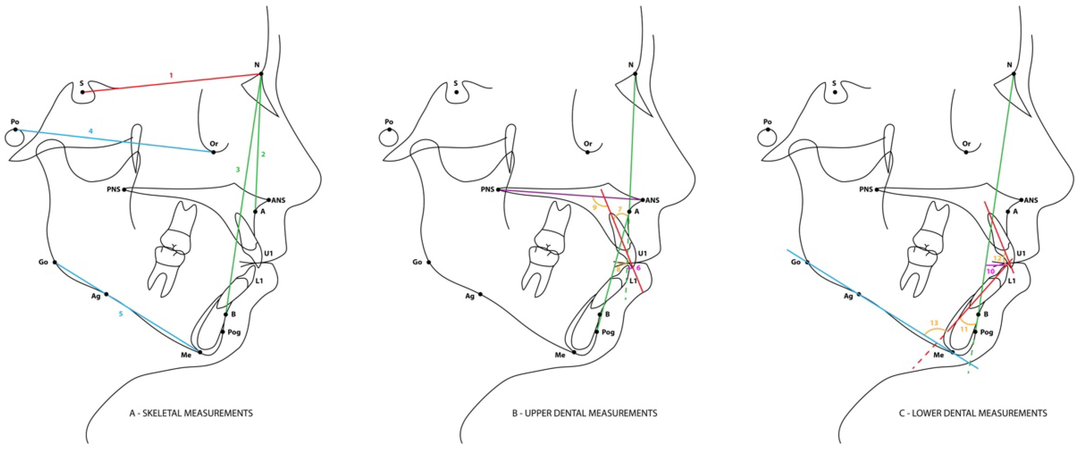
Figure 1. Main cephalometric landmarks and reference planes used in the study. S, sella; N, nasion; Po, porion; Or, orbital; ANS, anterior nasal spine; PNS, posterior nasal spine; A, A point; B, B point; Go, Gonion; Me, menton; Pog, pogonion; Ag, antegonial; U1, maxillary incisor; L1, mandibular incisor. (A) 1-SN, 2 NA, 3 NB, 4 FHP, 5 MP; (B) 6-U1–NA distance; 7-U1–NA angle; 8-U1–APog angle; 9-U1–PP angle; (C) 10-L1–NB distance; 11-L1–NB angle; 12-U1–L1 angle, 13-IMPA.
Applsci 15 10231 g001
Table 1. Sample characteristics (N = 154).
n%
GenderMale4227.3
Female11272.7
Age (years)10–196642.9
Minimum = 1120–293522.7
Maximum = 6630–391912.3
Mean = 26.940–492415.6
Standard deviation = 14.050–5974.5
60+31.9
Skeletal classificationClass I5435.1
Class II5233.8
Class III4831.2
Angle classificationClass I5938.3
Class II4026.0
Class III5535.7
Table 3. Frequency distribution of TDM signs/symptoms across the different skeletal and dental classifications.
TMD Signs/SymptomsChi-Squared Test
nNoYes
All sample154105 (68.2%)49 (31.8%)
By skeletal classification
Class I5439 (72.2%)15 (27.8%)p = 0.377
Class II5237 (71.2%)15 (28.8%)
Class III4829 (60.4%)19 (39.6%)
By Angle classification
Class I5940 (67.8%)19 (32,2%)p = 0.958
Class II4028 (70.0%)12 (30.0%)
Class III5537 (67.3%)18 (32.7%)
Table 4. Correlation coefficients between incisor inclination and other variables for all patients (n = 154) and stratified by the skeletal class. Additionally, a sub-sample of patients with overjet > 0 and overbite > 0 (n = 108) is also presented.
Incisor Inclination
U1–NAU1–APogU1–PP
Sagittal condylar guidance right (n = 154)R = −0.082R = −0.086R = −0.085
Skeletal Class I (n = 54)R= −0.200R= −0.135R= −0.236
Skeletal Class II (n = 52)R= −0.135R= −0.146R= −0.122
Skeletal Class III (n = 48)R= 0.072R= −0.034R= 0.095
Sagittal condylar guidance left (n = 154)R = −0.117 R = −0.070R = −0.135
Skeletal Class I (n = 54)R= −0.232R= −0.064R= −0.202
Skeletal Class II (n = 52)R= −0.192R= −0.226R= −0.197
Skeletal Class III (n = 48)R= −0.017R= 0.083R= −0.045
Overjet (n = 154)R = −0.162R = 0.481 ***R = −0.092
Skeletal Class I (n = 54)R= 0.103R= 0.207R= −0.077
Skeletal Class II (n = 52)R= 0.527 **R= 0.581 **R= 0.443 **
Skeletal Class III (n = 48)R= −0.126R= 0.267R= −0.133
Overbite (n = 154)R = −0.472 ***R = −0.129R = −0.468 ***
Skeletal Class I (n = 54)R= −0.289 *R= −0.241R= −0.369
Skeletal Class II (n = 52)R= −0.400 **R= −0.401 **R= −0.419 **
Skeletal Class III (n = 48)R= −0.419 **R= −0.187R= −0.461 **
Mandibular plane angle (n = 154)R = −0.034R = 0.188 *R = −0.057
Skeletal Class I (n = 54)R= 0.131R= 0.219R= 0.091
Skeletal Class II (n = 52)R= 0.057R= 0.104R= 0.039
Skeletal Class III (n = 48)R= −0.159R= −0.203R= −0.278
Sub-sample: overjet > 0 and overbite > 0 (n = 108)
Sagittal condylar guidance rightR = −0.156R = −0.160R = −0.177
Sagittal condylar guidance leftR = −0.122R = −0.159R = −0.167
OverjetR = 0.039R = 0.414 ***R = 0.066
OverbiteR = −0.483 **R = −0.233 *R = −0.451 **
Mandibular plane angleR = 0.047R = 0.169R = −0.035
Interincisal angleR = −0.498 **R = −0.872 **R = −0.596 **
R–Pearson Correlation Coefficient; *** p < 0.001, ** p < 0.01, * p < 0.05.
Table 5. Comparison of different outcomes based on TMD signs/symptoms in the total sample (n = 154) and in the sub-sample of patients with positive overjet and overbite (n = 108).
TMD Signs/Symptoms
No
(n = 105)
Yes
(n = 49)
p
Sagittal condylar guidance right49.30 (10.77)48.88 (14.70)0.755 (1)
Sagittal condylar guidance left48.31 (9.89)47.14 (15.22)0.664 (1)
Overjet2.82 (2.65)2.59 (2.92)0.820 (1)
Overbite2.16 (2.42)2.32 (2.41)0.709 (1)
U1–NA19.81 (8.24)20.18 (8.58)0.796 (2)
U1–APog24.26 (7.73)22.57 (7.44)0.204 (2)
U1–PP109.99 (9.01)108.96 (7.71)0.490 (2)
Mandibular plane angle24.89 (6.06)23.39 (7.36)0.183 (1)
Interincisal angle131.89 (10.12)134.06 (13.25)0.312 (2)
Sub-sample: overjet > 0 and overbite > 0 (N = 108)
U1–NA18.56 (8.11)17.73 (6.38)0.603 (2)
U1–APog24.76 (7.27)23.58 (6.71)0.427 (2)
U1–PP108.60 (9.24)106.55 (5.00)0.139 (2)
Results presented as mean (standard deviation); (1) p–Mann–Whitney test p-value; (2) p–Student’s t-test p-value.
Disclaimer/Publisher’s Note: The statements, opinions and data contained in all publications are solely those of the individual author(s) and contributor(s) and not of MDPI and/or the editor(s). MDPI and/or the editor(s) disclaim responsibility for any injury to people or property resulting from any ideas, methods, instructions or products referred to in the content.