Article

Oral Involvement and Pain Among Pemphigus Vulgaris Patients as a Clinical Indicator for Management—An Epidemiological and Clinical Study

1
Department of Oral Medicine, Sedation and Imaging, Hadassah Medical Center, Faculty of Dental Medicine, Hebrew University of Jerusalem, Jerusalem 9112102, Israel
2
Department of Dermatology and Venereology, Hadassah Medical Center, The Faculty of Medicine, Hebrew University of Jerusalem, Jerusalem 9112102, Israel
*
Author to whom correspondence should be addressed.
Appl. Sci. 2025, 15(18), 10145; https://doi.org/10.3390/app151810145
Submission received: 4 August 2025 / Revised: 6 September 2025 / Accepted: 15 September 2025 / Published: 17 September 2025
(This article belongs to the Section Applied Biosciences and Bioengineering)

Abstract

Oral lesions are the first sign in most cases of pemphigus vulgaris. Diagnostic delays involving multidisciplinary consultations, required for the evaluation of oral signs, have been reported. Our aims were to investigate whether there is a correlation between specific oral involvement sites and pain and other disease manifestations and treatment outcome as well as diagnostic delays. In this retrospective study, demographics and clinical data, including pain assessment using the Verbal Analogue Scale and treatment information, of 63 Pemphigus Vulgaris patients were analyzed; 43% of patients were examined by multiple physicians before diagnosis, and the mean time to diagnosis was 5.71 ± 8.96 months. A positive moderate strength correlation (Pearson Correlation = 0.5) between intra-oral and extra-oral Verbal Analogue Scale (VAS) symptom scores was noted. After 4–8 weeks of treatment there was a reduction in the mean number of oral sites and VAS scores from 4.71 ± 1.86 to 2.57 ± 1.34 and 7.64 ± 2.24 to 3.21 ± 3.2, respectively. Significant relationships between lip involvement at onset and both the number of involved oral sites and post-treatment symptoms were found. Lip and floor-of-mouth lesions were present in patients with significantly longer hospitalization periods. The results show that specific sites of oral involvement, mainly the tongue, lip and floor of the mouth, may be related to disease severity. Health-care providers should be aware of oral pain and detailed oral involvement which may affect treatment decisions.

1. Introduction

Oral vesicles and erosions and oral pain with a progressive nature are important signs of vesiculobullous diseases, in particular pemphigus vulgaris (PV). PV is the most prevalent form (70%) of the pemphigus group of diseases [1]. This rare autoimmune disease affects the skin and mucous membranes. Autoantibodies target specific desmosome proteins causing intraepidermal cleavage, resulting in vesicles. Painful erosions (teared vesicles) on mucous membranes are the first clinical sign, and if the disease is not treated it can cause severe morbidity or mortality [2,3]. Skin involvement is characterized by flaccid vesicles, mainly on the flexural areas, face, scalp, and extremities [1]. In the majority of cases the first site of involvement is within the oral cavity; therefore, oral signs, as evaluated by oral clinicians, are essential in disease management at onset or relapse. Despite the importance of oral lesion evaluation, diagnostic delays (time period between the appearance of the first sign or symptom and the definitive diagnosis) of approximately 6 months, involving numerous multidisciplinary consultations required for the evaluation of oral signs, have been reported. These delays increase health service utilization and costs [4] as treatment is usually long. Treatment aims are to promote the healing of blisters and erosions and reduce development of new lesions, reduce pain, improve function and life quality, and limit common side effects usually associated with long-term immunosuppressive or corticosteroid treatment. The first-line treatment includes corticosteroids or Anti-CD20 monoclonal antibodies. The protocols are variable, known to be “patient tailored” [5,6], and prediction of the disease outcome is challenging [7]. Mucosal involvement in general has been related to poor outcomes, and oral mucosal involvement has been correlated with an incomplete response to treatment, though existing studies have not specified which oral sites were studied [5]. The distribution of PV varies according to ethnic background—with an annual incidence of less than 0.76 per million in Finland [8] and up to 6.4 per million in Israel [9]. Higher incidences have also been reported in India, the Middle East, and Southeast Europe [10]. The current study was conducted in Israel, with a comparatively high incidence of PV [11]. Patients in this study were diagnosed and treated at the Hadassah Medical Center, a tertiary canter, with two collaborating specialists clinics—a dermatology clinic and an oral medicine. As oral signs and symptoms are usually the first to show or be noticed by oral clinicians and patients [10], their specific manifestations, as described by oral medicine teams, can contribute to disease management.
In this retrospective study, we aimed to examine the time between the first sign or symptom and definite diagnosis (diagnostic delays) in PV patients treated in our institution and to investigate the correlation between oral involvement (specific sites, pain and other disease manifestations) and treatment outcome measured by post-treatment signs, symptoms and days of hospitalization.

2. Methods

2.1. Patients

In this retrospective study, inclusion criteria were patients who were treated at the Hadassah Medical Center between 2012 and 2019, in the Oral Medicine and Dermatology Departments. The patients were diagnosed clinically by dermatologists and oral medicine specialists and by histopathological examination in the pathology department. Exclusion criteria were lack of definitive diagnosis or missing clinical data that was required for the study.

2.2. Data Recorded

Medical and demographic backgrounds were also analyzed in order to understand whether our study group was similar or different from other patient groups and whether the analysis results could be applied to other patient groups. The following data was retrieved from the records: (1) demographic details (age at disease onset, sex, ethnicity (Arab/Jewish) and medical history before PV treatment); (2) diagnostic delay, defined as the time in months from the first clinical sign or symptom of PV reported by the patient to the date of definite diagnosis as described by the medical file; (3) intensity of pain or discomfort measured using the self-reported Verbal Analogue Scale (VAS) from 0 to 10, recorded by the one of the two senior specialists (RC and NK) at the first visit (for intra- and or extra-oral lesions), and intra-oral symptoms 1–2 months after treatment initiation; (4) clinical features of PV—oral sites (lips, buccal mucosa, labial mucosa, hard palate, soft palate, tongue, gingiva, mouth floor), type of lesion (ulcerative/non-ulcerative), sites of extra-oral involvement, (skin, eyes, genital, esophagus larynx pharynx) prior to the beginning of the treatment for PV, and post-treatment hoarseness; (5) type of treatment provided; and (6) number of days of hospitalization.

2.3. Statistical Analysis

Descriptive statistics were used to summarize the demographic and clinical characteristics of the study population. Categorical variables were presented as frequencies and percentages, and continuous variables were reported as means with standard deviations (SD). Bivariate analyses were conducted to evaluate associations between oral site involvement and clinical outcomes such as pain severity (VAS), number of involved oral sites, and hospitalization duration. Continuous variables were analyzed using independent-sample t-tests. Given that the sample size exceeded 30, assumptions of normality were considered sufficiently met based on the Central Limit Theorem. Pearson Correlation coefficients were calculated to assess associations between continuous variables. Statistical significance was defined as p < 0.05. All analyses were performed using SPSS version 26.

3. Results

Files of 69 patients diagnosed with PV were initially reviewed and 6 were excluded due to missing clinical information.

3.1. Demographic Characteristics

Among the remaining 63 patients, 39 (61.9%) were females and 24 (38.1%) were males. Mean age at diagnosis was 48.7 years (Table 1). A total of 53 patients (84.1%) were Jews, and 10 patients (15.9%) were Arabs. The male to female ratio in these ethnic groups was 1:1.65 and 1:1.5, respectively, and average age at diagnosis was 51.06 (SD 13.6) and 43.00 (SD 13.5), respectively, with no statistically significant differences between the groups (younger, middle-aged (risk group), and older group distributions are presented in Table 1). Overall, 36 patients (57.1%) reported comorbidities at disease onset; the most frequent were cardiovascular diseases and hypertension, diabetes and hyperlipidemia. The other 27 patients (42.9%) had no chronic diseases (Table 1).

3.2. Diagnostic Delay

The interval between the appearance of a clinical sign and PV diagnosis was 5.71 ± 8.96 months, shorter for males (2.77 months, SD 1.92) and longer for females (5.41 months, SD 6.55). Most patients were examined by more than one physician before being diagnosed with PV. In total, 22% of the patients were self-referrals; 34.4% were examined by one physician and the rest were examined by two or more (Figure 1), with an average of 1.9 physicians. The physicians specialized as otorhinolaryngologists (45%), family physicians (32%), dermatologists (26%), dentists (24%), oral medicine specialists (8%) and others (11%) (data was available for 40 cases).
The presence of zero examining physicians before diagnosis designates self-referrals.

3.3. Clinical Characteristics and Pain at First Visit

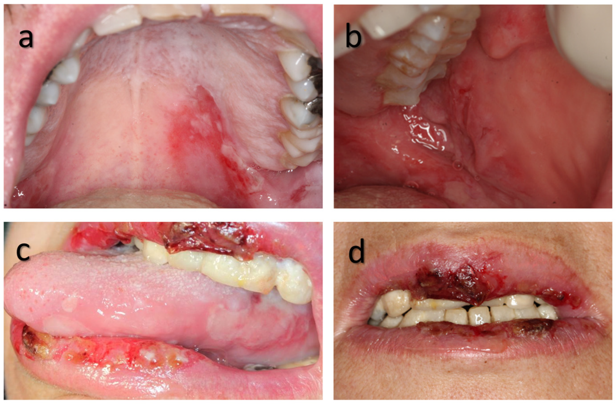
Overall, 62 patients (98.4%) had oral lesions (Figure 2).
Most patients had disease involvement at multiple oral sites, with a mean of 4.71 ± 1.86 sites (Table 2). The buccal mucosa was the most commonly involved oral site (80%) followed by the soft palate and tongue (75% and 56.7%, respectively) (Table 2).
In total, 96.6% complained of oral pain/discomfort with a mean VAS score of 7.64 ± 2.24. The mean VAS score was significantly higher in patients with labial mucosa and tongue involvement (Table 3).
Overall, 75.4% reported hoarseness. Regarding extra-oral involvement, the mean VAS score for pain/discomfort was 1.86 ± 1.9 SD, and there was a moderate positive strength correlation (Pearson Correlation = 0.5) between intra-oral and extra-oral VAS scores of pain/discomfort (p = 0.018).
In total, 53 (85.5%) had extra-oral involvement, 44 (71%) had cutaneous lesions, and all patients also had concomitant oral lesions; other sites were laryngeal (40%), intranasal (22%), genital (12%) and ocular (5%).

3.4. Treatment/Hospitalization

The mean number of hospitalization days for 58 patients was 30.57 ± 16.22. Systemic corticosteroid therapy was typically initiated with oral prednisone in combination with oral triamcinolone acetonide, at a cumulative dose equivalent to prednisone 1 mg/kg/day. The corticosteroid dosage was then gradually tapered by 5–10 mg/day every few days or weeks, depending on therapeutic response. Clinical assessment of the response included the daily number of new lesions, the presence or absence of a positive Nikolsky sign, and the rate of healing of newly developed lesions. Lack of clinical response within the first 7–14 days of treatment generally led to the addition of another systemic immunosuppressive agent, most commonly azathioprine or mycophenolate mofetil. Overall, 56 patients (96.6%) were treated with systemic steroids and 32 patients (50.8%) received Azathioprine as a steroid-sparing agent. Six patients (9.5%) displayed Azathioprine-related hepatotoxicity and therefore were given Mycophenolate Mofetil instead. None of the patients received Rituximab. Two patients (3.4%) with only oral involvement were treated with topical steroids and an antiseptic–analgesic mouth wash.

3.5. Oral Involvement and Disease Outcome/Severity

The mean total number of oral sites decreased to 2.57 ± 1.34 after four to eight weeks of treatment (Table 2). The mean VAS score of pain/discomfort for oral symptoms after treatment fell to 3.21 ± 3.2 (Table 2).
A positive correlation was found between tongue involvement and mean VAS scores for pain/discomfort at onset (Table 3), as well as between total number of oral sites before and after treatment (Pearson Correlation = 0.29, p = 0.044). A significant relationship was also found between lip involvement at onset and both the total number of involved oral sites after treatment and the VAS score for pain/discomfort after treatment (Table 3). Oral involvement of lips and the floor of mouth was noted in patients with significantly longer hospitalization periods (Table 3). A positive correlation was found between hospitalization period and the number of involved oral sites after treatment (Pearson Correlation = 0.371, p = 0.008). There was a moderate positive correlation between the intra- and extra-oral VAS scores for pain/discomfort (Pearson Correlation = 0.5, p = 0.018).
The Arab patients had higher rates of lip and palate involvement at onset compared to the Jewish patients (60% and 40% vs. 32% and 20%, respectively), but these, and the differences in other intra-oral features, were not statistically significant. All Arab patients had extra-oral involvement, compared to 82.7% of the Jewish patients; again, this difference was not statistically significant. The only significant difference in all examined parameters, i.e., demographic and clinical characteristics, was higher rates of hoarseness and laryngeal involvement among younger patients.

4. Discussion

Pemphigus vulgaris is a challenging disease for treatment as its symptoms (including oral pain), clinical manifestations and treatment outcomes are variable and usually non-predictable [12]. In this study we analyzed the clinical features of PV patients in a large tertiary hospital, in an area with a known high annual incidence of PV [9]. In the majority of patients, the oral cavity was involved from the very first stages. The combined team-work of our oral medicine and dermatology specialists allowed the analysis of clinical features including detailed pain intensity, oral involvement and relevant correlations.
The profile of the study population is similar to that reported in other studies, which means that the correlations reported here can be applied to other patient populations. The age and gender profiles of the study population were similar to or younger than those reported in the literature [5,13,14]. Almost two thirds of the study PV patients had comorbidities at disease onset, mainly hypertension, diabetes, and hyperlipidemia. The prevalence of comorbidities in our cohort is similar to that in the same age range in the general Israeli population (30%, 20% and 15–19%, respectively, for hypertension, diabetes, and hyperlipidemia) [15,16]. Other studies reported similar or additional comorbidities [3,17,18]. Although many of these PV-associated comorbidities may be age-related or influenced by geographic, life style-related or other possible factors, clinicians should always consider these comorbidities when managing PV, because PV treatments, mainly steroids, may have an impact on them [6].
Most of the PV patients in the current study consulted more than one physician before PV was considered. They were examined by otorhinolaryngologists, family physicians, dermatologists and dentists. The order in which patients saw these specialists probably reflects the main sites involved at disease onset [4], and a similar order has been published previously [19]. The mean interval between the appearance of the first clinical sign and PV diagnosis was consistent with reports of diagnostic delays ranging from 5.9 to 12 months [20], with an average delay of 6 months [5,21]. Robinson et al. emphasized the value of early diagnosis and prompt treatment, highlighting that prognosis also improves [21]. Less adverse effects and longer remissions were other reported advantages [22,23]. The published range of the number of consultants seen, reviewed by Petruzzi et al. [20], was 3.1 to 6.27, while in the current study the average was lower. Diagnostic delay and multiple consultations might be the result of clinician unfamiliarity with PV [24], in particular the oral manifestations [4], or due to the nonspecific clinical presentations and pain that PV has [25]. The predominance of oral ulcers as a presenting sign, as observed in this study and others [14], naturally leads to oral symptoms. Consequently, patients initially consulted physicians who commonly manage oral and laryngeal conditions—such as otorhinolaryngologists, family physicians, dermatologists, dentists, and oral medicine specialists.
While concurrent skin involvement with vesicles or erosions may suggest a differential diagnosis that includes chronic vesiculo-ulcerative diseases such as PV, mucous membrane pemphigoid, or erosive lichen planus, isolated oral involvement often leads to alternative diagnostic considerations. These may include pharyngitis, recurrent aphthous ulcerations, or recurrent intra-oral herpes infections—conditions more common in the general population and characterized by similarly nonspecific presenting symptoms—resulting in misdiagnosis [25].
The importance of recognizing oral signs and symptoms was highlighted by Daltaban et al. (2020) [14], who reported longer diagnostic delays in patients presenting exclusively with oral involvement compared to those with concurrent skin manifestations, largely due to misdiagnosis or lack of familiarity.
Our findings of fewer consultations prior to diagnosis may be attributed to a higher level of awareness of PV in Israel, where the disease is relatively more prevalent [11,13].
Oral involvement at presentation, found in more than 98% of PV patients in this study and in the majority of PV patients in previous studies [4,7,12,24], is an important sign of the disease, with the oropharyngeal mucosa often being the first site of involvement [5]. The intra-oral sites documented in this study were similar to those mentioned previously with the same or different orders [5,14,25]. The most common oral site of involvement in the current study is the buccal mucosa, which is the site under constant friction, triggering desquamation. We found an average of more than four oral sites involved per patient, while others reported two to over three sites among some of the patients [5]. The dominance of oral ulcers as a presenting sign obviously causes oral pain and symptoms. The vast majority of patients in the current study complained of oral symptoms, matching the high rates of oral symptoms in previous studies [5,14,25]. Many experienced soreness and in some the symptoms were severe enough to affect normal oral function and quality of life [14,26]. Oral symptoms may also be the eventual reason for referral to a health-care professional, expediting the diagnosis of PV [25]. The positive moderate strength correlation founded in this study between intra- and extra-oral VAS scores for pain/discomfort may be related to disease severity, as symptoms reflect the presence of erosive or ulcerated tissue. Self-report of pain intensity at the involved sites should be included in the clinical evaluation of PV patients, as this parameter could add to the evaluation of the disease severity. The higher prevalence of hoarseness in the current study compared to others [14,25] may reflect the difference in recording this sign; in the current study, the presence of hoarseness was a routine question for self-reporting, since some patients do not complain about it if not asked. In other studies, it may be included in the general description of laryngeal/pharyngeal symptoms. We suggest the inclusion of this question for any suspected PV patients and the consideration of hoarseness as a supporting sign in PV differential diagnosis when other clinical signs are not fully shown. Extra-oral involvement (cutaneous or mucosal) was noted in 85.5% of patients in the current study, and this is similar to the 83% reported by Daltaban et al. [14]. Lesion distribution was comparable to those reported by others [5,25,27]. Overall, through an analysis of the detailed oral involvement, we noted that its association with some parameters of the disease has not been described before, and as the current study group showed similar demographic features to other PV studies, the associations can be applied to other patients.
The mainstay of treatment during hospitalization was steroid regimens, which were the first-line treatment at the time of the study, and the dermatology department has much experience with these medications. As this treatment begins under hospitalization and continues on an outpatient basis when the disease manifestations are reduced and controlled, the number of hospitalization days as additional parameter for treatment outcome was recorded. Patients with lip involvement had longer hospitalization periods as well as worse post-treatment symptoms and more oral sites involved—all suggestive of more severe disease. Since the first signs of mucosal involvement often appear at the posterior part of the mouth and larynx, lip involvement in and of itself may be a sign that the disease has progressed anteriorly [28], which may imply progressed disease or, as mentioned, greater severity. The same applies to findings on the floor of the mouth, which was related to longer hospitalization. As mentioned, longer hospitalizations and a greater number of affected oral sites may be related to disease stage or severity. When patients have multiple oral sites at onset, full resolution of oral lesions should not be expected in the first period of treatment. The importance of mucosal lesions was emphasized by Cho et al. [7], reporting that mucosal involvement is a risk factor for poor clinical outcome in patients treated with rituximab. Arduino et al. [5] found lower rates of complete resolution following systemic treatments in patients with more extensive oral involvement but no detailed “oral involvement”.
We suggest that oral lesions should be examined carefully at onset. Patients with tongue involvement might experience higher levels of pain. Involvement of the lips or mouth floor may predict slower remission or severe disease. When a higher number of oral sites are involved, some will be present during the first weeks of treatment. Therefore, this kind of disease manifestation may imply the need for a different treatment protocol and expectations of a longer treatment period and close follow-up.
Although rituximab was not administered to any patient in our study, the findings remain relevant. In most clinical settings, rituximab is introduced in combination with short-term corticosteroids, which serve as a bridge therapy to control inflammation and symptoms until rituximab becomes effective—a process that may take up to 16 weeks. A more precise assessment of the severity of oral symptoms at disease onset may influence therapeutic decisions regarding the initial corticosteroid dosage, treatment duration, and optimal tapering schedule.
Moreover, there is a subset of pemphigus patients who cannot benefit from rituximab, including those with known hypersensitivity to its components, patients with severe heart failure, lactating women, and individuals with refractory pemphigus unresponsive to rituximab. These patients will likely require corticosteroids for a longer period, and our study findings may assist in designing more efficient corticosteroid-based treatment plans for them.
The assessment of oral site involvement is part of disease severity scoring systems; currently, each site is considered equally important [29]. We suggest that modified scoring systems for disease severity should give greater weight to lip and floor-of-mouth involvement. Furthermore, while existing scoring methods treat all oral sites equally, our results indicate that lesions of the lips, tongue, and floor of the mouth may carry greater prognostic significance. Accordingly, future studies should explore the development and validation of weighted scoring models that more accurately reflect the clinical impact of site-specific involvement.
Future research based on larger, prospective cohorts with comprehensive data collection and multivariate statistical modelling would be valuable to validate and expand upon these findings, thereby strengthening their clinical and translational relevance. Although our study did not employ advanced machine learning approaches due to its retrospective design and limited sample size, future investigations using techniques such as clustering or K-nearest neighbours may reveal hidden patterns in oral lesion distribution and pain profiles, enhancing phenotypic classification and supporting personalized management of pemphigus vulgaris.
Finally, integrating oral site involvement, VAS pain scores, and demographic factors into a predictive risk model for longer hospitalization could yield actionable clinical tools, including the potential development of user-friendly applications for real-world clinical practice.
The analysis of the disease phenotype and ethnicities (Arabs/Jews), sex and age groups showed no significant differences in all examined parameters except for the higher rates of hoarseness and laryngeal involvement among younger patients. As the majority of PV patients had oral involvement, clinicians should be familiar with PV signs. PV patients might have a higher rate of comorbidities at disease onset that must be taken into consideration, since most of the current systemic treatments can pose additional health risks. This retrospective study has several methodological limitations that should be acknowledged. First, the analyses were primarily limited to bivariate approaches without formal multivariable modelling to adjust for potential confounding factors such as age, sex, and comorbidities. Due to the retrospective design and sample size constraints, conducting advanced regression analyses was beyond the scope of this study. Consequently, there remains potential for residual confounding in the observed associations. Second, parametric tests were applied under the assumption of normality based on the Central Limit Theorem; however, some variables—including hospitalization duration—displayed skewed distributions, which may affect the robustness of statistical inferences. Additionally, assessments for multicollinearity among highly correlated variables were not performed, which could influence the interpretation of correlated outcomes. Despite these limitations, the findings provide exploratory insights into clinical and pain-related aspects of pemphigus vulgaris.
Future research employing larger, prospective cohorts with comprehensive data collection and multivariate statistical modelling would be valuable to validate and expand upon these results, thereby enhancing their clinical and translational applicability.

5. Conclusions

The majority of PV patients presented with oral involvement, and more than 40% were examined by multiple physicians prior to diagnosis, with a mean diagnostic delay of approximately 6 months. Improved clinician familiarity with the signs and symptoms of PV is essential to reduce unnecessary multidisciplinary consultations and shorten diagnostic delays.
A moderate positive correlation was observed between intra-oral and extra-oral VAS symptom scores. Following 4–8 weeks of treatment, both the mean number of affected oral sites and VAS scores decreased. Notably, lip involvement at onset was significantly associated with a greater number of oral sites affected and higher post-treatment symptom scores. In addition, lesions involving the lips and floor of the mouth were linked to significantly longer hospitalization periods.
Clinical evaluation at disease onset should therefore include patient self-reported pain intensity, the number of affected oral sites, and careful attention to lip and floor-of-mouth involvement. These factors appear to reflect disease severity, predict post-treatment symptom persistence, and correlate with longer hospitalization. Revision of current disease severity scoring systems to assign greater weight to these parameters—particularly anterior oral site involvement—is recommended.

Author Contributions

Conceptualization and design of study, R.C. and M.A.E.; investigation/acquisition of data, R.C., M.A.E. and N.K.; data curation and statistical analysis, R.C., M.A.E., M.C. and G.A.-W.; writing—original draft preparation, R.C., M.A.E. and Y.H.; writing—review and editing, R.C. and G.A.-W. All authors have read and agreed to the published version of the manuscript.

Funding

This research received no external funding.

Institutional Review Board Statement

The study was conducted in accordance with the Declaration of Helsinki and approved by the Institutional Ethics Committee of Hadassah Medical Center (protocol code #0717-18-HMO approval date 14 February 2019)) for studies involving humans.

Informed Consent Statement

Patient consent was waived as a retrospective study.

Data Availability Statement

Data is available upon request from the corresponding author, R. Czerninski—rakefet.czerninski@mail.huji.ac.il.

Conflicts of Interest

The authors declare no conflicts of interest.

Abbreviations

The following abbreviations are used in this manuscript:
PVPemphigus Vulgaris
VASVerbal Analogue Scale
FOMFloor of Mouth

References

  1. Joly, P.; Litrowski, N. Pemphigus group (vulgaris, vegetans, foliaceus, herpetiformis, brasiliensis). Clin. Dermatol. 2011, 29, 432–436. [Google Scholar] [CrossRef] [PubMed]
  2. Ellebrecht, C.T.; Maseda, D.; Payne, A.S. Pemphigus and Pemphigoid: From Disease Mechanisms to Druggable Pathways. J. Investig. Dermatol. 2021, 142, 907–914. [Google Scholar] [CrossRef] [PubMed]
  3. Kridin, K. Pemphigus group: Overview, epidemiology, mortality, and comorbidities. Immunol. Res. 2018, 66, 255–270. [Google Scholar] [CrossRef]
  4. Sirois, D.A.; Fatahzadeh, M.; Roth, R.; Ettlin, D. Diagnostic patterns and delays in pemphigus vulgaris: Experience with 99 patients. Arch. Dermatol. 2000, 136, 1569–1570. [Google Scholar] [CrossRef]
  5. Arduino, P.G.; Broccoletti, R.; Carbone, M.; Gambino, A.; Sciannameo, V.; Conrotto, D.; Cabras, M.; Sciascia, S.; Ricceri, F.; Baldovino, S.; et al. Long-term evaluation of pemphigus vulgaris: A retrospective consideration of 98 patients treated in an oral medicine unit in north-west Italy. J. Oral Pathol. Med. 2019, 48, 406–412. [Google Scholar] [CrossRef]
  6. Murrell, D.F.; Peña, S.; Joly, P.; Marinovic, B.; Hashimoto, T.; Diaz, L.A.; Sinha, A.A.; Payne, A.S.; Daneshpazhooh, M.; Eming, R.; et al. Diagnosis and management of pemphigus: Recommendations of an international panel of experts. J. Am. Acad. Dermatol. 2020, 82, 575–585.e1. [Google Scholar] [CrossRef]
  7. Cho, S.I.; Kim, J.W.; Lim, J.S.; Chung, J.H. Mucosal involvement is a risk factor for poor clinical outcomes and relapse in patients with pemphigus treated with rituximab. Dermatol. Ther. 2019, 32, e12814. [Google Scholar] [CrossRef] [PubMed]
  8. Hietanen, J.; Salo, O. Pemphigus: An epidemiological study of patients treated in Finnish hospitals between 1969 and 1978. Acta Derm.-Venereol. 1982, 62, 491–496. [Google Scholar] [CrossRef]
  9. Kridin, K.; Zelber-Sagi, S.; Bergman, R. Pemphigus Vulgaris and Pemphigus Foliaceus: Differences in Epidemiology and Mortality. Acta Derm.-Venereol. 2017, 97, 1095–1099. [Google Scholar] [CrossRef]
  10. Neville, B.D.; Damm, D.D.; Allen, C.M.; Chi, A.C. Pemphigus vulgaris in:Dermatologic Diseases. In Oral and Maxillofacial Pathology, 5th ed.; Elsevier: Maryland Heights, MO, USA, 2024; pp. 747–819. [Google Scholar]
  11. Schmidt, E.; Kasperkiewicz, M.; Joly, P. Pemphigus. Lancet 2019, 394, 882–894. [Google Scholar] [CrossRef]
  12. Malhi, K.; Chatterjee, D.; Mahajan, R.; Handa, S.; De, D. Direct immunofluorescence on plucked hair outer root sheath can predict relapse in pemphigus vulgaris. Int. J. Dermatol. 2024, 64, 367–376. [Google Scholar] [CrossRef] [PubMed]
  13. Kridin, K.; Schmidt, E. Epidemiology of Pemphigus. JID Innov. 2021, 1, 100004. [Google Scholar] [CrossRef] [PubMed]
  14. Daltaban, Ö.; Özçentik, A.; Karakaş, A.A.; Üstün, K.; Hatipoğlu, M.; Uzun, S. Clinical presentation and diagnostic delay in pemphigus vulgaris: A prospective study from Turkey. J. Oral Pathol. Med. 2020, 49, 681–686. [Google Scholar] [CrossRef]
  15. Israel Center for Disease Control (ICDC)-INHIS-4 Israel National Health Interview Survey INHIS-4 2019. Available online: https://www.gov.il/BlobFolder/reports/inhis-4-2019/he/files_publications_units_ICDC_INHIS-4-2019.pdf (accessed on 31 July 2025).
  16. Israel Diabetes Registry Report. Available online: https://www.gov.il/BlobFolder/generalpage/diabetes-2012-2022/he/files_publications_units_ICDC_diabetes-2012-2022.pdf (accessed on 31 July 2025).
  17. Pankakoski, A.; Kluger, N.; Sintonen, H.; Panelius, J. Clinical manifestations and comorbidities of pemphigus: A retrospective case-control study in southern Finland. Eur. J. Dermatol. 2022, 32, 480–486. [Google Scholar] [CrossRef]
  18. Baum, S.; Astman, N.; Berco, E.; Solomon, M.; Trau, H.; Barzilai, A. Epidemiological data of 290 pemphigus vulgaris patients: A 29-year retrospective study. Eur. J. Dermatol. 2016, 26, 382–387. [Google Scholar] [CrossRef]
  19. Hassona, Y.; Cirillo, N.; Taimeh, D.; Al Khawaldeh, H.; Sawair, F. Diagnostic patterns and delays in autoimmune blistering diseases of the mouth: A cross-sectional study. Oral Dis. 2018, 24, 802–808. [Google Scholar] [CrossRef]
  20. Petruzzi, M.; della Vella, F.; Squicciarini, N.; Lilli, D.; Campus, G.; Piazzolla, G.; Lucchese, A.; van der Waal, I. Diagnostic delay in autoimmune oral diseases. Oral Dis. 2023, 29, 2614–2623. [Google Scholar] [CrossRef]
  21. Robinson, N.; Yeo, J.; Lee, Y.; Aw, D. Oral Pemphigus Vulgaris: A Case Report and Literature Update. Ann. Acad. Med. Singap. 2004, 33 (Suppl. 4), 63S–68S. [Google Scholar] [CrossRef]
  22. Ljubojević, S.; Lipozenčić, J.; Brenner, S.; Budimčić, D. Pemphigus vulgaris: A review of treatment over a 19-year period. J. Eur. Acad. Dermatol. Venereol. 2002, 16, 599–603. [Google Scholar] [CrossRef]
  23. Kumar, S.; De, D.; Handa, S.; Ratho, R.; Bhandari, S.; Pal, A.; Kamboj, P.; Sarkar, S. Identification of factors associated with treatment refractoriness of oral lesions in pemphigus vulgaris. Br. J. Dermatol. 2017, 177, 1583–1589. [Google Scholar] [CrossRef] [PubMed]
  24. Ishii, N.; Maeyama, Y.; Karashima, T.; Nakama, T.; Kusuhara, M.; Yasumoto, S.; Hashimoto, T. A clinical study of patients with pemphigus vulgaris and pemphigus foliaceous: An 11-year retrospective study (1996–2006). Clin. Exp. Dermatol. 2008, 33, 641–643. [Google Scholar] [CrossRef] [PubMed]
  25. Shamim, T.; Varghese, V.I.; Shameena, P.M.; Sudha, S. Pemphigus vulgaris in oral cavity: Clinical analysis of 71 cases. Med. Oral Patol. Oral Cir. Bucal 2008, 13, E622–E626. [Google Scholar] [PubMed]
  26. Abdalla-Aslan, R.; Benoliel, R.; Sharav, Y.; Czerninski, R. Characterization of pain originating from oral mucosal lesions. Oral Surg. Oral Med. Oral Pathol. Oral Radiol. 2015, 121, 255–261. [Google Scholar] [CrossRef]
  27. Bardazzi, F.; Rucci, P.; Rosa, S.; Loi, C.; Iommi, M.; Di Altobrando, A. Comorbid diseases associated with pemphigus: A case-control study. J. Der Dtsch. Dermatol. Ges. 2021, 19, 1613–1619. [Google Scholar] [CrossRef]
  28. Mimouni, D.; Anhalt, G.J. Pemphigus. Dermatol. Ther. 2002, 15, 362–368. [Google Scholar] [CrossRef]
  29. Daniel, B.S.; Hertl, M.; Werth, V.P.; Eming, R.; Murrell, D.F. Severity score indexes for blistering diseases. Clin. Dermatol. 2012, 30, 108–113. [Google Scholar] [CrossRef] [PubMed]
Figure 1. Number of examining physicians before PV diagnosis and percentage of each group.
Applsci 15 10145 g001
Figure 2. PV oral involvement with symptomatic lesions. Upper panel: posterior oral sites: (a) erosion at hard and soft palate, (b) erosions at posterior buccal mucosa. Lower panel—severe conditions: (c) lip lesions, erosions at ventral and lateral sides of tongue; (d) lip lesions—erosions and crusts.
Applsci 15 10145 g002
Table 1. Demographic and medical characteristics of PV patients (N = 63).
N
GenderFemale39 (61.9)
Male24 (38.1)
OriginJewish53 (84.2)
Arab10 (15.8)
Age44 and younger22 (34.9)
45–6432 (50.7)
65 and up9 (14.4)
Medical history *Cardiovascular/hypertension15 (23.8)
Diabetes10 (15.8)
Hyperlipidemia10 (15.8)
Hypothyroidism4 (17.4)
Psychological disorders4 (17.4)
Other dermatologic disorders3 (4.7)
S/P malignancy3 (4.7)
GERD, Fibromyalgia 2, 1 (8.7, 1.6)
* Prior to PV treatment; patients can have multiple diagnoses.
Table 2. Clinical characteristics and pain at onset and after treatment.
At Onset *^After Treatment ^
Oral lesion site *
N = 63
N (% **); N (% **);
Buccal mucosa48 (80) 28 (55)
Soft Palate45 (75) 26 (50)
Tongue34 (57) 14 (27)
Labial mucosa32 (53) 12 (24)
Gingiva29 (48) 10 (20)
Lips22 (37) 7 (14)
Floor of mouth21 (35) 7 (14)
Hard palate14 (23) 2 (4)
Mean total number of lesion sites (SD)4.71 ± 1.862.57 ± 1.34
Symptoms discomfort/pain (mean VAS) (SD)7.64 ± 2.243.21 ± 3.2
* Onset = at first visit at clinic prior to beginning of the treatment. ** % calculation of percentage for each category—out of patients with known data. ^ Mean missing value: at onset: 3; after treatment: 11.37.
Table 3. Oral site involvement at onset, pain and clinical features.
Oral Lesion Sites at Onset,
Pain, Post-Treatment Sites and Hospitalization Period
Oral site involvement
at onset
Pain at
onset—
Mean VAS (SD)
Pain
after treatment—
Mean VAS (SD)
Sites
after treatment—
Mean (SD)
Hospitalization days—
Mean (SD)
Lip—positive8.10 (2.1)
(labial) *
3.38 (3.25)2.74 (1.6)40.68 (21.44)
Lip—negative5.29 (3.9)
(labial) *
1.13 (2.0)1.53 (1.2)25.84 (19.44)
p value0.0210.0290.0050.008
Tongue—positive 8.26 (2.21)---
Tongue—negative5.56 (3.32)- -
p value0.016 -
FOM—positive--42.81 (25.70)
FOM—negative--25.08 (11.56)
p value 0.001
FOM = floor of the mouth; * data on VAS at onset relate to labial involvement.
Disclaimer/Publisher’s Note: The statements, opinions and data contained in all publications are solely those of the individual author(s) and contributor(s) and not of MDPI and/or the editor(s). MDPI and/or the editor(s) disclaim responsibility for any injury to people or property resulting from any ideas, methods, instructions or products referred to in the content.