Article

In Vitro Photoprotective and Skin Aging-Related Enzyme In-Hibitory Activities of Cylindrospermum alatosporum (NR125682) and Loriellopsis cavernicola (NR117881) Extracts

by
Siphesihle Z. Sithole
1,
Albert O. Ikhane
2,*,
Foluso O. Osunsanmi
1,*,
Rebamang A. Mosa
2 and
Andrew R. Opoku
1
1
Department of Biochemistry and Microbiology, University of Zululand, Richards Bay 3900, South Africa
2
Department of Biochemistry, Genetics and Microbiology, University of Pretoria, Pretoria 0028, South Africa
*
Authors to whom correspondence should be addressed.
Appl. Sci. 2025, 15(17), 9718; https://doi.org/10.3390/app15179718
Submission received: 30 July 2025 / Revised: 31 August 2025 / Accepted: 2 September 2025 / Published: 4 September 2025

Abstract

The use of cyanobacteria has gained considerable interest in many industries, including the cosmetic industry, due to its rich array of bioactive metabolites. This study evaluates the in vitro photoprotective properties and the effect of Cylindrospermum alatosporum (NR125682) and Loriellopsis cavernicola (NR117881) extracts on slowing down the enzymes associated with skin aging. Various crude extracts were prepared using hexane, dichloromethane, and ethanol solvents. The resulting crude extract solvents were completely distilled to obtain their bioactive compounds, based on selected polarities. The sulfhydryl content of the crude extracts was determined and the aging-associated enzymes’ activity (collagenase, elastase, hyaluronidase, and tyrosinase) in the crude extracts was investigated. Furthermore, the in vitro photoprotective activity of the extracts was assessed by measuring UVA and UVB photoprotection. Most of the extracts contained varying amounts of sulfhydryl compounds (10.88–78.15 mg/g). All of the extracts demonstrated in vitro inhibitory activity against tyrosinase, hyaluronidase (IC50 6 µg/mL), and collagenase (IC50 50–70 µg/mL); weak elastase inhibitory activities were also observed. The crude extracts also showed appreciable UVA and UVB photoprotective activity. Meanwhile, L. cavernicola extracts demonstrated the highest UVB photoprotective activity (SPF 14.67–78.96). It is noteworthy that the crude extracts possessed anti-skin-aging potency with notable photoprotective capability.

1. Introduction

Human skin is a crucial barrier between internal organs and the external world, giving protection for the body’s internal organs [1]. However, the skin is perpetually exposed to environmental pollution and ultraviolet radiation A/B (UVR) as extrinsic factors, in particular UVA and UVB, as well as the intrinsic factors that determine the inevitable process of chronological aging [2]. These combined factors synergistically contribute to the multifaceted phenomenon of skin aging [3]. Skin aging is the progressive deterioration of key skin components such as collagen, elastin, tyrosine, and hyaluronic acid; as the years unfold, skin exhibits visible signs such as wrinkles, dryness, dermal atrophy, visible pores, loss of elasticity, rougher texture, and hyperpigmentation [4].
In their physiological state, these components maintain structural integrity [5], elasticity, pigmentation, and hydration [6]. However, when the skin is exposed to prolonged UVR, the level of reactive oxygen species (ROS) elevates, and the accumulation of these species subsequently leads to an induced skin-inflammatory response, DNA damage, reduction of antioxidant enzymes, and increased matrix metalloproteinase (MMP), which degrades collagen. In addition, UVR induces the overexpression of elastase, tyrosinase, and hyaluronidase, leading to skin aging [7].
In the quest for a more youthful appearance, individuals often resort to the use of rejuvenating procedures such as chemical peeling, hormonal replacement therapy, and many more. These methods are, however, linked to the occurrence of hyperpigmentation and an escalated susceptibility to the development of various skin and systemic malignancies [8]. There is global interest in anti-aging formulations that are composed of natural cosmetic ingredients such as hyaluronic acid and niacinamide, as opposed to their synthetic counterparts that may potentially be harmful to human health and the environment [9]. Exploring natural-origin sources for cosmetics formulation is therefore essential.
Biologically active substances, encompassing lipids, amino acids, polysaccharides, pigments, and antioxidants, are synthesized by flora, fauna, and microorganisms [10]. These bioactive substances can neutralize free radicals, absorb ultraviolet radiation, and chelate metal ions [11]. Cyanobacteria are photoautotrophic microorganisms known to generate a spectrum of secondary bioactive compounds including phenolic compounds, proteins, pigments, and MMP inhibitors [12,13], which could offer the potential to address skin aging without the adverse side-effects associated with conventional cosmetic products [14]. Leptolyngbya, Synechocystis, and Wollea genera have been reported to produce metabolites with radical-scavenging potential [15,16]. Additionally, phycobiliproteins (PBPs) and phenolics have also been reported from Arthospira, Spirulina, and Nostoc [17]. Mycosporine-like amino acids (MAAs) and scytonemin (SCY), both efficient UVR-absorbing compounds, have also been recorded in cyanobacteria [18,19]. This makes cyanobacteria a standout candidate for photoprotection compounds, and, while marine cyanobacteria has often been used in cosmetic formulations [16], fresh water, and soil, cyanobacteria also possess photoprotective metabolites [20].
In this study, we present our findings on the in vitro photoprotective and anti-skin-aging potential of dried crude extracts obtained via sequential solvent extraction of two freshwater cyanobacteria: Cylindrospermum alatosporum (NR125682) and Loriellopsis cavernicola (NR117881). It is important to state that the organic solvents utilized in the extraction process (hexane, dichloromethane, and 70% ethanol) were completely distilled to obtain the dried crude extracts utilized in this study.

2. Materials and Methods

2.1. Materials

Unless otherwise stated, all chemicals and reagents (including enzymes and their substrates) used in this study were of analytical grade and procured from Sigma Aldrich (Johannesburg, South Africa). All spectroscopic measurements were conducted on a BioTek (SYNERGY HT) plate reader. The following were utilized in this study: σ-phthaldehyde, glutathione, N-(3-[2-Furyl] acryloyl)-Leu-Gly-Pro-Ala (FALGPA), NaCl, SANA (N-succinyl-Ala-Ala-Ala-p-nitroanilide), kojic acid, and resveratrol.

2.2. Methodology

Details of the isolation and characterization of Cylindrospermum alatosporum (NR125682) and Loriellopsis cavernicola (NR117881), and the extraction and phytochemical constituents of the extracts, have been reported in [21]. Briefly, freshwater samples were enriched with BG-11 medium and cultivated for 21 days under continuous illumination. A sequence of re-plating was carried out to isolate single and pure colonies that were then identified and characterized through 16S rRNA sequencing. The cultivation, harvesting, sequential extraction, and complete distillation of the biomass were carried out to obtain the hexane, dichloromethane, and ethanol solvent-free dry extracts. The dried extracts used for the study were free of toxic solvents. Phytochemical analysis of the extracts (using gas chromatography–mass spectrometry [GC–MS]), phenol, and flavonoid contents, as well as the extracts’ antioxidant potential, have also been detailed by Ikhane, et al. [21].

2.2.1. Total Sulfhydryl Content

The method developed by Cohn and Lyle [22] was adapted to determine the sulfhydryl (SH) concentration of the cyanobacterial extracts. The extracts (10–50 µg/mL) were added to a test tube containing 50 μL of sodium hydrogen phosphate buffer (0.1 M, pH 8.0) and 10 μL of 1% σ-phthaldehyde. After 20 min of incubation at room temperature, the absorbance was measured spectrophotometrically at 420 nm. The total sulfhydryl content of the extracts was determined from the calibration curve of glutathione; the total sulfhydryl content was expressed as milligrams of glutathione equivalents per gram of dry extract (mg GE/g dry extract).

2.2.2. In Vitro Anti-Aging Enzyme Assays

Unless otherwise stated, the inhibitory enzyme activity of the extracts was calculated (after the respective experiment) using the following Formula (1) [22]:
% inhibition = (1 − B/A)
where A is the absorbance of the control experiment without the extract, and B is the absorbance of the test solution containing the extract (or a known inhibitor).
Collagenase Inhibitory Assay
The inhibitory activity of the extracts on collagenase was determined by monitoring the hydrolysis of N-(3-[2-Furyl] acryloyl)-Leu-Gly-Pro-Ala (FALGPA), used as an artificial substrate [23]. Stock solutions of Clostridium histolyticum collagenase (0.8 U/mg) and FALGPA (2 mM) were prepared in tricine buffer (50 mM, pH 7.5, containing 400 mM NaCl and 10 mM CaCl2). In their final concentrations in the buffer, the enzyme (0.1 U/mg) was pre-incubated with the extracts (0.07–0.2 mg/mL) at 37 °C for 15 min before adding the substrate, 0.8 mM FALGA, to initiate the reaction. The positive control was catechin. The reaction mixture was incubated at room temperature for 20 min. The collagenase activity was determined spectrophotometrically at 340 nm.
Elastase Inhibitory Assay
The inhibitory activity of the extracts on elastase was determined following the method described by Kraunsoe, et al. [24]. SANA (N-succinyl-Ala-Ala-Ala-p-nitroanilide), which releases color upon hydrolysis, was used as an artificial elastase substrate. In their final concentrations in 0.1 M Tris-HCl buffer, pH 8.0, the extract (0.07–0.2 mg/mL) was mixed with SANA (1 mM dissolved in 0.1 M Tris-HCl buffer, pH 8.0). The solutions were vortexed and pre-incubated at 25 °C for 10 min before adding 0.03 U/mg of porcine pancreatic elastase. After vortexing for 10 min, each solution was immersed in a water bath set to 25 °C. Catechin was used as the positive control. The enzyme (elastase) activity was spectrophotometrically measured at 410 nm.
Hyaluronidase Inhibitory Assay
The hyaluronidase inhibition assay described by Kim, et al. [25] was used to determine the extracts’ inhibitory activity on hyaluronidase. The microalgal extract (0.01–0.05 mg/mL) was pre-incubated (for 10 min at 37 °C) with 1.50 U/mg of bovine hyaluronidase in a 100 μL solution that contained 0.01% bovine serum albumin (BSA), 77 mM sodium chloride, and 20 mM sodium hydrogen phosphate buffer (pH 7.0). The reaction was initiated by adding 100 μL of sodium salt hyaluronic acid (0.03% in 300 mM sodium phosphate buffer, pH 5.35). The reaction mixture was incubated for 45 min at 37 °C. The undigested hyaluronic acid was precipitated using a 1 mL acid albumin solution (pH 3.75), containing 0.1% BSA, in 24 mM sodium acetate and 79 mM acetic acid. The solution was incubated at 37 °C for an additional 10 min. Epicatechin and catechin were used as positive controls. The activity of hyaluronidase was monitored spectrophotometrically at 600 nm.
Tyrosinase Inhibitory Assay
The tyrosinase-inhibitory potential of the extracts was evaluated as described by Tadtong, et al. [26]. Briefly, 120 μL of the extracts (0.01–0.05 mg/mL, dissolved in 20 mM phosphate buffer, pH 6.8) were added to 20 μL mushroom tyrosinase (500 U/mg dissolved in 20 mM sodium hydrogen phosphate buffer, pH 6.8). After 15 min pre-incubation at 25 °C, the reaction was initiated by the addition of 20 μL L-tyrosine (0.85 mM prepared in sodium hydrogen phosphate buffer, pH 6.8). The reaction mixture was incubated for 10 min at 25 °C. Kojic acid was used as the positive control. Tyrosinase activity was monitored by spectrophotometrically measuring dopachrome (DOPA-3,4-dihydroxyphenylalanine) formation at 470 nm.

2.2.3. In Vitro Photoprotective Activity

In Vitro UVB Photoprotection
The UVB photoprotective activity of the crude extract was assessed spectrophotometrically following the method described by Mansur, et al. [27]. The extracts (1.0 g) were dissolved in 100 mL of ethanol (99.9%) and ultrasonicated for 5 min. The extracts were filtered through cotton and the top 10 mL of the filtrate was discarded. Subsequently, 5 mL aliquots were prepared and diluted with 45 mL ethanol. Another 5 mL aliquot was taken from this solution and further diluted with 20 mL ethanol. The absorbance of the resulting solution was measured spectrophotometrically from 290 to 320 nm every 5 nm. Ethanol was used as a blank.
The sun protection factor (SPF) was calculated (2) according to the formula below:
S P F s p e c t r o p h o t o m e t r i c = C F × 290 320 E E λ × I λ × A b s   ( λ )
where EE represents erythemal effect spectrum; I—solar intensity spectrum; Abs—absorbance of sunscreen product; and CF—correction factor (=10). The values of EE × I were determined as constants.
In Vitro UVA Photoprotection
Resveratrol photo-degradation was evaluated spectrophotometrically at 306 nm at 30 min intervals over 2 h [28]. A solution of 0.1 mg/mL resveratrol and 0.04 g extract were mixed in petri dishes (diameter of 4.5 cm). Some dishes were then kept in the dark, while others were exposed to UV radiation (320–400 nm) at an exposure of 60.0 W.
The resveratrol degradation (3) was calculated according to the formula below:
C E = 1 R 1 · R · A l · ε C ο
where R = εE,305 nm/εZ,305 nm = 3.60 at 306 nm; CE represents concentration of resveratrol (μg·mL−1) at the time of the analysis; ε—molar extinction coefficient of resveratrol (μg·mL−1) = 27.775 L·mol−1·cm−1 at 306 nm; Co—initial concentration of resveratrol; l—light path; and A— measured absorbance.

2.2.4. Statistical Analysis

All of the tests were conducted in triplicate and reported as a mean ± standard deviation (SD). Data were subjected to one-way analysis of variance (ANOVA) using GraphPad Prism 8, and the least significant difference was performed. The results with a statistical difference of p ≤ 0.05 were considered acceptable.

3. Results

3.1. Total Sulfhydryl Content

The result presented in Figure 1 indicates that the ethanol extracts of both organisms contained the highest sulfhydryl content. The hexane extract of L. cavernicola appears to contain significantly more sulfhydryl than C. alatosporum’s; however, the inverse can be observed in their dichloromethane and ethanol extracts. Overall, both cyanobacteria display high sulfhydryl content. Additionally, given the polar nature of thiols, it is no surprise that ethanol demonstrates the highest concentration.

3.2. Enzyme-Inhibitory Activity of the Extracts

3.2.1. Inhibitory Effect on Collagenase and Elastase

The enzyme-inhibitory activity of the extracts was first tested on collagenase and elastase. Figure 2 demonstrates that the extracts had appreciable collagenase inhibitory activity. The ethanol extracts (IC50 values ≈ 50–60 µg/mL) (Table 1) exhibited superior anti-collagenase activity in comparison to the extracts from the other solvents. However, the extracts of both organisms exhibited limited efficacy as elastase inhibitors (Table 1); although no IC50 value was obtained, the extract from L. cavernicola exhibited the highest percentage inhibition, of 16.43%, compared to 12.3% of C. alatosporum (Tables S3 and S4).

3.2.2. Inhibitory Effect on Hyaluronidase and Tyrosinase

The enzyme-inhibitory activity of the extracts was also tested on hyaluronidase and tyrosinase. The results of the effect of the extracts on the two enzymes are presented in Table 1. The extracts of both organisms showed potency against hyaluronidase activity, with IC50 values consistently measuring at 6 µg/mL for all of the extracts. A low to moderate level of tyrosinase inhibition was also observed with all of the extracts. C. alatosporum displayed enhanced tyrosinase inhibition compared to L. cavernicola extracts; however, no IC50 was obtained with the tested concentrations (Tables S7 and S8).

3.3. In Vitro Photoprotective Activity

3.3.1. In Vitro UVB Photoprotection

The SPF values (Table 2) exhibit a dose-dependent increase in UVB photoprotection. Notably, the extracts of L. cavernicola exhibited superior UVB-blocking activity compared to the extracts of C. alatosporum, with the dichloromethane extract showing better efficacy in UVB photoprotection. UVB protection levels were categorized according to a review by Schalka and Reis [29], where values between 2 and 15 were considered low, 15–30 moderate, and values exceeding 30 indicated high UVB-blocking activity, as recognized by the U.S. Food and Drug Administration (FDA).

3.3.2. In Vitro UVA Photoprotection

The results presented in Figure 2 reveal that both extracts conferred protection to resveratrol against degradation with varying efficiency: the hexane and dichloromethane extracts of C. alatosporum demonstrated better resveratrol protection; the ethanol extracts offered minimal resveratrol protection.

4. Discussion

Aging is an inevitable process that progresses over time, and the skin, as a visible organ, directly exhibits signs of aging [30]. Skin aging involves a gradual decline in the organ’s structural components, driven by various factors such as genetic predisposition and extended exposure to ultraviolet radiation (UVR). Continuous UV radiation exposure leads to the generation of reactive oxygen species (ROS), which induces oxidative stress [7]. This oxidative stress, in turn, upregulates the expression of metalloproteinases, which degrade the extracellular matrix and contribute to the clinical manifestations of skin aging [7].
Collagen and elastin fibers are essential structural components of the skin; thus, a common sign of aging skin is a decrease in collagen and elastin content. The extracts of the two organisms under study, particularly the hexane extract of C. alatosporum, demonstrate inhibition of collagenase activity (Table 1). The integrity of collagen is known to be maintained by antioxidants due to the damaging effects of excessive ROS [31]. The estimated sulfhydryl content of the extracts (Figure 1) and other reported antioxidants [21] in the extracts could contribute to the efficiency of the extracts’ photoprotective activity. Sulfhydryl groups play an important role in the skin’s antioxidant defense mechanisms, potentially contributing to the management of skin aging [32]. In addition, we have previously reported that these extracts contain alkanes as primary constituents (see [21]). Further, according to a molecular docking model study [33], alkanes have a high fitting score in collagenase binding sites, hence the observed extracts’ ability to inhibit collagenase. Furthermore, the metal-chelating properties of the extracts may account for their strong inhibition against collagenase, as zinc ion is critical in activating site activity. Phenols are also recognized metal chelators, and the extracts contain a sizeable amount of phenols. Moreover, the strong electron-donating compounds found in the GC–MS report of the extracts may offer additional insights into the strong chelating property of the extracts (see [21]).
Hyaluronic acid has moisture-retention properties and has gained much interest in the cosmetics industry [34] due to its function in maintaining smooth, lubricated, and hydrated skin [35]. The results of this study indicate that the extracts are potent hyaluronidase inhibitors (Table 1). The literature reports that hyaluronidase inhibition correlates positively with phenolic content and antioxidant power [36]. We have reported earlier the presence of phenolic compounds in the extracts of the organisms [21].
Despite the observed poor inhibition of elastase and tyrosinase (Table 1), the extracts of C. alatosporum and L. cavernicola remain viable candidates for anti-aging formulations. Tyrosinase is a key regulator in the process of melanin production. Hyperactivity of tyrosinase can lead to issues such as hyperpigmentation, freckles, and melasma [37]. In contrast, inhibiting tyrosinase by 60% or more leads to unintended skin lightening, which is a prominent cause of skin cancer [38]. The moderate inhibition of tyrosinase allows for the effective management of skin-aging concerns, including hyperpigmentation, without compromising overall skin health. Crucially, the catalytic activity of tyrosinase depends on the presence of copper ions within the active site [39]. The inhibition of tyrosinase could be due to the metal-chelation activity [21] and sulfhydryl content (Figure 1) of the extracts of both organisms [40].
Photoprotection is an effective way of preventing irreversible skin damage by UVA (320–400 nm) and UVB (290–320 nm), both of which have been implicated in skin cancer development [41]. The SPF values of cosmetic products are indicative of their protection capacity against UVB [42]; SPF 50 products offer 98% protection from UVB light [43]. The observed concentration-dependent SPF values of the extracts (Figure 2) suggest enhanced photoprotection at higher concentrations. Additionally, the reported phenol and flavonoid content may account for the observed photoprotection.
Resveratrol degradation was employed to determine the UVA photoprotective potential of the extracts. Both extracts are UVA-photoprotective, with C. alatosporum extracts showing evidence of being a better photoprotective agent, although its photoprotective activity declines with time. These photoprotective activities may be linked to the flavonoid content of the extracts, as we have previously reported [21]. Flavonoids are capable of absorbing radiation in the UVB and UVA regions [44]. Moreover, the hydrophobic nature of the previously reported n-alkanes may prevent the penetration of UV, further strengthening the potential of these crude extracts for cosmetic formulations.

5. Conclusions

This study provides an evaluation of the photoprotective and anti-skin-aging properties of dried crude extracts from Cylindrospermum alatosporum and Loriellopsis cavernicola. The findings demonstrate that, while the extracts exhibited limited activity on elastase, they still hold substantial potential as candidates for anti-skin-aging formulations. Specifically, the extracts showed promising collagenase-inhibitory activity, moderate tyrosinase-inhibitory activity (Tables S7 and S8), and significant hyaluronidase-inhibitory activity. Additionally, the extracts exhibited moderate to considerable photoprotective activity. These multifaceted properties highlight the potential of these extracts in the development of effective photoprotective skin care products that reduce skin aging. Further research is necessary to establish the extracts’ cytotoxicity in skin cell lines, a topic that may be pursued in future studies. This work forms a baseline report for future work examining cyanobacterial extracts in cosmetics formulations.

Supplementary Materials

The following supporting information can be downloaded at https://www.mdpi.com/article/10.3390/app15179718/s1. Table S1—Collagenase-inhibitory activity of Cylindrospermum alatosporum (A) extract; Table S2—Collagenase-inhibitory activity of Loriellopsis cavernicola (B) extract; Table S3—Elastase-inhibitory activity of Cylindrospermum alatosporum (A) extract; Table S4— Elastase-inhibitory activity of Loriellopsis cavernicola (B) extract; Table S5—Hyaluronidase-inhibitory activity of Cylindrospermum alatosporum (A) extract; Table S6—Hyaluronidase-inhibitory activity of Loriellopsis cavernicola (B) extract; Table S7—Tyrosinase-inhibitory activity of Cylindrospermum alatosporum (A) extract; Table S8—Tyrosinase-inhibitory activity of Loriellopsis cavernicola (B) extract.

Author Contributions

Conceptualization, A.R.O.; methodology, S.Z.S. and A.O.I.; software, S.Z.S.; validation, S.Z.S., A.O.I. and F.O.O.; formal analysis, S.Z.S.; investigation, S.Z.S. and A.O.I.; resources, A.R.O.; data curation, S.Z.S. and A.O.I.; writing—original draft preparation, S.Z.S.; writing—review and editing, A.O.I., F.O.O., R.A.M. and A.R.O.; visualization, A.O.I.; supervision, F.O.O., R.A.M. and A.R.O.; project administration, A.R.O.; funding acquisition, A.R.O. All authors have read and agreed to the published version of the manuscript.

Funding

This research received no external funding.

Institutional Review Board Statement

Not applicable.

Informed Consent Statement

Not applicable.

Data Availability Statement

All data have been reported in the article.

Acknowledgments

We acknowledge the University of Zululand and the Department of Biochemistry and Microbiology for their support of this project.

Conflicts of Interest

The authors declare no conflicts of interest.

Abbreviations

The following abbreviations are used in this manuscript:
UVRUltraviolet radiation
ROSReactive oxygen species
MMPMatrix metalloproteinase
FALGPAN-(3-[2-Furyl] acryloyl)-Leu-Gly-Pro-Ala
SANAN-succinyl-Ala-Ala-Ala-p-nitroanilide
DOPA3,4-dihydroxyphenylalanine

References

  1. Mohd Zaid, N.A.; Sekar, M.; Bonam, S.R.; Gan, S.H.; Lum, P.T.; Begum, M.Y.; Mat Rani, N.N.I.; Vaijanathappa, J.; Wu, Y.S.; Subramaniyan, V. Promising natural products in new drug design, development, and therapy for skin disorders: An overview of scientific evidence and understanding their mechanism of action. Drug Des. Dev. Ther. 2023, 16, 23–66. [Google Scholar] [CrossRef] [PubMed]
  2. Aguilar-Toalá, J.; Liceaga, A. Identification of chia seed (Salvia hispanica L.) peptides with enzyme inhibition activity towards skin-aging enzymes. Amino Acids 2020, 52, 1149–1159. [Google Scholar] [CrossRef]
  3. Chaudhary, M.; Khan, A.; Gupta, M. Skin ageing: Pathophysiology and current market treatment approaches. Curr. Aging Sci. 2020, 13, 22–30. [Google Scholar] [CrossRef]
  4. Mohiuddin, A.K. Skin aging & modern age anti-aging strategies. Int. J. Clin. Dermatol. Res 2019, 7, 209–240. [Google Scholar]
  5. Costa, E.F.; Magalhães, W.V.; Di Stasi, L.C. Recent advances in herbal-derived products with skin anti-aging properties and cosmetic applications. Molecules 2022, 27, 7518. [Google Scholar] [CrossRef]
  6. Sriram, R.; Gopal, V. Aging Skin and Natural Bioactives that Impede Cutaneous Aging: A Narrative Review. Indian J. Dermatol. 2023, 68, 414–424. [Google Scholar] [CrossRef]
  7. Ansary, T.M.; Hossain, M.R.; Kamiya, K.; Komine, M.; Ohtsuki, M. Inflammatory molecules associated with ultraviolet radiation-mediated skin aging. Int. J. Mol. Sci. 2021, 22, 3974. [Google Scholar] [CrossRef]
  8. Fisher, J.F.; Mobashery, S. β-Lactams against the Fortress of the Gram-Positive Staphylococcus aureus Bacterium. Chem. Rev. 2020, 121, 3412–3463. [Google Scholar] [CrossRef]
  9. Juliano, C.; Magrini, G.A. Cosmetic ingredients as emerging pollutants of environmental and health concern. A mini-review. Cosmetics 2017, 4, 11. [Google Scholar] [CrossRef]
  10. Singh, S.K.; Kaur, R.; Bansal, A.; Kapur, S.; Sundaram, S. Chapter 8—Biotechnological exploitation of cyanobacteria and microalgae for bioactive compounds. In Biotechnological Production of Bioactive Compounds; Verma, M.L., Chandel, A.K., Eds.; Elsevier: Amsterdam, The Netherlands, 2020; pp. 221–259. [Google Scholar]
  11. Orfanoudaki, M.; Hartmann, A.; Karsten, U.; Ganzera, M. Chemical profiling of mycosporine-like amino acids in twenty-three red algal species. J. Phycol. 2019, 55, 393–403. [Google Scholar] [CrossRef] [PubMed]
  12. Chu, W.-L.; Phang, S.-M. Bioactive compounds from microalgae and their potential applications as pharmaceuticals and nutraceuticals. In Grand Challenges in Algae Biotechnology; Springer: Berlin/Heidelberg, Germany, 2020; pp. 429–469. [Google Scholar]
  13. Khan, F.; Akhlaq, A.; Rasool, M.H.; Srinuanpan, S. Cyanobacterial Bioactive compounds: Synthesis, extraction, and applications. In Pharmaceutical and Nutraceutical Potential of Cyanobacteria; Springer: Berlin/Heidelberg, Germany, 2024; pp. 215–243. [Google Scholar]
  14. Favas, R.; Morone, J.; Martins, R.; Vasconcelos, V.; Lopes, G. Cyanobacteria and microalgae bioactive compounds in skin-ageing: Potential to restore extracellular matrix filling and overcome hyperpigmentation. J. Enzym. Inhib. Med. Chem. 2021, 36, 1829–1838. [Google Scholar] [CrossRef]
  15. Nowruzi, B.; Sarvari, G.; Blanco, S. The cosmetic application of cyanobacterial secondary metabolites. Algal Res. 2020, 49, 101959. [Google Scholar] [CrossRef]
  16. Ručová, D.; Vilková, M.; Sovová, S.; Vargová, Z.; Kostecká, Z.; Frenák, R.; Routray, D.; Bačkor, M. Photoprotective and antioxidant properties of scytonemin isolated from Antarctic cyanobacterium Nostoc commune Vaucher ex Bornet & Flahault and its potential as sunscreen ingredient. J. Appl. Phycol. 2023, 35, 2839–2850. [Google Scholar] [CrossRef]
  17. Jerez-Martel, I.; García-Poza, S.; Rodríguez-Martel, G.; Rico, M.; Afonso-Olivares, C.; Gómez-Pinchetti, J.L. Phenolic profile and antioxidant activity of crude extracts from microalgae and cyanobacteria strains. J. Food Qual. 2017, 2017, 2924508. [Google Scholar] [CrossRef]
  18. Gao, X.; Jing, X.; Liu, X.; Lindblad, P. Biotechnological production of the sunscreen pigment scytonemin in cyanobacteria: Progress and strategy. Mar. Drugs 2021, 19, 129. [Google Scholar] [CrossRef]
  19. Pagels, F.; Guedes, A.C.; Vicente, A.A.; Vasconcelos, V. Cyanobacteria-based bioprocess for cosmetic products—Cyanobium sp. as a novel source of bioactive pigments. Phycology 2023, 3, 47–64. [Google Scholar] [CrossRef]
  20. Chen, Z.; Zhang, Y.; Wu, X.; Chen, L.; Li, X.; Wang, G. UV-B radiation increased the sensitivity of Tibetan soil cyanobacterium Loriellopsis cavernicola to the herbicide glyphosate. Chemosphere 2023, 335, 139141. [Google Scholar] [CrossRef]
  21. Ikhane, A.O.; Sithole, S.Z.; Cele, N.D.; Osunsanmi, F.O.; Mosa, R.A.; Opoku, A.R. In Vitro Antioxidant and In Silico Evaluation of the Anti-β-Lactamase Potential of the Extracts of Cylindrospermum alatosporum NR125682 and Loriellopsis cavenicola NR117881. Antioxidants 2024, 13, 608. [Google Scholar] [CrossRef]
  22. Cohn, V.H.; Lyle, J. A fluorometric assay for glutathione. Anal. Biochem. 1966, 14, 434–440. [Google Scholar] [CrossRef]
  23. Van Wart, H.E.; Steinbrink, D.R. A continuous spectrophotometric assay for Clostridium histolyticum collagenase. Anal. Biochem. 1981, 113, 356–365. [Google Scholar] [CrossRef]
  24. Kraunsoe, J.A.; Claridge, T.D.; Lowe, G. Inhibition of human leukocyte and porcine pancreatic elastase by homologues of bovine pancreatic trypsin inhibitor. Biochemistry 1996, 35, 9090–9096. [Google Scholar] [CrossRef]
  25. Kim, J.H.; Byun, J.C.; Bandi, A.K.R.; Hyun, C.-G.; Lee, N.H. Compounds with elastase inhibition and free radical scavenging activities from Callistemon lanceolatus. J. Med. Plants Res. 2009, 3, 914–920. [Google Scholar]
  26. Tadtong, S.; Amornrat, V.; Suwanna, V.; Sathaporn, N.; Sunit, S. Antityrosinase and antibacterial activities of mangosteen pericarp extract. J. Health Res. 2009, 23, 99–102. [Google Scholar]
  27. Mansur, J.d.S.; Breder, M.; Mansur, M.; Azulay, R.D. Determination of sun protection factor by spectrophotometry. An. Bras. Dermatol. 1986, 61, 121–124. [Google Scholar]
  28. Detoni, C.B.; Souto, G.D.; Da Silva, A.L.M.; Pohlmann, A.R.; Guterres, S.S. Photostability and skin penetration of different e-resveratrol-loaded supramolecular structures. Photochem. Photobiol. 2012, 88, 913–921. [Google Scholar] [CrossRef]
  29. Schalka, S.; Reis, V.M.S.d. Sun protection factor: Meaning and controversies. An. Bras. Dermatol. 2011, 86, 507–515. [Google Scholar] [CrossRef]
  30. Gu, Y.; Han, J.; Jiang, C.; Zhang, Y. Biomarkers, oxidative stress and autophagy in skin aging. Ageing Res. Rev. 2020, 59, 101036. [Google Scholar] [CrossRef]
  31. Beck, F.; Ilie, N. Antioxidants and collagen-crosslinking: Benefit on bond strength and clinical applicability. Materials 2020, 13, 5483. [Google Scholar] [CrossRef]
  32. Wei, M.; He, X.; Liu, N.; Deng, H. Role of reactive oxygen species in ultraviolet-induced photodamage of the skin. Cell Div. 2024, 19, 1. [Google Scholar] [CrossRef]
  33. Altyar, A.E.; Ashour, M.L.; Youssef, F.S. Premna odorata: Seasonal metabolic variation in the essential oil composition of its leaf and verification of its anti-ageing potential via in vitro assays and molecular modelling. Biomolecules 2020, 10, 879. [Google Scholar] [CrossRef]
  34. Bose, B.; Choudhury, H.; Tandon, P.; Kumaria, S. Studies on secondary metabolite profiling, anti-inflammatory potential, in vitro photoprotective and skin-aging related enzyme inhibitory activities of Malaxis acuminata, a threatened orchid of nutraceutical importance. J. Photochem. Photobiol. B Biol. 2017, 173, 686–695. [Google Scholar] [CrossRef]
  35. Chaiyana, W.; Chansakaow, S.; Intasai, N.; Kiattisin, K.; Lee, K.-H.; Lin, W.-C.; Lue, S.-C.; Leelapornpisid, P. Chemical constituents, antioxidant, anti-MMPs, and anti-hyaluronidase activities of Thunbergia laurifolia Lindl. leaf extracts for skin aging and skin damage prevention. Molecules 2020, 25, 1923. [Google Scholar] [CrossRef]
  36. Gębalski, J.; Graczyk, F.; Załuski, D. Paving the way towards effective plant-based inhibitors of hyaluronidase and tyrosinase: A critical review on a structure–activity relationship. J. Enzym. Inhib. Med. Chem. 2022, 37, 1120–1195. [Google Scholar] [CrossRef] [PubMed]
  37. Panzella, L.; Napolitano, A. Natural and bioinspired phenolic compounds as tyrosinase inhibitors for the treatment of skin hyperpigmentation: Recent advances. Cosmetics 2019, 6, 57. [Google Scholar] [CrossRef]
  38. LaBerge, G.S.; Duvall, E.; Grasmick, Z.; Haedicke, K.; Galan, A.; Leverett, J.; Baswan, S.; Yim, S.; Pawelek, J. Recent advances in studies of skin color and skin cancer. Yale J. Biol. Med. 2020, 93, 69. [Google Scholar]
  39. Zolghadri, S.; Bahrami, A.; Hassan Khan, M.T.; Munoz-Munoz, J.; Garcia-Molina, F.; Garcia-Canovas, F.; Saboury, A.A. A comprehensive review on tyrosinase inhibitors. J. Enzym. Inhib. Med. Chem. 2019, 34, 279–309. [Google Scholar] [CrossRef]
  40. Prade, R.A.; Terenzi, H.F. Role of sulfhydryl compounds in the control of tyrosinase activity in Neurospora crassa. Biochem. Genet. 1982, 20, 1235–1243. [Google Scholar] [CrossRef]
  41. Svobodová, A.; Vostálová, J. Solar radiation induced skin damage: Review of protective and preventive options. Int. J. Radiat. Biol. 2010, 86, 999–1030. [Google Scholar] [CrossRef]
  42. Sutar, M.P.; Chaudhari, S.R. Screening of in vitro sun protection factor of some medicinal plant extracts by ultraviolet spectroscopy method. J. Appl. Biol. Biotechnol. 2020, 8, 48–53. [Google Scholar] [CrossRef]
  43. Nascimento, L.B.d.S.; Tattini, M. Beyond photoprotection: The multifarious roles of flavonoids in plant terrestrialization. Int. J. Mol. Sci. 2022, 23, 5284. [Google Scholar] [CrossRef]
  44. Nunes, A.R.; Vieira, Í.G.; Queiroz, D.B.; Leal, A.L.A.B.; Maia Morais, S.; Muniz, D.F.; Calixto-Junior, J.T.; Coutinho, H.D.M. Use of flavonoids and cinnamates, the main photoprotectors with natural origin. Adv. Pharmacol. Pharm. Sci. 2018, 2018, 5341487. [Google Scholar] [CrossRef] [PubMed]
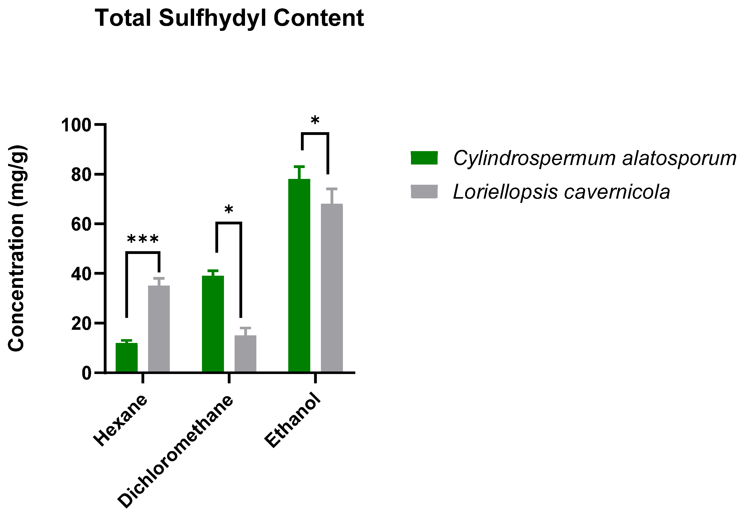
Figure 1. Total sulfhydryl content of the crude extracts of Cylindrospermum alatosporum and Loriellopsis cavernicola. The total sulfhydryl content of the extracts is presented in (mg GE/g dry extract), extrapolated from the calibration curve of glutathione after incubation at room temperature with 1% σ-phthaldehyde. p values: * 0.0332 *** 0.0002.
Applsci 15 09718 g001
Figure 2. Resveratrol (RSV) concentration after treatment with Cylindrospermum alatosporum and Loriellopsis cavernicola extracts. The extracts were incubated with RSV under UV radiation and in the dark. Resveratrol protection by the extracts was evaluated over time. HA—hexane extract of Cylindrospermum alatosporum; DA—dichloromethane extract of Cylindrospermum alatosporum; EA—ethanol extract of Cylindrospermum alatosporum; HB—hexane extract of Loriellopsis cavernicola; DB—dichloromethane extract of Loriellopsis cavernicola; EB—ethanol extract of Loriellopsis cavernicola.
Applsci 15 09718 g002
Table 1. Table 1 provides a summary of the inhibitory potential of Cylindrospermum alatosporum and Loriellopsis cavernicola extracts against collagenase, elastase, hyaluronidase, and tyrosinase. Both cyanobacteria performed similarly against the enzymes; however, the extracts of C. alatosporum appear the strongest, particularly its hexane and ethanol extracts.
IC50 (µg/mL)
HexaneDichloromethaneEthanolStandards
HAHBDADBEAEBECUAKA
Collagenase50 ± 1.5070 ± 3.44 **70 ± 5.93 **70 ± 6.23 **50 ± 6.3460 ± 5.34 **50 ± 2.30N/DN/D
ElastaseN/DN/DN/DN/DN/DN/DN/DN/DN/D
Hyaluronidase6 ± 0.276 ± 0.256 ± 0.556 ± 0.126 ± 0.456 ± 0.70N/DN/DN/D
TyrosinaseN/DN/DN/DN/DN/DN/DN/DN/D9 ± 0.50
HA—hexane extract of Cylindrospermum alatosporum; DA—dichloromethane extract of Cylindrospermum alatosporum; EA—ethanol extract of Cylindrospermum alatosporum; HB—hexane extract of Loriellopsis cavernicola; DB—dichloromethane extract of Loriellopsis cavernicola; EB—ethanol extract of Loriellopsis cavernicola; EC—epicatechin; UA—ursolic acid; KA—kojic acid; N/D—not determined. p value: ** 0.0021.
Table 2. Sun protection factor (SPF) values of Cylindrospermum alatosporum and Loriellopsis cavernicola extract.
Concentration
(µg/mL)
HAHBDADBEAEBProtection Level
UV-B Blocking
12011.65 ± 0.0431.12 ± 0.3611.25 ± 0.0049.98 ± 0.7017.14 ± 0.0514.67 ± 0.72Low to moderate
24021.30 ± 0.2831.92 ± 0.1114.96 ± 0.2651.74 ± 0.3121.28 ± 0.3622.93 ± 1.06Moderate
48039.04 ± 0.0333.39 ± 0.7420.58 ± 0.5152.04 ± 0.0222.88 ± 0.0125.12 ± 0.00Moderate to high
96059.76 ± 0.0149.3 ± 0.1826.24 ± 0.3678.96 ± 10.9053.20 ± 0.7027.30 ± 1.47Moderate to High
HA—hexane extract of Cylindrospermum alatosporum; DA—dichloromethane extract of Cylindrospermum alatosporum; EA—ethanol extract of Cylindrospermum alatosporum; HB—hexane extract of Loriellopsis cavernicola; DB—dichloromethane extract of Loriellopsis cavernicola; EB—ethanol extract of Loriellopsis cavernicola.
Disclaimer/Publisher’s Note: The statements, opinions and data contained in all publications are solely those of the individual author(s) and contributor(s) and not of MDPI and/or the editor(s). MDPI and/or the editor(s) disclaim responsibility for any injury to people or property resulting from any ideas, methods, instructions or products referred to in the content.