Article

Three-Dimensional Finite Element Analysis (FEM) of Tooth Stress: The Impact of Cavity Design and Restorative Materials

by
Yasemin Derya Fidancioğlu
1,
Sinem Alkurt Kaplan
2,*,
Reza Mohammadi
3,4 and
Hakan Yasin Gönder
2
1
Department of Pediatric Dentistry, Faculty of Dentistry, Necmettin Erbakan University, Konya 42090, Turkey
2
Department of Restorative Dentistry, Faculty of Dentistry, Necmettin Erbakan University, Konya 42090, Turkey
3
Faculty of Dentistry, Necmettin Erbakan University, Konya 42090, Turkey
4
Department of Oral and Maxillofacial Diseases, Faculty of Medicine, University of Helsinki, 00014 Helsinki, Finland
*
Author to whom correspondence should be addressed.
Appl. Sci. 2025, 15(17), 9677; https://doi.org/10.3390/app15179677
Submission received: 23 July 2025 / Revised: 17 August 2025 / Accepted: 19 August 2025 / Published: 3 September 2025

Abstract

Finite element analysis has been widely applied in restorative dentistry, but there is limited evidence directly comparing the biomechanical behavior of amalgam and bulk-fill composite resins in standardized cavity designs. This study aimed to evaluate the stress distribution in enamel, dentin, and restorative materials under different cavity configurations and filling materials. A 3D model of a maxillary molar was reconstructed from dental tomography using Geomagic Design X 2020. Four cavity models were created with Solidworks 2013: Class I (occlusal, Group A), Class II disto-occlusal (Group B), Class II mesio-occlusal (Group C), and Class II mesio-occluso-distal (Group D) cavities. Each model was restored with either amalgam or bulk-fill composite and a 600 N occlusal force was applied. Maximum principal stresses were analyzed with ABAQUS software. The highest stress was observed in the bulk-fill composite restoration of the Class II MO cavity (231 Mpa), whereas the lowest stress occurred in amalgam restoration of Class I cavity. Overall, amalgam restorations showed lower stress concentrations than bulk-fill composites, especially in complex cavity designs. These results suggest that cavity configuration and restorative material selection influence stress distribution and may impact the long-term biomechanical stability of restored teeth.

1. Introduction

Dental caries is one of the most common chronic diseases worldwide, and individuals remain susceptible throughout life. If untreated, carious lesions lead to structural damage and functional loss, requiring restorative intervention [1]. Over the years, a variety of restorative materials; including metals, ceramics, cements, and resin composites have been developed to replace tooth structure and restore function [2]. Among these, dental amalgam has been the standard material for more than a century due to its strength, wear resistance, ease of application, and cost effectiveness [3]. Despite these advantages, its poor esthetics, lack of adhesion, and mercury content have limited its acceptance in modern practice. Moreover, amalgam relies exclusively on mechanical retention, often requiring excessive removal of sound tooth structure, which conflicts with the principles of minimally invasive dentistry. Nevertheless, amalgam continues to be used, particularly in patients with special needs or in high volume public clinics, where its moisture tolerance and clinical reliability remain advantageous [4].
Resin composites emerged as the main alternative, offering superior esthetics and adhesive bonding to tooth structure. Progress in adhesive dentistry since the 1950s has facilitated their widespread use in both anterior and posterior restorations [5]. However, composite resins present challenges such as polymerization shrinkage, marginal leakage, and higher failure rates in extensive cavities compared to amalgam. Despite these shortcomings, they have become the most widely used in restorative material, largely driven by patient demand for tooth colored restorations [6,7].
Recent advances led to development of bulk-fill composites designed to simplify restorative procedures by allowing placement in increments up to 4 mm [8]. These materials exhibit improved translucency for deeper curing and incorporate modified fillers and monomers to reduce polymerization shrinkage. Clinically, bulk-fill composites reduce chair time and the risk of contamination between increments, which are significant advantages in posterior restorations. However, their long term performance remains influenced by multiple factors, including cavity type, number of bonded surfaces, and occlusal load [9,10].
In dentistry, it should be ensured that the forces occurring in the mouth are well recognized and analyzed, so that the forces can be distributed within physiological limits that would make the restorations compliant with the rules of oral rehabilitation. To this end, it is necessary to know the stress and strain against the forces. However, determining the behaviour of tissues and organs against coming forces is quite difficult, costly, and risky, and in some cases, impossible [11,12]. Considering these factors, it has become necessary to make models of living tissues and to determine the regions in such models where the forces are concentrated [13].
Finite element analysis (FEA) allows easy analysis of materials with complex geometries [14,15]. First used in the late 1960s when Ledney and Huang mathematically created a tooth model, FEA was introduced in dentistry in the 1970s by Farah et al. through his friends’ studies [15,16]. In the last 20 years, the finite element stress analysis has been frequently encountered in the literature [12,17].
FEA is a mathematical analysis method that, as mentioned, is easy to use to solve complex mechanical problems. FEM is known to be a powerful tool to design and optimize mechanical devices, as well as different human body parts or even human prostheses for foot, knee, or hip. This is especially true when their behavior is nonlinear [18]. It allows easier examination by dividing the region to be examined into small and simple elements, within which the solution can be provided [11,12,19]. Thus, with this method, analyses can be made in one dimension, two dimensions, and three dimensions.
Understanding how different cavity designs and restorative materials influence stress distribution is essential for improving restoration longevity and preserving tooth structure. While previous studies have investigated certain cavity types or specific restorative materials, there is limited evidence assessing all occlusal-including restorations (Class I, DO, MO, and MOD) simultaneously [20,21]. Addressing this gap is important to provide more comprehensive insight into how cavity configuration and material selection interact to affect biomechanical performance.
Therefore, the aim of this study was to evaluate the effects of different cavity designs and restorative materials on stress distribution within enamel, dentin, and restorative materials using three-dimensional finite element analysis (FEA). The null hypothesis was that cavity design and restorative material would not significantly influence stress distribution in the tooth–restoration complex.

2. Material and Methods

As an initial step, a three-dimensional image of an extracted permanent maxillary left first molar was obtained using a dental cone beam computed tomography (CBCT) (J Morita Mfg. Corp., Kyoto, Japan) (This study was unanimously approved by the Ethics Committee of Necmettin Erbakan University, Faculty of Dentistry). Tomography device irradiates at 90 kVp along with 5 mA current, for 17.5 s It. The voxel size of the device is 160 μm and its FOV is 40 × 40 mm.
With the Solidworks 2013 software (Solidworks Corp., Waltham, MA, USA), a 3D design software program, four different 3D cavity models were developed—Class 1 [occlusal (O), Group A], Class 2 [disto-occlusal (DO), Group B], Class 2 [mesio-occlusal (MO), Group C] and Class 2 [mesio-occlusal-distal (MOD), Group D] (Figure 1) all with a cavity angle of 95 degrees on the 3D model. Then, the bond gap thickness was regulated and the filling was modelled. Amalgam and bulk-fill composite were used as the filling materials. Their mechanical properties of dental tissues and restorative materials are shown in Table 1. Four cavities were modeled for each of these two materials, for a total of eight groups [Group A: amalgam-occlusal and Group A′: composite-occlusal; Group B: amalgam-disto-occlusal and Group B′: composite-disto-occlusal; Group C: amalgam-mesio-occlusal and Group C′: composite-mesio-occlusal; and Group D: amalgam-mesio-occlusal-distal and Group D′: composite-mesio-occlusal-distal).
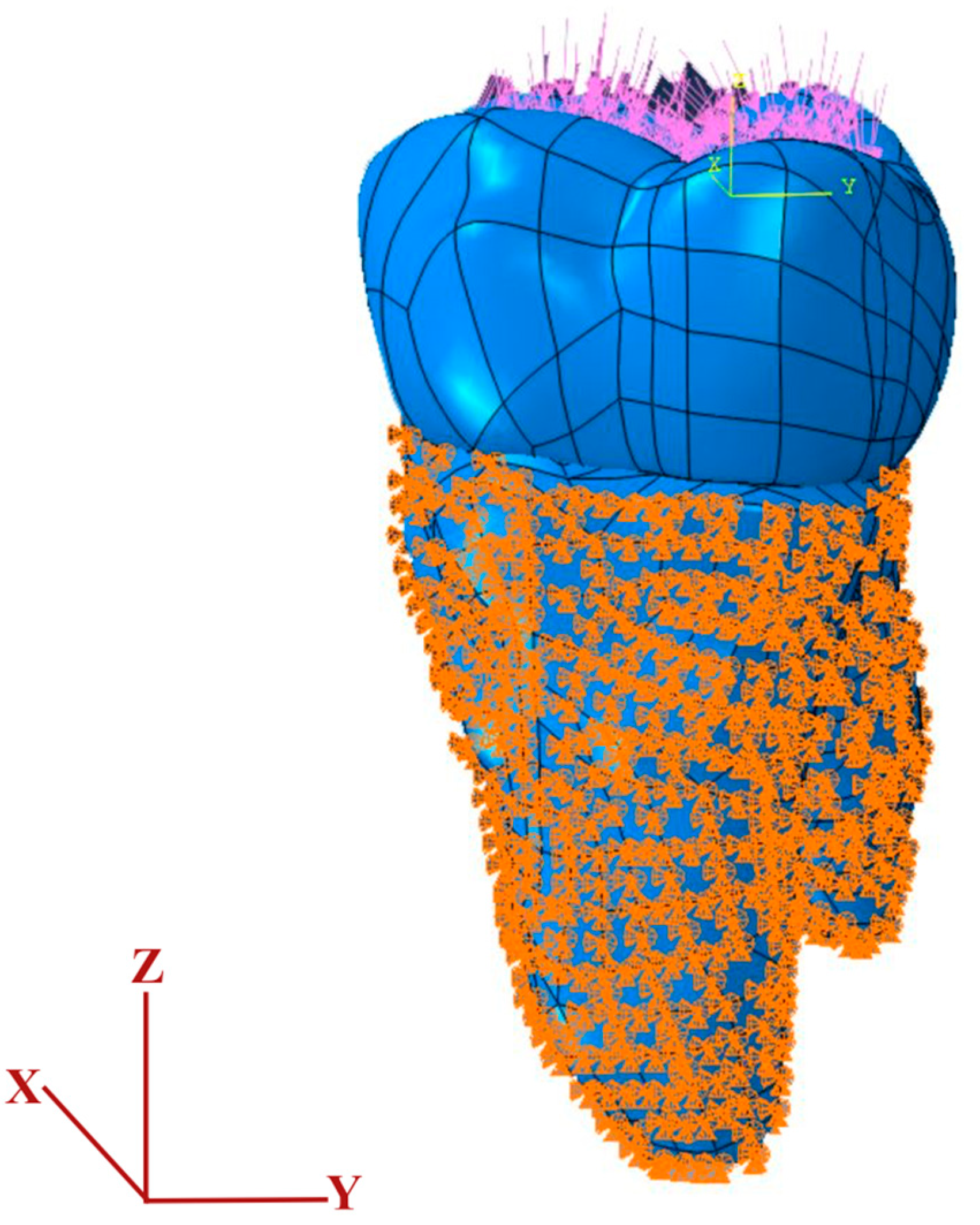
The restorative materials were defined in the simulation as isotropic and linearly elastic components. A vertical occlusal load of 600 N was applied in the FEA model, as it reflects the upper range of human masticatory force in the molar region—previous studies commonly use both normal chewing loads (~225 N) and maximum bite forces around 600 N to simulate realistic conditions [22,23]. The periodontal ligament (PDL) and alveolar bone were excluded from the model to simplify the analysis and isolate the biomechanical behavior of the tooth–restoration interface. This simplification is common in FEA research, although omitting the PDL can lead to slightly elevated stress values due to reduced damping. FEA simulations indicate that inclusion of the PDL significantly reduces force transmission compared to models without it, underscoring its shock-absorbing role [24]. Stress distribution was evaluated using finite element analysis (FEA) with Abaqus 2020 software (2020 Dassault Systèmes Simulia Corp., Johnston, RI, USA). Neither the periodontal ligament nor the surrounding jawbone structures were included in the model. Boundary conditions were established through the ‘Create Boundary Condition’ function in Abaqus, selecting mechanical constraints (symmetry/antisymmetry/encastre) under the load module. To simulate fixation, the tooth was constrained at the enamel–cementum junction, with all translational movements restricted (U1 = U2 = U3 = 0) from this junction to the apex. By applying these constraints to the region apical to the enamel–cementum junction (as shown in Figure 1), any three-dimensional displacement was effectively eliminated. The applied force represents the clinical loading conditions during routine chewing and biting (Figure 2).
The mesh properties used in the FEA for the tooth and restorations are shown in Table 2.
After the model produced in this study was transferred to the Abaqus program, meshing was performed by using Tet, Free (Algorithm, use default algorithm) in the mesh control section of the mesh tab. After this process, faulty areas were detected using verify mesh and the process was repeated several times in order to optimize the mesh quality, and the final mesh was decided (Table 2).
Table 1. Mechanical properties of dental tissues and restorative materials used in the 3D FEA models of the maxillary molars [20,25,26,27,28,29].
MaterialElastic Modulus (GPa)Poisson’s RatioTensile Strength (MPa)Compressive Strength (MPa)
Dentine 18.6000.3198.7297.0
Enamel 84.1000.3310.3384.0
Pulp 0.0020.45--
Amalgam 35.0000.353–5845–550
Bulk-fill composite12.0000.2542169.0
Adhesive system4.5000.3
Table 2. Nodes and elements for the different restorations tested.
ModelTotal ElementsTotal NodesMesh Type
A
(Amalgam-Occlusal Cavity)
7,066,4861,277,802Linear tetrahedral elements of C3D4
A′
(Composite-Occlusal Cavity)
7,235,8601,321,741Linear tetrahedral elements of C3D4
B
(Amalgam-Disto-occlusal Cavity)
7,404,6151,338,833Linear tetrahedral elements of C3D4
B′
(Composite-Disto-occlusal Cavity)
7,544,8621,381,242Linear tetrahedral elements of C3D4
C
(Amalgam-Mesio-occlusal Cavity)
7,275,5901,315,887Linear tetrahedral elements of C3D4
C′
(Composite-Mesio-occlusal Cavity)
7,354,6551,345,655Linear tetrahedral elements of C3D4
D
(Amalgam-Mesio-occluso-distal Cavity)
7,412,7391,342,075Linear tetrahedral elements of C3D4
D′
(Composite-Mesio-occluso-distal Cavity)
7,510,3281,379,288Linear tetrahedral elements of C3D4

3. Results

Finite element analysis was conducted to evaluate the stress distribution patterns generated by different cavity designs restored with either amalgam or bulk-fill composite. The findings demonstrated that both the restorative material and cavity configuration strongly influenced the magnitude and localization of stress concentrations within the enamel, dentin, restorative material, and adhesive layers.
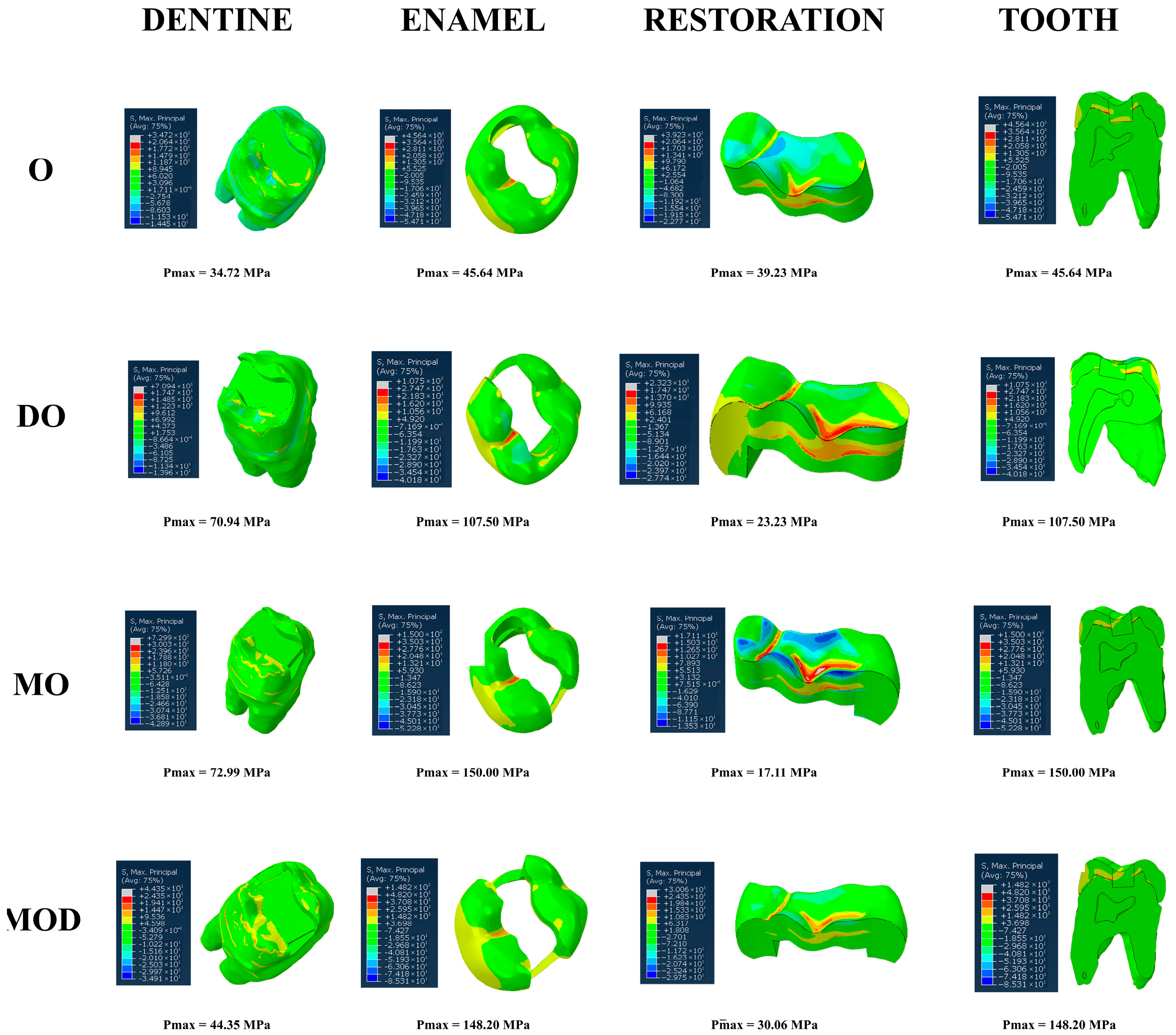
In amalgam restored models, stress values are generally lower and more evenly distributed across the tooth structure. The occlusal cavity (Group A) showed the lowest stress, with 45.64 MPa in enamel and 34.72 MPa in dentin. As the cavity design became more complex, stress levels increased, particularly in proximal cavities. The mesio-occlusal cavity (Group C) and the mesio-occluso-distal cavity (Group D) demonstrated the highest stresses, reaching 150 MPa and 148.20 MPa in enamel. Despite this increase, the stresses within the amalgam material itself remained low (17.11–39.23 MPa), reflecting amalgam’s ability to absorb occlusal loads and reduce transmission to tooth tissues. Stress concentrations were primarily observed at the internal line angles of cavity walls and along marginal ridges, yet no extreme localization occurred at the adhesive interface due to material’s reliance on mechanical retention (Figure 3 and Table 3).
Bulk-fill composite restorations exhibited a markedly different stress pattern. While simple occlusal cavities (Group A′) showed relatively low values (33.40 MPa in enamel and 24.56 MPa in dentin), stress increased sharply with more extensive preparations. The mesio-occlusal cavity (Group C′) generated the highest stress of all models, with 231 MPa in enamel and 157.90 MPa in dentin. A notable finding was the concentration of stress within the adhesive layer, which reached 211.80 MPa in Group C′, far exceeding the stresses observed in amalgam groups. Although stresses within the composite material itself were moderate (16.51–20.43 MPa), the high values at the interface indicate a potential risk of marginal breakdown and adhesive failure. Stress localization was most evident at the cervical margins of proximal boxes and at the base of the adhesive layer, suggesting vulnerability under functional loading (Figure 4 and Table 4).
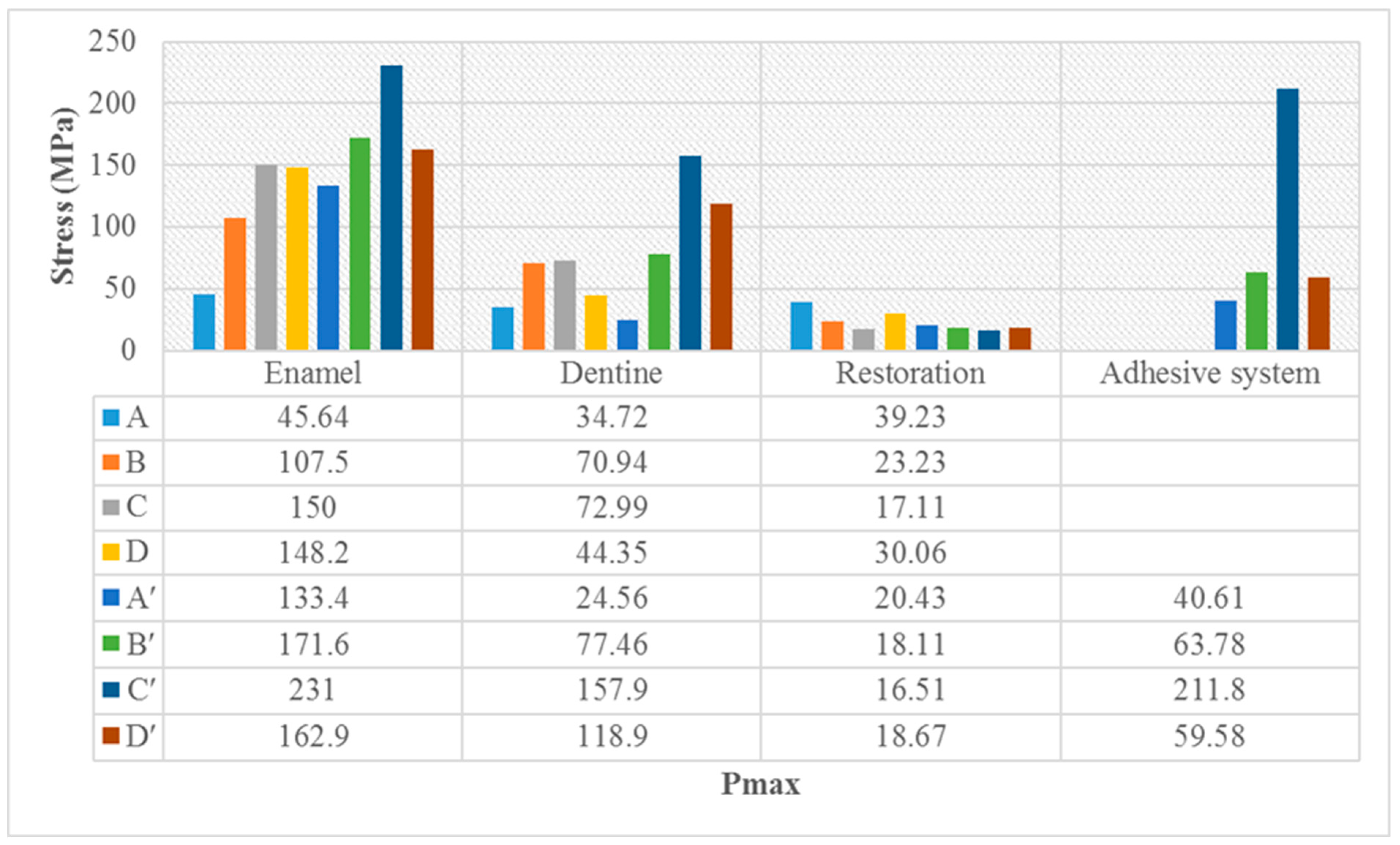
Across all cavity designs, amalgam restorations transmitted lower stresses to both enamel and dentin compared with bulk-fill composites. This difference became more pronounced in complex designs such as MO and MOD, where bulk-fill composite restorations produced significantly higher stresses at the adhesive interface. In contrast, amalgam restorations displayed more favorable stress distribution, likely contributing to greater biomechanical stability. These results support the clinical observation that amalgam restorations often demonstrate superior longevity in extensive caries, whereas bulk-fill composites may present higher susceptibility to debonding or secondary caries due to stress accumulation at adhesive interfaces (Figure 5).

4. Discussion

Due to the complexity of tooth structure and diversity of restorative materials, the mechanical performance of a restored tooth has been poorly understood so far. Although the mechanical benefits of curved junctions were reported by researchers, such junctions have not been used in dental restoration and cavity design [27]. Dental amalgam remains a widely used restorative material due to its ease of application and long-term clinical success. However, its lack of adhesion to tooth structures and reduced resistance of the remaining tooth increase the risk of crack propagation and fractures under repeated masticatory forces. Composite resins, on the other hand, possess an elastic modulus closer to natural tooth tissues, allowing for a more favorable stress distribution. Nevertheless, they have not consistently replaced amalgam in the restoration of non-vital teeth [30].
With the growing demand for aesthetics, light-cured composites have become increasingly common in posterior restorations. The traditional layering technique, however, is time-consuming, carries a risk of contamination, and may result in voids between layers. To overcome these challenges, bulk-fill composites were introduced. These materials can be polymerized in increments of 4–6 mm, reducing chair time and simplifying the restorative procedure [31]. For this reason, in the present study, bulk-fill resin composites were selected and compared with amalgam restorations to evaluate their biomechanical behavior.
For many years, many researchers have used finite element analysis as an alternative method to reduce costs in the design and optimization stages of mechanical problems. Finite element analysis has been widely used in industry to analyze engineering problems that are too complex to be solved by classical analytical methods, reducing the expense of experimental testing. One of the disadvantages of finite element analysis is that it requires a high computational cost, especially when it relies solely on the designer’s experience through simulations and trial and error in the process of placing proposed FE models [18].
The FEM techniques are totally non-intrusive and non-destructive, enabling researchers to investigate the biomechanical behavior of complex structures without altering or damaging the specimens. In addition, they allow global, non-contact measurement of displacement and strain fields, providing highly accurate data with sub-micrometric resolution. This makes FEM a valuable tool in dental biomechanics, as it offers detailed insight into stress distribution within both the restorative material and the surrounding dental tissues under various loading conditions. Such precision not only enhances the understanding of material behavior but also supports the development of more reliable and clinically relevant restorative strategies [30].
Similar finite element analyses have also been applied in prosthetic dentistry, particularly in evaluating the biomechanical performance of crowns, the ferrule effect, and implant-supported restorations. For instance, Eraslan et al. demonstrated that increasing ferrule height significantly reduced stress concentrations in endodontically treated teeth restored with crowns, highlighting the protective role of ferrule design [32]. Alberto et al. further showed that a full ferrule design in maxillary incisors restored with zirconia crowns and PEEK post–core systems improved stress distribution compared to partial ferrule designs [33]. In the implant field, Cosola et al. investigated ferrulized implant connections under fatigue loading and reported superior fracture resistance in designs with reinforced ferrule configurations [34]. Similarly, Epifania et al. analyzed crown stiffness and prosthetic screw absence in implant-supported systems, finding that stiffer crowns redirected stresses within the crown, whereas the lack of a screw increased stress at the abutment level [35]. These findings parallel our results by confirming that cavity design and restorative configuration directly influence stress distribution and long-term biomechanical stability. Integrating such insights from prosthodontics and implantology supports the broader applicability of FEM in restorative dentistry and reinforces the clinical significance of our findings.
Inside the oral cavity, the teeth and the restorations are subjected mainly to two types of stress: mechanical stress during functional activities and thermal stress due to temperature fluctuations. It is important to understand the stress distribution to enhance the longevity of the restorations [36]. Stresses generated during mastication and those related to polymerization shrinkage are also considered main causes of damage or failure of adhesive dental restorations [37,38].
The normal mastication force was calculated as 222 to 445 N (mean 322.50 N), and the highest 520 to 800 N (mean 660 N) in the molar region [22] and Ausiello et al. (2017) suggested that in a Class II MOD inlay, a 95° cavity-margin-angle under a 600 N load gives a more relevant picture of principal stress relief [38].
In Sengul et al. (2014), the stress applied to the primary molar tooth (tooth number 55) with an MO cavity and amalgam restoration was seen most in the enamel and least in the restoration [29]. In another inlay-FEA study by Darwich et al. (2021) [39] that used nanocomposite resin and lithium disilicate glass ceramic materials, the highest stress was observed on all the materials. Such stress was seen in the enamel and then in the dentine at least in the restorative material [39]. In the study of Tuncdemir et al. (2021) that used amalgam and bulk-fill composite resin, the stress distribution as a result of the force applied by the amalgam and composite restorations in Class I and Class II cavities was examined, and the highest stress was observed on the tooth with Class II composite restoration, while the lowest stress was observed on the tooth with Class I amalgam restoration and the stress on the tooth was less from the amalgam and more from the composite [40]. We obtained similar results in this study because the elastic modulus of the tooth was different from that of the restoration materials. The higher elastic modulus of amalgam than of the composite caused it to absorb more load and transmit less stress to the tooth.
In Yang H et al. (2018), which measured the stress distribution on occlusal, MO, MOD, and onlay composite, ceramic, and gold restorations, the highest stress was seen in the Class II-MOD restorations, while the lowest stress was seen in the onlay restoration [41]. In this study, the highest stress was observed in the Class II-MO composite restorations, and the lowest stress was seen on the tooth with Class I amalgam restoration.
Similar findings were reported by Gönder et al. (2023) [42], who investigated the stress distribution of bulk fill and conventional resin composites using finite element analysis. Their study demonstrated that when bulk fill composites were used instead of conventional resin composites, higher stress values were transmitted to the enamel, dentin, and adhesive interfaces, whereas the restorative material itself was subjected to lower stress levels. This was attributed to the higher modulus of elasticity of bulk fill composites, which causes a greater transfer of occlusal forces to the surrounding tooth structures. Our results are in agreement with these findings, as we also observed increased stress accumulation in the enamel and dentin tissues when bulk fill materials were used. This suggests that although bulk fill composites may provide certain clinical advantages, such as reduced application time, their mechanical behavior under load may lead to increased stress on the remaining tooth tissues, potentially affecting the longevity of restorations in the long term [42].
In this study, the amount of stress on the teeth was evaluated according to the most frequently applied cavity design differences between amalgam material, which has been used for many years, and bulk fill composite materials, which have become widespread recently. Evaluations were made with finite element analysis in a computer environment. This study has certain limitations that should be considered when interpreting the findings. The finite element model excluded periodontal ligament and alveolar bone structures, which may influence the actual clinical stress distribution. Additionally, the loading condition was simplified to a static vertical force of 600 N, while in vivo masticatory forces are dynamic and multidirectional. Other intraoral factors such as thermal cycling, humidity, fatigue loading, and individual patient variability were not simulated. Moreover, the material properties were assumed to be homogeneous, isotropic, and linearly elastic, which does not fully reflect the complex behavior of restorative materials and dental tissues in the oral environment. Future studies incorporating more realistic boundary conditions, fatigue testing, and clinical validation are necessary to better approximate real-world outcomes.
Although the null hypothesis of this study was that cavity design and restorative material would not significantly influence stress distribution in the tooth–restoration complex, the findings revealed the opposite. Both cavity design and material type significantly influenced stress patterns within the tooth structure and the restorative material. This outcome highlights that restorative decision-making cannot be isolated from biomechanical considerations. The findings of this study provide useful insights for clinical decision-making in restorative dentistry. Amalgam restorations demonstrated lower stress concentrations, particularly in simple cavity designs, suggesting that they may offer enhanced biomechanical stability under occlusal forces. However, the esthetic and biological limitations of amalgam should also be considered. Bulk-fill composites, while more prone to higher stress levels in complex cavity preparations, remain advantageous due to their esthetics, ease of use, and reduced chairside time. Clinicians should therefore carefully consider cavity design when selecting restorative materials: in more conservative cavity preparations, bulk-fill composites may be preferred for their esthetic benefits, whereas in extensive occlusal-involving designs, amalgam may provide more favorable stress distribution. Ultimately, a case-by-case evaluation, balancing biomechanical demands and patient expectations, is recommended for optimal clinical outcomes.

5. Conclusions

Within the limitations of this finite element analysis, where periodontal ligament and bone tissues were not modeled, it can be concluded that both cavity geometry and restorative material influence stress distribution in teeth. In Class I cavities, amalgam restorations produced lower stress in enamel but higher stress in dentin compared with bulk-fill composites. For Class II designs (MO, DO, MOD), amalgam demonstrated lower stress in both enamel and dentin, likely due to its higher elastic modulus and Poisson’s ratio, which allow greater stress absorption. Overall, amalgam showed more favorable stress adaptation than bulk-fill composites across cavity designs. These findings suggest that material selection should be guided not only by esthetic demands but also by cavity complexity and biomechanical requirements.

Author Contributions

Conceptualization, Y.D.F.; Methodology, Y.D.F., S.A.K., R.M. and H.Y.G.; Software, R.M.; Validation, R.M.; Formal analysis, R.M.; Investigation, R.M.; Resources, Y.D.F. and S.A.K.; Data curation, Y.D.F. and S.A.K.; Writing—original draft, Y.D.F., S.A.K. and H.Y.G.; Writing—review & editing, Y.D.F., S.A.K., R.M. and H.Y.G.; Supervision, H.Y.G.; Project administration, H.Y.G.; Funding acquisition, H.Y.G. All authors have read and agreed to the published version of the manuscript.

Funding

This research received no external funding.

Institutional Review Board Statement

In this study, extracted human teeth were used. This study was unanimously approved by the Ethics Committee for Non-Drug and Non-Medical Device Clinical Research at Necmettin Erbakan University, Faculty of Dentistry (approval number 2022/15-106 and approval date 24 February 2022).

Informed Consent Statement

Informed consent was obtained from all subjects involved in this study.

Data Availability Statement

The data presented in this study are available on request from the authors.

Conflicts of Interest

The authors declare no conflicts of interest.

References

  1. Selwitz, R.H.; Ismail, A.I.; Pitts, N.B. Dental caries. Lancet 2007, 369, 51–59. [Google Scholar] [CrossRef]
  2. Fejerskov, O.; Nyvad, B.; Kidd, E. (Eds.) Dental Caries: The Disease and İts Clinical Management; John Wiley & Sons: Hoboken, NJ, USA, 2015. [Google Scholar]
  3. Yadav, R.K.; Verma, U.P.; Tiwari, R.; Chaurasia, A. Mercury or mercury free restorations in oral cavity. Int. J. Public Health Sci. 2018, 7, 201–206. [Google Scholar] [CrossRef]
  4. Gönder, H.Y.; Demirel, M.G.; Mohammadi, R.; Alkurt, S.; Fidancioğlu, Y.D.; Yüksel, I.B. The Effects of Using Cements of Different Thicknesses and Amalgam Restorations with Different Young’s Modulus Values on Stress on Dental Tissue: An Investigation Using Finite Element Analysis. Coatings 2022, 13, 6. [Google Scholar] [CrossRef]
  5. Borgia, E.; Baron, R.; Borgia, J.L. Quality and survival of direct light-activated composite resin restorations in posterior teeth: A 5- to 20-year retrospective longitudinal study. J. Prosthodont. 2019, 28, e195–e203. [Google Scholar] [CrossRef] [PubMed]
  6. Sideridou, I.D.; Karabela, M.M.; Vouvoudi, E.C. Physical properties of current dental nanohybrid and nanofill light-cured resin composites. Dent. Mater. 2011, 27, 598–607. [Google Scholar] [CrossRef] [PubMed]
  7. Ferracane, J.L. Resin-based composite performance: Are there some things we can’t predict? Dent. Mater. 2013, 29, 51–58. [Google Scholar] [CrossRef]
  8. Yazici, A.R.; Kutuk, Z.B.; Ergin, E.; Karahan, S.; Antonson, S.A. Six-year clinical evaluation of bulk-fill and nanofill resin composite restorations. Clin. Oral Investig. 2022, 26, 417–426. [Google Scholar] [CrossRef]
  9. Rosatto, C.M.; Bicalho, A.A.; Veríssimo, C.; Bragança, G.F.; Rodrigues, M.P.; Tantbirojn, D.; Versluis, A.; Soares, C.J. Mechanical properties, shrinkage stress, cuspal strain and fracture resistance of molars restored with bulk-fill composites and incremental filling technique. J. Dent. 2015, 43, 1519–1528. [Google Scholar] [CrossRef]
  10. Benetti, A.R.; Havndrup-Pedersen, C.; Honoré, D.; Pedersen, M.K.; Pallesen, U. Bulk-fill resin composites: Polymerization contraction, depth of cure, and gap formation. Oper. Dent. 2015, 40, 190–200. [Google Scholar] [CrossRef]
  11. Ulusoy, M.; Aydın, A.K. Diş Hekimliğinde Hareketli Bölümlü Protezler, Cilt II; Ankara Üniversitesi Basımevi: Ankara, Turkey, 2003; pp. 687–689. [Google Scholar]
  12. Geng, J.P.; Tan, K.B.; Liu, G.R. Application of finite element analysis in implant dentistry: A review of the literature. J. Prosthet. Dent. 2001, 85, 585–598. [Google Scholar] [CrossRef]
  13. Çalıkkocaoğlu, S. Dişsiz Hastaların Protetik Tedavisi: Klasik Tam Protezler; Quintessence: Istanbul, Turkey, 2010. [Google Scholar]
  14. Şen, S.; Bayındır, Y.Z.; Güler, Ç. İnsan dişi kaplamalarında kullanılan farklı özelliklerdeki yapıştırıcı simanların gerilme dağılımına etkilerinin sonlu elemanlar yöntemi ile incelenmesi. Atatürk Üniversitesi Diş Hekim. Fakültesi Derg. 2012, 2012, 31–39. [Google Scholar]
  15. Mohammed, S.D.; Desai, H. Basic concepts of finite element analysis and its applications in dentistry: An overview. J. Oral Hyg. Health 2014, 2, 156. [Google Scholar]
  16. Shetty, P.; Hegde, A.; Rai, K. Finite element method–an effective research tool for dentistry. J. Clin. Pediatr. Dent. 2010, 34, 281–285. [Google Scholar] [CrossRef]
  17. Wakabayashi, N.; Ona, M.; Suzuki, T.; Igarashi, Y. Nonlinear finite element analyses: Advances and challenges in dental applications. J. Dent. 2008, 36, 463–471. [Google Scholar] [CrossRef]
  18. Gómez, F.S.; Lorza, R.L.; Bobadilla, M.C.; García, R.E. Improving the Process of Adjusting the Parameters of Finite Element Models of Healthy Human Intervertebral Discs by the Multi-Response Surface Method. Materials 2017, 10, 1116. [Google Scholar] [CrossRef]
  19. Adigüzel, O. Finite Element Analysis: Review Part I: The uses in dentistry, basic concepts and description of elements. Dent. J. Dicle 2010, 11, 18–23. [Google Scholar]
  20. Ausiello, P.; Ciaramella, S.; Fabianelli, A.; Gloria, A.; Martorelli, M.; Lanzotti, A.; Watts, D.C. Mechanical behavior of bulk direct composite versus block composite and lithium disilicate indirect Class II restorations by CAD-FEM modeling. Dent. Mater. 2017, 33, 690–701. [Google Scholar] [CrossRef] [PubMed]
  21. Ausiello, P.; Ciaramella, S.; Di Rienzo, A.; Lanzotti, A.; Ventre, M.; Watts, D.C. Adhesive class I restorations in sound molar teeth incorporating combined resin-composite and glass ionomer materials: CAD-FE modeling and analysis. Dent. Mater. 2019, 35, 1514–1522. [Google Scholar] [CrossRef]
  22. de Abreu, R.A.; Pereira, M.D.; Furtado, F.; Prado, G.P.; Mestriner, W., Jr.; Ferreira, L.M. Masticatory efficiency and bite force in individuals with normal occlusion. Arch. Oral Biol. 2014, 59, 1065–1074. [Google Scholar] [CrossRef]
  23. Liu, T.; Huang, Y.; Li, Y.; Meng, J.; Liu, Y.; Wei, Y.; Huang, Y.; Zhou, Q.; Yang, W.; Yan, F.; et al. Effect of different restorative design and materials on stress distribution in cracked teeth: A finite element analysis study. BMC Oral Health 2025, 25, 31. [Google Scholar] [CrossRef] [PubMed]
  24. Della Bona, A.; Borba, M.; Benetti, P.; Duan, Y.; Griggs, J.A. Three-dimensional finite element modelling of all-ceramic restorations based on micro-CT. J. Dent. 2013, 41, 412–419. [Google Scholar] [CrossRef] [PubMed]
  25. Pałka, K.; Bieniaś, J.; Dębski, H.; Niewczas, A. Finite element analysis of thermo-mechanical loaded teeth. Comput. Mater. Sci. 2012, 64, 289–294. [Google Scholar] [CrossRef]
  26. Jiang, W.; Bo, H.; Yongchun, G.; Longxing, N. Stress distribution in molars restored with inlays or onlays with or without endodontic treat-ment: A three-dimensional finite element analysis. J. Prosthet. Dent. 2010, 103, 6–12. [Google Scholar] [CrossRef] [PubMed]
  27. Babaei, B.; Shouha, P.; Birman, V.; Farrar, P.; Prentice, L.; Prusty, G. The effect of dental restoration geometry and material properties on biomechanical behaviour of a treated molar tooth: A 3D finite element analysis. J. Mech. Behav. Biomed. Mater. 2022, 125, 104892. [Google Scholar] [CrossRef]
  28. Rodrigues, M.D.; Soares, P.B.; Gomes, M.A.; Pereira, R.A.; Tantbirojn, D.; Versluis, A.; Soares, C.J. Direct resin composite restoration of endodontically-treated permanent molars in adolescents: Bite force and patient-specific finite element analysis. J. Appl. Oral Sci. 2020, 28, e20190544. [Google Scholar] [CrossRef]
  29. Sengul, F.; Gurbuz, T.; Sengul, S. Finite element analysis of different restorative materials in primary teeth restorations. Eur. J. Paediatr. Dent. 2014, 15, 317–322. [Google Scholar]
  30. Valin Rivera, J.L.; Gonçalves, E.; Vinicius Soares, P.; Milito, G.; Ricardo Perez, J.O.; Palacios Roque, G.F.; Valin Fernández, M.; Figueredo Losada, H.; Araújo Pereira, F.; Garcia del Pino, G.; et al. The Restored Premolars Biomechanical Behavior: FEM and Experimental Moiré Analyses. Appl. Sci. 2022, 12, 6768. [Google Scholar] [CrossRef]
  31. Gönder, H.Y.; Öz, C. Bulk-fill kompozit rezinler. Necmettin Erbakan Üniversitesi Diş Hekim. Derg. 2020, 2, 117–123. [Google Scholar] [CrossRef]
  32. Eraslan, O.; Aykent, F.; Yücel, M.T.; Akman, S. The finite element analysis of the effect of ferrule height on stress distribution at post-and-core-restored all-ceramic anterior crowns. Clin. Oral Investig. 2009, 13, 223–227. [Google Scholar] [CrossRef] [PubMed]
  33. Alberto, L.H.; Zhang, Z.; Duan, Y. Effect of Ferrule Design on Stress Distribution of Maxillary Incisor Rehabilitated with Ceramic Crown and PEEK Post–Core Material: A 3D Finite Element Analysis. Ceramics 2023, 6, 2256–2268. [Google Scholar] [CrossRef]
  34. Cosola, S.; Toti, P.; Babetto, E.; Covani, U.; Penarrocha-Diago, M.; Penarrocha-Oltra, D. In-vitro fatigue and fracture performance of three different ferrulized implant connections used in fixed prosthesis. J. Dent. Sci. 2021, 16, 397–403. [Google Scholar] [CrossRef]
  35. Epifania, E.; di Lauro, A.E.; Ausiello, P.; Mancone, A.; Garcia-Godoy, F.; Mendes Tribst, J.P. Effect of crown stiffness and prosthetic screw absence on the stress distribution in implant-supported restoration: A 3D finite element analysis. PLoS ONE 2023, 18, e0285421. [Google Scholar] [CrossRef] [PubMed]
  36. Agnihotri, H.; Bhatnagar, N.; Rao, G.V.; Jain, V.; Parkash, H.; Kar, A.K. Evaluation of the onset of failure under mechanical and thermal stresses on luting agent for metal–ceramic and metal crowns by finite element analysihons. Contemp. Clin. Dent. 2010, 1, 227–233. [Google Scholar] [CrossRef]
  37. Dejak, B.; Młotkowski, A. A comparison of stresses in molar teeth restored with inlays and direct restorations, including polymerization shrinkage of composite resin and tooth loading during mastication. Dent. Mater. 2015, 31, e77–e87. [Google Scholar] [CrossRef] [PubMed]
  38. Ausiello, P.; Ciaramella, S.; Garcia-Godoy, F.; Gloria, A.; Lanzotti, A.; Maietta, S.; Martorelli, M. The effects of cavity-margin-angles and bolus stiffness on the mechanical behavior of indirect resin composite class II restorations. Dent. Mater. 2017, 33, e39–e47. [Google Scholar] [CrossRef] [PubMed]
  39. Darwich, A.; Adel, O.; Nazha, H. Finite element analysis of onlay restorations: Influence of design and onlay material on stress distribution in mandibular molars. SSRN Electron. J. 2021. [Google Scholar] [CrossRef]
  40. Tuncdemir, M.T.; Yeşilyurt, N.G.; Arıkan, M. Comparison of the Stress Distribution in Class I and Class II Amalgam and Bulk-Fill Composite Restorations Using CAD-FEM Modeling. Int. J. Periodontics Restor. Dent. 2021, 41, e1–e9. [Google Scholar] [CrossRef]
  41. Yang, H.; Park, C.; Shin, J.H.; Yun, K.D.; Lim, H.P.; Park, S.W.; Chung, H. Stress distribution in premolars restored with inlays or onlays: 3D finite element analysis. J. Adv. Prosthodont. 2018, 10, 184–190. [Google Scholar] [CrossRef]
  42. Gönder, H.Y.; Mohammadi, R.; Harmankaya, A.; Yüksel, İ.B.; Fidancıoğlu, Y.D.; Karabekiroğlu, S. Teeth Restored with Bulk–Fill Composites and Conventional Resin Composites; Investigation of Stress Distribution and Fracture Lifespan on Enamel, Dentin, and Restorative Materials via Three-Dimensional Finite Element Analysis. Polymers 2023, 15, 1637. [Google Scholar] [CrossRef]
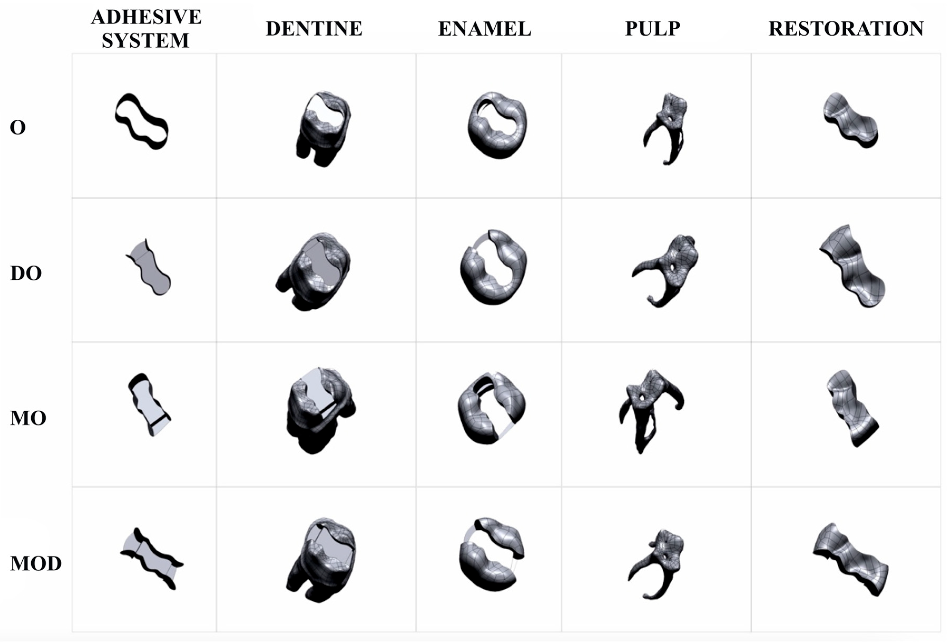
Figure 1. Class 1 [occlusal (O), Group A], Class 2 [disto-occlusal (DO), Group B], Class 2 [mesio-occlusal (MO), Group C], and Class 2 [mesio-occlusal-distal (MOD), Group D]. All groups: Adhesive layer, Dentine, Enamel, Pulp and Restoration.
Applsci 15 09677 g001
Figure 2. Load and boundary conditions.
Applsci 15 09677 g002
Figure 3. Maximum principal stress (Pmax) distribution in the enamel, dentine, and restoration with the use of amalgam as a restorative material.
Applsci 15 09677 g003
Figure 4. Maximum principal stress (Pmax) distribution in the enamel, dentine, and restoration with the use of bulk-fill composite resin as a restorative material.
Applsci 15 09677 g004
Figure 5. Maximum principal stress (Pmax) distribution in the enamel, dentine, restoration, and adhesive system in each group.
Applsci 15 09677 g005
Table 3. Maximum principal stress (Pmax) distribution in the enamel, dentine, and restoration with the use of amalgam.
Cavity TypeDentinEnamelRestorationTooth
Occlusal (O)34.7245.6439.2345.64
Disto-occlusal (DO)70.94107.5023.23107.50
Mesio-occlusal (MO)72.99150.0017.11150.00
Mesio-occluso-distal (MOD)44.35148.2030.06148.20
Table 4. Maximum principal stress (Pmax) distribution in the enamel, dentine, and restoration with the use of bulk-fill composite resin.
Cavity TypeDentinEnamelRestorationToothAdhesive System
Occlusal (O)24.5633.4020.4333.4040.61
Disto-occlusal (DO)77.46171.6018.11171.6063.78
Mesio-occlusal (MO)157.90231.0016.51231.00211.80
Mesio-occluso-distal (MOD)118.90162.9018.67162.9059.58
Disclaimer/Publisher’s Note: The statements, opinions and data contained in all publications are solely those of the individual author(s) and contributor(s) and not of MDPI and/or the editor(s). MDPI and/or the editor(s) disclaim responsibility for any injury to people or property resulting from any ideas, methods, instructions or products referred to in the content.