Article

Formation of Ordered Ionic Salt Agglomerates Through Evaporative Crystallization in Hanging Drop Systems

1
Lasers Department, National Institute for Lasers, Plasma and Radiation Physics, 409 Atomistilor Street, 077125 Bucharest-Magurele, Romania
2
Academy of Romanian Scientists (AOSR), Ilfov Street 3, 050044 Bucharest, Romania
3
Geological Institute of Romania, 1 Caransebes Street, 012271 Bucharest, Romania
*
Authors to whom correspondence should be addressed.
Appl. Sci. 2025, 15(17), 9280; https://doi.org/10.3390/app15179280 (registering DOI)
Submission received: 14 July 2025 / Revised: 12 August 2025 / Accepted: 20 August 2025 / Published: 23 August 2025
(This article belongs to the Section Optics and Lasers)

Abstract

Featured Application

Spheroidal agglomerates of sodium chloride (NaCl) can serve as microchambers for thermal and/or chemical reactions, featuring infrared-transparent walls. Alternatively, they may function as templates for faceted structures. Facet lenses and facet mirrors are optical devices capable of dividing a single light beam into multiple distinct beams. Utilizing agglomerated NaCl crystals as facet templates, we fabricated faceted carbonaceous materials. This methodology offers promising avenues for the synthesis of faceted graphene and carbon nanotube-based structures, which may be integrated as functional components within various types of sensors. Convex or concave faceted colloidal photonic crystals exhibit simultaneous Bragg diffraction of white light, enabling the splitting, focusing, and dispersion of both transmitted and reflected beams. Such materials hold potential for incorporation during the development of spectrometers.

Abstract

This study introduces novel experimental systems that facilitate the nucleation, growth, aggregation, and agglomeration of ionic salt solutions, leading to structurally and functionally distinctive crystal formations. Through evaporative crystallization in hanging drops—including layered binary solutions—a range of macroscopic agglomerates were produced, such as hollow spheroidal NaCl/NiSO4 structures, octahedral NaCl films, pentagonally arranged CdSO4 spherulites, and NH4Cl dendritic shells. Additionally, NaCl spheroids were used as templates to fabricate carbon-based morphologies and colloidal photonic crystals with convex or concave geometries, which were subsequently analyzed optically. The study reveals that crystallization and self-assembly, whether independently or synergistically applied, can yield complex architectures with potential applications in advanced device manufacturing beyond conventional processing methods.

1. Introduction

Self-assembly and crystallization are intriguing processes in which ions, molecules, or particles spontaneously organize themselves into structured patterns without external intervention. These phenomena play a pivotal role in various scientific domains, including nanotechnology and materials science. Their principles are fundamental to the design of advanced materials, pharmaceuticals, and emerging nanotechnologies. However, what characteristics do they share, and what distinguishes them? Can these processes operate synergistically, and if so, in what manner? These are among the key questions that this article seeks to explore.
Self-assembly is a process by which components spontaneously organize themselves into structured and functional arrangements without external guidance [1,2,3]. It is a phenomenon observed in various natural and synthetic systems, from the microscopic world of molecules and proteins to macroscopic structures like certain types of assembled nanomaterials. Self-assembly demonstrates nature’s ability to create complex systems through simple rules and interactions [1,2,3]. There are more specific forms of self-assembly, such as molecular self-assembly [4,5] which has as its driving force hydrogen bonding (attraction between hydrogen atoms and electronegative atoms), pi-stacking (interactions between aromatic rings), and hydrophobic forces (non-polar molecules aggregate to avoid water); supramolecular self-assembly [6,7], which has as its driving force non-covalent interactions (hydrogen bonding and van der Waals forces) or host–guest chemistry (molecules form complexes with specific shapes and functions); and colloidal self-assembly [8,9] which has as its driving force van der Waals forces, electrostatic interactions (charges on the particles can lead to attraction or repulsion), depletion attraction (caused by the presence of polymers or other small particles in the solution), capillary forces, or external fields: magnetic, electric, light, or gravity fields. Colloidal self-assembly can also be classified in nanoparticle self-assembly [10,11] and hard-sphere colloid self-assembly [12,13]. Self-assembly is a powerful tool in nanotechnology, enabling the creation of complex structures and devices with a wide range of applications, such as drug delivery systems (for targeted delivery of pharmaceuticals), nanotechnology (creating nanoscale structures and devices), biomolecular condensates (formation of cellular structures), molecular machines (creating devices at the molecular level), catalysis (enhancing chemical reactions), material science (developing new materials with unique properties), photonic crystals (used in optical devices), sensors (for detecting chemical or biological substances), cosmetics (to create structured materials with specific properties), and many others [9,10,11,12,13].
However, there have been only a few attempts at using ionic salt crystals as building blocks in a self-assembly phenomenon. Potential solutions may lie in the phenomenon of crystal aggregation [14], where a crystallizing solution could produce a quasi-ordered solid, with each individual crystal acting as a unit of a colloidal crystal. In the fields of crystallography and mineralogy, a crystal aggregate [15,16,17] refers to a collection of crystals that have grown together, often forming a mass devoid of the distinct external morphology characteristic of individual crystals. These aggregates can present a variety of structural forms, including granular, fibrous, botryoidal, lamellar (layered), bladed (elongated and flattened), and dendritic (branching in a tree-like manner) forms. Similarly to individual crystals, crystal aggregates may exhibit symmetrical elements such as rotational axes, roto-inversion axes, a center of symmetry, and mirror planes. The properties of these aggregates, including their structural configuration, morphology, shape, single-crystal size, habit and integrity are determined by phenomena such as nucleation [18,19,20,21], growth [22,23], and agglomeration [24,25]. Since self-assembly typically occurs in a liquid medium, our research is confined to the crystallization of ionic salts from solution.
Nucleation in solution may occur through the cooling or evaporation of a saturated solution or through the presence of impurities within the solution. Even crystal seeds function as impurities, thereby promoting secondary crystallization. The rate of crystal growth is markedly influenced by variables such as the temperature, pressure, and the availability of spatial and nutrient resources during the crystallization process. Agglomeration proceeds through a three-step mechanism wherein individual single crystals collide, form a loosely bound aggregate, and ultimately coalesce through the formation of solid bridges, facilitated by the supersaturation of the surrounding medium.
The final configuration and morphology of a crystal aggregate are influenced by numerous factors. Even in a straightforward scenario where three drops of same crystallizable solution evaporate under identical environmental conditions, the resulting crystal aggregates exhibit three distinct structures: a crystalline film, a dendritic formation, and a spherical agglomerate [26]. This variability arises from the processes of nucleation, growth, and agglomeration, which, regardless of the simplicity or complexity of the setup, are fundamentally governed by two principal factors: supersaturation and the width of the metastable zone.
Supersaturation [27] is a non-equilibrium physical state in which a solution contains a greater amount of a solute than permitted by its equilibrium solubility. At low levels of supersaturation, crystal growth occurs at a faster rate than nucleation, resulting in a larger distribution of crystal sizes. Conversely, at high supersaturation levels, nucleation dominates, leading to the formation of smaller crystals. Effective control of supersaturation is critical for achieving the desired product characteristics, particularly the final crystal size distribution and crystalline phase.
The metastable zone width [28] is defined as the level of supersaturation at which the first crystal becomes observable within the solution. It represents the upper limit of supersaturation at which the solution remains stable with respect to nucleation. The metastable zone width is influenced by variables such as the temperature, solution composition, cooling or evaporation rates, the presence of impurities, and mechanical effects [29,30,31,32,33,34,35]. Even minor alterations in the experimental conditions can push the system beyond its stability threshold, triggering sudden crystallization. Furthermore, the metastable zone width, along with the structure, morphology, and degree of ordering of the crystal agglomerates, is significantly impacted by the nature and geometry of the interface between the crystallization zone and the external environment [36,37,38]. Various types of interfaces, including liquid/gas (e.g., aerosols) [39,40], liquid/liquid (e.g., emulsions and microfluidic devices) [41,42], liquid/solid/gas (e.g., sessile or hanging drops and droplets, thin or thick films) [43,44,45,46], and liquid/solid (e.g., confined systems like capillaries, membranes, or porous solids) [47,48,49,50] interfaces, can produce vastly different crystal agglomerates, even for the same ionic salt solution under identical experimental conditions.
The structure and morphology of crystal aggregates have been extensively studied to elucidate their distinctive properties resulting from aggregation [51,52] or to identify the conditions under which they develop patterned structures. Patterns formed through the aggregation of crystals of specific ionic salts, as reported in the scientific literature, include two-dimensional configurations such as grids [53], ripples [54], flower-like shapes [55], stains [56], dendritic formations [57], and snowflake-like structures [58]. Furthermore, three-dimensional formations, such as spherical agglomerates [59], layered aggregates [60], ring shells [61], spherical shells [62], and mesocrystals [63], have also been observed. In addition, crystallization and self-assembly have been synergistically combined by employing colloidal crystals as templates to crystallize various salts within their pores, leading to the creation of ionic salt inverse-opals [64,65]. The study of simultaneous crystallization and agglomeration/self-assembly phenomena holds potential applications in various fields, including the fabrication of biomimetic materials [66,67], the development of soft crystal sensors [68], and advancements in medical diagnostics [69].
This article aims to explore the mechanisms of nucleation, growth, aggregation, and agglomeration of crystals formed from specific ionic salt solutions through the evaporative crystallization of hanging drops, employing innovative experimental setups where gravitational effects are of significant importance. Particular emphasis will be placed on analyzing the degree of structural ordering within the resulting crystal agglomerates. Furthermore, the study will outline several compelling applications derived from the findings, including the development of facet materials [70,71] and light-diffractive optical devices with concave and convex features [72,73].

2. Materials and Methods

2.1. Materials

Analytical-grade ionic salts in a crystalline powder form, commercially available, were utilized alongside sub-micron SiO2 spheres with an average diameter of 0.264 mm (5% w/v) provided as an aqueous colloidal solution by microParticles GmbH, Berlin, Germany. The ionic salts were dissolved in deionized water, while the colloidal SiO2 solution was utilized as received without further modification.
For the purposes of crystallization and self-assembly, a variety of substrates were employed, including smooth, clean glass cylinders with a diameter of 7 mm, glass and steel spheres with a diameter of 10 mm, and commercially available glass microscope slides. Polystyrene flakes (Mw 35,000) sourced from Sigma-Aldrich Chemie GmbH, St. Louis, MO, USA, were used in the fabrication of structured substrates.
The deposition of ionic salt and colloidal solutions onto the substrates was performed using standard, commercially available 1 mL KD-JECT III syringes equipped with G 29 needles.

2.2. Methods

2.2.1. Preparation of Salt Solution

Deionized water was placed in a vessel, into which an excess amount of powdered salt was introduced. The mixture was vigorously stirred to ensure the dissolution of the salt and subsequently left undisturbed, resulting in a clear solution in equilibrium with a residual salt deposit that settled at the bottom of the container. In this equilibrium state, the concentration of the solution, denoted as c0, corresponded to the saturation concentration, cs, at the ambient temperature, T. A portion of the clear solution was extracted using a syringe and utilized to produce saturated solution hanging drops. Alternatively, a small quantity of deionized water was added to the syringe to dilute the solution, thereby generating an unsaturated solution, which was used to form unsaturated solution hanging drops.

2.2.2. Formation of Hanging Drops

The substrate was initially positioned in a horizontal orientation. Subsequently, a droplet of either an ionic salt or colloidal solution was carefully formed at the syringe’s tip. The syringe was used to produce droplets with volumes ranging from 2 to 12 μL. Each droplet was delicately transferred onto the underside of the substrate by making light contact. Additional droplets were incrementally added to the pre-existing drop hanging from the substrate until the drop approached its maximum equilibrium volume, just prior to detachment and falling. The maximum volume of the hanging drops was approximately 60 μL when glass cylinders served as substrates and approximately 180 μL when microscope glass slides were employed. For evaporation rate or supersaturation measurements, the experimental setup was adapted to rest on the platen of an electronic scale.

2.2.3. Layered Binary Fluid Hanging Drop Formation

The process commenced by suspending the substrate in a horizontal orientation. A droplet of deionized water was then carefully formed at the tip of a syringe and subsequently transferred to the bottom surface of the substrate by gently touching it. Additional droplets were incrementally added to the existing droplet until its maximum diameter was attained. Maintaining contact between the syringe tip and the hanging drop, a portion of the droplet’s volume was extracted to produce a hanging drop with the desired volume and a fixed (maximum) diameter. Subsequently, a droplet of a specified ionic salt solution, prepared in accordance with the procedure outlined in Section 2.2.1, was formed at the syringe tip and delicately transferred to the bottom of the hanging water drop. Additional solution droplets were introduced until the target volume was achieved.

2.2.4. Introducing a SiO2 Colloidal Crystal Fragment into a Hanging Drop

The procedure began with the deposition of a small SiO2 colloidal crystal, measuring several hundred micrometers in size, onto a hydrophobic substrate. Subsequently, a small droplet of the solution was formed at the syringe needle tip and carefully brought into contact with the surface of the colloidal crystal while it was observed under a magnifying lens. The colloidal crystal was transferred to the syringe tip. Finally, the colloidal crystal was transferred into the hanging drop by gently bringing the syringe tip into contact with the surface of the hanging drop.

2.2.5. Fabrication of Faceted Convex Carbonaceous Materials

A NaCl spheroidal hollow agglomerate, fabricated using the hanging drop method, was horizontally positioned with its opening facing upward on a metal substrate containing a small hole, the diameter of which was smaller than that of the spheroid. Sucrose powder was then added to the spheroid until it was completely filled. This assembly was introduced into a furnace preheated to 400 °C and allowed to remain there for 30 min. Afterward, the sample was removed from the furnace, naturally cooled, and then subjected to ultrasonication in distilled water for 1–2 min. This process yielded a spheroidal, faceted carbonaceous material suitable for further analysis.

2.2.6. Fabrication of Faceted Convex Colloidal Photonic Crystals

A NaCl spheroidal hollow agglomerate, prepared using the hanging drop method, was horizontally positioned with its opening facing upward on a metal substrate. The substrate contained a small hole on its surface, narrower than the spheroid’s diameter. Polystyrene powder was poured into the spheroid until it was completely filled. The assembly was then placed in a furnace set at 170 °C for 2 min. Following this, the sample was removed from the furnace, naturally cooled, and then subjected to ultrasonication in distilled water for 1–2 min, resulting in a spheroidal, faceted polystyrene material ready to serve as a template for SiO2 colloid self-assembly. For this purpose, the faceted polystyrene sample was vertically positioned with its faceted surface facing downward (while its non-faceted side was anchored on the bottom tip of a vertical fiber). Two droplets of a SiO2 colloidal solution (~20 μL) were applied by touching the faceted surface. The colloid self-assembled within the pores of the polystyrene template over the course of several hours under normal laboratory conditions (T = 25 °C, RH = 60%).

2.2.7. Fabrication of Faceted Concave Colloidal Photonic Crystals

A few flakes of polystyrene were deposited onto a glass slide and then placed in a furnace. The polystyrene was heated to 170 °C until it formed a molten drop approximately 1–2 mm in height. Subsequently, a NaCl spheroid was placed vertically onto the molten polystyrene using thin tweezers, with its opening facing upward. The spheroid partially sank into the molten polystyrene without filling its interior. Once the sample was removed from the furnace and allowed to cool and solidify, it was washed to dissolve the NaCl, resulting in a faceted concave polystyrene sample. This sample was then kept in a horizontal position, and two droplets of a SiO2 colloidal solution were introduced into the faceted concavity. The solution was left to evaporate under standard laboratory conditions (T = 25 °C, RH = 60%) for 2–3 h. As a result of evaporative self-assembly, a faceted concave colloidal photonic crystal was formed.

2.3. Investigations

Macroscale observations of the crystal agglomerates were conducted using a zoom camera and optical microscopy. To simultaneously capture both the interior and exterior features of the photographed objects, the samples were placed on a flat mirror. This setup allowed for the recording of both the objects and their reflections in a single photograph.
Four distinct optical microscopes were employed to analyze the reflected and transmitted light. One of them was endowed with a linear polarizer.
A scanning electron microscope (SEM) (Apreo S, Thermo Fisher Scientific, Auburn, AL, USA) was utilized to examine the structures and morphologies of the samples.
UV–Vis reflectance spectra were obtained via a fiber optic system using an AvaLight-DHc light source and an AvaSpec-ULS2048CL-EVO high-resolution spectrometer, both provided by Avantes, Apeldoorn, The Netherlands.
Atomic force microscopy (AFM) (XE-100, Park Systems Inc., Santa Clara, CA, USA) was employed to investigate the morphology of the structural units within the aggregates.

3. Results and Discussion

3.1. Evaporative Crystallization from a Hanging Drop

When a drop of a colloidal solution containing hard spheres is suspended from a substrate (Figure 1a [74]), it forms a high-quality spheroidal colloidal crystal upon solvent evaporation (Figure 1b [74]). This phenomenon is attributed to the negligible static friction coefficient at the liquid/air interface and the gravitational force driving the self-assembly process (Figure 1c) [74,75,76].
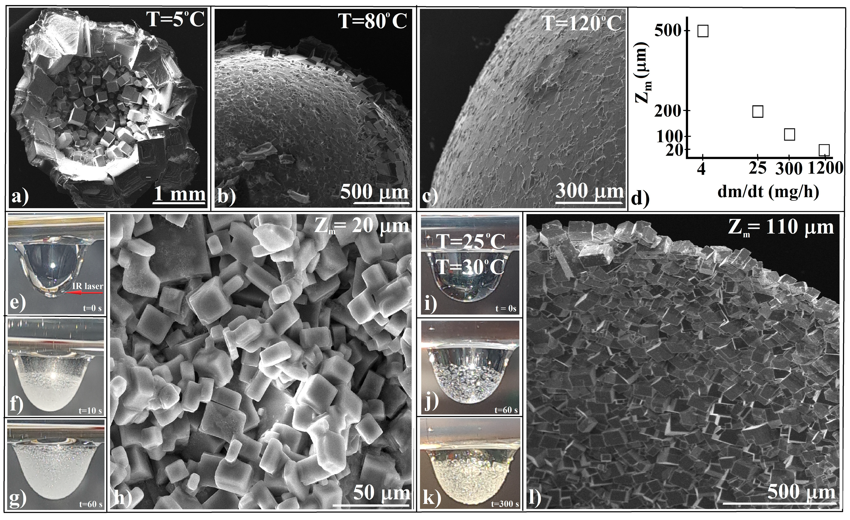
In a large-volume hanging drop of a NaCl solution (Figure 1d), with an initial concentration, c0, lower than its saturation concentration, cs, gravity induces a notable gradient in the solute distribution and a convective flow along the drop height [77,78]. This configuration results in a reduced critical supersaturation value, the concentration at which spontaneous crystallization occurs. Consequently, the growth rate significantly surpasses the nucleation rate, leading to the formation of a small number of crystals (even just one, as shown in Figure 1d) with large dimensions and flat faces at the end of the droplet’s evaporation. This outcome is consistent with our previous work and the findings of other researchers [79,80,81]. In this configuration, the induction time (τ) is around one hour. When maintaining the experimental conditions (Figure 1e) but starting with a saturated solution (c0 = cs), the sub-micron crystals which exist in a dynamic equilibrium of dissolution and crystallization rapidly grow (τ = 0.5 h) and aggregate at the drop’s bottom (Figure 1e). The aggregate growth is periodic rather than continuous, implying that new critical supersaturation levels must be reached for additional crystals to nucleate and grow. Using a magnifying lens or camera, new single crystals can be observed nucleating at the liquid/gas or liquid/solid/gas interface, sliding down along the drop’s liquid/air interface, and contacting the already existing crystals. We consider that gravity and the negligible friction at the liquid/air interface have the same roles as in the case of colloidal crystals (Figure 1c). Once the growing aggregate reaches the substrate, the new crystals spread as an efflorescence layer out from the triple-line contact of the drop until complete solvent evaporation. This phenomenon is entirely reproducible and independent of experimental parameters such as the ambient pressure, temperature, relative humidity, mechanical and temperature shocks, and the substrate’s nature and shape.
The crystal agglomerate formed a spheroidal shell (bell-like) (Figure 1f) with a thickness equivalent to that of a single crystal. Each crystal from the spheroid composition was oriented with its (111) crystallographic planes approximately normal to the radial direction of the spheroid (Figure 1g,h). The crystals exhibited significant size dispersion, ranging from 150 to 200 μm, while maintaining a well-preserved habit. The spheroids demonstrated impressive mechanical resistance, with well-bonded crystals capable of withstanding falls from over one meter in height and enduring ultrasonic treatment in ethanol without disintegrating. These pore-free spheroids are ideal for depositing non-polar liquids, powders, or melts. Their high mechanical resistance and their absence of pores were attributed to solid bridges that welded the individual crystals together at their interfaces, transforming the aggregates into agglomerates in the third stage of the agglomeration process [82].
Although several studies have documented the formation of spherical agglomerates, they have not extensively explored the arrangement of individual crystals within these agglomerates. Typically, these formations are referred to as crusts, shells, spherical aggregates, or spherical crystals. In colloidal crystal synthesis, a temperature increase or evaporation rate decrease lead to the better ordering of the colloidal spheres [83,84]. Also, the temperature and evaporation rate have been shown to affect the quantity, size, and growth of ionic and protein crystals [12,85,86]; thus we studied the role of the temperature and evaporation rate in the NaCl agglomeration process. By maintaining the setup architecture, large-volume hanging drops, and initial concentration (c0 = cs), we investigated the changes in the crystallization of spheroidal crystal agglomerates under conditions of a fixed temperature, small fluctuations in the temperature, and shock-induced variations in the drop temperature. The induction time (τ) was employed for the qualitative comparison of these phenomena.
In an evaporative crystallization process where the evaporation is not affected by temperature variation (as in our case), the evaporation rate increases with a rise in the temperature, significantly influencing the crystal growth [32]. Crystals that grow more slowly (at lower temperatures) tend to be larger, while when drops experience rapid evaporation (at higher temperatures) [87] this can reduce their sizes to a few tens of micrometers. This can be observed in the SEM images of NaCl spheroids grown at T = 5 °C (Figure 2a), T = 80 °C, and T = 120 °C (Figure 2b,c). The overall dependence of the crystal size on the drop evaporation rate in our experiments is depicted in Figure 2d.
Evaporative crystallization of a hanging drop of a NaCl solution at different ambient temperatures revealed the following: (a) The formation of a spheroidal crystal agglomerate shell was independent of the temperature, even during boiling. (b) The number of crystals in the agglomerate composition increased with a rising temperature, while their size decreased as the temperature increased. This implies that the ratio of the nucleation rate to the growth rate increased with the temperature. (c) The cubic crystals’ habit became less distinguishable at higher temperatures. (d) The degree of ordering of the crystals decreased with an increasing temperature.
We believe these observations indicate competition between crystal nucleation and growth, balanced by the evaporation rate [32], with the temperature as a visible experimental parameter. Though the results were reproducible, these experiments did not allow for a generalization of the approach. However, they can give us an idea of the direction to follow.
When subjecting saline drops to a thermal shock by impacting them with an infrared pulsed laser beam (photo/thermal conversion [88,89,90]) (laser wavelength of λ = 10.6 μm, power density of P = 3 kW/cm2, pulse duration of t = 0.5 s, Figure 2e), rapid heating of a shell at the liquid/gas interface of the drop occurred, leading to explosive boiling, intense evaporation, a rapid increase in saturation, and fast crystallization (τ = 10 s, Figure 2f) of numerous small crystals with a size of ~20 μm (Figure 2h). The crystals aggregated in a disordered manner, forming a spheroidal agglomerate shell (Figure 2g).
Even a small temperature fluctuation during crystallization is known to increase the likelihood of numerous crystals nucleating. In a new experimental setup, a minor variation in the temperature between the substrate and the droplet (Figure 2i) resulted in significant effects. The induction time decreased (Figure 2j) in comparison with that shown in Figure 1b, which implies earlier agglomeration (Figure 2k) and subsequently the production of a larger volume of the agglomerate. A high-quality spheroidal crystal agglomerate was obtained, characterized by a larger number of well-defined crystals (Figure 2l).
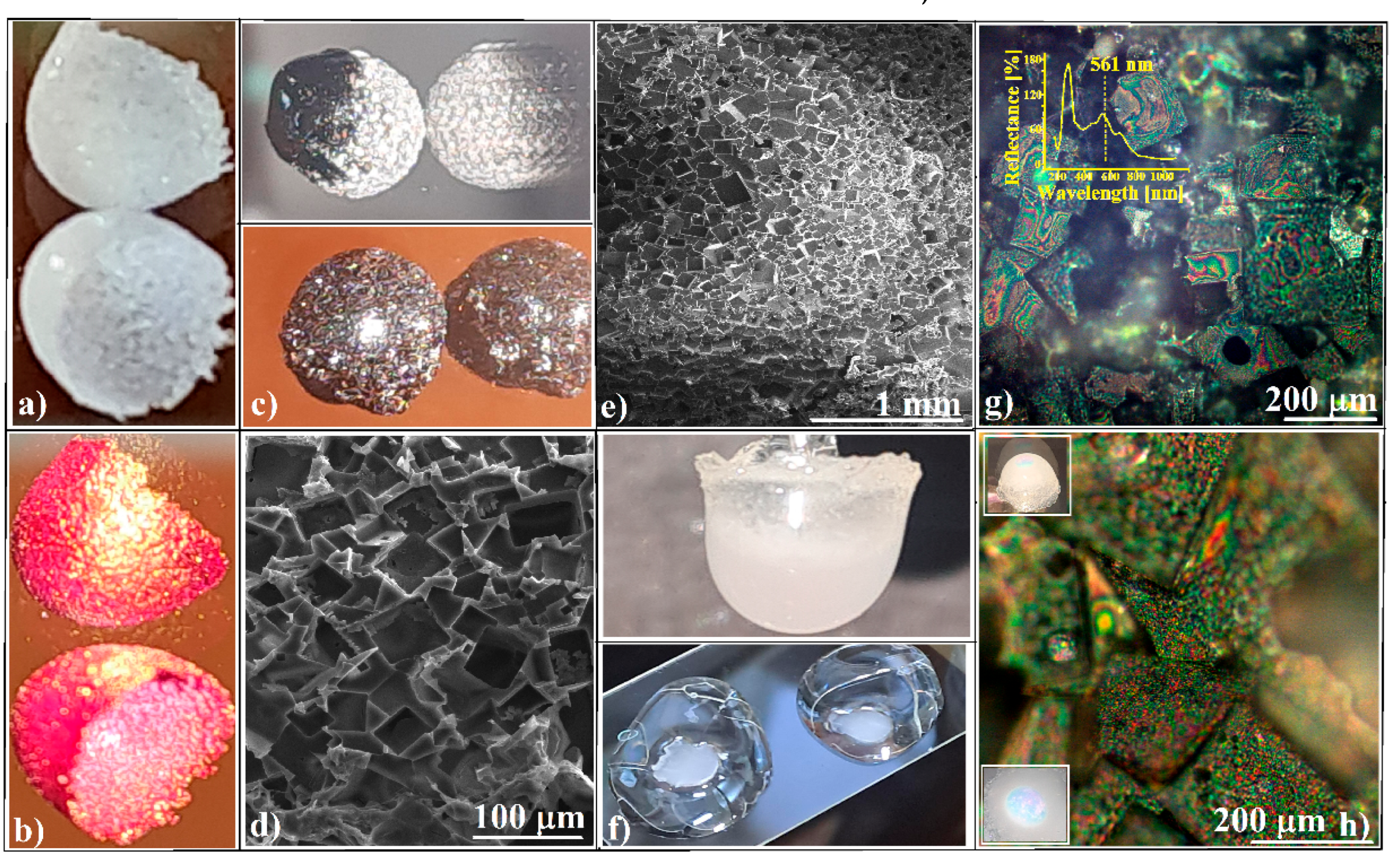
Neither a sudden shock nor a slight temperature fluctuation significantly enhanced the ordering of single crystals or altered the shape of the spheroids, as observed. However, these NaCl spheroids (Figure 3a) remained transparent in the infrared region [91]. Their lack of pores makes them ideal for use as sample cells for non-aqueous liquids (Rose Bengal from ethanol, Figure 3b) in IR spectroscopy or IR laser beam irradiation in specific setups [92]. The size of the single crystals (ranging from tens to hundreds of micrometers) within the spheroids’ composition suggests that terahertz spectroscopy could be an appropriate method for investigating their agglomeration phenomenon [93,94]. Their faceted texture makes them suitable for use as simple optical devices (such as facet lenses or facet mirrors [70,71,95]) or as more complex ones (like facet graphenes [96] or oriented carbon nanotubes [97]), which could be accomplished by using them as faceted reaction chambers for some carbon precursor materials [98,99]. Although the use of crystals and crystal agglomerates as templates is not a novel approach in the synthesis of materials with special properties [100,101], the employment of three-dimensional ordered crystal agglomerates, as in our case, significantly increases the complexity of the resulting materials. A faceted carbonaceous sample obtained by us through the thermal treatment of sucrose (T = 400 °C, in air, for t = 30 min) is shown in Figure 3c,d. Similarly, by melting and then cooling polystyrene powder in contact with the inner or outer surface of a spheroid, faceted convex and concave polystyrene structures can be fabricated (Figure 3e). These structures can be used as substrates and templates for colloidal self-assembly (of a 264 nm SiO2 colloid, in our case) (Figure 3f), resulting in faceted convex or concave colloidal photonic crystals (Figure 3g,h). These nearly optical devices exhibit vivid colors which can be seen in optical microscopy images, colors generated by the Bragg diffraction of white light. A geometric optical phenomenon, the simultaneous observation of transmitted (red) and reflected (green) light when the structures’ reflections in a plane mirror were photographed (insets in Figure 3h), was also observed. UV-Vis retroreflection spectroscopy (inset in Figure 3g) showed a broad band centered at λ = 561 nm, which is characteristic of the (111) family planes of a 264 nm SiO2 colloidal crystal [74,75]. Its broadness was attributed to a wide filling factor and the different orientations of the facets relative to the direction of the incident light.
However, all of these examples are still at the proof-of-concept level. Significant efforts are needed to improve their quality and transform them into final applications.
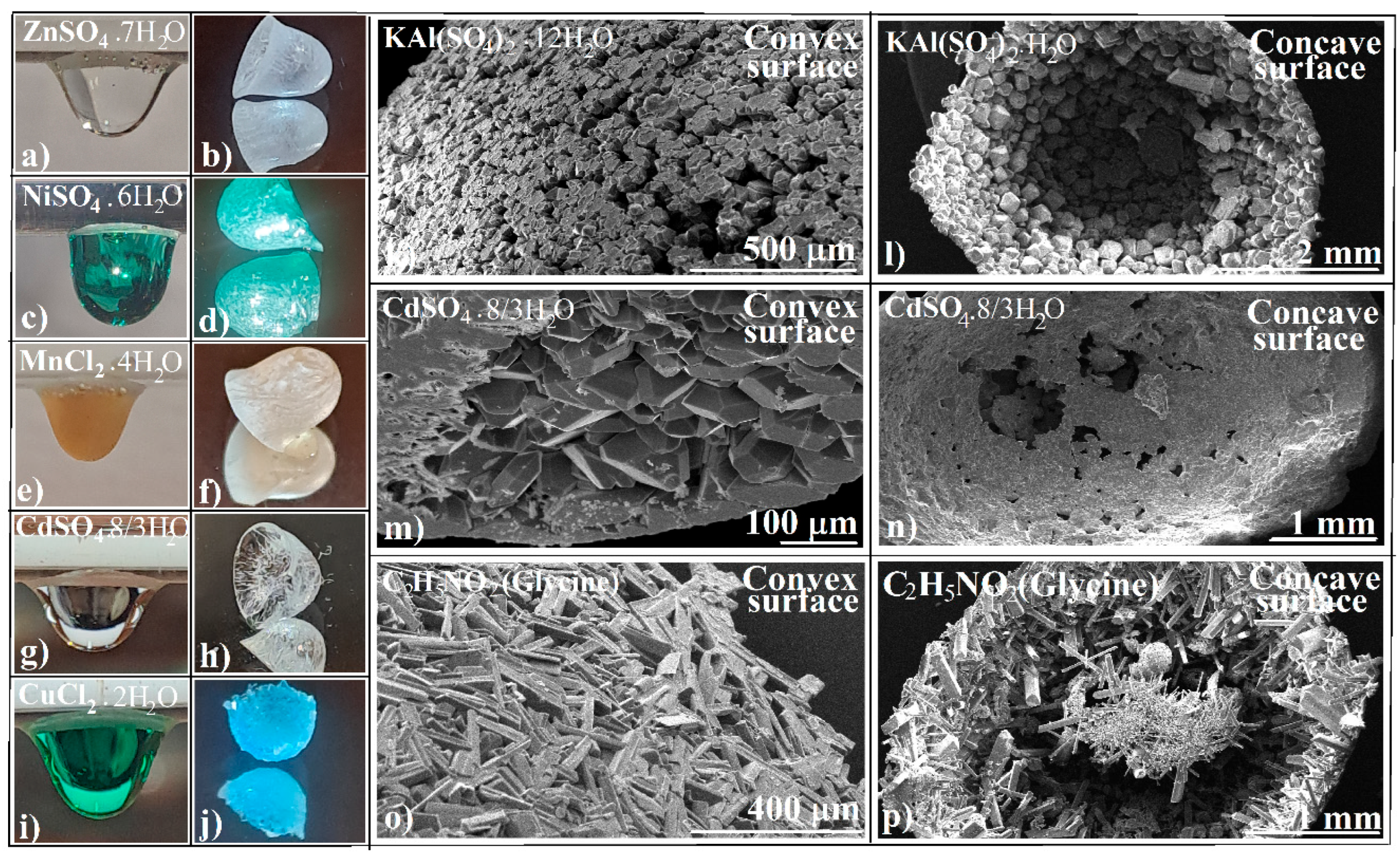
These experimental features and proposed applications apply to NaCl and KCl. In the aggregation process of these anhydrous salts, water may serve solely as a transport medium. Hydrated salts [102,103,104], on the other hand, can retain water within their crystals and form intra-aggregate bonds, potentially leading to the production of various types of agglomerates or distinct crystal arrangements. In our experiments, hanging drops of some hydrated ionic salt solutions, where c0 = cs, with a varying water content in the crystallization solution, were allowed to crystallize by drop evaporation at T = 25 °C and RH = 60% (Figure 4a–j).
Direct observation of the crystallization of the drops revealed that (a) all the ionic salts crystallized into a spheroidal crystal agglomerate shell (Figure 4a–j). (b) The overall evaporation time varied, ranging from a few hours to a few tens of hours, and did not correlate with the water content in the crystallization solution. (c) The number and size of single crystals within the agglomerate did not correlate with the water content in the crystallization solution. However, the number of crystals increased and their size decreased when crystallization began with a temperature fluctuation, as shown in Figure 2i.
SEM images taken of some of the final agglomerates (Figure 4k–p) revealed that crystals with a habit different from that of cubic NaCl organized themselves in a similar manner. They were oriented such that the radial direction of the spheroid was normal to one of their (111) crystallographic planes, resulting in a relatively continuous and smooth surface.
However, the saturation concentration cs was no longer a general threshold point in the formation of the crystal agglomerate shell. Some hydrated salts began to form a shell starting from a more diluted solution. Moreover, the CdSO4 solution formed a crystal agglomerate shell with a 2D spherulitic structure, but only if the initial solution concentration c0 was lower than its saturation concentration, cs.
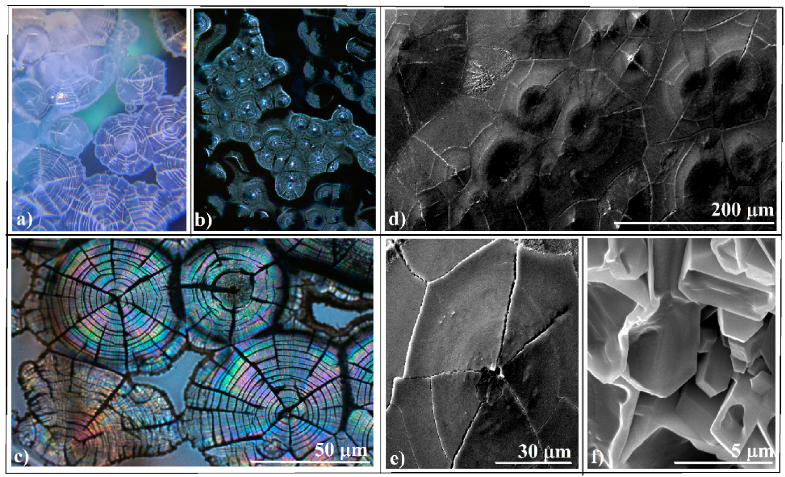
Spherulites [105,106] are crystalline structures that radiate from a central point, forming spherical domains. They can develop in metals, polymers, organic molecules, and biopolymers and crystallize from melts. There are a few documented instances of spherulites forming from evaporating solutions of ionic salts, such as CaCO3 [107], BaCO3, and SrCO3, often studied in the context of bio-mineralization. However, there is no information about the formation of spherulites from cadmium sulfate; thus we were surprised to observe bouquets (small clusters) of spherulites blooming in the transmitted (Figure 5a) and reflected (Figure 5b) light of the optical microscope. These structures acquired colors when polarized light was used (Figure 5c), demonstrating properties of optical birefringence [108,109]. Both optical microscopy and SEM investigations (Figure 5d,e) confirmed that we were indeed dealing with a bidimensional (2D) spherulitic structure. The convex side presented spherulites while the concave one (Figure 5f) presented the normal crystal habit of CdSO4·8/3H2O.
However, there were only a few spherulites on the spheroid surface. Their packing tendency led us to believe that a much larger number of spherulites could be organized into an array, a pattern, or even a 2D hierarchically organized crystal, given the proper conditions.
At the end of this chapter, it can be concluded that the evaporative crystallization of ionic salt solutions in a maximally voluminous hanging drop results in the development of a hollow, spheroidal (bell-like) crystal agglomerate. Within this structure, the (111) crystallographic planes of the individual crystals comprising the agglomerate are generally oriented perpendicular to the spheroid’s radius. This phenomenon occurs when the initial solutions are saturated and contain sub-micron crystals in a dynamic state of dissolution and crystallization equilibrium. However, hydrated ionic salts can form spheroids in starting solutions with a concentration below saturation. In this case, CdSO4 forms spherulitic crystals.

3.2. Evaporative Crystallization in a Layered Binary-Solution Hanging Drop

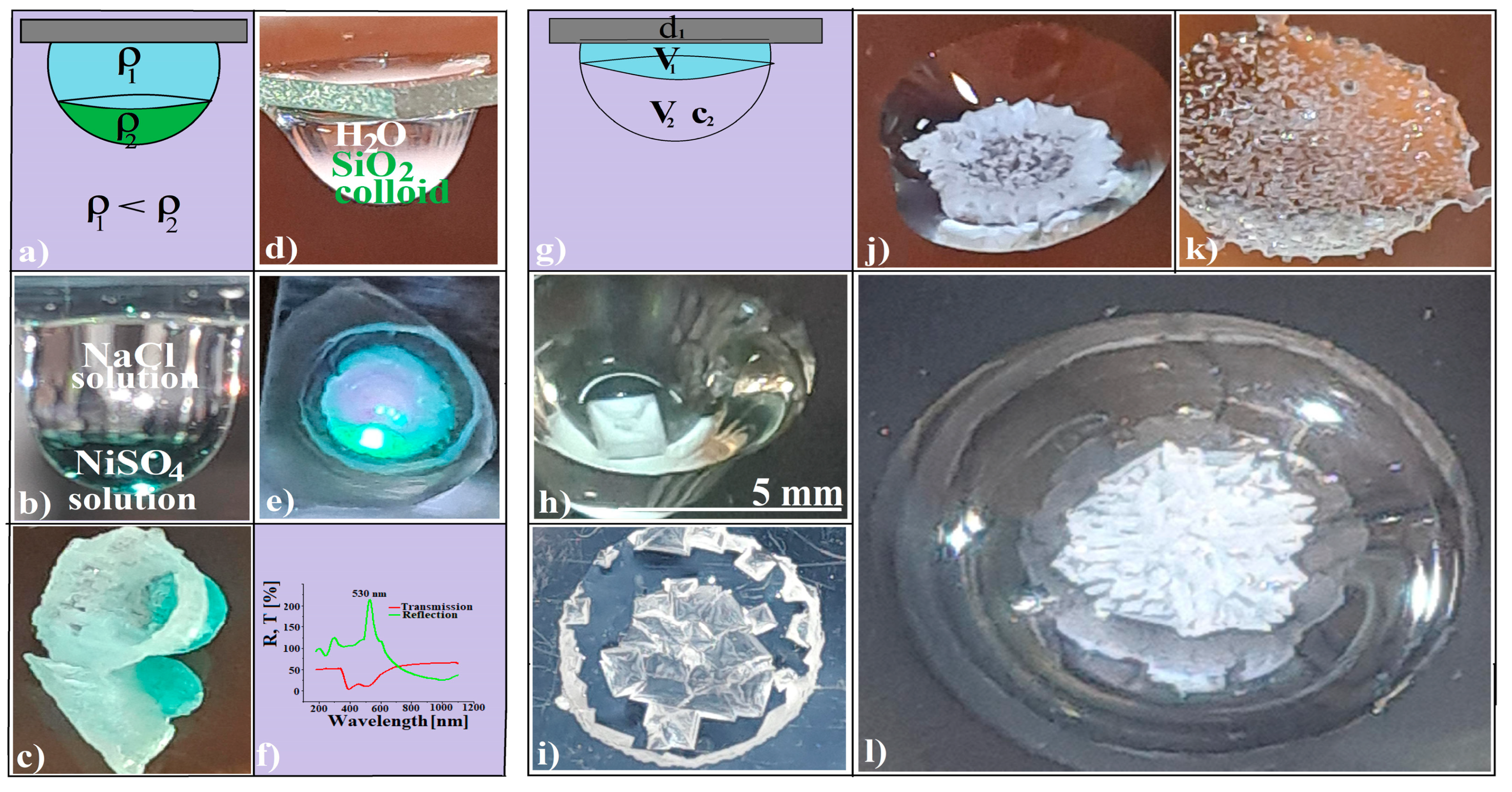
Most research on evaporative crystallization in droplets focuses on single-component droplets or droplets containing several miscible liquids in a homogeneous mixture [110,111,112]. However, two or more distinct liquids can remain in equilibrium in a suspended drop if their densities differ. A liquid will float if it is less dense than the liquid it is placed in and sink if it is denser (Figure 6a). In this case, the system can be considered a stratified fluid, where the fluid density varies in the vertical direction [113,114].
If the liquids are crystallizable solutions (such as saturated solutions of NaCl and NiSO4, see Figure 6b) that are allowed to evaporate under normal ambient conditions, various competing phenomena develop in the droplet. New flows and gradients, different from those in a single-component suspended drop, influence the nucleation and growth of crystals in the final agglomerate, forming two distinct and united crystal shells (Figure 6c). This results from the competition between local solution saturation generated by droplet evaporation and the much slower process of solution interdiffusion. It was surprising for us to see that this approach also worked in the case of a SiO2 (264 nm) colloid droplet hanging from a suspended water drop (Figure 6d), which finally formed a high-quality colloidal crystal film with a slightly curved bump in its center (Figure 6e). UV-Vis spectroscopy performed on such films (Figure 6e) revealed the existence of intense transmission and reflection Bragg diffraction bands. The slight shift in the transmission band towards smaller wavelengths relative to the reflection band was caused by the different investigation setups used, with a light incident angle of θ = 0° for reflected and θ = 45° for transmitted light.
Even more surprising was the observation that the evaporation of a concentrated NaCl solution in the same experimental setup, a suspended layered binary-solution drop (Figure 6g), induced nucleation, growth, and organization of an unique crystal (Figure 6h), a few crystals forming a compact group (Figure 6i) or organized as a ring (Figure 6j), a single layer of crystals (Figure 6k), or a 3D crystal aggregate with radial symmetry (Figure 6l).
The formation of the agglomerate observed in Figure 6l can be explained by DLA (diffusion-limited aggregation) theory [115], which describes the agglomeration process of particles moving randomly until they encounter an existing structure to which they adhere. Over time, this results in the formation of complex, tree-like structures known as dendrites. However, although NaCl is known to form dendrites, particularly in a gel medium [116], the crystal agglomerate in Figure 6l, with its short branches and radial symmetry, is quite unusual. The symmetry of the crystal pathways, influenced by the geometry of the drop’s bottom, may have contributed to the unique shape and structure of this NaCl agglomerate.
Intrigued by the poor reproducibility and lack of control over the aggregate formation shown in Figure 6h–l, we attempted to identify the specific features of the setup responsible for these issues. We found that the water drop’s surface contact diameter (d1) and volume (V1), as well as the saline drop’s volume (V2) and its concentration (c2), were the experimental parameter values that ensured decent (>90%) reproducibility and control over the aggregate shapes. Their values are provided in Table 1. The significance of the d1 value should be emphasized. It represents the maximum contact diameter that a hanging drop can achieve when spreading on a substrate. This value remains constant for the same substrate. In most cases, d1 is approximately 7 mm for water and water-based solutions and glass slide substrates; however, it may vary for glass slides manufactured by different producers. In such cases, it is necessary to experimentally determine new, appropriate values for V1 and V2.
It is remarkable that the experimental values from Table 1 form a pattern. Considering the shapes of the resulting agglomerates based on their dimensionality—ranging from 0D (unique crystal and large-sized crystal agglomerate), 1D (crystal ring), and 2D (crystal layer) to 3D (dendrite)—their transition from 0D to 3D appears to be governed by a function of functions, where V1 is a linear function and V2 a quadratic one. This insight provides us with an idea of the complexity of crystal agglomeration.
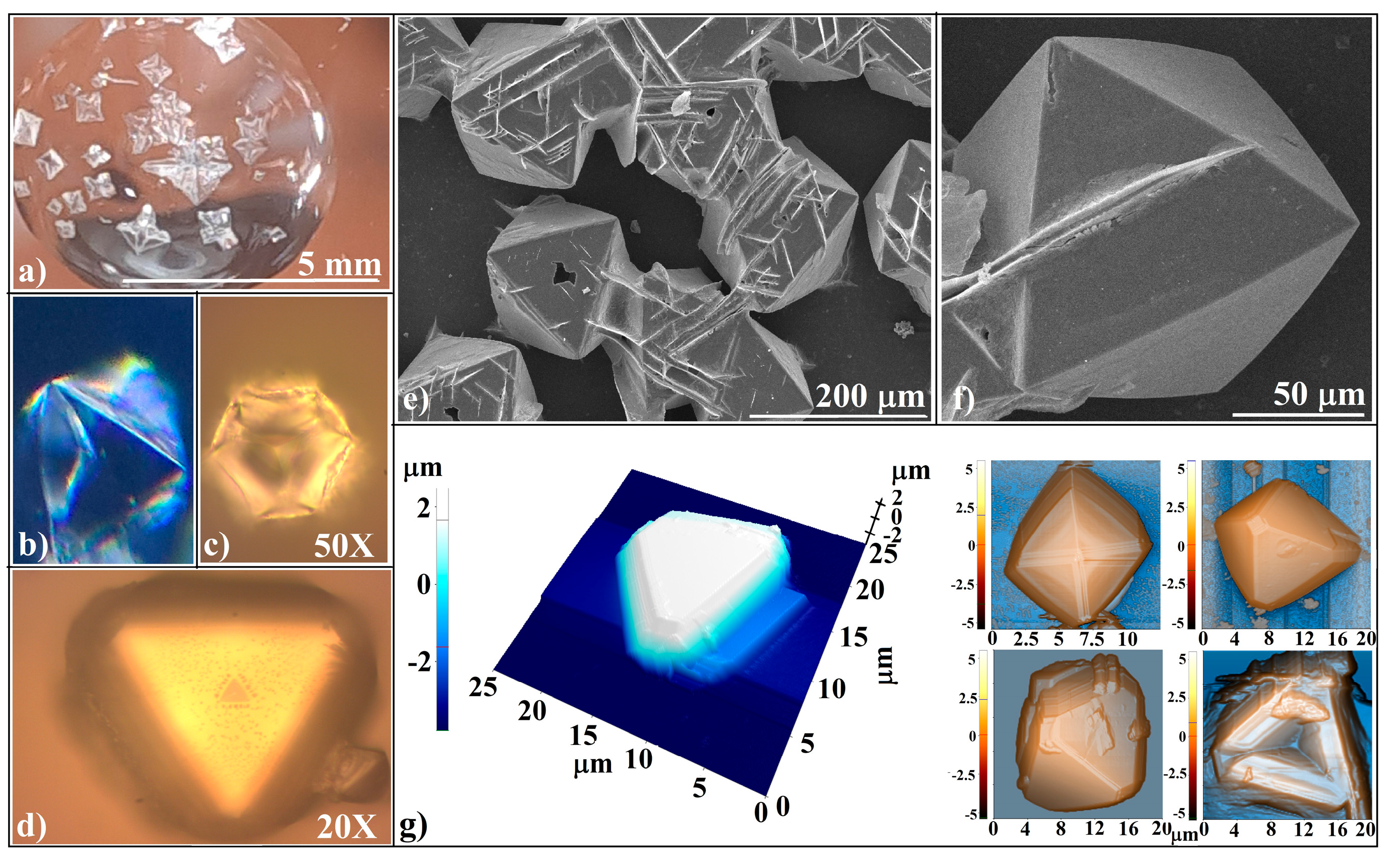
We noted that in the case of crystal layers, crystals with unusual shapes (Figure 7a) formed at the water/NaCl solution interface and slid down to the bottom of the drop, forming aggregates. The change in the NaCl habit has often been reported to be a result of adding organic molecules to the NaCl solution or crystallization in gels, at the water/air interface, or in confined volumes [117,118,119,120,121] and is generally attributed to the different growth rates of the NaCl crystal facets caused by the varying accessibility of chlorine and sodium ions near the crystal. We consider that in a layered binary hanging drop, the cause is the difference in the chlorine and sodium ion concentrations near the crystallization zone due to their different rates of diffusion from the solution to this zone.
The investigations revealed not only a wide distribution of crystal sizes but also the presence of crystals with varying habits. Optical microscopy images (Figure 7b–d) displayed prismatic crystals shaped like pyramids and hopper pyramids. Scanning electron microscopy (SEM) images (Figure 7e,f) identified these structures as being octahedral, and atomic force microscopy (AFM) images (Figure 7g) further confirmed the octahedral morphology of the crystals. Additionally, the AFM images highlighted the presence of intriguing features, such as hopper pyramidal cavities, where the decomposition of white light (not depicted here) was observed under optical microscopy. Furthermore, the SEM images revealed unexpected straight-line patterns on the surfaces of the octahedral crystals. A small fragment of these patterns, visible on the lower left crystal in the AFM group (Figure 7f), suggests the presence of solid, straight, parallel walls, which could potentially be utilized for Bragg diffraction of light in innovative experimental setups. However, considerable effort is required to achieve precise morphological and structural control of these crystal aggregates, which are formed through the evaporative crystallization of NaCl in a layered binary solution in a hanging drop configuration.
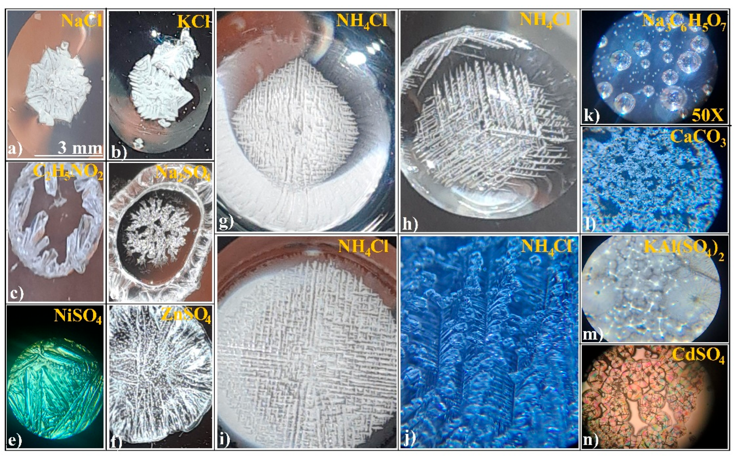
Experiments performed on a limited number of ionic salts suggest that evaporative crystallization in a layered water/salt solution hanging drop setup (d = 7 mm, V1 = 20–30 μL, V2 = 60–150 μL, c2 = 95% cs) results in either a dendritic agglomerate (Figure 8e–f) or a spherulitic array (Figure 8k–n). Impressive structures of highly ordered NH4Cl resembling a colloidal crystal lattice can be seen in Figure 8g–j.
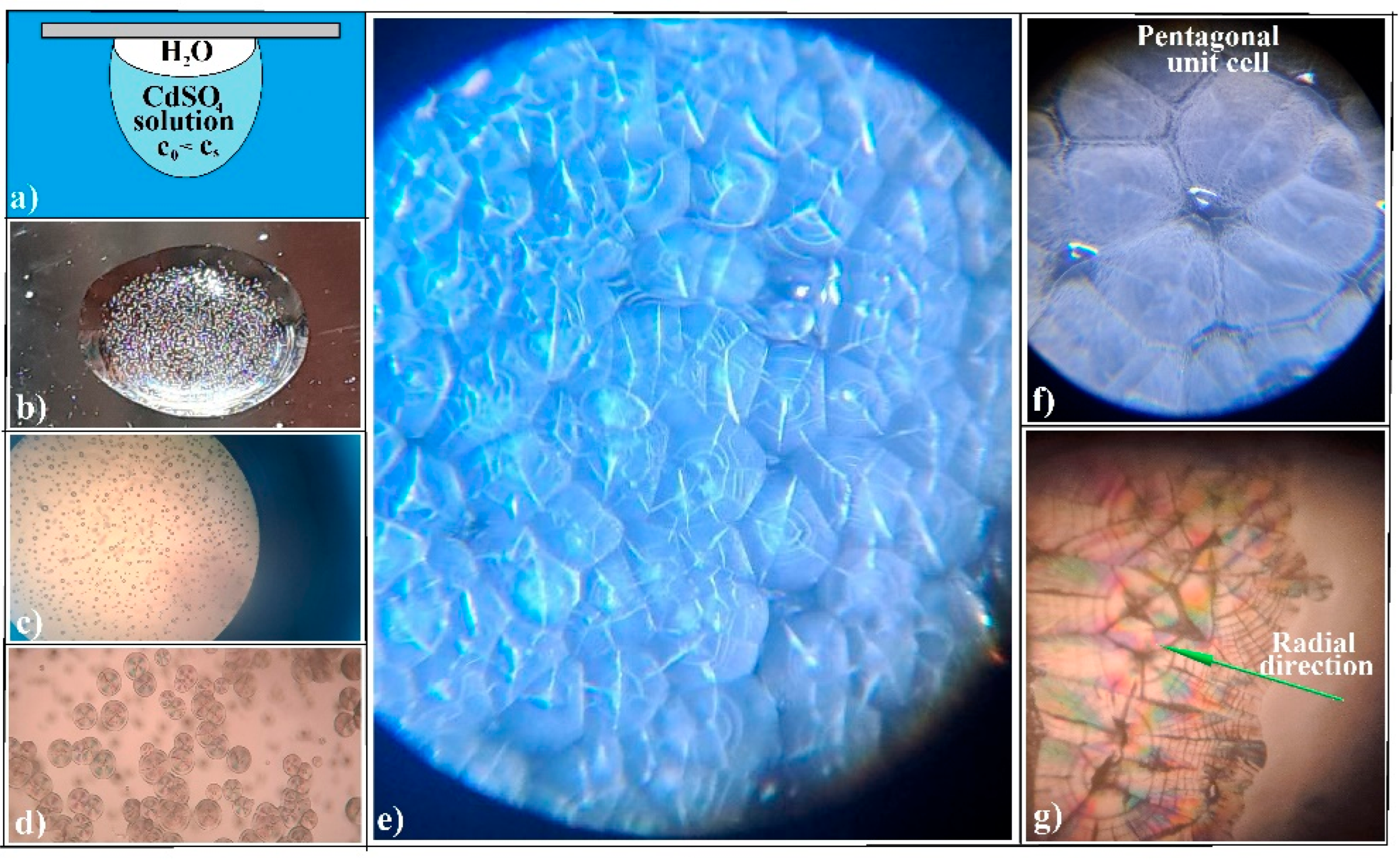
Furthermore, in this setup (Figure 9a), CdSO4 significantly increased the density of spherulites (Figure 9b–d), forming a compact film (Figure 9e). The entropy acted as the driving force, organizing them into a pentagonal lattice (Figure 9f). Each spherulite exhibited impressive, orientated colors when viewed under transmitted polarized light (Figure 9g). Unfortunately, these colors were ephemeral, lasting less than an hour under usual ambient conditions (T = 25 °C, RH = 60%).
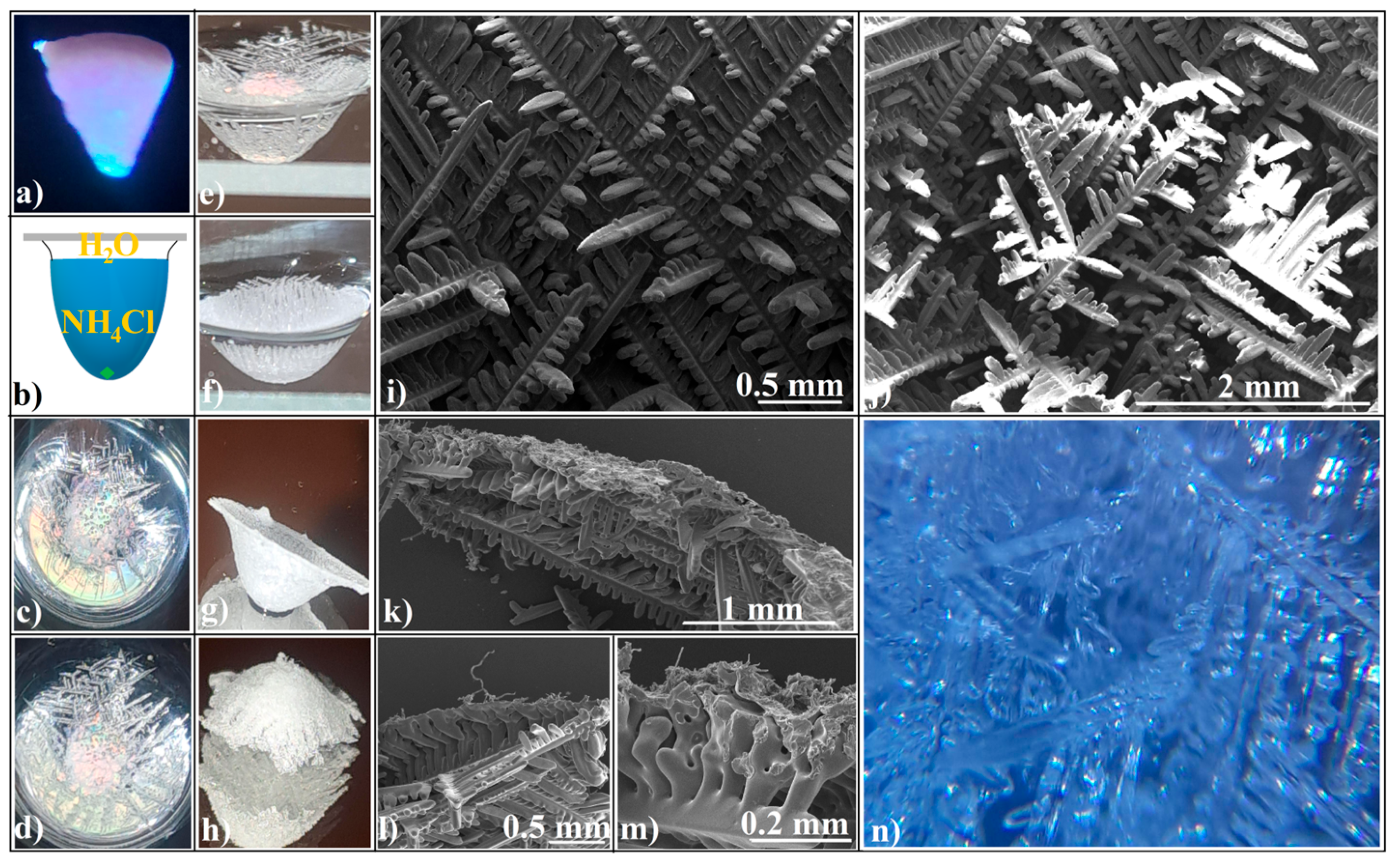
As we can see, this setup has proven to be extremely productive in generating new patterns and ordered structures. It achieves this by inducing new flows and gradients in a drop and by separating diffusion and evaporation, which are opposite phenomena affecting the achievement of critical supersaturation. Unfortunately, the most promising candidate for forming ordered agglomerates, namely NH4Cl, produced significant efflorescence during its evaporative crystallization. As a result, only a small portion of its solution contributed to the construction of a hollow spheroid, and even then, the dendritic structure was of a poor quality. However, we discovered that if a small fragment (hundreds of micrometers in size) of a colloidal crystal self-assembled from 264 nm SiO2 spheres (Figure 10a) [74,75] was placed at the bottom of a maximum-volume layered hanging drop (Figure 10b), it acted as a localized catalyst for an inhomogeneous nucleation process (Figure 10c–f). Consequently, most of the efflorescence transformed into useful crystals, contributing to the construction of a hollow spheroidal crystal (Figure 10g,h). The resulting structure featured a wall formed by high-quality orthogonal dendrites (Figure 10i) and a “bunch” of these on the spheroid’s bottom (Figure 10j).
Cross-sectional SEM images of the spheroid walls revealed their intricate microstructural features (Figure 10k–m), where NH4Cl dendrites formed a 3D pattern resembling a woven fabric. A normal reflection optical microscopy image (Figure 10n) of the spheroid wall further highlighted this complexity. These observations suggest that such structures could serve as an excellent starting point for utilizing ionic salts as building blocks in the fabrication of ordered materials with unique properties.
However, it is not constructive to interpret the agglomeration of ionic salt crystals within the framework of the self-assembly phenomenon because of the following:
(a)
The structural units of crystalline agglomerates can undergo transformations, altering their shape and size, which adds a layer of complexity to the system. This adaptability is quite different from the static colloidal spheres typically employed in hard-sphere self-assembly processes, where the spheres maintain their shape and size throughout. It is like comparing the fluidity of a shapeshifter to the rigidity of Lego blocks. The crystallization process allows for a more flexible and adaptive structure, which can be advantageous in various applications.
(b)
The attachment of these structural units (ionic salt single crystals) to the agglomerate is more akin to what occurs in spin coating—a specific technique for synthesizing colloidal films—where the spheres are driven along the substrate by the centrifugal force, rather than from a wide angle as in self-assembly. This becomes even more intriguing when considering crystal agglomeration in a hanging drop, where the substrate or air/water interface is curved, not flat, as it is in spin coating.
These observations suggest that we are either dealing with two independent phenomena or that both are part of a more complex one. However, we found that the “self-assembly” of ionic salts is possible with the appropriate setups. The most challenging task is selecting the optimal experimental parameters from a vast array of possibilities. Numerous static and dynamic parameters influence the final form of a crystal agglomerate. Most of these parameters are unknown, leading to the belief that crystallization is a weakly reproducible and hard-to-control “stochastic phenomenon”. The deductive approach seems to be useless. However, as Roy Glauber (awarded the Nobel Prize in Physics, 2005) said, “Too many kids in school get the notion that science is deductive, and deductive science is almost never creative. Real ideas arrive via intuition, via guesswork, and we’re guessing all the time” [122]. Thus, it may be difficult, but it could be worth it.

4. Conclusions

At the conclusion of this article, we have reached the conviction that crystal agglomeration and self-assembly phenomena are integral components of a broader and more complex process, one that offers a refined and intricate perspective on reality. Evaporative crystallization of ionic salts in a hanging drop can generate crystal agglomerates with unusual packing, symmetry, and shapes in two or three dimensions, such as hollow spheroidal NaCl agglomerates, octahedral NaCl films, pentagonally arranged CdSO4 spherulites, and NH4Cl dendritic shells, structures that are more often expected to be obtained through a self-assembly process rather than through a crystallization one. Whether acting independently or in synergy, crystallization and self-assembly have the potential to generate macroscopic structures with remarkable properties, which may be of significant interest for the development of future devices. Ordered crystal agglomerates, besides their own properties, can act as templates for both self-assembly phenomena and classical material processing technologies. Furthermore, there may be the opportunity to produce faceted devices with material structures such as ionic salts or biomolecules that cannot be produced through conventional mechanical, thermal, or chemical processes through alternative means. In particular, a simple hanging drop, specifically an initially layered binary-solution hanging drop, can function as a microreactor, wherein control over confinement or time-competing phenomena contribute an additional layer of complexity to the resulting structures.

Author Contributions

Conceptualization and formal analysis, I.S.; methodology, I.S.; validation, I.S.; investigation, I.S., I.U., I.A., S.B. and M.D.; resources, F.D. and M.D.; writing—original draft preparation, I.S.; writing—review and editing, I.S., C.T.F. and I.P.; supervision, I.S.; project administration, F.D.; funding acquisition, I.P. All authors have read and agreed to the published version of the manuscript.

Funding

This research was funded by the Romanian Minister of Education and Research under the Romanian National Nucleu Program LAPLAS VII, contract no. 30N/2023 and Project PN23390403/2023.

Conflicts of Interest

The authors declare no conflicts of interest.

References

  1. Whitesides, G.M.; Grzybowski, B. Self-assembly at all scales. Science 2002, 295, 2418–2421. [Google Scholar] [CrossRef]
  2. Whitesides, G.M.; Boncheva, M. Beyond molecules: Self-assembly of mesoscopic and macroscopic components. Proc. Natl. Acad. Sci. USA 2002, 99, 4769–4774. [Google Scholar] [CrossRef]
  3. Grzybowski, B.A.; Wilmer, C.E.; Kim, J.; Browne, K.P.; Bishop, K.J.M. Self-assembly: From crystals to cells. Soft Matter 2009, 5, 1110–1128. [Google Scholar] [CrossRef]
  4. Pochan, D.; Scherman, O. Introduction: Molecular Self-Assembly. Chem. Rev. 2021, 121, 13699–13700. [Google Scholar] [CrossRef] [PubMed]
  5. Huie, J.C. Guided molecular self-assembly: A review of recent efforts. Smart Mater. Struct. 2003, 12, 264–271. [Google Scholar] [CrossRef]
  6. Cheng, H.; Liu, R.; Zhang, R.; Huang, L.; Yuan, Q. Recent advances in supramolecular self-assembly derived materials for high-performance supercapacitors. Nanoscale Adv. 2023, 5, 2394–2412. [Google Scholar] [CrossRef]
  7. Shcherbina, M.A.; Chvalun, S.N. Driving Forces of the Self-Assembly of Supramolecular Systems: Partially Ordered Mesophases. Russ. J. Phys. Chem. 2018, 92, 1161–1170. [Google Scholar] [CrossRef]
  8. Li, Q.; Jonas, U.; Zhao, X.S.; Kapp, M. The forces at work in collodial self-assembly: A review on fundamental interactions between collodial particles. Asia-Pac. J. Chem. Eng. 2008, 3, 255–268. [Google Scholar] [CrossRef]
  9. Xu, Z.; Wang, L.; Fang, F.; Fu, Y.; Yin, Z. A Review on Colloidal Self-Assembly and their Applications. Curr. Nanosci. 2016, 12, 725–746. [Google Scholar] [CrossRef]
  10. Thorkelsson, K.; Bai, P.; Xu, T. Self-assembly and applications of anisotropic nanomaterials: A review. Nano Today 2015, 10, 48–66. [Google Scholar] [CrossRef]
  11. Böker, A.; He, J.; Emrick, T.; Russell, T.P. Self-assembly of nanoparticles at interfaces. Soft Matter 2007, 3, 1231–1248. [Google Scholar] [CrossRef] [PubMed]
  12. Xia, Y.; Gates, B.; Yin, Y.; Lu, Y. Monodispersed Colloidal Spheres: Old Materials with New Applications. Adv. Mater. 2000, 12, 693–713. [Google Scholar] [CrossRef]
  13. Cai, Z.; Li, Z.; Ravaine, S.; He, M.; Song, Y.; Yin, Y.; Zheng, H.; Teng, J.; Zhang, A. From colloidal particles to photonic crystals: Advances in self-assembly and their emerging applications. Chem. Soc. Rev. 2021, 50, 5898–5951. [Google Scholar] [CrossRef] [PubMed]
  14. Wieser, P.E.; Vukmanovic, Z.; Kilian, R.; Ringe, E.; Holness, M.B.; Maclennan, J.; Edmonds, M. To sink, swim, twin, or nucleate: A critical appraisal of crystal aggregation processes. Geology 2019, 47, 948–952. [Google Scholar] [CrossRef]
  15. Klein, C. Mineral. Britannica. Available online: https://www.britannica.com/science/mineral-chemical-compound (accessed on 5 March 2025).
  16. García-Ruiz, J.M.; Otálora, F. Crystal Growth in Geology: Patterns on the Rocks. In Handbook of Crystal Growth; Nishinaga, T., Rudolph, P., Eds.; Elsevier B.V.: Amsterdam, The Netherlands, 2015; Volume II, pp. 1–43. ISBN 9780444633033. [Google Scholar] [CrossRef]
  17. Ortoleva, P.; Chen, Y.; Chen, W. Agates, Geodes, Concretions and Orbicules: Self-Organized Zoning and Morphology. In Fractals and Dynamic Systems in Geoscience; Kruhl, J.H., Ed.; Springer: Berlin/Heidelberg, Germany, 1994; Volume 1, pp. 283–305. ISBN 978-3-662-07304-9. [Google Scholar] [CrossRef]
  18. Rasmuson, Å.C. Introduction to Crystallization of Fine Chemicals and Pharmaceuticals. In Molecules: Nucleation, Aggregation and Crystallization; Sedzik, J., Riccio, P., Eds.; World Scientific Publishing: London, UK, 2009; Volume 1, pp. 145–172. ISBN 978-981-4470-49-0. [Google Scholar] [CrossRef]
  19. Davey, R.; Garside, J. From Molecules to Crystallizers—An Introduction to Crystallization; Oxford University Press: New York, NY, USA, 2000; Volume 1, pp. 1–96. ISBN 0198504896. [Google Scholar] [CrossRef]
  20. Kalikmanov, V.I. Classical Nucleation Theory. In Nucleation Theory. Lecture Notes in Physics; Springer: Dordrecht, The Netherlands, 2013; Volume 860, pp. 1–316. ISBN 978-90-481-3643-8. [Google Scholar] [CrossRef]
  21. Revalor, E.; Hammadi, Z.; Astier, J.-P.; Grossier, R.; Garcia, E.; Hoff, C.; Furuta, K.; Okustu, T.; Morin, R.; Veesler, S. Usual and unusual crystallization from solution. J. Cryst. Growth 2010, 312, 939–946. [Google Scholar] [CrossRef]
  22. Nancollas, G.H.; Purdie, N. The kinetics of crystal growth. Q. Rev. Chem. Soc. 1964, 18, 1–20. [Google Scholar] [CrossRef]
  23. Jackson, K.A. Crystal growth kinetics. Mater. Sci. Eng. 1984, 65, 7–13. [Google Scholar] [CrossRef]
  24. Heisel, S.; Holtkötter, J.; Wohlgemuth, K. Measurement of agglomeration during crystallization: Is the differentiation of aggregates and agglomerates via ultrasonic irradiation possible? Chem. Eng. Sci. 2019, 210, 115214. [Google Scholar] [CrossRef]
  25. David, R.; Paulaime, A.-M.; Espitalier, F.; Rouleau, L. Modelling of multiple-mechanism agglomeration in a crystallization process. Powder Technol. 2003, 130, 338–344. [Google Scholar] [CrossRef]
  26. Shahidzadeh-Bonn, N.; Rafaï, S.; Bonn, D.; Wegdam, G. Salt Crystallization during Evaporation: Impact of Interfacial Properties. Langmuir 2008, 24, 8599–8605. [Google Scholar] [CrossRef]
  27. Barrett, M.; McNamara, M.; Hao, H.X.; Barrett, P.; Glennon, B. Supersaturation tracking for the development, optimization and control of crystallization processes. Chem. Eng. Res. Des. 2010, 88, 1108–1119. [Google Scholar] [CrossRef]
  28. Kim, K.-J.; Mersmann, A. Estimation of metastable zone width in different nucleation processes. Chem. Eng. Sci. 2001, 56, 2315–2324. [Google Scholar] [CrossRef]
  29. Misyura, S.Y. Different modes of heat transfer and crystallization in a drop of NaCl solution: The influence of key factors on the crystallization rate and the heat transfer coefficient. Int. J. Therm. Sci. 2021, 159, 106602. [Google Scholar] [CrossRef]
  30. Yang, H.; Florence, A.J. Relating induction time and metastable zone width. CrystEngComm 2017, 19, 3966–3978. [Google Scholar] [CrossRef]
  31. Misyura, S. The dependence of evaporation and crystallization kinetics on dynamic and thermal background. AIChE J. 2020, 66, e16282. [Google Scholar] [CrossRef]
  32. Qu, J.; Escobar, L.; Li, J.; Rao, Z.; Xu, B. Experimental study of evaporation and crystallization of brine droplets under different temperatures and humidity levels. Int. Commun. Heat Mass Transf. 2020, 110, 104427. [Google Scholar] [CrossRef]
  33. Ancheyta-Palacios, M.; Sánchez-Santiago, K.; Escalera Santos, G.J.; Carreón, Y.P.; González-Gutiérrez, J. Gravity-driven crystallization in protein and NaCl droplets. Colloids Surf. A Physicochem. Eng. Asp. 2025, 707, 135821. [Google Scholar] [CrossRef]
  34. Fontana, P.; Schefer, J.; Pettit, D. Characterization of sodium chloride crystals grown in microgravity. J. Cryst. Growth 2011, 324, 207–211. [Google Scholar] [CrossRef]
  35. Bunio, L.B.; Wang, J.; Kannaiyan, R.; Gates, I.D. Evaporation and crystallization of NaCl-water droplets suspended in air by acoustic levitation. Chem. Eng. Sci. 2022, 251, 117441. [Google Scholar] [CrossRef]
  36. Page, A.J.; Sear, R.P. Crystallization controlled by the geometry of a surface. J. Am. Chem. Soc. 2009, 131, 17550–17551. [Google Scholar] [CrossRef]
  37. Mann, S.; Archibald, D.D.; Didymus, J.M.; Douglas, T.; Heywood, B.R.; Meldrum, F.C.; Reeves, N.J. Crystallization at Inorganic-organic Interfaces: Biominerals and Biomimetic Synthesis. Science 1993, 261, 1286–1292. [Google Scholar] [CrossRef] [PubMed]
  38. Krog, N.; Larsson, K. Crystallization at Interfaces in Food Emulsions—A General Phenomenon. Eur. J. Lipid Sci. Technol. 1992, 94, 55–57. [Google Scholar] [CrossRef]
  39. Ford, I.J. Models of Crystallisation in Evaporating Droplets. MRS Online Proc. Libr. 1995, 398, 637–642. [Google Scholar] [CrossRef]
  40. Armstrong Green, N.C.; Haddrell, A.E.; Gregson, F.K.A.; Lewis, D.; Church, T.; Reid, J.P. Studies of the Crystallization and Dissolution of Individual Suspended Sodium Chloride Aerosol Particles. J. Phys. Chem. A 2024, 128, 4315–4323. [Google Scholar] [CrossRef]
  41. Tanaka, M.; Yamanaka, S.; Shirakawa, Y.; Shimosaka, A.; Hidaka, J. Preparation of porous particles by liquid–liquid interfacial crystallization. Adv. Powder Technol. 2011, 22, 125–130. [Google Scholar] [CrossRef]
  42. Coupland, J.N. Crystallization in emulsions. Curr. Opin. Colloid Interface Sci. 2002, 7, 445–450. [Google Scholar] [CrossRef]
  43. Kumar, V.; Dash, S. Patterns during Evaporative Crystallization of a Saline Droplet. Langmuir 2022, 38, 10265–10273. [Google Scholar] [CrossRef]
  44. Sperling, M.; Gradzielski, M. Droplets, Evaporation and a Superhydrophobic Surface: Simple Tools for Guiding Colloidal Particles into Complex Materials. Gels 2017, 3, 15. [Google Scholar] [CrossRef] [PubMed]
  45. Charlesworth, D.H.; Marshall, W.R., Jr. Evaporation from drops containing dissolved solids. AIChE J. 1960, 6, 9–23. [Google Scholar] [CrossRef]
  46. Fontana, P.; Pettit, D.; Cristoforetti, S. Sodium chloride crystallization from thin liquid sheets, thick layers, and sessile drops in microgravity. J. Cryst. Growth 2015, 428, 80–85. [Google Scholar] [CrossRef]
  47. Bormashenko, E.; Roy, P.K.; Shoval, S.; Legchenkova, I. Interfacial Crystallization within Liquid Marbles. Condens. Matter 2020, 5, 62. [Google Scholar] [CrossRef]
  48. Li, Q.; Wang, Z.; Guo, H.; Zhao, J.; Luo, H.; Huang, X. A perspective view of salt crystallization from solution in porous media: Morphology, mechanism, and salt efflorescence. Sci. Rep. 2024, 14, 23510. [Google Scholar] [CrossRef] [PubMed]
  49. Desarnaud, J.; Derluyn, H.; Carmeliet, J.; Bonn, D.; Shahidzadeh, N. Metastability Limit for the Nucleation of NaCl Crystals in Confinement. J. Phys. Chem. Lett. 2014, 5, 890–895. [Google Scholar] [CrossRef] [PubMed]
  50. Naillon, A.; Duru, P.; Marcoux, M.; Prat, M. Evaporation with sodium chloride crystallization in a capillary tube. J. Cryst. Growth 2015, 422, 52–61. [Google Scholar] [CrossRef]
  51. Zhang, H.; Zhao, Z.; Turley, A.T.; Wang, L.; McGonigal, P.R.; Tu, Y.; Li, Y.; Wang, Z.; Kwok, R.T.K.; Lam, J.W.Y.; et al. Aggregate Science: From Structures to Properties. Adv. Mater. 2020, 32, 2001457. [Google Scholar] [CrossRef]
  52. Bika, D.G.; Gentzler, M.; Michaels, J.N. Mechanical properties of agglomerates. Powder Technol. 2001, 117, 98–112. [Google Scholar] [CrossRef]
  53. Sandu, I.; Urzica, I.; Dumitru, M.; Banici, A.; Fleaca, C.; Dumitrache, F. Crystallization from Thin Films on a Hot Plate: A New Approach of Solid Surface Pattering. J. Mater. Sci. Chem. Eng. 2019, 7, 9–18. [Google Scholar] [CrossRef]
  54. Sandu, I.; Urzica, I.; Niculescu, A.M.; Fleaca, C.T.; Dumitrache, F.; Badiceanu, M. Self-organisation of single-crystals as ripple patterns through laser ablation of ionic salt solutions. Appl. Surf. Sci. 2017, 417, 160–164. [Google Scholar] [CrossRef]
  55. Zhang, Y.; Liu, Y.; Ji, X.; Banks, C.E.; Song, J. Flower-like agglomerates of hydroxyapatite crystals formed on an egg-shell membrane. Colloids Surf. B 2011, 82, 490–496. [Google Scholar] [CrossRef]
  56. Shahidzadeh, N.; Schut, M.; Desarnaud, J.; Prat, M.; Bonn, D. Salt stains from evaporating droplets. Sci. Rep. 2015, 5, 10335. [Google Scholar] [CrossRef]
  57. Giri, A.; Dutta Choudhury, M.; Dutta, T.; Tarafdar, S. Multifractal Growth of Crystalline NaCl Aggregates in a Gelatin Medium. Cryst. Growth Des. 2013, 13, 341–345. [Google Scholar] [CrossRef]
  58. Pathak, B.; Christy, J.; Sefiane, K.; Gozuacik, D. Complex Pattern Formation in Solutions of Protein and Mixed Salts Using Dehydrating Sessile Droplets. Langmuir 2020, 36, 9728–9737. [Google Scholar] [CrossRef]
  59. Chen, L.; Wang, J. Continuous Production of NaCl Spherical Particles via Emulsion Crystallization in Microfluidic Devices. Crystal Growth Des. 2023, 23, 8918–8928. [Google Scholar] [CrossRef]
  60. Rodriguez-Navarro, A.; Garcia-Ruiz, J.M. Model of textural development of layered crystal aggregates. Eur. J. Mineral. 2000, 12, 609–614. [Google Scholar] [CrossRef]
  61. Mailleur, A.; Pirat, C.; Simon, G.; Fulcrand, R.; Colombani, J. Ring shells obtained from pure water drops evaporating on a soluble substrate. Colloids Surf. A 2022, 651, 129724. [Google Scholar] [CrossRef]
  62. Zhang, X.; Chen, H.; Wang, Z.; Wang, N.; Zang, D. Evaporation of Saline Droplets on a Superhydrophobic Substrate: Formation of Crystal Shell and “Legs”. Materials 2023, 16, 5168. [Google Scholar] [CrossRef]
  63. Cölfen, H.; Antonietti, M. Mesocrystals: Inorganic superstructures made by highly parallel crystallization and controlled alignment. Angew. Chem. Int. Ed. 2005, 44, 5576–5591. [Google Scholar] [CrossRef]
  64. Sandu, I.; Dumitru, M.; Fleaca, C.T.; Dumitrache, F. Rise and side infiltration in opals and porous materials for their skin-free replica synthesis. Mater. Res. Express 2019, 6, 046201. [Google Scholar] [CrossRef]
  65. Hetherington, N.B.J.; Kulak, A.N.; Kim, Y.-Y.; Noel, E.H.; Snoswell, D.; Butler, M.; Meldrum, F.C. Porous Single Crystals of Calcite from Colloidal Crystal Templates: ACC Is Not Required for Nanoscale Templating. Adv. Funct. Mater. 2011, 21, 948–954. [Google Scholar] [CrossRef]
  66. Wang, Y.; Liu, Z.; Pan, H.; Tang, R. Biomineralization inspired crystal growth for biomimetic materials preparation. J. Cryst. Growth 2023, 603, 127029. [Google Scholar] [CrossRef]
  67. Qin, K.; Zheng, Z.; Wang, J.; Pan, H.; Tang, R. Biomineralization strategy: From material manufacturing to biological regulation. Giant 2024, 19, 100317. [Google Scholar] [CrossRef]
  68. Kato, M.; Ito, H.; Hasegawa, M.; Ishii, K. Soft crystals-flexible response systems with high structural order. Chem. A Eur. J. 2019, 25, 5099–5329. [Google Scholar] [CrossRef] [PubMed]
  69. Rapis, E. A change in the physical state of a non-equilibrium blood plasma protein film in patients with carcinoma. Tech. Phys. 2002, 47, 510–512. [Google Scholar] [CrossRef]
  70. Li, L.; Xia, Y.; Zeng, M.; Fu, L. Facet engineering of ultrathin two-dimensional materials. Chem. Soc. Rev. 2022, 51, 7327–7343. [Google Scholar] [CrossRef]
  71. Ejima, H.; Yanai, N.; Best, J.P.; Sindoro, M.; Granick, S.; Caruso, F. Near-incompressible faceted polymer microcapsules from metal-organic framework templates. Adv. Mater. 2013, 25, 5767–5771. [Google Scholar] [CrossRef]
  72. Chen, C.-C.; Chang, J.-Y.; Chi, G.-C. Design of GaN convex diffractive microlenses. Opt. Laser Technol. 2002, 34, 569–573. [Google Scholar] [CrossRef]
  73. Churin, E.; Bayvel, P.; Smirnitskii, V.; Timofeev, F.; Midwinter, J. Concave grating and convex mirror double dispersion spectrograph for optical network applications. Appl. Opt. 1997, 36, 7822–7825. [Google Scholar] [CrossRef]
  74. Sandu, I.; Antohe, I.; Fleaca, C.T.; Dumitrache, F.; Urzica, I.; Brajnicov, S.; Iagaru, R.; Sava, B.A.; Dumitru, M. Shaping in the Third Direction: Self-Assembly of Convex Colloidal Photonic Crystals on an Optical Fiber Tip by Hanging Drop Method. Polymers 2024, 16, 33. [Google Scholar] [CrossRef]
  75. Sandu, I.; Antohe, I.; Fleaca, C.T.; Dumitrache, F.; Urzica, I.; Dumitru, M. Shaping in the Third Direction: Colloidal Photonic Crystals with Quadratic Surfaces Self-Assembled by Hanging-Drop Method. Polymers 2024, 16, 1931. [Google Scholar] [CrossRef]
  76. Sandu, I.; Dumitru, M.; Fleaca, C.T.; Dumitrache, F. Hanging colloidal drop: A new photonic crystal synthesis route. Photonics Nanostruct. Fundam. Appl. 2018, 29, 42–48. [Google Scholar] [CrossRef]
  77. Li, Y.; Diddens, C.; Lv, P.; Wijshoff, H.; Versluis, M.; Lohse, D. Gravitational effect in evaporating binary microdroplets. Phys. Rev. Lett. 2019, 122, 114501. [Google Scholar] [CrossRef]
  78. Ma, X.; Sefiane, K.; Bennacer, R.; Lapert, X.; Bakir, F. Solutal and Gravitational Effects during Binary Mixture Droplets Evaporation. Microgravity Sci. Technol. 2024, 36, 17. [Google Scholar] [CrossRef]
  79. Sandu, I.; Iordache, I.; Fleaca, C.; Dumitrache, F.; Niculescu, A. The Influence of Gravity on Single Crystallization in Large Volume Drops. J. Crystalliz. Proc. Technol. 2014, 4, 206–211. [Google Scholar] [CrossRef]
  80. Çelikbilek, M.; Ersundu, A.E.; Aydın, S. Crystallization Kinetics of Amorphous Materials. In Advances in Crystallization Processes; Mastai, Y., Ed.; InTech Europe: Rijeka, Croatia, 2012; Volume 1, pp. 127–162. ISBN 978-953-51-4297-3. [Google Scholar] [CrossRef]
  81. Pettit, D.; Fontana, P. Comparison of sodium chloride hopper cubes grown under microgravity and terrestrial conditions. npj Microgravity 2019, 5, 25. [Google Scholar] [CrossRef]
  82. Gielen, B.; Jordens, J.; Thomassen, L.C.J.; Braeken, L.; Van Gerven, T. Agglomeration Control during Ultrasonic Crystallization of an Active Pharmaceutical Ingredient. Crystals 2017, 7, 40. [Google Scholar] [CrossRef]
  83. Im, S.H.; Park, O.O. Effect of Evaporation Temperature on the Quality of Colloidal Crystals at the Water—Air Interface. Langmuir 2002, 18, 9642–9646. [Google Scholar] [CrossRef]
  84. Liu, G.Q.; Wang, Z.S.; Ji, Y.H. Influence of growth parameters on the fabrication of high-quality colloidal crystals via a controlled evaporation self-assembly method. Thin Solid Film. 2010, 518, 5083–5090. [Google Scholar] [CrossRef]
  85. Chen, R.-Q.; Lu, Q.-Q.; Cheng, Q.-D.; Ao, L.-B.; Zhang, C.-Y.; Hou, H.; Liu, Y.-M.; Li, D.-W.; Yin, D.-C. An ignored variable: Solution preparation temperature in protein crystallization. Sci. Rep. 2015, 5, 7797. [Google Scholar] [CrossRef]
  86. Astier, J.-P.; Veesler, S. Using Temperature To Crystallize Proteins: A Mini-Review. Cryst. Growth Des. 2008, 8, 4215–4219. [Google Scholar] [CrossRef]
  87. Quilaqueo, M.; Aguilera, J.M. Crystallization of NaCl by fast evaporation of water in droplets of NaCl solutions. Food Res. Int. 2016, 84, 143–149. [Google Scholar] [CrossRef]
  88. Sassen, K. Infrared (10.6-μm) radiation induced evaporation of large water drops. J. Opt. Soc. Am. 1981, 71, 887–891. [Google Scholar] [CrossRef]
  89. Park, B.S.; Armstrong, R.L. Laser droplet heating: Fast and slow heating regimes. Appl Opt. 1989, 28, 3671–3680. [Google Scholar] [CrossRef]
  90. Ferraz-Albani, L.A.; Baldelli, A.; Knapp, C.J.; Jäger, W.; Vehring, R.; Nobes, D.S.; Olfert, J.S.; Kostiuk, L.W. Enhanced Evaporation of Microscale Droplets With an Infrared Laser. J. Heat Transf. 2017, 139, 011503. [Google Scholar] [CrossRef]
  91. Kaplunov, I.; Kropotov, G.; Rogalin, V.; Shahmin, A. Optical properties of alkali halide crystals. J. Phys. Conf. Ser. 2020, 1697, 012253. [Google Scholar] [CrossRef]
  92. Nistor, S.V.; Nistor, L.C.; Teodorescu, S.V.; Ciucur, G.; Chis, I.; Drăgulinescu, D.; Grigorescu, D.; Grigoriu, C. Output couplers of ultra-transparent KCl and NaCl in a pulsed CO2 laser. Opt. Laser Technol. 1989, 21, 377–380. [Google Scholar] [CrossRef]
  93. Chen, L.; Ren, G.; Liu, L.; Guo, P.; Wang, E.; Zhu, Z.; Yang, J.; Shen, J.; Zhang, Z.; Zhou, L.; et al. Probing NaCl hydrate formation from aqueous solutions by terahertz time-domain spectroscopy. Phys. Chem. Chem. Phys. 2020, 22, 17791–17797. [Google Scholar] [CrossRef]
  94. Ajito, K.; Ueno, Y.; Kim, J.Y.; Sumikama, T. Capturing the Freeze-Drying Dynamics of NaCl Nanoparticles Using THz Spectroscopy. J. Am. Chem. Soc. 2018, 140, 13793–13797. [Google Scholar] [CrossRef] [PubMed]
  95. Liu, L.; Engel, T.; Flury, M.; Visser, M. Faceted structure: A design for desired illumination and manufacture using 3D printing. OSA Contin. 2018, 1, 26–39. [Google Scholar] [CrossRef]
  96. Joung, D.; Nemilentsau, A.; Agarwal, K.; Dai, C.; Liu, C.; Su, Q.; Li, J.; Low, T.; Koester, S.J.; Cho, J.H. Self-Assembled Three-Dimensional Graphene-Based Polyhedrons Inducing Volumetric Light Confinement. Nano Lett. 2017, 17, 1987–1994. [Google Scholar] [CrossRef] [PubMed]
  97. He, D.; Li, H.; Li, W.; Haghi-Ashtiani, P.; Lejay, P.; Bai, J. Growth of carbon nanotubes in six orthogonal directions on spherical alumina microparticles. Carbon 2011, 49, 2273–2286. [Google Scholar] [CrossRef]
  98. Wang, B.; Jin, P.; Yue, Y.; Ji, S.; Li, Y.; Luo, H. Synthesis of NaCl single crystals with defined morphologies as templates for fabricating hollow nano/micro-structures. RSC Adv. 2015, 5, 5072–5076. [Google Scholar] [CrossRef]
  99. Zhang, Z.; Feng, J.; Jiang, Y.; Liu, P.; Zhang, Q.; Wei, R.; Chen, X.; Feng, J. Self-Sacrificial Salt Templating: Simple Auxiliary Control over the Nanoporous Structure of Porous Carbon Monoliths Prepared through the Solvothermal Route. Nanomaterials 2018, 8, 255. [Google Scholar] [CrossRef]
  100. Li, J.; Lu, D.; Kondamareddy, K.K.; Gu, W.; Liu, Y.; Su, Y.; You, Z.; Fan, H.; Ho, W. Fabrication of Hexagonal Prism-Shaped Double S-Scheme Cu2O@CdS/ZnS Heterojunctions Utilizing Zeolite Templates: Enhanced Photocatalytic Hydrogen Production and Mechanism Insight. Cryst. Growth Des. 2024, 24, 8769–8781. [Google Scholar] [CrossRef]
  101. Jalaly, M.; Hosseini, R.; Bakhshi, A.; Chehelamirani, M. Self-assembly synthesis of 3D graphene/nano-Fe3O4 hybrid aerogels with improved mechanical and thermal properties. J. Alloys Cpds. 2022, 902, 163718. [Google Scholar] [CrossRef]
  102. Sögütoglu, L.-C.; Steiger, M.; Houben, J.; Biemans, D.; Fischer, H.R.; Donkers, P.; Huinink, H.; Adan, O.C.G. Understanding the Hydration Process of Salts: The Impact of a Nucleation Barrier. Cryst. Growth Des. 2019, 19, 2279–2288. [Google Scholar] [CrossRef]
  103. Rammelberg, H.U.; Schmidt, T.; Ruck, W. Hydration and dehydration of salt hydrates and hydroxides for thermal energy storage—Kinetics and energy release. Energy Procedia 2012, 30, 362–369. [Google Scholar] [CrossRef]
  104. Tian, F.; Qu, H.; Zimmermann, A.; Munk, T.; Jørgensen, A.C.; Rantanen, J. Factors affecting crystallization of hydrates. J. Pharm. Pharmacol. 2010, 62, 1534–1546. [Google Scholar] [CrossRef] [PubMed]
  105. Gránásy, L.; Pusztai, T.; Tegze, G.; Warren, J.A.; Douglas, J.F. Growth and form of spherulites. Phys. Rev. E. 2005, 72, 011605. [Google Scholar] [CrossRef]
  106. Liu, X.; Contal, C.; Schmutz, M.; Krafft, M.P. Two-Dimensional Radial or Ring-Banded Nonbirefringent Spherulites of Semifluorinated Alkanes Coexistent with Close-Packed Self-Assembled Surface Nanodomains. Langmuir 2018, 34, 15126–15133. [Google Scholar] [CrossRef]
  107. Beck, R.; Andreassen, J.-P. Spherulitic Growth of Calcium Carbonate. Cryst. Growth Des. 2010, 10, 2934–2947. [Google Scholar] [CrossRef]
  108. Raimo, M. An optical test to unveil twisting of birefringent crystals in spherulites. R. Soc. Open Sci. 2019, 6, 181215. [Google Scholar] [CrossRef]
  109. Clough, S.; Van Aartsen, J.J.; Stein, R.S. Scattering of Light by Two-Dimensional Spherulites. J. Appl. Phys. 1965, 36, 3072–3085. [Google Scholar] [CrossRef]
  110. Edwards, A.M.J.; Atkinson, P.S.; Cheung, C.S.; Liang, H.; Fairhurst, D.J.; Ouali, F.F. Density-driven flows in evaporating binary liquid droplets. Phys. Rev. Lett. 2018, 121, 184501. [Google Scholar] [CrossRef] [PubMed]
  111. Koldeweij, R.B.J.; van Capelleveen, B.F.; Lohse, D.; Visser, C.W. Marangoni-driven spreading of miscible liquids in the binary pendant drop geometry. Soft Matter 2019, 15, 8525–8531. [Google Scholar] [CrossRef] [PubMed]
  112. Pahlavan, A.A.; Yang, L.; Bain, C.D.; Stone, H.A. Evaporation of binary-mixture liquid droplets. The formation of picoliter pancake shapes. Phys. Rev. Lett. 2021, 127, 024501. [Google Scholar] [CrossRef]
  113. Phillips, O.M. On flow induced by diffusion in a stably stratified fluid. Deep-Sea Res. 1970, 17, 435–443. [Google Scholar] [CrossRef]
  114. Stewart, R.W. The problem of diffusion on a stratified fluid. Adv. Geophys. 1959, 6, 303–311. [Google Scholar] [CrossRef]
  115. Zhang, Q.; Amooie, A.; Bazant, M.Z.; Bischofberger, I. Growth morphology and symmetry selection of interfacial instabilities in anisotropic environments. Soft Matter 2021, 17, 1202–1209. [Google Scholar] [CrossRef]
  116. Yang, Z.; Zhang, J.; Zhang, L.; Fu, B.; Tao, P.; Song, C.; Shang, W.; Deng, T. Self-assembly in Hopper shaped crystals. Adv. Funct. Mater. 2020, 30, 1908108. [Google Scholar] [CrossRef]
  117. van der Voort, E.; Hartman, P. The role of water in the habit change from cube to octahedron of alkali halide crystals. J. Cryst. Growth 1990, 104, 450–456. [Google Scholar] [CrossRef]
  118. Qin, Y.; Yu, D.; Zhou, J. DNA action on the growth habit modification of NaCl crystals. CrystEngComm 2017, 19, 5356–5360. [Google Scholar] [CrossRef]
  119. Pramanik, S.K.; Ganguly, B. Crystal habit modification of sodium chloride using habit modifiers: A dive into more than 50 years of research & development on crystal habit. CrystEngComm 2025, 27, 2439–2451. [Google Scholar] [CrossRef]
  120. Desarnaud, J.; Derluyn, H.; Carmeliet, J.; Bonn, D.; Shahidzadeh, N. Hopper growth of salt crystals. J. Phys. Chem. Lett. 2018, 9, 2961–2966. [Google Scholar] [CrossRef]
  121. Zhang, J.; Zhang, S.; Wang, Z.; Zhang, Z.; Wang, S.; Wang, S. Hopper-like single crystals of sodium chloride grown at the interface of metastable water droplets. Angew. Chem. Int. Ed. 2011, 50, 6044–6047. [Google Scholar] [CrossRef]
  122. Roy, J.G.; John, L.H.; Theodor, W.H. Transcript from an Interview with the 2005 Physics Laureates. Available online: https://www.nobelprize.org/prizes/physics/2005/glauber/225060-2005-physics-laureates-interview-transcript (accessed on 7 March 2025).
Figure 1. Photographs of (a) SiO2 colloidal drop hanging from a fiber and (b) SiO2 colloidal photonic crystal reflecting Bragg-diffracted white light; (c) schematic of gravity’s influence on colloidal self-assembly in a hanging drop; successive photographs presenting (d) evaporative crystallization in a hanging drop of NaCl solution, with c0 < cs between starting time t0 and completion of drop evaporation tf, where τ marks moment when first crystal was observed, and (e) evaporative crystallization in a hanging drop of NaCl solution, where c0 = cs and t0, tf, and τ have same meanings as above; (f) photographic image of NaCl spheroid and (g) scanning electron microscope (SEM) image of outer surface and (h) SEM image of inner surface.
Applsci 15 09280 g001
Figure 2. SEM images of NaCl spheroids formed at various ambient temperatures: (a) T = 5 °C, (b) T = 80 °C, (c) T = 120 °C. (d) The dependence of the crystal size on the drop’s evaporation rate. (eh) Evaporative crystallization of a hanging drop of a NaCl solution, initially irradiated with a pulsed infrared laser beam, and an SEM image of the resulting spheroid. (il) Evaporative crystallization of a hanging drop of a NaCl solution, with an initially small temperature difference between the substrate temperature (T = 25 °C) and the drop temperature (T = 30 °C), and the SEM image of the resulting spheroid.
Applsci 15 09280 g002
Figure 3. Photographs of spheroids and their corresponding reflections in plane mirror: (a) empty NaCl spheroid, (b) NaCl spheroid with Rose Bengal dye film formed on its inner surface, (c) NaCl spheroid filled with carbonaceous material (top image), and faceted carbonaceous material spheroid after NaCl removal (bottom image). (d) SEM image of carbonaceous material’s surface. (e) SEM image of polystyrene faceted surface after NaCl removal. (f) Photographs of convex (top image) and concave (bottom image) polystyrene templates during SiO2 colloid self-assembly in attached (hanging) or deposed (sitting) drops, respectively. (g,h) Optical microscopy images of faceted polystyrene concave SiO2 colloidal photonic crystal; yellow inset in (g) shows retroreflection UV-Vis spectrum, whereas those in (h) show geometric optic phenomenon (upper inset in (h) captures plane mirror image of convex spheroid and lower inset shows image of concave one).
Applsci 15 09280 g003
Figure 4. (aj) Photographs of hanging drops of hydrated ionic salt solution and their corresponding spheroidal crystal agglomerates, including photographs of both them directly and their plane mirror images. (kp) SEM images of specific ionic salt crystal agglomerates (Glycine was also ionized with zwitterionic structure), showing both outer and inner surfaces.
Applsci 15 09280 g004
Figure 5. Optical microscopy images of CdSO4·8/3H2O spherulites grown on the outer surface of spherical crystal agglomerates: (a) transmission, (b) reflection, (c) transmission of polarized light, (d,e) SEM images of CdSO4·8/3H2O spherulites, and (f) an SEM image of the inner surface of the CdSO4·8/3H2O spheroids.
Applsci 15 09280 g005
Figure 6. (a) Schematic setup of a layered binary-solution hanging drop. (b) Image of a hanging drop of layered NaCl/NiSO4 aqueous solutions. (c) Direct and plane mirror images of a NaCl/NiSO4·6H2O crystal agglomerate. (d) Image of a layered binary hanging drop of a H2O/SiO2 colloid (e) SiO2 colloidal crystal film. (f) UV-Vis reflection and transmission spectra of the SiO2 colloidal crystal film. (g) Schematic setup of a hanging drop of a layered H2O/salt solution. Photographs of crystals formed by varying the water and NaCl solution volumes, producing (h) a single NaCl crystal, (i) a few large-sized crystals, (j) a ring of crystals, (k) a single layer of crystals, and (l) a compact dendrite of NaCl crystals.
Applsci 15 09280 g006
Figure 7. (a) Image of the aggregation of NaCl crystals in a hanging drop of a layered binary solution. Octahedral NaCl crystals: (bd) optical microscopy images, (e,f) SEM images, (g) AFM images.
Applsci 15 09280 g007
Figure 8. Photographic images of specific ionic salt crystal agglomerates: (af) dendritic structures, (gi) orthogonal NH4Cl dendrite arrays, (j) optical microscopy of NH4Cl dendrites, (kn) spherulitic arrays.
Applsci 15 09280 g008
Figure 9. (a) Schematic of CdSO4·8/3H2O spherulitic array formation in a layered hanging drop. Photographic images of the CdSO4·8/3H2O spherulitic array during its initial stages: (b) Rear view through the glass slide, (c,d) front view. Microscopy images of (e) a compact CdSO4·8/3H2O spherulitic array, (f) CdSO4·8/3H2O spherulites with a pentagonal lattice, (g) radial orientation of the colors in a CdSO4·8/3H2O spherulite array under polarized transmitted light.
Applsci 15 09280 g009
Figure 10. (a) SiO2 colloidal photonic crystal. (b) Schematic setup used for NH4Cl crystallization. (cf) Photo images of the experimental setup along with the crystals agglomeration process. (g,h) Hollow, spheroidal, orthogonal, dendritic NH4Cl agglomerates: direct and plane mirror images. SEM images of a NH4Cl spheroid: (i) normal to the inner wall; (j) on the spheroid’s bottom; (km) a wall cross-section; (n) a normal reflection optical microscopy image of the inner wall.
Applsci 15 09280 g010
Table 1. Dependency of NaCl agglomerate’s shape on water and solution layer volumes.
T = 25 °C
RH = 60%
V1
[μL]
V2
[μL]
c1
[% wt.]
d1
[mm]
C2
[% wt.]
Single crystal (Figure 6h)0 ÷ 10150–1700795
Aggregate with few crystals (Figure 6i)0 ÷ 1050–600795
Ring (Figure 6j)10 ÷ 2015–300795
Single layer (Figure 6k)10 ÷ 2050–600795
Dendrite (Figure 6l)20 ÷ 3060–1500795
Lack of reproducibility and control>30
Disclaimer/Publisher’s Note: The statements, opinions and data contained in all publications are solely those of the individual author(s) and contributor(s) and not of MDPI and/or the editor(s). MDPI and/or the editor(s) disclaim responsibility for any injury to people or property resulting from any ideas, methods, instructions or products referred to in the content.