Review

A Review of the Most Commonly Used Additive Manufacturing Techniques for Improving Mandibular Resection and Reconstruction Procedures

by
Paweł Turek
1,*,
Małgorzata Zaborniak
1,
Katarzyna Grzywacz-Danielewicz
1,
Michał Bałuszyński
2,
Bogumił Lewandowski
2,3,
Janusz Kluczyński
4 and
Natalia Daniel
5
1
Faculty of Mechanical Engineering and Aeronautics, Rzeszow University of Technology, 35-959 Rzeszow, Poland
2
Department of Maxillofacial Surgery, University Clinical Hospital named after Fryderyk Chopin in Rzeszów, 35-055 Rzeszów, Poland
3
Collegium Medicum, University of Rzeszów, 35-315 Rzeszów, Poland
4
Institute of Robots & Machine Design, Faculty of Mechanical Engineering, Military University of Technology, Gen. S. Kaliskiego St., 00-908 Warsaw, Poland
5
Faculty of Mechatronics, Armament and Aviation, Institute of Rocket Technology and Mechatronics, Military University of Technology, 2 gen. S. Kaliskiego Street, 00-908 Warsaw, Poland
*
Author to whom correspondence should be addressed.
Appl. Sci. 2025, 15(17), 9228; https://doi.org/10.3390/app15179228
Submission received: 4 July 2025 / Revised: 15 August 2025 / Accepted: 15 August 2025 / Published: 22 August 2025
(This article belongs to the Special Issue Feature Review Papers in Additive Manufacturing Technologies)

Abstract

Background: Mandibular defects caused by trauma or tumor resection pose significant challenges in both functional and aesthetic reconstruction. Additive manufacturing (AM) technologies offer promising solutions for surgical planning and personalized treatment. Objectives: This review aims to evaluate current trends in the application of AM technologies for mandibular resection and reconstruction, with a particular focus on material selection, clinical integration, and technology-specific advantages. Methods: A structured literature review was performed using PubMed, Scopus, Web of Science, and Google Scholar. Studies published between January 2020 and May 2025 were screened using the following inclusion criteria: original peer-reviewed English-language research involving AM in mandibular surgery. The exclusion criteria included review articles, non-English sources, and non-mandibular studies. A total of 77 studies met the inclusion criteria and were analyzed in this review. Results: Based on the literature review conducted from 2020 to 2025, the most common restorative methods for the mandible using additively manufactured models include reconstruction with a titanium surgical plate bent to the curvature of the edges and angle of the mandible or a personalized titanium or PEEK surgical plate made directly based on the patient’s diagnosis. Implants made of Ti-6AL-4V ELI and bioceramic scaffolds are also used in the reconstruction process. They are developed based on patient diagnostic data and effectively replace the loss of mandibular bone structure. In addition, based on models and surgical guides created using additive manufacturing techniques, the performance of autogenous grafts from the fibula or iliac crest has improved significantly when used with a titanium implant plate. Conclusions: Additive manufacturing supports highly personalized and accurate mandibular reconstruction. The advantages of these methods include a reduced overall duration of procedures, a lower health risk for patients due to less reliance on general anesthesia, a near perfect match between the implant and the remaining hard tissues, and satisfactory aesthetic outcomes. However, success depends on the appropriate selection AM technology and material, particularly in load-bearing applications.

1. Introduction

1.1. Clinical Background and Rationale

Mandibular integrity is essential to maintain respiratory function, proper chewing, speech articulation, and facial aesthetics. Anatomically, the mandible is the only movable bone in the craniofacial complex and plays a central role in oral and maxillofacial biomechanics. Mandibular discontinuity requently results from traumatic injuries, oncological resections, or congenital abnormalities. Among these, trauma and tumors represent the leading clinical indications for reconstruction [1,2]. Mandibular fractures are among the most common facial injuries, which often require stabilization with titanium fixation plates. Oncological resections, including segmental mandibulectomy, require complex reconstructive strategies due to the volume and geometry of lost bone tissue [2]. Various methods have been developed to address mandibular defects, including the use of reconstruction plates, nonvascularized bone grafts, and free vascularized flaps [3,4,5,6]. Although titanium plates are simple to implement, complications such as soft tissue shrinkage, plate exposure, and chronic inflammation are frequent [4,5]. These issues can ultimately lead to functional loss or deformities, such as Andy Gump-type appearance [6]. Therefore, autogenous grafts—particularly fibular, iliac, and scapular flaps—are used to improve long-term outcomes. The sagittal fibular flap remains the most commonly applied, but the scapular flap, especially in Japan, is also well established due to its favorable vascular anatomy and versatility in composite reconstruction.

1.2. Challenges and the Role of Additive Manufacturing

Mandibular reconstruction procedures are among the most complex in maxillofacial surgery. Postoperative complications include airway obstruction, swallowing disorders, malocclusion, and unsatisfactory aesthetic outcomes [1]. These challenges have driven the development of computer-aided surgical planning and patient-specific medical devices. In this context, additive manufacturing (AM)—commonly called 3D printing—and Reverse Engineering (RE) have gained recognition for their ability to enhance surgical precision and workflow efficiency [7,8,9,10,11]. Recent analyses indicate that maxillofacial surgery and dentistry account for approximately 58.3% of AM applications in medical modeling, followed by orthopedics (23.7%) [12]. These specialties routinely employ virtual and physical models derived from CT or CBCT data for preoperative planning, resection guidance, and implant fabrication [13,14,15]. AM-based approaches are increasingly used in procedures such as hemisection [16], segmental mandibulectomy [17], and tumor resection in the head and neck [18], offering the potential for improved anatomical fidelity, shorter operative times, and better prosthetic integration.

1.3. The Gap in the Literature and the Aim of the Study

Despite the rapid adoption of AM in mandibular reconstruction, the available literature is highly fragmented. Many publications focus on isolated cases, specific materials, or single AM techniques, with limited comparative analysis in clinical contexts. Additionally, there is a lack of consolidated knowledge on the technology–material combinations best suited to different types of mandibular defects, particularly in load-bearing applications versus template-based navigation. This review addresses this gap by systematically analyzing current trends (2020–2025) in the use of AM technologies for mandibular reconstruction. The evaluation evaluates the most frequently applied techniques, materials (e.g., titanium, PEEK, bioceramics), and clinical use cases, and highlights advantages, limitations, and directions for future development.

1.4. Methodology of Literature Selection

This review is based on a structured literature search conducted in PubMed, Scopus, Web of Science, and Google Scholar (Figure 1). The search terms included combinations of ‘mandibular reconstruction’, ‘additive manufacturing’, ‘3D printing’, ‘surgical guide’, ‘personalized implant’, ‘titanium’, ‘PEEK’, and “bioceramic scaffold”. The inclusion criteria were as follows: original peer-reviewed studies published between January 2020 and May 2025, written in English, and focused on AM applications in mandibular resection or reconstruction. Review articles, non-English sources, and non-peer-reviewed content (e.g., abstracts) were excluded. A total of 77 studies that met these criteria were analyzed. Although this is a narrative review, the literature search and study selection were conducted in accordance with the general principles outlined in the PRISMA 2020 guidelines.

2. Reconstruction of Mandibular Geometry, CAD Modeling of Surgical Guide and Implants for Additive Manufacturing

Reconstructing the anatomical geometry of the mandible is crucial for planning and performing surgical procedures. Skills in interpreting DICOM data and numerical data processing techniques, including filtering, interpolation, segmentation, and reconstruction, are essential. Additionally, expertise in CAD modeling and the ability to write data in stereolithography (STL) format are critical for designing surgical guides and implants for additive manufacturing. Figure 2 presents a schematic diagram illustrating the reconstruction and design process of mandibular anatomical structures, surgical templates, and implants for planning and performing surgical procedures.

2.1. Process of Reconstructing Mandibular Geometry from DICOM Data

The diagnosis is typically based on data obtained from multidetector or cone beam computed tomography (CT) systems, which identify injuries or diseases that affect mandible bone structures. The data collected from these tomographic systems are stored in DICOM format. This format includes essential information, such as the dimensions and thickness of the sections depicted in each image. Each element of the created image corresponds to the average radiation attenuation coefficient of the tissue volume element in the imaged layer. This numerical value is expressed using the standard computed tomography Hounsfield unit (HU) scale. Certain artifacts may appear as 2D image distortions during the diagnosis of the mandibular area. These distortions can be due to various factors, including patient movement, the presence of metal components (such as implants and prostheses), or excessive radiation exposure. Such artifacts can adversely affect the spatial and contrast resolution quality of the DICOM data [19,20]. Insufficient spatial and contrast resolution can significantly hinder the accurate interpretation of the results. Filtering and data interpolation methods enhance the quality of the DICOM data. Filtering techniques often aim to remove noise and sharpen edges, thus increasing contrast resolution [21]. Furthermore, 2D image interpolation involves calculating additional pixel values based on the intensity of neighboring pixels, which helps improve spatial resolution [22].
The filtered and cleaned data are now ready for further processing, which involves segmentation, extracting groups of pixels with similar intensity from the entire data set. DICOM processing software enables data segmentation through various methods, which can be performed manually, semi-automatically, or automatically [23,24]. These methods enable the extraction of a segmented contour of the anatomical structure. The most commonly used semi-automatic methods for segmenting mandibular geometry include the following:
  • Region-based segmentation methods (such as region growth, region splitting, and watershed segmentation) [24];
  • Thresholding methods (including global thresholding and local thresholding) [13];
  • Clustering methods (such as K-means, fuzzy, and hierarchical clustering) [25].
The development of artificial intelligence (AI) has enabled the automation of segmenting the anatomical structure from the rest of the skull [26]. AI methods are increasingly utilized because they offer superior performance and precision compared to traditional methods.
Various reconstruction methods are used to visualize the 3D model of the mandible. For recent publications, two main categories have been identified: contour-based and voxel-based methods [27,28]. However, the voxel-based approach is more commonly used to reconstruct the geometry of anatomical structures [13,24]. This preference is primarily because most software applications for anatomical reconstruction employ this method. Many voxel-based techniques rely on the Marching Cubes algorithm [29,30]. This algorithm divides the space into cubes that cover one or more voxels. The algorithm then checks the nodes of each designated cube for a specific isovalue. Depending on whether the value of a node is greater or smaller than the isovalue, polygons corresponding to the isosurface between these points are inserted into the cube. Each cube has 256 possible orientations relative to the surface; however, only 15 unique canonical orientations can be identified. Based on the application of the algorithm, a three-dimensional model is generated in STL format [31]. When a 3D-STL file is generated, the object is represented using triangulated surfaces. A 3D-STL file represents a surface by defining the normal vector for each facet and specifying the coordinates of the three vertices that form a triangle. The triangles can vary in size and shape. When the conversion process is executed correctly, the normal vector should always point outward from the approximated solid. When creating a 3D-STL file, thousands or even millions of triangles may be generated, resulting in a more accurate representation of the curvilinear surface. Differences in mapping accuracy are characterized by angular deviation and chord deviation. The chord deviation is typically defined as the maximum standard linear deviation measured perpendicularly from the surface of the designed 3D-CAD model to the nearest triangular surface of the resulting 3D-STL model. This deviation controls the maximum allowable error between the generated 3D-STL model and the original 3D-CAD design. Therefore, the deviation of the chord is a key parameter that determines the accuracy of the generation of the 3D-STL model. The chord deviation also regulates the maximum angle between the normal vectors of any two adjacent triangles in the mesh. This parameter improves the accuracy of the generation of 3D-STL models, particularly in areas with rounded surfaces. In extreme cases, regions with rounding may be chamfered without adequately adjusting for angular deviation. This situation arises when the rounding radius matches the value of the chord deviation.

2.2. Process of CAD Modeling of Surgical Guides and Implants for Additive Manufacturing

With the development of 3D modeling tools, CAD systems have enabled a wide range of engineering analyses, including the fabrication of finished products through modern manufacturing techniques, such as additive methods. There are three basic types of 3D modeling, edge (skeletal), surface, and solid, based on which surgical guides and implants are developed within the mandibular area. The design process for the mandible is carried out on diagnostic data obtained before surgery. Having a model of the anatomical structure of the mandible developed before surgery enables the conduct of preliminary analyzes, including estimating the volume and extent of the resection [13]. In addition, the process of designing resection templates to fit the geometry of the mandible is carried out, as well as surgical guides to facilitate the harvesting of a free flap, such as a fibula flap [32] or from the iliac plate [33], and to restore the continuity of the mandible with it. The volume of the free flap to be harvested is estimated on a digital analysis of a 3D model of the mandible. The grafts are anastomosed to the mandibular stumps, with reconstruction plates filling the post-resection area of the mandible. However, for such a procedure, the patient must be positively qualified by a cardiologist due to the significant stress on the body from the hours-long procedure. When the patient is found to have an excessively high probability of perioperative complications, the most standard method that allows the restoration of mandibular continuity is the use of classic [13] (shaped during surgery) or individualized [34], based on the patient’s digital mandibular geometry, reconstruction plates or implants usually made of titanium. An invaluable technique in the models or implants is the use of a mirror image of a healthy part of the mandible [35,36]. Sometimes, however, the progression does not allow for the use of this technique.

3. Results

In total, 77 studies published between January 2020 and May 2025 met the inclusion criteria: 26 cases concern books and literature reviews related to the research problem, and 51 strictly concern specific research articles. A summary of the 51 research articles is presented in Table 1.
The largest group of publications focused on PBF technologies (n = 20), primarily for the fabrication of titanium implants and surgical templates. VPP (n = 11) and MEX (n = 11) were frequently applied for producing surgical guides and anatomical models, while MJT (n = 5) was mainly used for educational models and high-resolution templates. BJT was reported in only four in vitro studies. No relevant studies were found for DED or SHL in mandibular reconstruction.

4. The Additive Manufacturing of Anatomical Models, Surgical Templates, and Implants in the Mandible Area

Additive manufacturing (AM) methods are rapidly gaining popularity for creating complex models. These methods divide a digital design model into layers before building the object. The thickness of each layer depends on the specific AM technique used [7]. The AM process involves adding material layer by layer until the complete model is completed. The time it takes to produce the final model can vary widely, ranging from several hours to several days. This difference depends on the manufacturing technology, the size of the parts, and the geometric complexity [7,31]. There are several technologies for fabricating models through additive manufacturing. In collaboration with the International Organization for Standardization (ISO) and the American Society for Testing and Materials (ASTM), two standards have been developed: ISO/ASTM 52,900 [37] and ISO/ASTM 52,910 [38] describing the AM technology currently used. At present, seven AM technologies have been defined: vat polymerization (VPP), powder bed fusion (PBF), material extrusion (MEX), direct energy deposition (DED), sheet lamination (SHL), material jetting (MJT), and binder jetting (BJT). The differences between these technologies lie in how successive layers are cured and in the types of materials used. A review of the literature on the use of AM technologies to support resection processes and reconstruct mandibular geometry was performed, covering studies from 2020 to 2025. Careful attention was paid to the materials used and the application of the manufactured 3D models.

4.1. Material Extrusion Technology

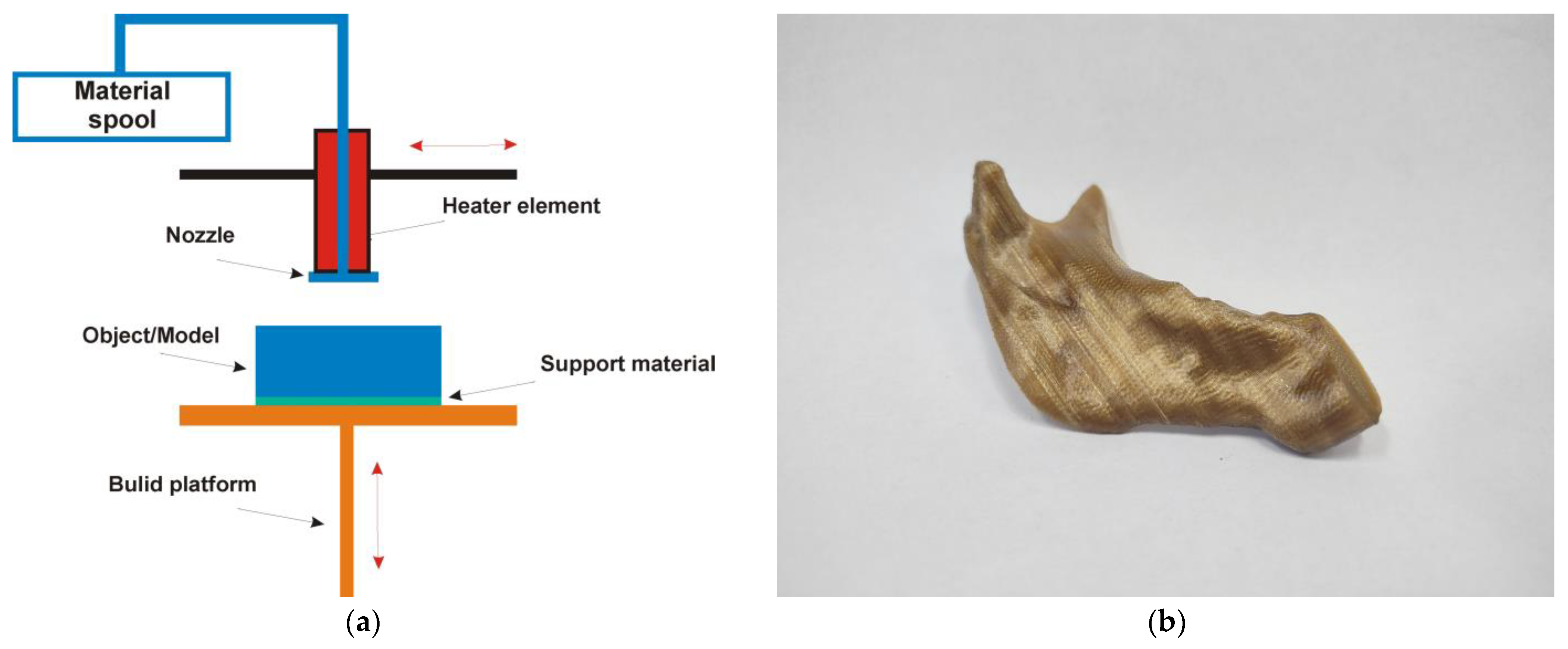
Material extrusion (MEX) is a 3D-printing technology in which a continuous thermoplastic material is pushed through a heated nozzle, melting it (Figure 3a). The 3D printer deposits the material on a work platform along a defined path, where the filament cools and solidifies to form a solid object. The MEX technology mainly includes the following manufacturing methods: Fused Granulate Fabrication (FGF), Fused Filament Fabrication (FFF), and Fused Deposition Modeling (FDM). In the literature review from 2020 to 2025, MEX technology produced the most common anatomical models of the mandible (Figure 3b), surgical guides, implants, and scaffolds. Table 2 presents the current research conducted on models manufactured using MEX techniques.
The publication [39] discusses the creation of models for the manufacture of reconstruction plates of Acrylonitrile Butadiene Styrene (ABS) material for the treatment of acute trauma in maxillofacial surgery. The authors highlight the advantages of 3D models, which aid in visualizing and matching osteosynthetic materials. However, they also address the challenges associated with the manufacturing of ABS models, particularly its tendency to melt during drilling procedures. Although ABS presents certain disadvantages, such as thermal deformation during drilling procedures, it remains a widely used material for producing anatomical models due to its affordability, accessibility, and good dimensional stability for nonimplant applications. In clinical practice, such trade-offs are often acceptable when the model is used only for visualization or plate pre-bending rather than implantation. Likewise, PLA and PCL are favored for their biocompatibility and ease of extrusion, although their lower mechanical strength limits their use in load-bearing roles. These considerations illustrate the importance of selecting materials based on the specific application, whether structural, temporary, or visual.
Polylactic acid (PLA)-based templates are preferred over other metal materials because their hydrophobic properties prevent rapid dissolution of gentamicin [38]. Based on a study in rabbits, the authors demonstrated the feasibility of two bioengineered scaffolds for reconstructing mandibular defects. In mandibular reconstruction, PLA-based materials [40,42] are commonly used to fix tissue and position bone fragments. When studying the 3D-printing process with PLA and its mechanical properties, research has shown that 3D-printed PLA parts exhibit superior impact resistance, tensile strength, and compressive strength. They show an increase in fit and a reduction in treatment time. MEX technology, which uses ABS, PLA, and polycaprolactone (PCL) materials, commonly prepares surgical guides [33,35,44] for preoperative and intraoperative use, as well as anatomical models for pre-bending fixation plates in mandibular reconstruction. Based on the literature review, an effective repair of significant segmental bone defects is sought. The reconstruction of a mandibular defect should consider its anatomical shape and aesthetic effect, while also restoring physiological function. The predominant material in the development of mandibular implants is titanium. On the other hand, based on modern research, polyetheretherketone (PEEK) or other polymers from the polyether ketone family are becoming a preferred bone substitute because of their higher chemical resistance, radiopacity, and mechanical properties in comparison to those of human bone. Despite this, research on the use of PEEK implants in mandibular reconstruction is scarce [41,45]. In the future, it will be necessary to determine which MEX materials and fabrication techniques offer the best cost–benefit ratio for each type of procedure and application.
Taking into account articles published between January 2020 and May 2025 on the applicability of MEX technology in the creation of models to support resection and reconstruction of the mandibular geometry, the following results can be presented:
  • MEX is instrumental in the production of customized surgical guides [42]. These guides, including implant surgical stents, are specifically designed to ensure the precise placement of implants or other surgical instruments during various procedures. The tailored nature of these guides means they can accommodate the unique anatomy of each patient, significantly improving the accuracy and efficiency of the surgical process. By facilitating proper alignment and positioning [37], MEX-generated surgical guides not only enhance surgical outcomes but also contribute to a reduction in procedural complications [36,44].
  • MEX technology is not always used for the creation of implants [41]; it serves an essential role in the fabrication of temporary implants or scaffolds designed for bone regeneration [45]. Utilizing biocompatible materials, MEX can create structures that provide support and stability for healing tissues. These temporary implants are crucial in situations where immediate structural integrity is needed while the body initiates its healing processes [44].
  • MEX technology is used in the development of advanced scaffolds for bone grafting procedures [38]. These scaffolds create an optimal environment for new bone growth by providing a framework that encourages cell proliferation and tissue integration [40]. The customized nature of MEX allows for the design of scaffolds that can match the specific anatomical requirements of different patients, promoting successful outcomes in bone restoration and regeneration efforts. This application of MEX not only enhances the efficacy of bone grafts but also accelerates the healing process, ultimately benefiting patient recovery.
Based on the presented results, the most significant advantages of MEX technology in the creation of models to support resection and reconstruction of the mandibular geometry are related to cost-effectiveness [42,44], reduced surgical time [33,37,43], customization, and improved accuracy of surgical planning [35,41,42]. In the case of limitations, these include material limitations [37], porosity [40], surface finishing [36,38], and mechanical properties [45].

4.2. Material Jetting Technology

Material jetting (MJT) operates like a standard inkjet printer. However, instead of printing a single layer of ink, multiple layers are built on top of each other to create a solid part. The print nozzle ejects hundreds of tiny droplets of photopolymer. Then it cures or solidifies them using ultraviolet (UV) light (Figure 4a). After depositing and curing one layer, the platform lowers the thickness of that layer. The process is repeated to construct the 3D object. The MJT technology mainly includes the following manufacturing methods: Nanoparticle Jetting (NPJ), PolyJet, and Multijet Printing (MJP). In the literature review from 2020 to 2025, MJT technology produced the most common anatomical models of the mandible (Figure 4b), surgical guides, implants, and scaffolds. Table 3 presents the current research conducted on models manufactured using MJT techniques.
Additive manufacturing using light-polymerized resins is gaining popularity in the medical field due to improved 3D-printing quality and increased manufacturing speed. This technology allows the creation of both finished and highly personalized anatomical models [46], as well as surgical guides [48] and models for preoperative simulations [42]. In a study involving dogs, the authors [39] demonstrated that 3D-printed resin implants offer significant advantages over traditional methods for mandibular reconstruction. These advantages include increased implant strength and a faster, more efficient approach to implant design. Furthermore, the authors of another publication [47] confirmed that light polymerized resins exhibit high dimensional precision, consistent repeatability, low surface roughness, and minimal cytotoxicity.
Taking into account articles published between January 2020 and May 2025 on the applicability of MJ technology in the creation of models to support resection and reconstruction of the mandibular geometry, the following results can be presented:
  • By manufacturing a physical replica of a patient’s mandible, surgeons can engage in detailed preoperative planning. This tactile model allows for a thorough assessment of the unique anatomical features and variations of the patient’s mandible [42]. Surgeons can practice complex procedures, visualize surgical options, and anticipate potential complications, ultimately enhancing surgical outcomes and reducing operative time.
  • MJT can be utilized to fabricate custom prosthetics or surgical guides explicitly tailored for mandible reconstruction [42]. By capturing the intricacies of the patient’s anatomy, these models enable the design of prosthetic devices that fit securely and function effectively [48], improving patient comfort and functionality following surgical interventions.
  • The highly accurate models generated via MJ are invaluable resources for both patient education and medical training [46]. They can help patients better understand their condition by providing a tangible representation of their anatomy, elucidating the surgical process, and setting realistic expectations for outcomes [39]. Additionally, these models serve as practical training tools for medical professionals, allowing them to refine their skills in a hands-on manner without the risks associated with live procedures. This approach fosters improved competence and confidence in tackling real-world clinical challenges [47].
Based on the presented results, the most significant advantages of MJ technology in the creation of models to support resection and reconstruction of the mandibular geometry are related to high resolution and accuracy [42,46], multi-material 3D printing [46], and good material properties [47]. In the case of limitations, these include material limitations [39], cost [48], and limited build size [46].

4.3. Powder Bed Fusion Technology

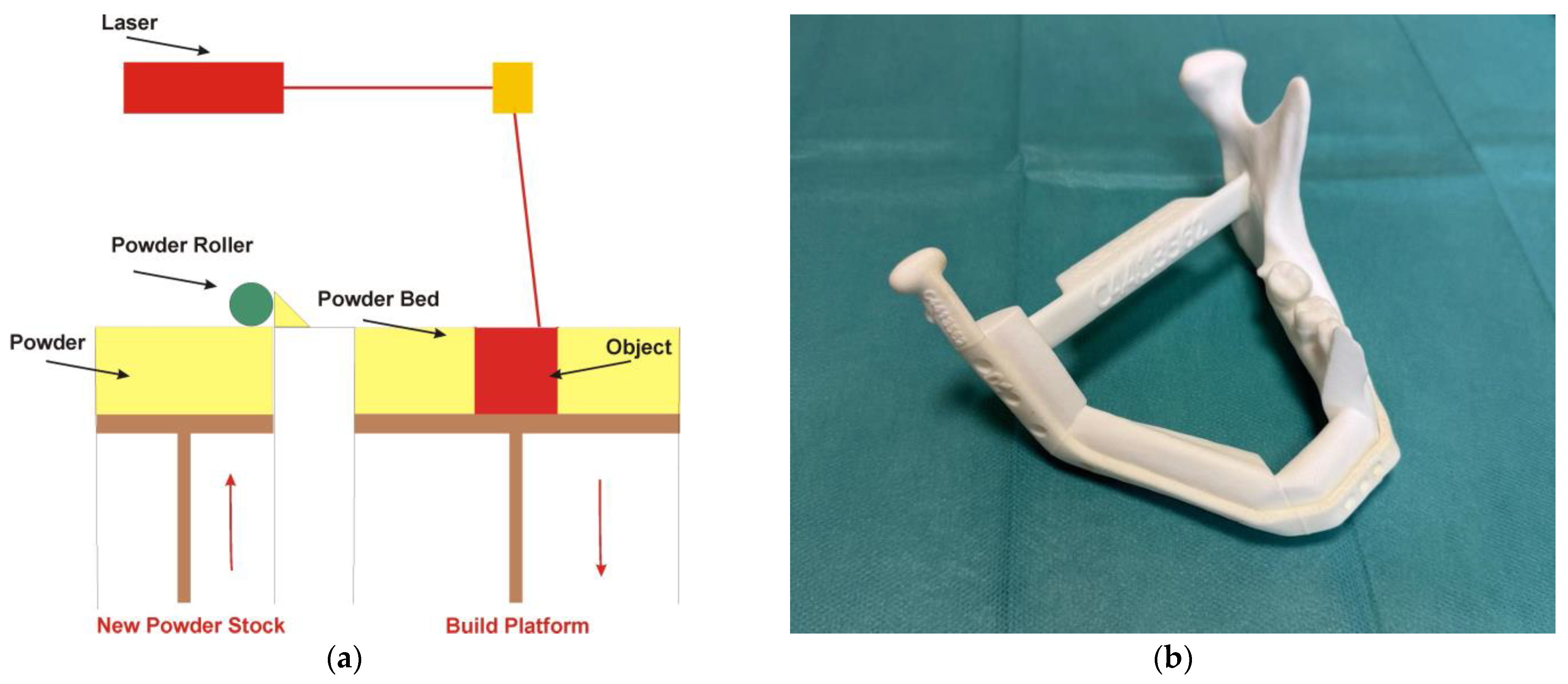
Powder bed fusion (PBF) is a technology that uses thermal energy to fuse powdered materials into a solid shape selectively (Figure 5a). The method begins by spreading a granular 3D-printing medium, such as metal, ceramics, or polymers, across the surface of a work platform to create a layer of powder. Using data from a CAD file, the powder bed fusion machine applies thermal energy to specific areas of the powder layer, causing the granules to fuse into a solid layer. After this, the work platform is lowered, and a new thin layer of powder is added. This process is repeated for each subsequent layer until the entire design is completely 3D-printed. PBF technology mainly includes the following manufacturing methods: selective laser sintering (SLS), selective laser melting (SLM), Direct Metal Laser Sintering (DMLS), Electron beam melting (EBM), Micro Selective Laser Sintering (µSLS), and Cold Metal Fusion. In the review of the literature from 2020 to 2025, PBF technology produced the most common anatomical models of the mandible, surgical guides (Figure 5b), implants, and scaffolds. Table 4 presents the current research conducted on models manufactured using PBF techniques.
AM technologies from the PBF group are becoming increasingly crucial in reconstructive medicine, particularly maxillofacial surgery. Their growing popularity is primarily attributed to titanium alloys such as Ti-6Al-4V ELI [18,53,54,55,56,58,59,60,65], which have been clinically used for years due to their high biocompatibility, corrosion resistance, and suitable mechanical properties. The available publications indicate that we are increasingly engaged in research and established clinical procedures focused on reconstructing the mandible and nearby craniofacial structures. Personalized implants produced using techniques such as SLM [17,51,52,53,54,57,58,59,62], DMLS [55,63,64], or EBM [18,56,60,65] have been used successfully in real-life cases of bone defect reconstruction, effectively integrating with the bone tissue. While part of PBF technology, SLS technology is used mainly in anatomical planning and modeling because it is limited to polymeric materials [49,50,61]. The introduction of PEEK-based materials has expanded their applications, allowing the creation of biocompatible scaffolds used in bone regeneration research [49].
Taking into account articles published between January 2020 and May 2025 on the applicability of PBF technology in the creation of models to support resection and reconstruction of the mandibular geometry, the following results can be presented:
  • One of the standout features of PBF technology is its ability to produce patient-specific implants that fit precisely within the contours of an individual’s mandible [49,50,51,52,53,54,55,56]. This level of customization is pivotal, as it not only enhances the mechanical integration of the implant with the surrounding bone but also improves the overall clinical outcomes for patients undergoing mandible reconstruction [18,58,59,60,61]. By accurately replicating the patient’s anatomical structures [63], the implants can restore both function and aesthetics more effectively than traditional implant methods [59,63].
  • In addition, surgical templates are manufactured using PBF technology [57]. One of the primary functions of surgical templates is to facilitate the accurate positioning of plates, screws, and bone grafts during the intricate process of mandibular reconstruction [57,64]. The templates act as crucial guides for performing osteotomies, which are surgical cuts made to reshape or reconstruct the mandible [62]. Their incorporation into surgical practices not only enhances the precision of surgical interventions but also promotes superior patient outcomes [57,62].
Based on the presented results, the most significant advantages of PBF technology in the creation of models to support resection and reconstruction of the mandibular geometry are related to improved biocompatibility [17,53,55], enhanced mechanical properties [49], reduced risk of infection [59], and shorter treatment times [49,55,56]. In the case of limitations, these include cost and time [52,53,54], porosity and surface roughness [18,65], and residual stress [63,64].

4.4. Vat Polymerization Technology

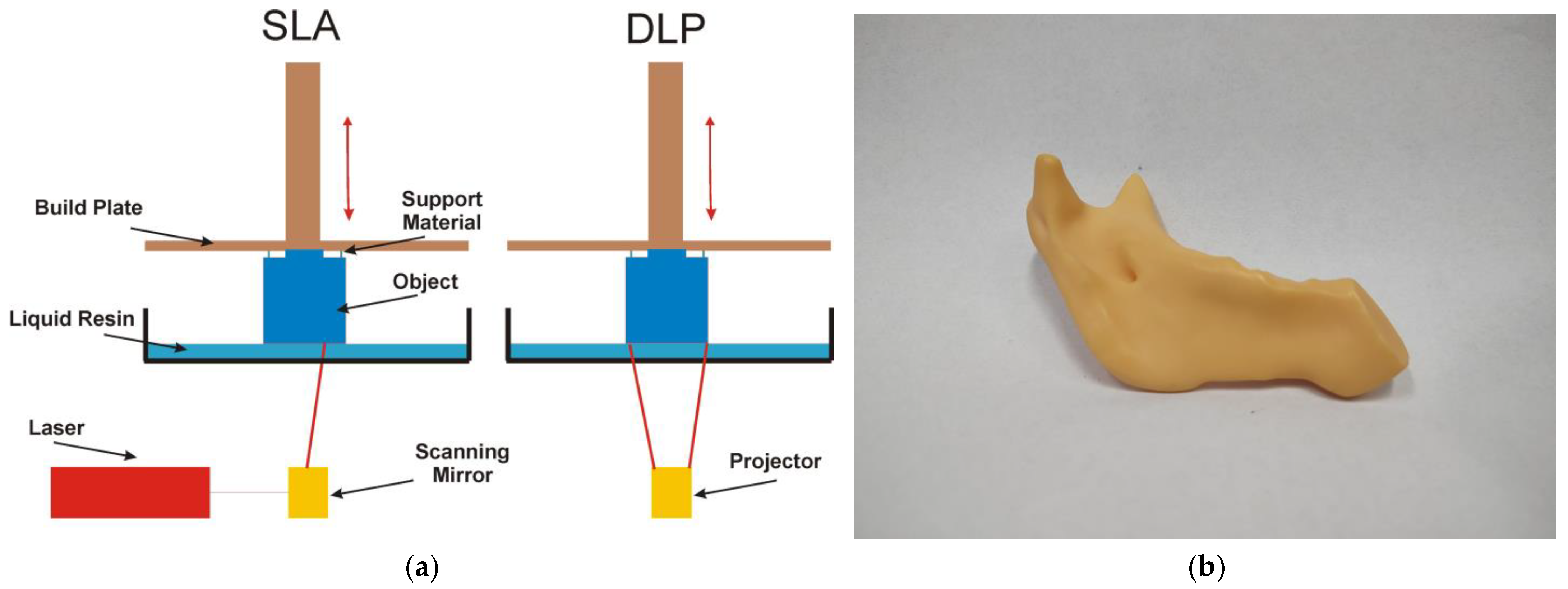
Vat photopolymerization (VPP) is a technology used to create 3D objects by selectively curing liquid photopolymer resin through light-activated polymerization (Figure 6a). In most 3D printers that utilize this method, the liquid photopolymer is contained in a vat, with the working platform partially submerged beneath the surface. According to information from the CAD file, the 3D printer directs a light source to specific points, selectively hardening the liquid photopolymer into a solid layer. After each layer is created, the work platform is submerged back into the resin, and this process is repeated to build additional layers until the entire object is manufactured. The VPP technology mainly includes the following manufacturing methods: Stereolitography (SLA), Digital Light Processing (DLP), Masked Stereolitography (MSLA), Microstereolitography (µSLA), Digital Light Synthesis (DLS), Digital Composite Manufacturing (DCM), Two-Photon Polymerization (TPP), Lithography-based Metal Manufacturing (LMM), Low Force Stereolitography (LFS), and Projection Microstereolitography (PµSL). In the review of the literature from 2020 to 2025, VPP technology produced the most common anatomical models of the mandible, surgical guides (Figure 6b), implants, and scaffolds. Table 5 presents the current research conducted on models manufactured using VPP techniques.
The DLP and SLA methods lead the way in manufacturing models that utilize VPP technology for medical applications. The DLP method primarily produces scaffolds to regenerate damaged or missing tissues [69]. These structures, made with appropriate resin-ceramic mixtures, are undergoing animal tests [42,68,71]. Furthermore, 3D-printed models created using the DLP method are used in dental applications, such as surgical templates [73]. Stereolithography technology 3D printing is widely utilized in maxillofacial surgery to plan mandibular reconstruction due to its high precision in reproducing anatomical structures [50]. SLA models serve various purposes, including analyzing the precision of reconstructing three-dimensional anatomical structures and fitting and pre-bending titanium reconstruction plates [66]. Additionally, these models are used as surgical templates for planning cuts and drilling holes for implant plates and in implant treatment [67]. Since 2020, there have also been reports in the literature on the use of photopolymer resin prints as titanium-coated implants.
Taking into account articles published between January 2020 and May 2025 on the applicability of VPP technology in the creation of models to support resection and reconstruction of the mandibular geometry, the following results can be presented:
  • Personalized guides that, based on a virtual surgical plan, fit perfectly to the surface of the patient’s mandible. The surgeon uses them to make precise and safe bone cuts during resection (removal of the diseased fragment) [66,69,72]. This allows cuts to be made with millimeter precision, ensuring that the graft will fit perfectly into the defect site. Additionally, they allow for the precise positioning and immobilization of the bone graft in the defect site in the mandible [67,70].
  • Test models are used by surgeons for practical, preoperative testing and fitting of implants (e.g., titanium plates) and for simulating the procedure. This avoids surprises during the actual operation and significantly reduces its duration [50,73].
  • Specific scaffolds that promote bone regeneration [42,68,71,74].
Based on the presented results, the most significant advantages of VPP technology in the creation of models to support resection and reconstruction of the mandibular geometry are related to improved surgical precision and accuracy [66,67,68,69], reduced operative time [42,68,69], and enhanced team communication [50,73]. In the case of limitations, these include cost and time [50,72] and material limitations of the surgical guides [67,70].

4.5. Binder Jetting Technology

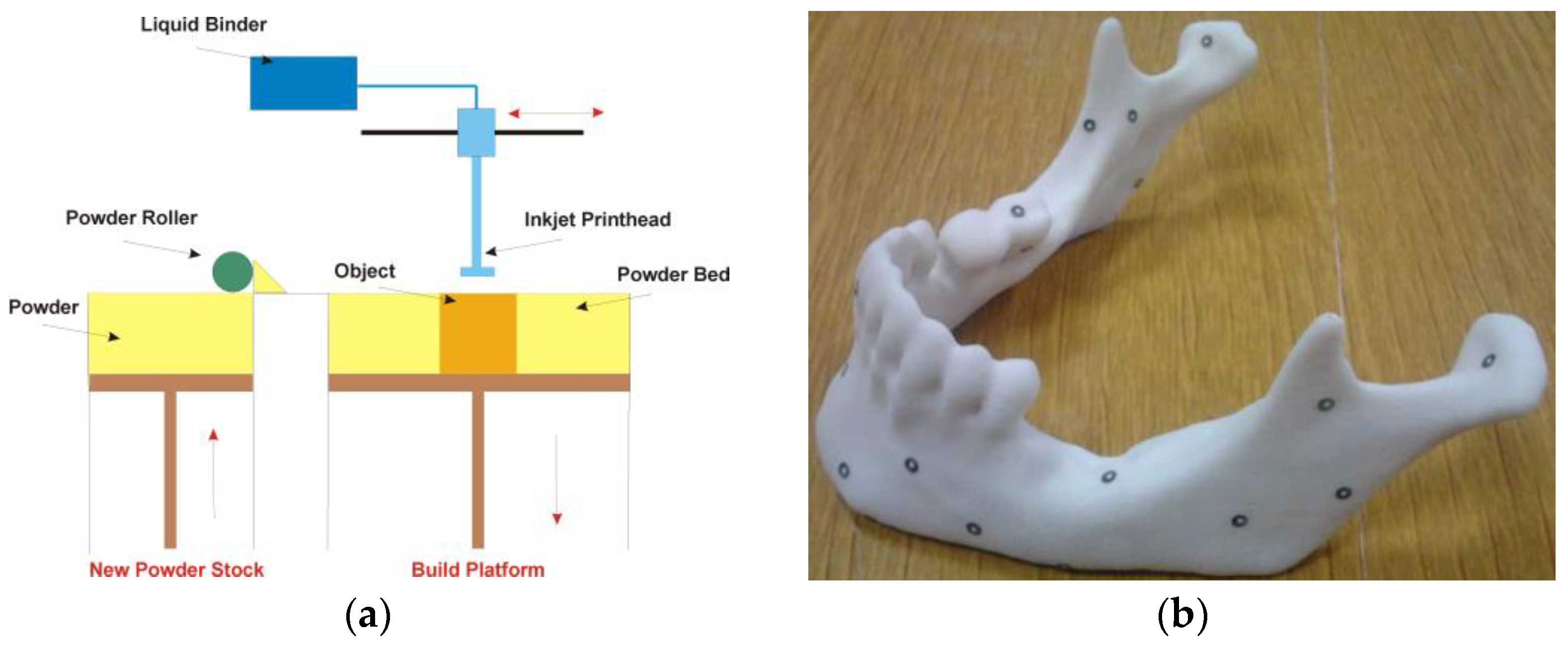
Binder jetting (BJT) is a 3D-printing technology in which a liquid bonding agent selectively binds areas of a powder bed (Figure 7a). This technology is similar to selective laser sintering (SLS), which begins with an initial layer of powder on the working platform. However, instead of using a laser to sinter the powder as in SLS, binder jetting moves a print nozzle over the powder surface, depositing binder droplets that are typically 80 μm in diameter. These droplets bond the powder particles to form each layer of the object. After a layer is 3D-printed, the powder bed is lowered, and a new layer of powder is spread over the previously 3D-printed layer. This process is repeated until the entire object is formed. The BJT technology mainly includes the following manufacturing methods: Multi Jet Fusion (MJF), Metal Jet (MJ), Selective Absorption Fusion (SAF), High-Speed Sintering (HSS), Polymer Binder Jetting, Metal Binder Jetting, and Sand Binder Jetting. In the literature review from 2020 to 2025, BJT technology produced the most common anatomical models of the mandible (Figure 7b). Table 6 presents current research on models manufactured using BJT techniques.
Although extensive research has been conducted on the creation of suitable scaffold structures using BJT technology, most of it has been limited to laboratory studies [76,77]. In the past five years, no publications have demonstrated the direct application of surgical templates, implants, and scaffolds for the resection and reconstruction of mandibular geometry. No studies were observed using DED and SHL technologies to fabricate mandibular anatomical models, surgical templates, implants, or scaffolds.
Based on the presented results, the most significant advantages of BJ technology in the creation of models to support resection and reconstruction of the mandibular geometry are related to cost-effectiveness for low to medium volumes and fast production [61,75]. In the case of limitations, these include dimensional accuracy [61,75], mechanical properties [61], and material limitations [75].
Although directed energy deposition (DED) and sheet lamination (SHL) are among the AM technologies, no studies between 2020 and 2025 were identified that applied these methods to mandibular reconstruction. Their omission reflects the current lack of clinical or experimental implementation in this field, rather than their technological irrelevance.

5. Discussion

In mandibular reconstruction procedures, CAD modeling and additive manufacturing play crucial roles in both surgical planning and final performance. Regardless of the reconstruction method, whether using autogenous grafts, standard or individualized titanium plates, or implants, visualizing volumetric data from the patient’s medical information enables the creation of a three-dimensional anatomical model. This model offers several benefits:
  • It helps the surgeon prepare more effectively for the procedure;
  • It increases the precision of the surgery;
  • It aids in the selection of appropriate surgical tools;
  • It facilitates thorough consultations with other medical professionals prior to the procedure;
  • It allows for a more detailed presentation of the procedure to the patient, enabling better discussion of its course;
  • It can reduce the overall surgery time under general anesthesia;
  • It helps minimize blood loss during the procedure;
  • It reduces the risk of intraoperative complications.
Models created using AM techniques are increasingly utilized in reconstructive surgery for the mandible following procedures such as hemisection [70,73], mandibulectomy [50,57], and removal of head and neck tumors [43,62,66]. The challenge of restoring aesthetics after extensive mandibular resection is well recognized in the medical community, mainly due to the complex spatial shape of the bone [57,62,64]. This complexity makes it challenging to achieve an aesthetic restoration that accurately mimics the original appearance and function of the stomatognathic system. Other functions of the stomatognathic system must be considered, particularly the accurate alignment of the occlusion with implants [64]. Compared to traditional reconstructive methods, AM technologies are more tolerable to patients and offer improved precision in replicating mandible function and facial appearance before surgical removal [37,48,65,69]. By manufacturing a model based on a CT scan, the surgeon can preoperatively plan the extent of the procedure and the type of cuts to be made in the structure corresponding to the patient’s mandible [57,62]. The model can also be used to determine the size of the mandibular fragment to be resected [13,41].
Three-dimensional visualization and AM techniques are often essential in mandibular hemisection, where only the condylar region remains [13,57]. When reconstructing significant bone defects, it is crucial to accurately map the edges and angles of the mandible on the affected side to ensure optimal soft tissue aesthetics. The surgery planning is conducted during the digital visualization stage. To facilitate this, resection guides are typically designed based on the digital model of the mandible and then produced using AM techniques [62,66]. They are generally made using PBF and VPP technologies, which involve titanium and photocurable resins. An implant model can also be designed and manufactured to replace the bone defect. They are made most often using PBF technology with Ti-6AL-4V ELI. However, if complete mandibular reconstruction is not planned, traditional surgical techniques are used to fuse interrupted sections of the mandible using a titanium plate [59]. This procedure can also be enhanced by creating and manufacturing, using AM techniques, an individualized surgical plate geometrically tailored to fit the unique shape. If individualized surgical plates are not planned for a patient, standard ones are used instead. These classic surgical plates are bent on an anatomical mandible model manufactured using AM techniques before surgery [13,37,66]. Digital mirroring methods are also used, which involve superimposing the healthy half of the mandible onto the tumor-affected half. As a result, a digital model of the mandible is produced that excludes the affected portion [13]. This digitally developed model is then manufactured using AM techniques, allowing for the precise bending of the surgical plate. Although the human face is naturally asymmetric, the mirroring process is so effective that patients achieve satisfactory facial aesthetics after the procedure [55]. An overview of the clinical applications and suitability of various AM technologies used in mandibular reconstruction is presented in Table 7.
A mandible reconstruction method worth special attention is the transplantation of an autogenous fibula [13,60], an iliac crest [51], or, in some cases, particularly in Japan, a scapular flap. The scapular flap offers a reliable pedicle length and the possibility of harvesting composite tissues. This procedure involves creating a 3D-printed model of the patient’s mandible based on the 3D model obtained from a CT scan. Then, on the model prepared in this way, a classic titanium surgical plate is bent to achieve a shape that is in line with the course of the posterior mandibular branch, the angle of the mandible, and the edge of the lower mandibular body. A second model is created in parallel on a CT scan of the sagittal or iliac crest. Based on the three-dimensional representation, a resection template is designed in a CAD environment and manufactured using AM technology, allowing the surgeon to remove the required amount of bone tissue from the fibula or iliac crest area. Then, during mandibular surgery, the bone is reconstructed immediately after the excision of the pathologically affected bone fragment. For this purpose, a titanium surgical plate is first implanted, which fuses the left fragment of the mandible, and then an autogenous graft (taken from the fibula or the iliac crest) is shaped on its basis, allowing the aesthetics to be maintained after healing postoperative wounds and the appearance of bone remodeling processes and adaptation of soft tissues to the new titanium scaffolding.
Currently, there is significant interest in the literature in fields related to tissue engineering, particularly in the mandibular area [42,56,58]. Research is focused on developing suitable spatial structures for porous substrates that create an environment conducive to the regeneration of damaged tissues. Cell scaffolds are primarily produced using additive manufacturing (AM) technologies. These scaffolds are often made of ceramics [42,74]. Once these scaffolds are prepared, stem cells are introduced, allowing tissue development to regenerate bone defects. The most common research on bone structure in the mandibular area is based primarily on in vivo studies conducted in animals [38,39].
Despite the numerous advantages of using additive manufacturing in mandibular reconstruction, several limitations must also be considered. One of the primary disadvantages is the high cost and limited accessibility of AM technologies, especially in low-resource settings. The process of image acquisition, segmentation, CAD modeling, and 3D printing requires specialized equipment and trained personnel, which may not be available in all clinical environments. Additionally, the manufacturing time for custom implants or guides may delay urgent surgical procedures. Another concern is the variability in the mechanical properties of some printed polymers, such as PLA or PCL, which may limit their long-term performance and biocompatibility in load-bearing applications. Furthermore, regulatory approval processes for patient-specific implants, particularly those made from novel or hybrid materials, can be time-consuming and restrictive, potentially delaying clinical implementation. Lastly, inaccuracies can arise during data processing or STL conversion, which may affect the final fit of the implant or guide if not correctly validated. These limitations underscore the need for standardized protocols and further clinical validation.
In terms of clinical outcomes, several studies reported favorable results associated with the use of additively manufactured surgical guides and implants. For example, studies involving titanium implants manufactured using SLM or DMLS technology have shown high integration success rates (>90%) and low complication rates at short- and mid-term follow-up [18,52,54,58]. Patient satisfaction related to postoperative aesthetics and function was also reported to be high, particularly in cases involving personalized implant geometry or digitally planned resections [44,62,65]. However, due to differences in study design, follow-up duration, and reporting standards, a formal meta-analysis was not feasible within the scope of this review. Future systematic reviews and clinical records will be needed to better quantify the long-term success rates of AM-based mandibular reconstructions.
This review provides a descriptive synthesis of the literature on the use of additive manufacturing in mandibular reconstruction but does not constitute a systematic review. The selection of studies was limited to English-language publications from 2020 to mid-2025 and focused on peer-reviewed journal articles and books indexed in major databases. As a result, some relevant studies may have been intentionally excluded. Furthermore, the heterogeneity of the available literature—in terms of study design, endpoints, and reporting standards—makes it challenging difficult to conduct a quantitative comparison or meta-analysis. Many reviewed articles focus on technical feasibility or prototyping rather than long-term clinical outcomes, which limits the strength of the clinical recommendations. Finally, while efforts have been made to assess the clinical relevance of each technology and material, variability in reporting quality and the lack of standardized outcome metrics pose inherent challenges to critical evaluation.

6. Conclusions

Reconstruction of mandibular continuity remains a clinically and technically demanding procedure due to the complex anatomical geometry and the need for precise aesthetic and functional restoration. Additive manufacturing (AM), combined with medical imaging and computer-aided design, allows patient-specific modeling that facilitates surgical planning, including the estimation of resection margins, preoperative bending of fixation plates, and fabrication of customized implants and scaffolds. These technologies have contributed to a reduction in surgical time and improved surgical precision.
This review highlights that not all AM technologies are suitable for load-bearing mandibular reconstruction. Material extrusion (e.g., FFF/FDM with PLA, ABS, PCL) and vat photopolymerization (e.g., SLA, DLP) are most appropriate for surgical guides or anatomical templates because of their limited mechanical properties. On the contrary, powder bed fusion methods (e.g., SLM, DMLS, EBM) with Ti-6Al-4V ELI alloys, or selectively PEEK using high temperature MEX or SLS, are recommended for structural reconstructions requiring long-term biomechanical performance. Proper alignment between the selected material, the manufacturing process, and the intended clinical function remains a key determinant of success.
Despite these advances, the field faces challenges beyond technical feasibility. Regulatory uncertainty regarding the approval and validation of patient-specific implants, especially in AM workflows, remains a significant hurdle. Ethical concerns related to data protection and consent in AI-assisted planning must be addressed through standardized protocols. Furthermore, the high cost of equipment, limited reimbursement pathways, and variability in institutional infrastructure restrict broader clinical adoption.
Future research should focus on high-quality clinical trials, long-term outcome reporting, and the development of integrated frameworks that address not only the technical but also regulatory and economic aspects of AM in mandibular reconstruction.

Author Contributions

Conceptualization, P.T., M.Z., K.G.-D., M.B., and B.L.; methodology, P.T., M.Z., K.G.-D., and M.B.; writing—original draft preparation, P.T., M.Z., K.G.-D., M.B., J.K., and N.D.; writing—review and editing, P.T., B.L., and J.K.; visualization, J.K. and N.D.; supervision, P.T. All authors have read and agreed to the published version of the manuscript.

Funding

This work was partially co-financed by the Military University of Technology under research project: UGB 22-016.

Institutional Review Board Statement

Not applicable.

Informed Consent Statement

Not applicable.

Data Availability Statement

The original contributions presented in this study are included in this article; further inquiries can be directed to the corresponding author.

Conflicts of Interest

The authors declare no conflicts of interest.

References

  1. Govind, A.; Lugo, R.; Pogrel, M.A. Mandibular Reconstruction. Oral. Maxillofac. Surg. 2025, 645–672. [Google Scholar] [CrossRef]
  2. Huang, N.; Wang, P.; Gong, P.; Huang, B. The progress in reconstruction of mandibular defect caused by osteoradionecrosis. J. Oncol. 2023, 2023, 1440889. [Google Scholar] [CrossRef]
  3. Ritschl, L.M.; Fichter, A.M.; Grill, F.D.; Hart, D.; Hapfelmeier, A.; Deppe, H.; Mücke, T. Bone volume change following vascularized free bone flap reconstruction of the mandible. J. Cranio-Maxillofac. Surg. 2020, 48, 859–867. [Google Scholar] [CrossRef] [PubMed]
  4. Mashrah, M.A.; Aldhohrah, T.; Abdelrehem, A.; Sakran, K.A.; Ahmad, H.; Mahran, H.; Wang, L. Survival of vascularized osseous flaps in mandibular reconstruction: A network meta-analysis. PLoS ONE 2021, 16, e0257457. [Google Scholar] [CrossRef] [PubMed]
  5. Bauer, E.; Mazul, A.; Zenga, J.; Graboyes, E.M.; Jackson, R.; Puram, S.V.; Pipkorn, P. Complications after soft tissue with plate vs bony mandibular reconstruction: A systematic review and meta-analysis. Otolaryngol.–Head. Neck Surg. 2021, 164, 501–511. [Google Scholar] [CrossRef]
  6. Lilly, G.L.; Petrisor, D.; Wax, M.K. Mandibular rehabilitation: From the Andy Gump deformity to jaw-in-a-day. Laryngoscope Investig. Otolaryngol. 2021, 6, 708–720. [Google Scholar] [CrossRef]
  7. Gibson, I.; Rosen, D.W.; Stucker, B. Additive Manufacturing Technologies; Springer: Cham, Switzerland, 2021. [Google Scholar]
  8. Amaya-Rivas, J.L.; Perero, B.S.; Helguero, C.G.; Hurel, J.L.; Peralta, J.M.; Flores, F.A.; Alvarado, J.D. Future trends of additive manufacturing in medical applications: An overview. Heliyon 2024, 10, e26641. [Google Scholar] [CrossRef]
  9. Allawi, N.I. Application of Reverse Engineering and Rapid Prototyping for Reconstruction of Human Mandible. arXiv 2021, arXiv:2106.15489. [Google Scholar] [CrossRef]
  10. Kumar, D.; Yadav, P.K.; Bhaskar, J. 3D Modelling of Human Joints Using Reverse Engineering for Biomedical Applications. In Advances in Manufacturing and Industrial Engineering; Springer: Singapore, 2021; pp. 865–875. [Google Scholar] [CrossRef]
  11. Salmi, M. Additive manufacturing processes in medical applications. Materials 2021, 14, 191. [Google Scholar] [CrossRef]
  12. Oren, D.; Dror, A.A.; Bramnik, T.; Sela, E.; Granot, I.; Srouji, S. The power of three-dimensional printing technology in functional restoration of rare maxillomandibular deformity due to genetic disorder: A case report. J. Med. Case Rep. 2021, 15, 197. [Google Scholar] [CrossRef]
  13. de Koning, S.B.; Ter Braak, T.P.; Geldof, F.; van Veen, R.L.P.; van Alphen, M.J.A.; Karssemakers, L.H.E.; Karakullukcu, M.B. Evaluating the accuracy of resection planes in mandibular surgery using a preoperative, intraoperative, and postoperative approach. Int. J. Oral Maxillofac. Surg. 2021, 50, 287–293. [Google Scholar] [CrossRef]
  14. Oldhoff, M.G.E.; Mirzaali, M.J.; Tümer, N.; Zhou, J.; Zadpoor, A.A. Comparison in clinical performance of surgical guides for mandibular surgery and temporomandibular joint implants fabricated by additive manufacturing techniques. J. Mech. Behav. Biomed. Mater. 2021, 119, 104512. [Google Scholar] [CrossRef] [PubMed]
  15. Probst, F.A.; Liokatis, P.; Mast, G.; Ehrenfeld, M. Virtual planning for mandible resection and reconstruction. Innov. Surg. Sci. 2023, 8, 137–148. [Google Scholar] [CrossRef] [PubMed]
  16. Shaheen, S.R.; Sridevi, E.; Sankar, A.S.; Krishna, V.S.S.; Sridhar, M.; Sankar, K.S. Contemporary era of Three-dimensional printing in pediatric dentistry: An overview. J. Oral Res. Rev. 2023, 15, 72–79. [Google Scholar] [CrossRef]
  17. Kim, M.K.; Ham, M.J.; Kim, W.R.; Kim, H.G.; Kwon, K.J.; Kim, S.G.; Park, Y.W. Investigating the accuracy of mandibulectomy and reconstructive surgery using 3D customized implants and surgical guides in a rabbit model. Maxillofac. Plast. Reconstr. Surg. 2023, 45, 8. [Google Scholar] [CrossRef]
  18. Xia, Y.; Feng, Z.C.; Li, C.; Wu, H.; Tang, C.; Wang, L.; Li, H. Application of additive manufacturing in customized titanium mandibular implants for patients with oral tumors. Oncol. Lett. 2020, 20, 51. [Google Scholar] [CrossRef]
  19. Cao, L.; Liu, X.; Qu, T.; Cheng, Y.; Li, J.; Li, Y.; Guo, J. Improving spatial resolution and diagnostic confidence with thinner slice and deep learning image reconstruction in contrast-enhanced abdominal CT. Eur. Radiol. 2023, 33, 1603–1611. [Google Scholar] [CrossRef]
  20. Miranda-Viana, M.; Fontenele, R.C.; Nogueira-Reis, F.; Farias-Gomes, A.; Oliveira, M.L.; Freitas, D.Q.; Haiter-Neto, F. DICOM file format has better radiographic image quality than other file formats: An objective study. Braz. Dent. J. 2023, 34, 150–157. [Google Scholar] [CrossRef]
  21. Dino, J.A.; Barani, S.; Poornapushpakala, S.; Subramoniam, M. Implementation of Various Filtering Technique for Noise Removal in DICOM Images of Colon Carcinoma. In Proceedings of the 2024 Global Conference on Communications and Information Technologies (GCCIT), Bangalore, India, 25–26 October 2024; pp. 1–5. [Google Scholar] [CrossRef]
  22. Fajar, A.; Sarno, R.; Fatichah, C.; Fahmi, A. Reconstructing and resizing 3D images from DICOM files. J. King Saud. Univ. Comput. Inf. Sci. 2022, 34, 3517–3526. [Google Scholar] [CrossRef]
  23. Qiu, B.; van der Wel, H.; Kraeima, J.; Glas, H.H.; Guo, J.; Borra, R.J.; van Ooijen, P.M. Automatic segmentation of mandible from conventional methods to deep learning—A review. J. Pers. Med. 2021, 11, 629. [Google Scholar] [CrossRef]
  24. Irshad, T.B.; Pascoletti, G.; Bianconi, F.; Zanetti, E.M. Mandibular bone segmentation from CT scans: Quantitative and qualitative comparison among software. Dent. Mater. 2024, 40, e11–e22. [Google Scholar] [CrossRef]
  25. Syuhada, F.; Fatichah, C.; Fabroyir, H. Image Enhancement for Visualization Jaw Bone in DICOM CT Scans using Windowing Strategies, Adaptive Denoising Bilateral Filtering, and Sharpening. In Proceedings of the 2024 7th International Seminar on Research of Information Technology and Intelligent Systems (ISRITI), Yogyakarta, Indonesia, 11–12 December 2024; pp. 564–569. [Google Scholar]
  26. Gruber, L.J.; Egger, J.; Bönsch, A.; Kraeima, J.; Ulbrich, M.; van den Bosch, V.; Puladi, B. Accuracy and precision of mandible segmentation and its clinical implications: Virtual reality, desktop screen and artificial intelligence. Expert. Syst. Appl. 2024, 239, 122275. [Google Scholar] [CrossRef]
  27. Lahoud, P.; EzEldeen, M.; Beznik, T.; Willems, H.; Leite, A.; Van Gerven, A.; Jacobs, R. Artificial intelligence for fast and accurate 3-dimensional tooth segmentation on cone-beam computed tomography. J. Endod. 2021, 47, 827–835. [Google Scholar] [CrossRef] [PubMed]
  28. Belikova, K.; Rogov, O.Y.; Rybakov, A.; Maslov, M.V.; Dylov, D.V. Deep negative volume segmentation. Sci. Rep. 2021, 11, 16292. [Google Scholar] [CrossRef] [PubMed]
  29. Wang, J.; Huang, Z.; Yang, X.; Jia, W.; Zhou, T. Three-dimensional reconstruction of jaw and dentition CBCT images based on improved marching cubes algorithm. Procedia CIRP 2020, 89, 239–244. [Google Scholar] [CrossRef]
  30. Chalazoniti, A.; Lattanzi, W.; Halazonetis, D.J. Shape variation and sex differences of the adult human mandible evaluated by geometric morphometrics. Sci. Rep. 2024, 14, 8546. [Google Scholar] [CrossRef]
  31. Zhou, L.; Miller, J.; Vezza, J.; Mayster, M.; Raffay, M.; Justice, Q.; Bernat, J. Additive manufacturing: A comprehensive review. Sensors 2024, 24, 2668. [Google Scholar] [CrossRef]
  32. Li, J.; Li, X.; Ma, K.; Sun, J.; Bai, N.; Liu, Y. Rehabilitation of long-term mandibular defects by whole-process digital fibula flap combining with implants: A case report. J. Prosthodont. 2023, 32, 187–195. [Google Scholar] [CrossRef]
  33. Xu, G.; Jia, J.; Xiong, X.; Peng, L.; Bu, L.L.; Wang, X. Mandibular reconstruction with the contralateral vascularized iliac flap using individual design: Iliac crest used to reconstruct the ramus and the anterior border of the iliac wing used to reconstruct the inferior border: A case report. Front. Surg. 2022, 9, 924241. [Google Scholar] [CrossRef]
  34. Zhong, S.; Shi, Q.; Van Dessel, J.; Gu, Y.; Sun, Y.; Yang, S.; Politis, C. Biomechanical validation of structural optimized patient-specific mandibular reconstruction plate orienting additive manufacturing. Comput. Methods Programs Biomed. 2022, 224, 107023. [Google Scholar] [CrossRef]
  35. Wu, P.; Hu, L.; Li, H.; Feng, L.; Liu, Y.; Zhang, S.; Lu, R.J. Clinical application and accuracy analysis of 3D printing guide plate based on polylactic acid in mandible reconstruction with fibula flap. Ann. Transl. Med. 2021, 9, 460. [Google Scholar] [CrossRef] [PubMed]
  36. Moiduddin, K.; Mian, S.H.; Ameen, W.; Alkindi, M.; Ramalingam, S.; Alghamdi, O. Patient-specific surgical implant using cavity-filled approach for precise and functional mandible reconstruction. Appl. Sci. 2020, 10, 6030. [Google Scholar] [CrossRef]
  37. Silva, J.; Lago, C.; Soares Pereira, R.; Furtado da Costa, M.; Nascimento Martins, K.; de Luna Campos, G.; de Souza Andrade, E. The Use of 3D Model Printing for Acute Planning in Oral and Maxillofacial Traumatology. Open Dent. J. 2024, 18, e18742106326414. [Google Scholar] [CrossRef]
  38. Ferrari, M.; Taboni, S.; Chan, H.; Townson, J.; Gualtieri, T.; Franz, L.; Ruaro, A.; Mathews, S.; Daly, M.; Douglas, C.; et al. Hydrogel-chitosan and polylactic acid-polycaprolactone bioengineered scaffolds for reconstruction of mandibular defects: A preclinical in vivo study with assessment of translationally relevant aspects. Front. Bioeng. Biotechnol. 2024, 12, 1353523. [Google Scholar] [CrossRef]
  39. Baumgartner, D.; Schramel, J.P.; Kau, S.; Eberspächer-Schweda, M.C.; Unger, E.; Oberoi, G.; Peham, C. 3D printed plates based on generative design biomechanically outperform manual digital fitting and conventional systems printed in photopolymers in bridging mandibular bone defects of critical size in dogs. Front. Vet. Sci. 2023, 10, 1165689. [Google Scholar] [CrossRef]
  40. Slavin, B.V.; Ehlen, Q.T.; Costello, J.P.; Nayak, V.V.; Bonfante, E.A.; Jalkh, E.B.B.; Runyan, C.M.; Witek, L.; Coelho, P.G. 3D Printing Applications for Craniomaxillofacial Reconstruction: A Sweeping Review. ACS Biomater. Sci. Eng. 2023, 9, 6586–6609. [Google Scholar] [CrossRef]
  41. Li, Y.; Li, Z.; Tian, L.; Li, D.; Lu, B.; Shi, C.; Niu, Q.; Liu, F.; Kong, L.; Zhang, J. Clinical application of 3D-printed PEEK implants for repairing mandibular defects. J. Craniomaxillofac Surg. 2022, 50, 621–626. [Google Scholar] [CrossRef]
  42. Kim, J.-W.; Yang, B.-E.; Hong, S.-J.; Choi, H.-G.; Byeon, S.-J.; Lim, H.-K.; Chung, S.-M.; Lee, J.-H.; Byun, S.-H. Bone Regeneration Capability of 3D Printed Ceramic Scaffolds. Int. J. Mol. Sci. 2020, 21, 4837. [Google Scholar] [CrossRef]
  43. Katschnig, M.; Wallner, J.; Janics, T.; Burgstaller, C.; Zemann, W.; Holzer, C. Biofunctional Glycol-Modified Polyethylene Terephthalate and Thermoplastic Polyurethane Implants by Extrusion-Based Additive Manufacturing for Medical 3D Maxillofacial Defect Reconstruction. Polymers 2020, 12, 1751. [Google Scholar] [CrossRef]
  44. Damecourt, A.; Nieto, N.; Galmiche, S.; Garrel, R.; de Boutray, M. In-house 3D treatment planning for mandibular reconstruction by free fibula flap in cancer: Our technique, European Annals of Otorhinolaryngology. Head. Neck Dis. 2020, 137, 501–505. [Google Scholar] [CrossRef]
  45. Olate, S.; Uribe, F.; Huentequeo-Molina, C.; Goulart, D.R.; Sigua-Rodriguez, E.A.; Alister, J.P. Mandibular Angle Contouring Using Porous Polyethylene Stock or PEEK-based Patient Specific Implants. A Critical Analysis. J. Craniofac Surg. 2021, 32, 242–246. [Google Scholar] [CrossRef] [PubMed]
  46. Chokshi, S.; Gangatirkar, R.; Kandi, A.; DeLeonibus, M.; Kamel, M.; Chadalavada, S.; Gupta, R.; Munigala, H.; Tappa, K.; Kondor, S.; et al. Medical 3D Printing Using Material Jetting: Technology Overview, Medical Applications, and Challenges. Bioengineering 2025, 12, 249. [Google Scholar] [CrossRef]
  47. Schneider, K.H.; Oberoi, G.; Unger, E.; Janjic, K.; Rohringer, S.; Heber, S.; Agis, H.; Schedle, A.; Kiss, H.; Podesser, B.K.; et al. Medical 3D printing with polyjet technology: Effect of material type and printing orientation on printability, surface structure and cytotoxicity. 3D Print. Med. 2023, 9, 27. [Google Scholar] [CrossRef] [PubMed]
  48. Šimić, L.; Kopačin, V.; Mumlek, I.; Butković, J.; Zubčić, V. Improved technique of personalised surgical guides generation for mandibular free flap reconstruction using an open-source tool. Eur. Radiol. Exp. 2021, 5, 30. [Google Scholar] [CrossRef]
  49. Maruf, D.A.; Ren, J.; Cheng, K.; Xin, H.; Lewin, W.; Pickering, E.; Clark, J.R. Evaluation of osseointegration of plasma treated polyaryletherketone maxillofacial implants. Sci. Rep. 2025, 15, 1895. [Google Scholar] [CrossRef]
  50. Msallem, B.; Vavrina, J.J.; Beyer, M.; Halbeisen, F.S.; Lauer, G.; Dragu, A.; Thieringer, F.M. Dimensional Accuracy in 3D Printed Medical Models: A Follow-Up Study on SLA and SLS Technology. J. Clin. Med. 2024, 13, 5848. [Google Scholar] [CrossRef]
  51. Li, Y.; Liu, H.; Wang, C.; Yan, R.; Xiang, L.; Mu, X.; Hu, M. 3D printing titanium grid scaffold facilitates osteogenesis in mandibular segmental defects. NPJ Regen. Med. 2023, 8, 38. [Google Scholar] [CrossRef]
  52. Haroun, F.; Benmoussa, N.; Bidault, F.; Lassau, N.; Moya-Plana, A.; Leymarie, N.; Gorphe, P. Outcomes of mandibular reconstruction using three-dimensional custom-made porous titanium prostheses. J. Stomatol. Oral. Maxillofac. Surg. 2023, 124, 101281. [Google Scholar] [CrossRef]
  53. Yang, H.J.; Oh, J.H. Reconstruction of mandibular contour defect using patient-specific titanium implant manufactured by selective laser melting method. J. Craniofacial Surg. 2022, 33, 2055–2058. [Google Scholar] [CrossRef]
  54. Van Kootwijk, A.; Moosabeiki, V.; Saldivar, M.C.; Pahlavani, H.; Leeflang, M.A.; Niar, S.K.; Zadpoor, A.A. Semi-automated digital workflow to design and evaluate patient-specific mandibular reconstruction implants. J. Mech. Behav. Biomed. Mater. 2022, 132, 105291. [Google Scholar] [CrossRef]
  55. Nemtoi, A.; Covrig, V.; Nemtoi, A.; Stoica, G.; Vatavu, R.; Haba, D.; Zetu, I. Custom-Made Direct Metal Laser Sintering Titanium Subperiosteal Implants in Oral and Maxillofacial Surgery for Severe Bone-Deficient Patients—A Pilot Study. Diagnostics 2022, 12, 2531. [Google Scholar] [CrossRef] [PubMed]
  56. Lim, H.K.; Choi, Y.J.; Choi, W.C.; Song, I.S.; Lee, U.L. Reconstruction of maxillofacial bone defects using patient-specific long-lasting titanium implants. Sci. Rep. 2022, 12, 7538. [Google Scholar] [CrossRef] [PubMed]
  57. Xu, X.; Cheng, K.J.; Liu, Y.F.; Fan, Y.Y.; Wang, J.H.; Wang, R.; Dong, X.T. Experimental validation of finite element simulation of a new custom-designed fixation plate to treat mandibular angle fracture. Biomed. Eng. OnLine 2021, 20, 15. [Google Scholar] [CrossRef] [PubMed]
  58. Farajpour, H.; Bastami, F.; Bohlouli, M.; Khojasteh, A. Reconstruction of bilateral ramus-condyle unit defect using custom titanium prosthesis with preservation of both condyles. J. Mech. Behav. Biomed. Mater. 2021, 124, 104765. [Google Scholar] [CrossRef] [PubMed]
  59. Koper, D.C.; Leung, C.A.; Smeets, L.C.; Laeven, P.F.; Tuijthof, G.J.; Kessler, P.A. Topology optimization of a mandibular reconstruction plate and biomechanical validation. J. Mech. Behav. Biomed. Mater. 2021, 113, 104157. [Google Scholar] [CrossRef]
  60. Lang, J.J.; Bastian, M.; Foehr, P.; Seebach, M.; Weitz, J.; von Deimling, C.; Burgkart, R. Improving mandibular reconstruction by using topology optimization, patient specific design and additive manufacturing?—A biomechanical comparison against miniplates on human specimen. PLoS ONE 2021, 16, e0253002. [Google Scholar] [CrossRef]
  61. Msallem, B.; Sharma, N.; Cao, S.; Halbeisen, F.S.; Zeilhofer, H.-F.; Thieringer, F.M. Evaluation of the Dimensional Accuracy of 3D-Printed Anatomical Mandibular Models Using FFF, SLA, SLS, MJ, and BJ Printing Technology. J. Clin. Med. 2020, 9, 817. [Google Scholar] [CrossRef]
  62. Park, J.H.; Odkhuu, M.; Cho, S.; Li, J.; Park, B.Y.; Kim, J.W. 3D-printed titanium implant with pre-mounted dental implants for mandible reconstruction: A case report. Maxillofac. Plast. Reconstr. Surg. 2020, 42, 28. [Google Scholar] [CrossRef]
  63. Grecchi, F.; Zecca, P.A.; Macchi, A.; Mangano, A.; Riva, F.; Grecchi, E.; Mangano, C. Full-Digital Workflow for Fabricating a Custom-Made Direct Metal Laser Sintering (DMLS) Mandibular Implant: A Case Report. Int. J. Environ. Res. Public Health 2020, 17, 2693. [Google Scholar] [CrossRef]
  64. Mangano, C.; Bianchi, A.; Mangano, F.G.; Dana, J.; Colombo, M.; Solop, I.; Admakin, O. Custom-made 3D printed subperiosteal titanium implants for the prosthetic restoration of the atrophic posterior mandible of elderly patients: A case series. 3D Print. Med. 2020, 6, 1. [Google Scholar] [CrossRef]
  65. Moiduddin, K.; Mian, S.H.; Ahmed, N.; Ameen, W.; Al-Khalefah, H.; Mohammed, M.K.; Umer, U. Integrative and multi-disciplinary framework for the 3D rehabilitation of large mandibular defects. Int. J. Adv. Manuf. Technol. 2020, 106, 3831–3847. [Google Scholar] [CrossRef]
  66. Tatti, M.; Carta, F.; Bontempi, M.; Deriu, S.; Mariani, C.; Marrosu, V.; Foddis, E.; Gerosa, C.; Marongiu, G.; Saba, L.; et al. Segmental Mandibulectomy and Mandibular Reconstruction with Fibula-Free Flap Using a 3D Template. J. Pers. Med. 2024, 14, 512. [Google Scholar] [CrossRef]
  67. Cho, R.Y.; Byun, S.H.; Park, S.Y.; On, S.W.; Kim, J.C.; Yang, B.E. Patient-specific plates for facial fracture surgery: A retrospective case series. J. Dent. 2023, 137, 104650. [Google Scholar] [CrossRef]
  68. Zhang, B.; Yin, X.; Zhang, F.; Hong, Y.; Qiu, Y.; Yang, X.; Gou, Z. Customized bioceramic scaffolds and metal meshes for challenging large-size mandibular bone defect regeneration and repair. Regen. Biomater. 2023, 10, rbad057. [Google Scholar] [CrossRef]
  69. Kivovics, M.; Pénzes, D.; Moldvai, J.; Mijiritsky, E.; Németh, O. A custom-made removable appliance for the decompression of odontogenic cysts fabricated using a digital workflow. J. Dent. 2022, 126, 104295. [Google Scholar] [CrossRef]
  70. Türker, H.; Aksoy, B.; Özsoy, K. Fabrication of Customized dental guide by stereolithography method and evaluation of dimensional accuracy with artificial neural networks. J. Mech. Behav. Biomed. Mater. 2022, 126, 105071. [Google Scholar] [CrossRef] [PubMed]
  71. Ryu, J.-I.; Yang, B.-E.; Yi, S.-M.; Choi, H.G.; On, S.-W.; Hong, S.J.; Lim, H.-K.; Byun, S.-H. Bone Regeneration of a 3D-Printed Alloplastic and Particulate Xenogenic Graft with rhBMP-2. Int. J. Mol. Sci. 2021, 22, 12518. [Google Scholar] [CrossRef] [PubMed]
  72. Lee, H.; Choi, S.; Chae, Y.K.; Jung, J.; Choi, S.C.; Nam, O.H. Customized surgical guide with a bite block and retraction arm for a deeply impacted odontoma; a technical note. J. Stomatol. Oral Maxillofac. Surg. 2021, 122, 456–457. [Google Scholar] [CrossRef]
  73. Yousefi, F.; Shokri, A.; Farhadian, M.; Vafaei, F.; Forutan, F. Accuracy of maxillofacial prototypes fabricated by different 3-dimensional printing technologies using multi-slice and cone-beam computed tomography. Imaging Sci. Dent. 2021, 51, 41. [Google Scholar] [CrossRef] [PubMed]
  74. Xu, F.; Ren, H.; Zheng, M.; Shao, X.; Dai, T.; Wu, Y.; Liu, Y. Development of biodegradable bioactive glass ceramics by DLP printed containing EPCs/BMSCs for bone tissue engineering of rabbit mandible defects. J. Mech. Behav. Biomed. Mater. 2020, 103, 103532. [Google Scholar] [CrossRef]
  75. Peker Ozturk, H.; Ayyıldız, S. Comparison of different 3D printers in terms of dimensional stability by image data of a dry human mandible obtained from CBCT and CT. Int. J. Artif. Organs. 2024, 47, 49–56. [Google Scholar] [CrossRef]
  76. Simchi, A.; Petzoldt, F.; Hartwig, T.; Hein, S.B.; Barthel, B.; Reineke, L. Microstructural development during additive manufacturing of biomedical grade Ti-6Al-4V alloy by three-dimensional binder jetting: Material aspects and mechanical properties. Int. J. Adv. Manuf. Technol. 2023, 127, 1541–1558. [Google Scholar] [CrossRef]
  77. Kuah, K.X.; Salehi, M.; Huang, Z.; Zhang, S.X.; Seet, H.L.; Nai, M.L.S.; Blackwood, D.J. Surface modification with phosphate and hydroxyapatite of porous magnesium scaffolds fabricated by binder jet additive manufacturing. Crystals 2022, 12, 1850. [Google Scholar] [CrossRef]
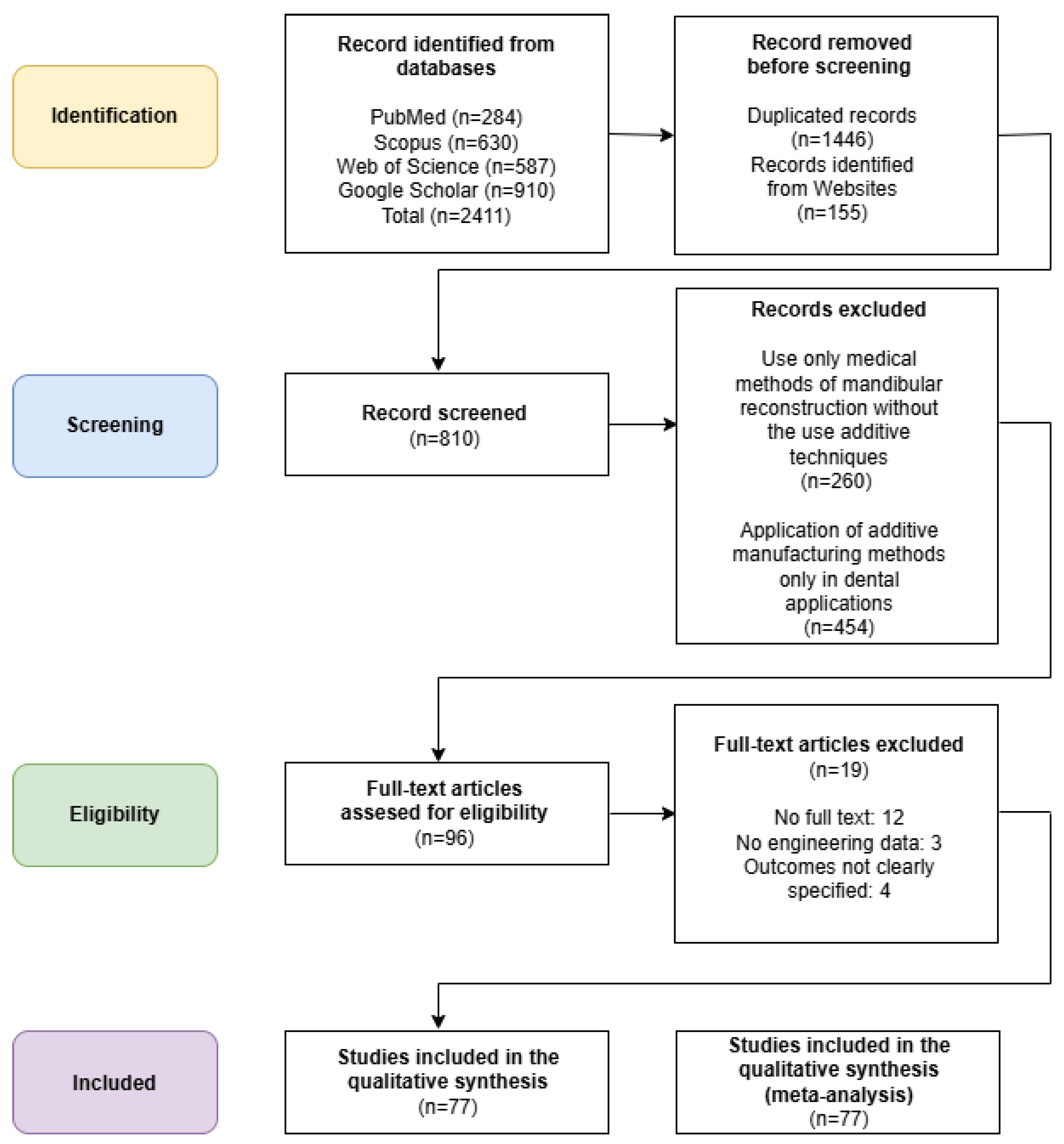
Figure 1. The PRISMA flow diagram for study selection.
Applsci 15 09228 g001
Figure 2. Digital workflow for mandibular reconstruction using additive manufacturing (AM) technologies. The flow chart illustrates the complete sequence from medical imaging acquisition (CT), image segmentation and mirroring, virtual surgical planning (CAD), resection guide design, and fabrication of personalized implants.
Applsci 15 09228 g002
Figure 3. Use of 3D-printed anatomical mandible models in preoperative planning. (a) Example of a patient-specific anatomical model fabricated by FDM technology, used to visualize mandibular geometry and simulate the surgical approach (b) Manual bending of a standard titanium plate onto the printed model prior to surgery, ensuring precise fit and reducing intraoperative adjustment.
Applsci 15 09228 g003
Figure 4. Digital mirroring and segmentation for planning mandibular reconstruction. (a) Three-dimensional model of the affected mandible with pathological bone marked for resection. (b) Result of digital mirroring, where the healthy contralateral side is superimposed to reconstruct the missing anatomical region.
Applsci 15 09228 g004
Figure 5. Patient-specific implant design and manufacturing using powder bed fusion (PBF) technology. (a) Three-dimensional model of a titanium implant designed to restore mandibular continuity after resection. (b) Implant additively manufactured and fabricated from Ti-6Al-4V ELI alloy using PBF, offering high mechanical strength and biocompatibility.
Applsci 15 09228 g005
Figure 6. Application of 3D printing in the reconstruction of the mandibular fibula flap. (a) A 3D-printed anatomical mandible model with a pre-bent titanium reconstruction plate prepared before surgery. (b) A surgical resection guide designed using CAD tools and printed with high precision to assist in the removal of bone from the fibula.
Applsci 15 09228 g006
Figure 7. Additive manufacturing of ceramic scaffolds for mandibular bone tissue engineering. (a) CAD design of a porous scaffold tailored to a mandibular defect, optimized for osteoconduction and vascularization. (b) Example of a 3D-printed bioceramic scaffold intended for in vivo testing in animal models.
Applsci 15 09228 g007
Table 1. Application of additive technology in the mandibular area.
AM TechnologyNumber of Studies Identified (2020–2025)Main Applications
MEX11Anatomical models, surgical guides, temporary implants/scaffolds
MJT5Anatomical models, surgical guides, educational/training models
PBF20Personalized titanium/PEEK implants, surgical guides, anatomical models
VPP11Surgical guides, anatomical models, bioceramic scaffolds
BJT4Anatomical models (in vitro)
DED/SHL0No relevant publications found
Table 2. Application of MEX additive technology in the mandibular area.
YearAM Method and
3D Printer
MaterialApplicationReference
2024FDMABSBiomodel for forming a reconstruction plate[38]
2024FFF/FDM
Prusa i3 MK3S+/MK4
PLAImplantable scaffolds
(rabbit studies)
[39]
2023FFF/FDM
3DGence Industry F340
PCL, PLA
(bioceramics additives)
Biocomposite bone scaffolds[40]
2022FFF/FDM
INTAMSYS FUNMAT HT Enhanced
PEEKImplants[41]
2022FDM
Not specified
PLAResection guides[33]
2021FDM
Not specified
PLAResection guides[35]
2020FDM
Value3D MagiX MF2000
PLACreating osteotomy templates[42]
2020FFF/FDM
HAGE3D
PET-GMaxillofacial implants[43]
2020FFF/FDM
Raise3D® model N2
PLAModels of the mandible and fibula (proximal part)[44]
2020FFF/FDM
Apium M220
PEEKMandibular angle implants of different shapes and sizes[45]
2020FDM
Stratasys FDM machine
ABSSurgical plate[36]
Table 3. Application of MJT additive technology in the mandibular area.
YearAM Method and
3D Printer
MaterialApplicationReferenceStudy Type/Experimental Context
2025PolyJet
J5 MediJet 3D
MED610, MED615Planning and educational models[46]Educational and training models; no clinical application
2023PolyJet
Connex3 Objet500
Vero Pure White RGD835
TangoPlus FLX930
Mandible implants
(studies conducted
on dogs)
[39]Animal study (canine model); not used in human clinical setting
2023 PolyJet
Connex3
Objet500
Vero Clear RGD810, VeroPureWhite RGD837 MED610 Determining the quality and properties of scaffolds[47]In vitro mechanical testing; no clinical use
2021PolyJet
Objet30
VeroGlaze MED620Mandibular osteotomy templates[48]Prototyping of surgical templates; no patient-based outcomes
2020PolyJet
Cubicon Lux
Photoreactive ceramic–resin compositeCreating osteotomy templates[42]Experimental template development; not applied clinically
Table 4. Application of PBF additive technology in the mandibular area.
YearAM Method and
3D Printer
MaterialApplicationReferenceStudy Type/Sample Size/Clinical Outcome
2025SLS
EOS P800
PEEKImplants used in mandibular bone regeneration
(research conducted on a group of sheep)
[49]Animal study (sheep); n = not specified
2024SLS
Fuse 1
PA12Models for analyzing the accuracy of the reconstruction of three-dimensional anatomical models[50]In vitro study; model validation
2023SLM
SLM 280
Commercialized pure titanium (CP-Ti, SLM solution)Implant and surgical guide[17]Animal study (rabbits); n = not specified
2023SLM
Concept Laser
Titanium powder
(not specified)
Personalized grid scaffold used in mandibular bone regeneration[51]Preclinical feasibility study; n = not specified
2023SLM
(not specified)
Pure titaniumPersonalized implants used in mandibular bone regeneration[52]Clinical case series; n = few (exact n not stated)
2022SLM
MetalSys250
Ti-6AL-4V ELI powderPersonalized implants used in mandibular bone regeneration[53]Case series; n = few (not stated); favorable outcome
2022SLM
SLM Solutions Group AG
Ti-6AL-4V ELI powderPersonalized implants used in mandibular bone regeneration[54]Case report/series; n not reported
2022DMLS
TruPrint 1000
Ti-6AL-4V ELI powderPersonalized implants used in mandibular bone regeneration[55]Clinical feasibility; n ≈ small
2022EBM
Electron Beam Melting A1 Arcam
Ti-6AL-4V ELI powderPersonalized implants used in mandibular bone regeneration[56]Clinical case series; n = not stated
2021SLS
Sinterstation HiQ/HS
Nylon powderModel used for creating a surgical guide[57]In vitro prototype only
2021SLM
SLM 280 Twin 2x400W
Ti-6AL-4V ELI powderPersonalized grid scaffold used in mandibular bone regeneration[58]Experimental model (clinical translation suggested)
2021SLM
AM250
Ti-6AL-4V ELI powderCondyle-restricting devices and fixation plates[57]Prototype application; no clinical trial reported
2021SLM
(not specified)
Ti-6AL-4V ELI powderModels for analyzing the topology of the mandibular reconstruction plate[59]Clinical trial; n = 20; excellent fit and surgical outcome; minimal complications
2021EBM
FIT Production GmbH
Ti-6AL-4V ELI powderPersonalized implants used in mandibular bone regeneration[60]Case-based approach; n = not specified
2020SLS
EOSINT P 385
PA 2200
(polyamide powder)
Models for analyzing the accuracy of the reconstruction of three-dimensional anatomical models[61]In vitro model comparison
2020SLM
(not specified)
Titanium alloy
(not specified)
Resection guide and personalized mandibular implant[62]Case report; n = 1
2020DMLS
DMP Dental100
Ti-6AL-4V ELI powderPersonalized implants used in mandibular bone regeneration[63]Clinical feasibility; n not specified
2020DMLS
ProX-DMP100 3D System
Titanium alloy
(not specified)
Surgical guide with subperiosteal implants[64]Case study; outcome not detailed
2020 EBM
Arcam printer
(not specified)
Ti-6AL-4V ELI powderPersonalized grid scaffold used in mandibular bone regeneration[18]Preclinical experiment
2020EBM
Electron Beam Melting A2 Arcam
Ti-6AL-4V ELI powderPersonalized implants with a scaffold used in mandibular bone regeneration[65]Clinical application; n = not stated
Table 5. Application of VPP additive technology in the mandibular area.
YearAM Method and
3D Printer
MaterialApplicationReferenceStudy Type/Sample Size/Clinical Outcome
2024SLA
3D Form 2 Formlabs
Resin
(not specified)
Model used for creating a surgical guide[66]Prototype/preclinical use
2024SLA
Form 3B+
Standard Gray (resin)/BioMed Clear (resin)Models for analyzing the accuracy of the reconstruction of three-dimensional anatomical models[50]In vitro evaluation
2023SLA
HALOT-SKY 3D
Medical grade resinSurgical guide to assist in drilling holes for the implant plate[67]Clinical assistance tool, n not specified
2023DLP
Shaoxing Xunshi Technology
A mix of bioceramic powders and photosensitive acrylicBioceramic scaffolds used in bone tissue regeneration (research conducted on a group of rabbits)[68]Animal study (rabbits), n = not specified
2022 DLP
RapidShape S30
SHERprint-orthoSurgical guide to decompress odontogenic cysts[69]Case-based clinical use, n = 1
2021SLA
(not specified)
Biocompatible resinSurgical guide for implant treatment[70]Clinical case study
2021DLP
Cubicon Lux
Photoreactive ceramic-resin compositeBiocompatible scaffolds to improve mandibular bone regeneration (research conducted on a group of dogs)[71]Animal study (dogs), n not specified
2021DLP
Nextdent 5100
NextDent SG resinSurgical guide to assist in the surgical extraction of a retained tooth[72]Clinical feasibility study
2021DLP
Planmeca Creo
Detax Freeprint model resinModels for analyzing the accuracy of the reconstruction of three-dimensional anatomical models[73]In vitro validation
2020DLP
Cubicon Lux
Photoreactive ceramic-resin compositeBiocompatible scaffolds used in mandibular bone regeneration (studies conducted on a group of dogs)[42]Animal study (dogs),
not specified
2020DLP
ACME DLP 3D
Ceramic suspensionsBioceramic scaffolds used in bone tissue regeneration (research conducted on a group of rabbits)[74]Animal study (rabbits), successful integration
Table 6. Application of BJ additive technology in the mandibular area.
YearAM Method and
3D Printer
MaterialApplicationReferenceStudy Type/Sample Size/Clinical Outcome
2024Polymer Binder Jetting
ZCorp 3D printer
ABS150A manufactured anatomical model of the mandible was used to assess geometric accuracy.[75]In vitro accuracy validation study
2023Binder Jetting
(not specified)
Ti-6Al-4VManufactured scaffold structure.[76]Material study
2022Binder Jetting
(not specified)
Mg-Zn-ZrManufactured scaffold structure.[77]Material study after Hydroxyapatite Coating
2020Binder Jetting
(not specified)
ProJet CJP 660Pro
ZP151A manufactured anatomical model of the mandible was used to assess geometric accuracy.[61]In vitro model-based study; no clinical involvement
Table 7. Matrix of AM technologies and their applicability to specific clinical tasks in mandibular reconstruction.
Application AreaMaterial Extrusion (FDM/FFF)Vat Photopolymerization (SLA/DLP)Powder Bed Fusion (SLM/DMLS)Binder JettingMEX (PEEK)
Anatomical Models✅ Common (PLA/ABS)✅ High-resolution models⚠️ Rare use (costly)✅ Cost-effective⚠️ Rare
Surgical Guides⚠️ Less common (low precision)✅ Preferred (photopolymers)✅ Used (metal guides)⚠️ Less precise⚠️Emerging
Reconstructive Titanium Implants❌ Not applicable❌ Not applicable✅ Standard clinical choice❌ Not applicable❌ Not applicable
Patient-Specific PEEK Implants❌ Not applicable❌ Not applicable⚠️ Experimental via LS❌ Not applicable✅ Preferred
Bioceramic Scaffolds (research)⚠️ Experimental⚠️ Experimental⚠️ Experimental✅ Used in trials❌ Not applicable
✅—Recommended/widely used
⚠️—Limited or experimental use
❌—Not suitable or unsupported
Disclaimer/Publisher’s Note: The statements, opinions and data contained in all publications are solely those of the individual author(s) and contributor(s) and not of MDPI and/or the editor(s). MDPI and/or the editor(s) disclaim responsibility for any injury to people or property resulting from any ideas, methods, instructions or products referred to in the content.