Article

Three-Dimensional-Printed Models: A Novel Approach to Ultrasound Education of the Placental Cord Insertion Site

1
Discipline of Medical Radiation Science, Curtin Medical School, Curtin University, Perth, WA 6845, Australia
2
Vestrum Ultrasound for Women, Bunbury, WA 6230, Australia
3
Curtin Medical Research Institute (Curtin MRI), Curtin University, Perth, WA 6845, Australia
*
Author to whom correspondence should be addressed.
Appl. Sci. 2025, 15(15), 8221; https://doi.org/10.3390/app15158221
Submission received: 21 June 2025 / Revised: 19 July 2025 / Accepted: 23 July 2025 / Published: 24 July 2025

Abstract

Assessment of the placental cord insertion (PCI) is a vital component of antenatal ultrasound examinations. PCI can be complex, particularly in cases of abnormal PCI, and requires proficient sonographer spatial perception. The current literature describes the increasing potential of three-dimensional (3D) modelling to enhance spatial awareness and understanding of complex anatomical structures. This study aimed to evaluate sonographers’ confidence in ultrasound assessment of the PCI and the potential benefit of novel 3D-printed models (3DPMs) of the PCI in ultrasound education. Sonographers employed at a large private medical imaging practice in Western Australia were invited to participate in a face-to-face presentation of two-dimensional (2D) ultrasound images, ultrasound videos, and 3DPMs of normal cord insertion (NCI), marginal cord insertion (MCI), and velamentous cord insertion (VCI). Our objective was to determine the benefit of 3DPMs in improving sonographers’ confidence and ability to spatially visualise the PCI. Thirty-three participants completed questionnaires designed to compare their confidence in assessing the PCI and their ability to spatially visualise the anatomical relationship between the placenta and PCI, before and after the presentation. There was a significant association between a participant’s year of experience and their confidence levels and spatial awareness of the PCI prior to the demonstration. The results showed the 3DPMs increased participant confidence and their spatial awareness of the PCI, with no significant association with years of experience. Additionally, participating sonographers were asked to rate the 3DPMs as an educational device. The 3DPMs were ranked as being a more useful educational tool for spatially visualising the NCI, MCI, and VCI than 2D ultrasound images and videos. Most participants responded favourably when asked whether the 3DPMs would be useful in ultrasound education, with 75.8%, 84.8%, and 97% indicating the models of NCI, MCI, and VCI, respectively, would be extremely useful. Our study has demonstrated a potential role for 3DPMs of the PCI in ultrasound education, supplementing traditional 2D educational resources.

1. Introduction

During pregnancy, the umbilical cord acts as a lifeline for the foetus connecting it to the placenta. The cord can insert into the placenta in a variety of locations, referred to as the placental cord insertion (PCI) site. In most pregnancies, the cord inserts toward the middle of the placenta (NCI); sometimes, it inserts towards the placental edge (MCI) and occasionally into the foetus membranes instead of the placenta (VCI). The latter two types of PCI are associated with potential maternal and foetal complications, and as such, the PCI location should be assessed during routine antenatal ultrasound examinations. In 2023, Ward et al. reviewed the current practice of PCI documentation in Australia, discussing the associated challenges [1]. Whilst the PCI can be efficiently evaluated with two-dimensional and colour Doppler ultrasound imaging [2,3], ultrasound assessment of the PCI site can be complex, particularly when the cord insertion is abnormal, and this requires proficient sonographer spatial perception. Despite the availability of excellent two-dimensional (2D) resources, visualising anatomical spatial relationships remains challenging [4]. The production of three-dimensional printed models (3DPMs) can address this problem by providing clear visualisation of the spatial relationships [5].
Three-dimensional printing technology is showing great potential in medical applications; in particular, it has been shown that 3DPMs offer exceptional dimensional awareness [6]. Three-dimensional anatomical models are now widely used in many aspects of medicine [7,8,9,10,11] including education [12,13,14], pre-surgical planning [15,16,17,18,19], and patient communication [20,21,22], due to the advancements in 3D printing technology, reduction in costs, and ease of access to 3D printing facilities [7,18]. Patient-specific or personalised 3D models can be printed at a 1:1 scale based on imaging datasets, mainly computed tomography (CT), magnetic resonance imaging (MRI), and ultrasound [15]. Further, these models can be printed with different materials, depending on their usefulness for clinical applications [20,23].
The current literature describes the production of a variety of 3DPMs for a diverse array of medical applications with superior advantages reported when compared to the use of standard 2D or 3D image visualisations [7,21,24]. In the field of obstetrics, raw data obtained from 3D ultrasound imaging is being used to create 3DPMs to facilitate antenatal assessment, maternal and paternal foetal attachment, and presurgical planning for foetal malformations [25,26]. To the best of our knowledge, no studies have described the creation of 3DPMs that depict the PCI. This study aimed to address this research gap by assessing the potential value of 3DPMs of the PCI in ultrasound education. Our 3DPMs are patient-specific, based on images of selected patient cases; thus, the models reflect realistic physiological and pathological processes. We hypothesised that our 3DPMs would serve as a useful adjunct to the currently available PCI site educational resources of 2D ultrasound images and videos.

2. Materials and Methods

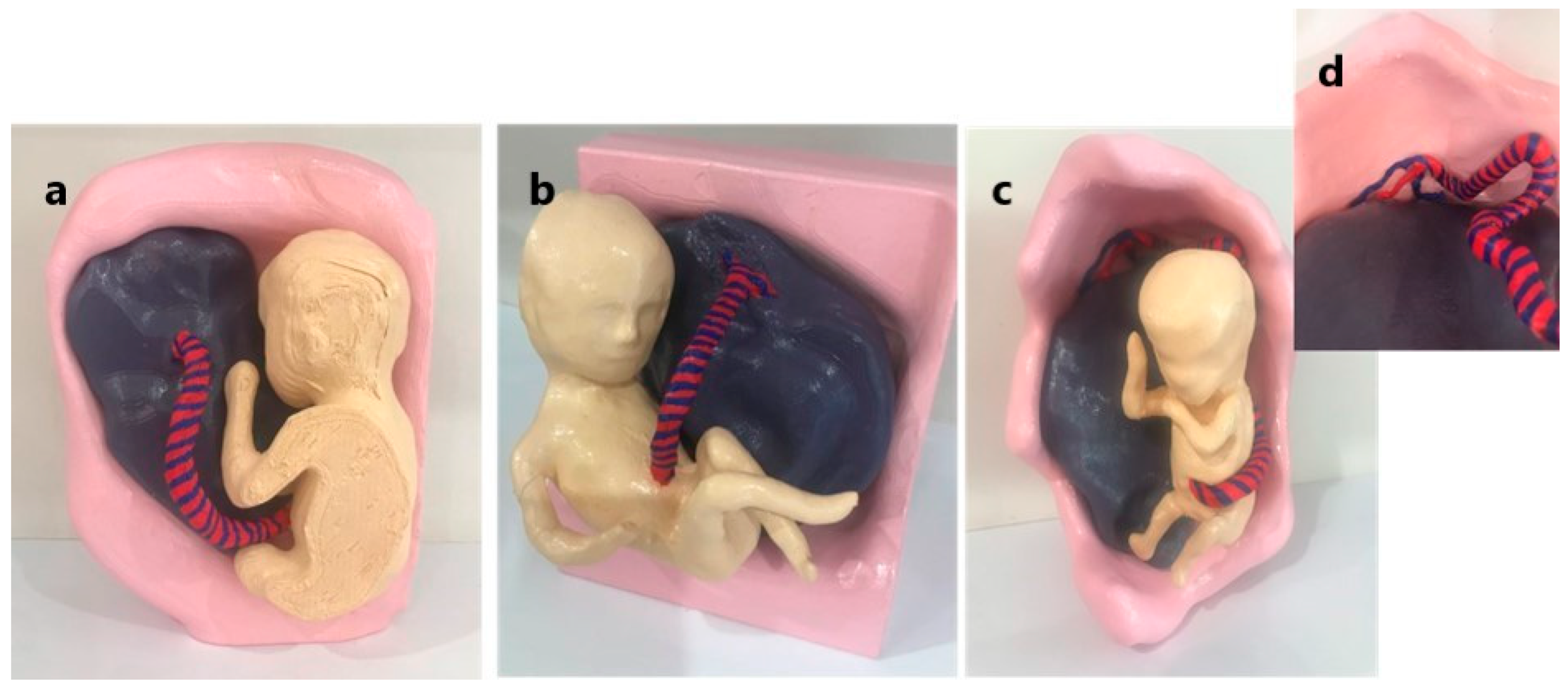
2.1. Three-Dimensional Model Development

Three 3D models, representing normal, marginal, and velamentous placental cord insertions, were developed using the raw ultrasound data of three selected patients at Vestrum Ultrasound for Women (VUW) who provided informed consent. We acquired 3D static images of the NCI, MCI, and VCI (the gestational ages of the foetuses were 20 weeks 3 days, 16 weeks 2 days, and 12 weeks 4 days, respectively) using a GE Voluson E10 ultrasound machine (GE Healthcare, Solingen, Germany) at VUW. These images in Digital Imaging and Communications in Medicine (DICOM) format were imported into an open-source software 3D Slicer (www.slicer.org), version 5.4.0. Segmentation of the images for each PCI was performed manually to accurately delineate the boundaries of the uterus, placenta, foetus, and umbilical cord. The segmented volume data was converted to Standard Tessellation Language (STL) files.
The 3DPMs were printed using a Raised 3D N2 Plus fused deposition modelling printer with a printing speed of 70 mm/s and an xy resolution of 12 microns, utilising polylactic acid material. Each of the three models comprised four STL files (foetus, umbilical cord, placenta, and uterus), which were printed in a ratio of 1:1 for the NCI and MCI models and a ratio of 1:2 for the VCI model due to the earlier gestation of the foetus. The individual components of the models were glued together and painted schematically, resulting in 3DPMs demonstrating the anatomical relationship of the placenta, cord, and foetus. Figure 1 presents the three completed 3DPMs.

2.2. Participant Recruitment and Data Collection

Thirty-three sonographers were recruited from a large private medical imaging practice in Western Australia through convenience sampling. Each participant attended a face-to-face demonstration of normal, marginal, and velamentous cord insertions utilising 2D ultrasound images, ultrasound videos, and our 3DPMs. As far as we are aware, our 3DPMs are the first printed models demonstrating the PCI, and the participants had no prior exposure to our models. As such, we believe there to be no cause for participant bias towards the models. The sonographers were initially asked to complete a questionnaire (Supplementary File S1) establishing the following:
  • Informed consent;
  • Demographics (years of experience as a sonographer);
  • Confidence in assessing the PCI with ultrasound and ability to spatially visualise the placenta and PCI utilising a five-level Likert-scale rating (with 1 indicating extremely poor and 5 indicating excellent).
Ultrasound images and videos of the three classifications of cord insertion were then presented in the form of a Power Point presentation (Supplementary File S2), followed by a hands-on demonstration of the 3DPM. Participants were then asked to complete a subsequent questionnaire (Supplementary File S3). A one-to-three ranking scale was utilised to establish which of the three methods demonstrating the PCI (2D ultrasound images, 2D ultrasound videos, and 3DPM), if any, best enhanced the participants’ ability to spatially visualise the relationship between the placenta and the PCI. Five-level Likert-scale questions (with 1 indicating not at all and 5 indicating very much) were used to determine whether the 3DPMs had the following characteristics:
  • Increased confidence in identifying the NCI, MCI, and VCI with ultrasound;
  • Improved the understanding of the structural relationship between the placenta and the PCI;
  • Enhanced the ability to spatially visualise the placenta and the PCI.
Participants’ opinions on the usefulness of the 3DPMs in ultrasound education and their likelihood of recommending the 3DPMs as an educational device were assessed using five-level Likert-scale questions (with 1 indicating not useful/not likely and 5 indicating extremely useful/likely).
Participants were given the option of providing additional comments at the end of the second questionnaire. The demonstration, inclusive of questionnaire completion, took approximately 15 min for each participant to complete.

2.3. Statistical Analysis

Data analysis was performed using IBM SPSS Statistics v26 (IBM Corporation, Armonk, NY, USA), Microsoft Excel v2209 (Microsoft Corporation, Redmond, WA, USA), and G*Power v3.1.9.7 (Heinrich-Heine-Universität, Düsseldorf, Germany) [27]. Post hoc analysis using one-way ANOVA in G*Power with an effect size of 0.61, level of significance 5%, and sample size of 33, calculated the power of our study to be 86%. The Shapiro–Wilk test demonstrated the ordinal data for the Likert-scale questions and the ranked order for the rating scale questions were not normally distributed (p < 0.05, p < 0.001, respectively). Cronbach’s alpha was used to test for internal consistency of the Likert-scale questions with α = 0.977 for the pre-demonstration survey and α = 0.938 for the post-demonstration survey, indicating high internal consistency and thus reliability of the survey questions. Additionally, individual questions were deemed reliable as removal of the questions independently from the analysis resulted in a Cronbach’s alpha ranging from 0.97 to 0.978 (pre-demonstration) and 0.928 to 0.941 (post-demonstration). Descriptive statistics were used to determine the mean and standard deviation of the Likert- and rating- scale questions. The chi-square test with degrees of freedom (df) was used to assess association between rankings of the 2D ultrasound images, videos, and the 3DPMs across the participants. Kruskal–Wallis one-way ANOVA was conducted to assess the association between participants’ years of experience and their questionnaire responses, with Dunn’s post hoc tests used to assess the association between the three groups of sonographer experience and responses. A p-value < 0.05 was considered statistically significant. Eta squared (η2) was used to measure the effect size of significant Kruskal–Wallis tests. Qualitative data from the free-text section of the questionnaire was analysed thematically, with data categorised into five themes.

2.4. Ethics Approval

This study was approved by the Curtin University Human Research Ethics Committee (HRE2021-0629), SKG Radiology clinical standards committee, and VUW.

3. Results

A total of 33 sonographers were recruited in this study. The participants’ years of experience were categorised into three groups, with Group 1 having ≤3 years of experience (n = 12), Group 2 having 4 to 10 years of experience (n = 9), and Group 3 having more than 10 years of experience (n = 12).
The results indicated that the participants’ confidence in assessing the PCI and their ability to spatially visualise the placenta and PCI for all PCI classifications, pre-demonstration, was higher than average (mean = 2.5). The association between years of experience and all responses, except for “ability to spatially visualise placenta and PCI while performing ultrasound” and “ability to spatially visualise placenta and MCI while performing ultrasound”, was statistically significant (Table 1). It is noted that the mean rating for both the confidence and spatial awareness decreased with increasing PCI complexity.
We further analysed the data to establish relationships between the grouped years of experience and participants’ responses. There was a significant association between a sonographer’s experience level and (i) their confidence in assessing the PCI and (ii) their ability to spatially visualise the placenta and PCI, with those sonographers in group 1 (least experienced participants) being less confident and less able to spatially visualise the placenta and PCI than those in group 2 (p-values < 0.05 for all PCI types). There was no significant association when comparing groups 2 and 3 (Table 2).
Following the demonstration, participants were asked to rank the 2D ultrasound images, 2D ultrasound videos, and 3DPMs from 1 to 3 (with 1 being least helpful and 3 being most helpful) to determine which method of PCI demonstration was the most effective in terms of spatially visualising the PCI. The results showed most participants found the ultrasound images to be the least useful method for spatially visualising the NCI, MCI, and VCI (ranked 1 in 81.8%, 78.8%, and 84.8%, respectively) and the 3DPMs the most beneficial (ranked 3 in 69.7%, 75.8%, and 78.8%, respectively). Table 3 documents the ranking of the images, videos, and 3DPMs for each PCI category. There was no significant association between the ranking and the participant’s years of experience, df = 4.
On a scale of 1–5 (with 1 being “not at all” and 5 being “very much”), the mean values were above average for each question listed in Table 4. The SD values indicate a high variability in all responses, which may be due to the low sample size. Following the demonstration of the 3DPMs, the sonographers indicated that their confidence, understanding, and ability to spatially visualise the PCI increased in proportion to the PCI complexity, as illustrated in Table 4 and Figure 2, Figure 3 and Figure 4.
There was no significant association between a sonographer’s years of experience and the 3DPMs in terms of improving their confidence, understanding, or ability to spatially visualise the placenta and PCI (Table 5).
Most sonographers indicated they felt the models of each PCI type would be useful in ultrasound education (Figure 5) with 75.8%, 84.8%, and 97% indicating the models of normal, marginal, and velamentous cord insertions, respectively, would be extremely useful. Participants were also asked how likely they would be to recommend the models as an educational device with 69.7%, 72.7%, and 81.8% indicating they would recommend normal, marginal, and velamentous cord insertion models, respectively (Figure 6).
Table 6 presents the participants’ mean ratings for the usefulness of the 3DPMs in ultrasound education. There was no significant association between these opinions and the participants’ years of experience (Table 6), and statistical significance was not reached when comparing the grouped years of experience (Supplementary Table S4).
Eleven participants provided comments in the free-text section of the questionnaire (Table 7). The feedback was categorised into five themes: (i) use of 3DPMs of the PCI as educational tools; (ii) improved visualisation of the PCI; (iii) extended use of the 3D models; (iv) general comments; and (v) limitations, with several sonographers providing comments applicable to multiple themes.

4. Discussion

Comprehensive understanding of complex anatomy can be challenging in all aspects of medical imaging. Imaging modalities primarily produce 2D images, which limits the spatial perception of anatomical relationships [28]. Studies have demonstrated medical students find visualising anatomy in three dimensions particularly challenging [29]. Even when 3D imaging is undertaken, interpreting 3D images on flat 2D screens can impede understanding of complicated anatomy [28,30,31]. The production of 3DPMs can bypass this problem by providing clear visualisation of spatial relationships [5].
Backhouse et al. stated that despite the availability of excellent 2D resources, visualising anatomical spatial relationships remains challenging [4]. This suggests 2D resources on their own may not be adequate for teaching anatomy, one of the most onerous subjects for students due to the need for spatial visualisation [32]. Halpern et al. suggested the use of 3D models would improve a student’s understanding of a 2D ultrasound image resulting in increased confidence in recognising anatomy [33]. In our study, participants indicated the 3DPMs were more beneficial in terms of visualising the NCI, MCI, and VCI than the more traditional methods of 2D ultrasound images and videos. Our findings concur with the literature that 3DPMs increase spatial visual orientation [34] and assist with understanding anatomical relationships [35].
3DPMs are being progressively valued in ultrasound education with their ability to accurately depict anatomical structures [17,36] and provide both a visual and tactile representation which improves understanding, particularly of complex anatomy [7,19]. As supported by our participants’ responses, assessment of the PCI with ultrasound becomes more difficult with increasing complexity of the PCI, with an NCI being the most straightforward to visualise, and a VCI, due to its intricate anatomy, the most problematic to assess. Backhouse et al. investigated the effect a 3DPM of an orbit had on optometry students’ education. The response of their participants was extremely positive, with the students indicating the model enhanced their ability to spatially visualise anatomical relationships, preferring the 3D models over traditional resources [4]. In the present study, most participants indicated the 3DPMs would be beneficial in ultrasound education, increasing their confidence in assessment and spatial awareness of the PCI. Interestingly, statistical significance was not achieved when comparing these responses and years of experience, suggesting the 3DPMs may be beneficial for sonographers with varying levels of practice.
In their literature review, Halpern et al. found that 14 of 16 studies included in their review demonstrated that 3D modelling applications enhanced student education in ultrasound [33]. Tan et al. showed that 3D models enhanced the understanding of and contributed significantly to anatomical education [34]. Interestingly, a study by Yi et al. that evaluated the educational value of 3DPMs, 3D images, and 2D images in assessing the ventricular system of the brain showed that not only did their participants rate their 3DPMs higher than 3D images, but the models distinctly increased interest and enthusiasm among the students in their research [37]. Our findings, in conjunction with the current literature, support a positive role for 3DPMs in ultrasound education.
The free-text section of our questionnaire introduced new concepts for the use of 3D models in obstetric ultrasound education with two participants suggesting 3DPMs of placental location and placental developmental variation would be beneficial. Our 3DPMs could also provide enhanced patient understanding of placental and PCI anatomy and pathology.
In 2022, Lau et al. explored the concept of virtual reality (VR) in education of congenital heart disease with reported comparable benefits of 3DPMs and VR models [24]. VR was preferred over 3DPMs in a 2023 study by Awari et al. [38]. As a future direction, it would be interesting to investigate the role of VR models in ultrasound education of the PCI. Long-term retention studies would also be prudent.
As with all advancements in medical education, cost and feasibility must be considered when assessing the practicality of introducing 3DPMs into ultrasound education. Three-dimensional printing technology is being used increasingly in medical and educational applications, with manufacturing costs varying greatly. The current literature describes decreasing costs [4,7,18]; however, the expenditure is relative to the model being created and the materials used. Perhaps the most significant cost is time, with the printing process sometimes taking days to complete. In our study, the print time for the individual components of the models ranged from 1.0 to 28.5 h, according to the size of the part (Supplementary Table S5), with a total cost of approximately AUD 50 per model, inclusive of materials and printing, making our models an affordable and readily accessible adjunct to ultrasound education. A further consideration is receipt of approval of the 3DPMs from educational facilities.
We would like to acknowledge some limitations of our study. As a small cohort from a single private radiology practice in Western Australia was recruited through convenience sampling, our results may not be representative of the entire population of sonographers employed at this practice or indeed sonographers employed by other ultrasound facilities. The validity of our results would benefit from a larger group of participants from a diverse range of ultrasound settings including both private and public departments offering general ultrasound examinations and/or specialised obstetric ultrasound. There are innumerable anatomical variations of a VCI with the aberrant vascular anatomy being unique to each patient. Our study demonstrated a single example of a VCI. Production of multiple 3DPMs demonstrating a variety of VCIs would be valuable. Our demonstration was not pilot tested; thus, although Cronbach’s alpha indicated reliability of the survey questions, we cannot be sure of the comprehensibility of the PowerPoint presentation and the 3D model demonstration. Finally, while participants of this study have responded positively to the value of the 3DPMs in education, feedback from ultrasound educators is required to support the potential integration of 3DPMs into ultrasound training programs.

5. Conclusions

Three-dimensional printing technology is rapidly evolving, and the benefits of printed models are being increasingly appreciated. This study has shown that 3DPMs of the PCI could be beneficial in ultrasound education. Our participants indicated improved confidence in assessing the PCI and enhanced spatial orientation of the PCI, particularly when it is complex, following the demonstration of the models. The 3DPMs were the preferred method of demonstrating the PCI over more traditional resources of 2D ultrasound images and ultrasound videos. We believe our 3DPMs could be a valuable and low-cost addition to ultrasound education, pending training program approval. Future studies should assess whether 3DPMs of the PCI improve diagnostic accuracy in clinical practice.

Supplementary Materials

The following supporting information can be downloaded at: https://www.mdpi.com/article/10.3390/app15158221/s1, File S1: pre-demonstration questionnaire; File S2: Powerpoint presentation; File S3: post-demonstration questionnaire; Table S4: Participant opinion of 3DPMs comparing grouped years of experience, p-values; Table S5: Specifications of the individual components of the 3D printed models.

Author Contributions

Conceptualisation: S.W., Z.S. and S.M.; writing—original and draft preparation, S.W.; project administration, Z.S. and S.M.; project supervision, Z.S. and S.M.; writing—review and editing, S.W. and Z.S. All authors have read and agreed to the published version of the manuscript.

Funding

This research received no external funding.

Institutional Review Board Statement

The study was conducted in accordance with the Declaration of Helsinki, and approved by Curtin University Human Research Ethics Committee (approval number: HRE2021-0629, and date of approval: 8 October 2021).

Informed Consent Statement

Informed consent was obtained from all participants involved in this study.

Data Availability Statement

The original contributions presented in this study are included in the article. Further inquiries can be directed to the corresponding author.

Acknowledgments

The authors thank Yin How Wong, Taylor’s University, Malaysia for facilitating the printing of the 3D PCI models. We also thank SKG Radiology for hosting the demonstrations, the sonographers who participated in this survey, and Andrea Liddiard, Vestrum Ultrasound for Women, for her clinical assistance with image data collection.

Conflicts of Interest

The authors declare no conflicts of interest.

References

  1. Ward, S.; Sun, Z.; Maresse, S. Current practice of placental cord insertion documentation in Australia—A sonographer survey. Australas J. Ultrasound Med. 2023, 26, 157–168. [Google Scholar] [CrossRef] [PubMed]
  2. Allaf, M.B.; Andrikopoulou, M.; Crnosija, N.; Muscat, J.; Chavez, M.R.; Vintzileos, A.M. Second trimester marginal cord insertion is associated with adverse perinatal outcomes. J. Matern Fetal Neonatal Med. 2019, 32, 2979–2984. [Google Scholar] [CrossRef] [PubMed]
  3. Wax, I.R.; Craig, W.Y.; Pinette, M.G.; Wax, J.R. Second-Trimester Ultrasound-Measured Umbilical Cord Insertion-to-Placental Edge Distacne: Determining an Outcome-Based Threshold for Identifying Marginal Cord Insertion. J. Ultrasound Med. 2020, 39, 351–358. [Google Scholar] [CrossRef] [PubMed]
  4. Backhouse, S.; Taylor, D.; Armitage, J.A. Is This Mine to Keep? Three-dimensional Printing Enables Active, Personalized Learning in Anatomy. Anat. Sci. Educ. 2019, 12, 518–528. [Google Scholar] [CrossRef] [PubMed]
  5. Loke, Y.H.; Harahsheh, A.S.; Krieger, A.; Olivieri, L.J. Usage of 3D models of tetralogy of Fallot for medical education: Impact on learning congenital heart disease. BMC Med. Educ. 2017, 17, 54. [Google Scholar] [CrossRef] [PubMed]
  6. Liang, J.; Ma, Q.; Zhao, X.; Pan, G.; Zhang, G.; Zhu, B.; Xue, Y.; Li, D.; Lu, B. Feasibility Analysis of 3D Printing With Prenatal Ultrasound for the Diagnosis of Fetal Abnormalities. J. Ultrasound Med. 2022, 41, 1385–1396. [Google Scholar] [CrossRef] [PubMed]
  7. Bastawrous, S.; Wake, N.; Levin, D.; Ripley, B. Principles of three-dimensional printing and clinical applications within the abdomen and pelvis. Abdom. Radiol. 2018, 43, 2809–2822. [Google Scholar] [CrossRef] [PubMed]
  8. Cui, H.; Yu, L.; Li, H.; Wang, H.; Liang, W.; Wang, H.; Wang, H. Evaluation of placental growth potential and placental bed perfusion by 3D ultrasound for early second-trimester prediction of preeclampsia. J. Assist. Reprod. Genet. 2022, 39, 1545–1554. [Google Scholar] [CrossRef] [PubMed]
  9. Chen, J.; He, H.; Song, T.; Su, C.; Chen, D. Utilizing 3D Printing Model of Placenta Percreta to Guide Obstetric Operation [3D]. Obstet. Gynecol. 2017, 129, 42S. [Google Scholar] [CrossRef]
  10. Hu, C.; Zhang, W.; Li, P. 3D Printing and Its Current Status of Application in Obstetrics and Gynecological Diseases. Bioengineering 2023, 10, 299. [Google Scholar] [CrossRef] [PubMed]
  11. You, Y.; Niu, Y.; Sun, F.; Huang, S.; Ding, P.; Wang, X.; Zhang, X.; Zhang, J. Three-dimensional printing and 3D slicer powerful tools in understanding and treating neurosurgical diseases. Front. Surg. 2022, 9, 1030081. [Google Scholar] [CrossRef] [PubMed]
  12. Kiesel, M.; Beyers, I.; Kalisz, A.; Joukhadar, R.; Wöckel1, A.; Herbert, S.-L.; Curtaz, C.; Wulff, C. A 3D printed model of the female pelvis for practical education of gynecological pelvic examination. 3D Print. Med. 2022, 8, 13. [Google Scholar] [CrossRef] [PubMed]
  13. Recker, F.; Jin, L.; Veith, P.; Lauterbach, M.; Karakostas, P.; Schäfer, V.S. Development and Proof of Concept of a Low-Cost Ultrasound Training Model for Diagnosis of Giant Cell Arteritis Using 3D Printing. Diagnostics 2021, 11, 1106. [Google Scholar] [CrossRef] [PubMed]
  14. Recker, F.; Remmersmann, L.; Jost, E.; Jimenez-Cruz, J.; Haverkamp, N.; Gembruch, U.; Strizek, B.; Schäfer, V.S. Development of a 3D-printed nuchal translucency model: A pilot study for prenatal ultrasound training. Arch. Gynecol. Obstet. 2024, 310, 2055–2064. [Google Scholar] [CrossRef] [PubMed]
  15. Sun, Z.; Wong, Y.H.; Yeong, C.H. Patient-Specific 3D-Printed Low-Cost Models in Medical Education and Clinical Practice. Micromachines 2023, 14, 464. [Google Scholar] [CrossRef] [PubMed]
  16. Sun, Z.; Wee, C. 3D Printed Models in Cardiovascular Disease: An Exciting Future to Deliver Personalized Medicine. Micromachines 2022, 13, 1575. [Google Scholar] [CrossRef] [PubMed]
  17. Wittek, A.; Strizek, B.; Recker, F. Innovations in ultrasound training in obstetrics. Arch. Gynecol. Obstet. 2024, 311, 871–880. [Google Scholar] [CrossRef] [PubMed]
  18. Yasli, M.; Dabbagh, S.R.; Tasoglu, S.; Aydin, S. Additive manufacturing and three-dimensional printing in obstetrics and gynecology: A comprehensive review. Arch. Gynecol. Obstet. 2023, 308, 1679–1690. [Google Scholar] [CrossRef] [PubMed]
  19. Meyer-Szary, J.; Luis, M.S.; Mikulski, S.; Patel, A.; Schulz, F.; Tretiakow, D.; Fercho, J.; Jaguszewska, K.; Frankiewicz, M.; Pawlowska, E.; et al. The Role of 3D Printing in Planning Complex Medical Procedures and Training of Medical Professionals-Cross-Sectional Multispecialty Review. Int. J. Environ. Res. Public Health 2022, 19, 3331. [Google Scholar] [CrossRef] [PubMed]
  20. Traynor, G.; Shearn, A.I.; Milano, E.G.; Ordonez, M.V.; Nieves, M.; Forte, V.; Caputo, M.; Schievano, S.; Mustard, H.; Wray, J.; et al. The use of 3D-printed models in patient communication: A scoping review. J. 3D Print. Med. 2022, 6, 3–23. [Google Scholar] [CrossRef] [PubMed]
  21. Lee, S.Y.; Squelch, A.; Sun, Z. Investigation of the Clinical Value of Four Visualization Modalities for Congenital Heart Disease. J. Cardiovasc. Dev. Dis. 2024, 11, 278. [Google Scholar] [CrossRef] [PubMed]
  22. Biglino, G.; Koniordou, D.; Gasparini, M.; Capelli, C.; Leaver, L.-K.; Khambadkone, S.; Schievano, S.; Taylor, A.M.; Wray, J. Piloting the Use of Patient-Specific Cardiac Models as a Novel Tool to Facilitate Communication During Cinical Consultations. Pediatr. Cardiol. 2017, 38, 813–818. [Google Scholar] [CrossRef] [PubMed]
  23. Jin, Z.; Li, Y.; Yu, K.; Liu, L.; Fu, J.; Yao, X.; Zhang, A.; He, Y. 3D Printing of Physical Organ Models: Recent Developments and Challenges. Adv. Sci. 2021, 8, 2101394. [Google Scholar] [CrossRef] [PubMed]
  24. Lau, I.; Gupta, A.; Ihdayhid, A.; Sun, Z. Clinical Applications of Mixed Reality and 3D Printing in Congenital Heart Disease. Biomolecules 2022, 12, 1548. [Google Scholar] [CrossRef] [PubMed]
  25. Coté, J.J.; Coté, B.P.; Badura-Brack, A.S. 3D printed models in pregnancy and its utility in improving psychological constructs: A case series. 3D Print. Med. 2022, 8, 16. [Google Scholar] [CrossRef] [PubMed]
  26. Coté, J.J.; Thomas, B.; Marvin, J. Improved maternal bonding with the use of 3D-printed models in the setting of a facial cleft. J. 3D Print. Med. 2018, 2, 97–102. [Google Scholar] [CrossRef]
  27. Faul, F.; Erdfelder, E.; Buchner, A.; Lang, A.-G. Statistical power analyses using GPower 3.1: Tests for correlation and regression analyses. Behav. Res. Methods. 2009, 41, 1149–1160. [Google Scholar] [CrossRef] [PubMed]
  28. Lau, I.; Sun, Z. Dimensional accuracy and clinical value of 3d printed models in congenital heart disease: A systematic review and meta-analysis. J. Clin. Med. 2019, 8, 1483. [Google Scholar] [CrossRef] [PubMed]
  29. Young, J.C.; Quayle, M.R.; Adams, J.W.; Bertram, J.F.; McMenamin, P.G. Three-Dimensional Printing of Archived Human Fetal Material for Teaching Purposes. Anat. Sci. Edu. 2019, 12, 90–96. [Google Scholar] [CrossRef] [PubMed]
  30. Lauridsen, H.; Hansen, K.; Nørgård, M.Ø.; Wang, T.; Pedersen, M. From tissue to silicon to plastic: Three-dimensional printing in comparative anatomy and physiology. R. Soc. Open Sci. 2016, 3, 150643. [Google Scholar] [CrossRef] [PubMed]
  31. Hadeed, K.; Acar, P.; Dulac, Y.; Cuttone, F.; Alacoque, X.; Karsenty, C. Cardiac 3D printing for better understanding of congenital heart disease. Arch. Cardiovasc. Dis. 2018, 111, 1–4. [Google Scholar] [CrossRef] [PubMed]
  32. Chen, S.; Pan, Z.; Wu, Y.; Gu, Z.; Li, M.; Liang, Z.; Zhu, H.; Yao, Y.; Shui, W.; Shen, Z.; et al. The role of three-dimensional printed models of skull in anatomy education: A randomized controlled trail. Sci. Rep. 2017, 7, 575. [Google Scholar] [CrossRef] [PubMed]
  33. Halpern, S.A.; Brace, E.J.; Hall, A.J.; Morrison, R.G.; Patel, D.V.; Yuh, J.Y.; Brolis, N.V. 3-D modeling applications in ultrasound education: A systematic review. Ultrasound Med. Biol. 2022, 48, 188–197. [Google Scholar] [CrossRef] [PubMed]
  34. Tan, L.; Wang, Z.; Jiang, H.; Han, B.; Tang, J.; Kang, C.; Zhang, N.; Xu, Y. Full color 3D printing of anatomical models. Clin. Anat. 2022, 35, 598–608. [Google Scholar] [CrossRef] [PubMed]
  35. WFUMB/ISUOG. WFUMB/ISUOG Statement on the Safe Use of Doppler Ultrasound During 11–14 Week Scans (or Earlier in Pregnancy). Ultrasound Med. Biol. 2013, 39, 373. [Google Scholar] [CrossRef] [PubMed]
  36. Clark, A.E.; Biffi, B.; Sivera, R.; Dall’Asta, A.; Fessey, L.; Wong, T.-L.; Paramasivam, G.; Dunaway, D.; Schievano, S.; Lees, C.C. Developing and testing an algorithm for automatic segmentation of the fetal face from three-dimensional ultrasound images. R. Soc. Open Sci. 2020, 7, 201342. [Google Scholar] [CrossRef] [PubMed]
  37. Yi, X.; Ding, C.; Xu, H.; Huang, T.; Kang, D.; Wang, D. Three-Dimensional Printed Models in Anatomy Education of the Ventricular System: A Randomized Controlled Study. World Neurosurg. 2019, 125, 891–901. [Google Scholar] [CrossRef] [PubMed]
  38. Awori, J.; Friedman, S.D.; Howard, C.; Kronmal, R.; Buddhe, S. Comparative effectiveness of virtual reality (VR) vs. 3D printed models of congenital heart disease in resident and nurse practitioner educational experience. 3D Print Med. 2023, 9, 2. [Google Scholar] [CrossRef] [PubMed]
Figure 1. The completed 3D-printed models representing the (a) NCI, (b) MCI, (c), VCI and a (d) close-up of the VCI inserting into the foetal membranes. 3D: three-dimensional, NCI: normal cord insertion, MCI: marginal cord insertion, VCI: velamentous cord insertion.
Applsci 15 08221 g001
Figure 2. Rating of the 3DPMs in improving confidence in assessing the PCI (%). The values 1–5 represent the Likert scale, while the numbers in the y-axis indicate the percentage of participants providing these scores. NCI: normal cord insertion, MCI: marginal cord insertion, VCI: velamentous cord insertion.
Applsci 15 08221 g002
Figure 3. Rating of the 3DPMs in improving the understanding of the structural relationships between the PCI and placenta (%). The values 1–5 represent the Likert scale, while the numbers in the y-axis indicate the percentage of participants providing these scores. NCI: normal cord insertion, MCI: marginal cord insertion, VCI: velamentous cord insertion.
Applsci 15 08221 g003
Figure 4. Rating of the 3DPMs in improving ability to spatially visualise the placenta and PCI (%). 1–5 indicates Likert scale. NCI: normal cord insertion, MCI: marginal cord insertion, VCI: velamentous cord insertion.
Applsci 15 08221 g004
Figure 5. Rating of the usefulness of 3DPMs of the PCI in ultrasound education (%). The values 1–5 represent the Likert scale, while the numbers in the y-axis indicate the percentage of participants providing these scores. NCI: normal cord insertion, MCI: marginal cord insertion, VCI: velamentous cord insertion.
Applsci 15 08221 g005
Figure 6. Ratings of the participants’ recommendation of 3DPMs of the PCI (%). The values 1–5 represent the Likert scale, while the numbers in the y-axis indicate the percentage of participants providing these scores. NCI: normal cord insertion, MCI: marginal cord insertion, VCI: velamentous cord insertion.
Applsci 15 08221 g006
Table 1. The mean ratings for participant confidence and ability to spatially visualise the PCI, pre-demonstration.
Question
Please Rate Your…
Mean (SD)p-ValueEffect Size η2
Confidence in assessing PCI with ultrasound3.87 (1.01)0.0050.28
Confidence in identifying NCI with ultrasound4.03 (0.98)<0.0010.40
Confidence in identifying MCI with ultrasound3.79 (1.02)0.0020.34
Confidence in identifying VCI with ultrasound3.58 (1.03)0.0020.35
Ability to spatially visualise placenta and PCI while performing ultrasound4.00 (1.00)0.17
Ability to spatially visualise placenta and NCI while performing ultrasound4.09 (1.10)0.0020.35
Ability to spatially visualise placenta and MCI while performing ultrasound3.84 (1.08)0.22
Ability to spatially visualise placenta and VCI while performing ultrasound3.55 (1.15)0.0070.26
PCI: placental cord insertion. NCI: normal cord insertion, MCI: marginal cord insertion, VCI: velamentous cord insertion.
Table 2. Participant confidence in assessing and spatially visualising the PCI, pre-demonstration, according to years of experience, with p-values shown.
Group 1 vs. Group 2
p-Value (Effect Size η2)
Group 1 vs. Group 3
p-Value
Group 2 vs. Group 3
p-Value
Confidence in assessing the PCI with ultrasound0.002 (0.32)0.021 0.337
Confidence in identifying NCI with ultrasound<0.001 (0.39)0.0260.104
Confidence in identifying MCI with ultrasound<0.001 (0.36)0.019 0.222
Confidence in identifying VCI with ultrasound<0.001 (0.40)0.033 0.126
Ability to spatially visualise the placenta and PCI0.005 (0.30)0.077 0.236
Ability to spatially visualise the placenta and NCI<0.001(0.38)0.14 0.265
Ability to spatially visualise the placenta and MCI0.009 (0.28)0.41 0.481
Ability to spatially visualise the placenta and VCI0.003 (0.34)0.02 0.433
PCI: placental cord insertion. NCI: normal cord insertion, MCI: marginal cord insertion, VCI: velamentous cord insertion.
Table 3. Ranking for method of demonstration for each PCI type with p-values shown, (n = 33).
PCI Type Ranking 1 n (%)Ranking 2 n (%)Ranking 3 n (%)p-Value
NCIImage27 (81.8)3 (9.1)3 (9.1)0.348
Video3 (9.1)23 (69.7)7 (21.2)0.785
Model3 (9.1)7 (21.2)23 (69.7)0.862
MCIImage26 (78.8)5 (15.2)2 (6.1)0.341
Video3 (9.1)24 (72.7)6 (18.2)0.824
Model4 (12.1)4 (12.1)25 (75.8)0.452
VCIImage28 (84.8)2 (6.1)3 (9.1)0.483
Video029 (87.9)4 (12.1)0.492
Model5 (15.2)2 (6.1)26 (78.8)0.586
PCI: placental cord insertion. NCI: normal cord insertion, MCI: marginal cord insertion, VCI: velamentous cord insertion.
Table 4. Mean ratings of the 3DPMs based on participants’ responses (n = 33).
Question:
Have the 3DPMs Improved Your…
MeanSD
Confidence in assessing the PCI with ultrasound3.841.14
Confidence in assessing NCI with ultrasound3.731.28
Confidence in assessing MCI with ultrasound3.841.23
Confidence in assessing a VCI with ultrasound4.360.89
Understanding of the structural relationship between the placenta and NCI3.331.34
Understanding of the structural relationship between the placenta and MCI3.821.26
Understanding of the structural relationship between the placenta and VCI4.450.90
Ability to spatially visualise the placenta and NCI3.641.41
Ability to spatially visualise the placenta and MCI3.941.25
Ability to spatially visualise the placenta and VCI4.391.00
3DPMs: three-dimensional-printed models, PCI: placental cord insertion, NCI: normal cord insertion, MCI: marginal cord insertion, VCI: velamentous cord insertion.
Table 5. 3DPMs’ role in improving participant confidence, understanding, and ability to spatially visualise the placenta and cord insertion types according to years of experience, p-values.
Question:
Have the 3D Models Improved Your …
p-Value
Confidence in assessing the PCI with ultrasound?0.223
Confidence in identifying an NCI with ultrasound0.092
Confidence in identifying an MCI with ultrasound0.157
Confidence in identifying a VCI with ultrasound0.099
Understanding of the structural relationship between an NCI and the placenta0.906
Understanding of the structural relationship between an MCI and the placenta0.272
Understanding of the structural relationship between a VCI and the placenta1.000
Ability to spatially visualise the placenta and an NCI0.192
Ability to spatially visualise the placenta and an MCI0.714
Ability to spatially visualise the placenta and a VCI0.960
3D: three-dimensional, PCI: placental cord insertion, NCI: normal cord insertion, MCI: marginal cord insertion, VCI: velamentous cord insertion.
Table 6. Mean ratings of the usefulness of 3DPMs in ultrasound education based on participants’ opinions, n = 33.
OpinionsMeanSDp-Value
Do you think the 3D model of the NCI would be useful in ultrasound education?4.550.970.605
Do you think the 3D model of the MCI would be useful in ultrasound education?4.730.720.601
Do you think the 3D model of the VCI would be useful in ultrasound education?4.970.171.000
Would you recommend the 3D model of the NCI as an educational device?4.421.090.582
Would you recommend the 3D model of the MCI as an educational device?4.540.830.930
Would you recommend the 3D model of the VCI as an educational device?4.810.390.622
3D: three-dimensional, NCI: normal cord insertion, MCI: marginal cord insertion, VCI: velamentous cord insertion.
Table 7. Thematic analysis of qualitative data.
ThemeFeedbackTotal
Use of 3DPMs of the PCI as educational toolsUseful teaching resources for trainees and junior staff (n = 4)
Useful educational tool to explain different types of PCI (n = 2)
n = 6
Improved visualisation of the PCIBetter visualisation and understanding of PCI (n = 6)
The VCI model was particularly helpful (n = 2)
n = 8
Extended use of the 3D modelsThe 3D models could help spatially visualise placental location (n = 1) and placental developmental variations (n = 1)n = 2
General commentsCould be helpful in explaining our images/videos of the PCI to radiologist (n = 1)
Excellent for dyslexic students who require a more visual approach to study (n = 1)
The videos and 3D models pair nicely together (n = 1)
n = 3
LimitationsCost and availability (n = 1)n = 1
3DPMs: three-dimensional-printed models, PCI: placental cord insertion, VCI: velamentous cord insertion, 3D: three-dimensional.
Disclaimer/Publisher’s Note: The statements, opinions and data contained in all publications are solely those of the individual author(s) and contributor(s) and not of MDPI and/or the editor(s). MDPI and/or the editor(s) disclaim responsibility for any injury to people or property resulting from any ideas, methods, instructions or products referred to in the content.

