Article

Novel Probiotic Strain Lactiplantibacillus plantarum CNTA 628 Modulates Lipid Metabolism and Improves Healthspan in C. elegans

by
Ignacio Goyache
1,2,†,
Lorena Valdés-Varela
3,†,
Raquel Virto
3,
Miguel López-Yoldi
2,
Noelia López-Giral
3,
Ana Sánchez-Vicente
3,
Fermín I. Milagro
1,2,4,5,* and
Paula Aranaz
2,4
1
Department of Nutrition, Food Science and Physiology, Faculty of Pharmacy and Nutrition, University of Navarra, 31008 Pamplona, Spain
2
Center for Nutrition Research, University of Navarra, c/Irunlarrea 1, 31008 Pamplona, Spain
3
CNTA, Ctra. NA-134 Km. 53, 31570 San Adrián, Spain
4
Navarra Institute for Health Research (IdiSNA), 31008 Pamplona, Spain
5
Centro de Investigación Biomédica en Red de la Fisiopatología de la Obesidad y Nutrición (CIBEROBN), Instituto de Salud Carlos III, 28029 Madrid, Spain
*
Author to whom correspondence should be addressed.
These authors contributed equally to this work.
Appl. Sci. 2025, 15(14), 8007; https://doi.org/10.3390/app15148007
Submission received: 3 June 2025 / Revised: 9 July 2025 / Accepted: 14 July 2025 / Published: 18 July 2025
(This article belongs to the Special Issue Probiotics, Prebiotics, Postbiotics: From Mechanisms to Applications)

Abstract

The call for new approaches to prevent and treat metabolic syndrome-related diseases has led to research on the use of lacto-fermentative probiotics with beneficial metabolic properties like Lactobacilli. Here, we characterize the probiotic properties of a novel strain, Lactiplantibacillus plantarum CNTA 628, and investigate its potential anti-obesity and health-promoting activities in the Caenorhabditis elegans model, additionally elucidating the molecular mechanisms involved. Lactiplantibacillus plantarum CNTA 628 exhibited sensitivity to the entire spectrum of antibiotics analyzed, gastric and intestinal resistance in vitro, β-galactosidase and bile-salt hydrolysate activities, and the capacity to form biofilms and produce SCFAs. In addition, it reduced the binding of the pathogenic E. coli O157:H7 to intestinal epithelial cells (Caco-2) and exerted immune-modulating effects in cellular models. Supplementation with this probiotic significantly reduced C. elegans fat accumulation by more than 18% under control and high-glucose conditions, lowered senescence, improved oxidative stress, and significantly enhanced lifespan without affecting the development of the worms. Gene expression analyses evidenced that L. plantarum CNTA 628 plays a role in regulating daf-22 and maoc-1 gene expression, both linked to beta-oxidation pathways. Our results demonstrate the health-benefiting properties of this novel strain and suggest its potential as probiotic candidate for the prevention and treatment of metabolic syndrome-related conditions.

1. Introduction

Dysbiosis is defined as an imbalance in the intestinal microbiota composition and has been associated with obesity [1,2] and other metabolic disorders such as type 2 diabetes or metabolic dysfunction-associated liver disease (MASLD) [3,4,5]. Conversely, some genera of bacteria or even specific bacterial strains—mainly lactic acid bacteria (LAB)—have been shown to produce beneficial effects, including weight loss, reduced inflammation, and healthier hormonal profiles. This has led to their consideration as possible probiotics of interest in metabolic diseases [6,7]. In fact, probiotics—defined by the WHO as “live organisms which, when administered in adequate amounts, confer a benefit to the host”—are currently a hot topic in research as gut microbiota modulators for the prevention and/or treatment of metabolic syndrome and obesity.
Lactiplantibacillus plantarum is a Gram-positive LAB widely recognized for its health-promoting properties and versatility in industrial applications [8,9,10]. Regarding its use within the food industry, L. plantarum is considered a safe probiotic with the ability to prolong food shelf life and improve flavor characteristics, and it shows antimicrobial and antioxidant properties [9]. Notably, L. plantarum has emerged as a promising probiotic candidate for managing obesity and metabolic syndrome, supported by extensive evidence from studies in different rodent models and humans [10]. Thus, various strains of L. plantarum have demonstrated beneficial effects such as the modulation of the gut microbiota, enhancement of gut barrier function, reduction in systemic inflammation, and improvement of metabolic markers, including fat accumulation reduction, the maintenance of glucose homeostasis and lipid metabolism [10,11,12,13,14,15,16,17,18]. These effects are attributed to its strong resilience in gastrointestinal conditions, its ability to adhere to the intestinal mucosa, and its role in regulating key metabolic hormones like leptin and adiponectin. Moreover, clinical trials suggest that the health benefits observed in animal models may translate to humans [19,20]. Thus, evidence suggests that L. plantarum supplementation can lead to a decreased body mass index in adults relative to placebo, and this effect is frequently accompanied by an improvement of obesity-related inflammatory markers [19]. However, differences in the effectiveness of different strains of this probiotic have been reported, which makes it difficult to obtain robust results. Although further clinical studies are needed to clarify strain-specific actions and optimal dosing, L. plantarum stands out as a compelling candidate for adjunctive strategies in metabolic health.
Several research groups are actively seeking new strains of Lactiplantibacillus plantarum with potential probiotic effects against physiological and metabolic disturbances related to obesity. Importantly, beneficial properties such as the improvement in carbohydrate and lipid metabolism are often strain-specific. While many bacterial species exhibit general health-promoting effects, the efficacy and mechanisms of action can differ markedly between strains within the same species [21]. This variability affects their survival in gastrointestinal conditions, adhesion to the intestinal mucosa, immune modulation, and metabolic or antimicrobial activities. Therefore, thorough functional characterization—including both in vitro and in vivo assessments—is essential to identify the specific properties and molecular mechanisms of each probiotic strain before its application in human health.
In this sense, the use of simple screening models such as Caenorhabditis elegans makes it possible to analyze the effects of new probiotic strains on parameters related to the metabolic syndrome, such as lipogenesis, oxidative stress, aging and life expectancy, and unveil the molecular mechanisms behind these effects [22]. This in vivo model has been instrumental in identifying probiotic properties of various lactic acid bacteria species, including species of Lactiplantibacillus plantarum [16,23,24]. These findings provide a foundational basis for their development as potential probiotics targeting metabolic diseases associated with obesity.
Here, we present the in vitro characterization of a novel lactic acid bacteria strain, Lactiplantibacillus plantarum CNTA 628, isolated from homemade chorizo and describe its health-promoting effects using the validated obesity and probiotics screening model, Caenorhabditis elegans, along with the underlying mechanism of action responsible for these effects.

2. Materials and Methods

2.1. The Kinetic Characterization of the Microbial Growth of L. plantarum CNTA 628

The bacterial strain utilized in this research was Lactobacillus plantarum CNTA 628, isolated from homemade chorizo. L. plantarum CNTA 628 is deposited at the “Colección Española de Cultivos Tipo (CECT)” under the accession number CECT 30972.
Researchers cultured the strain in MRS broth (Merck, Darmstadt, Germany) at 37 °C under an anaerobic environment. A single colony was used to inoculate 10 mL of MRS broth, which, after 19 h, was used to inoculate 50 mL of fresh MRS broth (0.125 mL). The inoculated media were incubated at 37 °C in an oxygen-free environment for 48 h. Viable cell counts were determined at specific intervals using the plate count method, and microbial counts were reported as Log CFU/mL. The pH of the cultures was measured directly with a pH meter Basic 20+ (Crison Instruments S. A., Barcelona, Spain). The kinetic data were processed using Python 3.9 (Python Software Foundation, Python Language Reference, version 3.9.13) and fitted to the four-parameter Gompertz model [25].

2.2. Resistance to Gastrointestinal Tract Conditions

This methodology was conducted as described previously [26,27].

2.3. Phenotypic Analysis of Antibiotic Resistance

The antibiotic resistance profile of the candidate probiotic was evaluated according to the ISO 10932/IDF 223 guidelines [28]. Minimal inhibitory concentrations (MICs) for different antibiotics were measured using the microdilution technique in 96-well microtiter plates. The MIC was identified as the lowest concentration of antibiotic that prevented visible bacterial growth. Following MIC determination, the strain was categorized as resistant or susceptible in accordance with the guidelines set by the European Food Safety Authority [29]. Three separate experiments were performed to determine the MIC values.

2.4. Potencial Probiotics’capacity to Generate Short-Chain Fatty Acids (SCFAs)

Uncontrolled-pH batch cultures were conducted in a defined medium as described previously [30]. The media were supplemented with 2% of either Synergy 1 or P95 (both prebiotic substrates) or glucose as a positive control. Each medium type was placed in different tubes, with an additional tube without a carbon source as a negative control. The tubes were inoculated with L. plantarum CNTA 628 and fermentations were conducted under anaerobic conditions at 37 °C for a duration of 48 h. Samples were taken at 0 and 48 h for SCFA quantification by GC-MS. The cultures were centrifuged, and the supernatants were filtered and frozen [30]. All experiments were performed in triplicate.

2.5. Assessment of β-Galactosidase and Bile Salt Hydrolase (BSH) Activities

The β-galactosidase [31] and BSH [32] activities of L. plantarum CNTA 628 were assessed as previously described [27].

2.6. Capability to Generate a Biofilm

The ability to produce biofilms of L. plantarum CNTA 628 was evaluated as previously described [33]. Briefly, overnight cultures of L. plantarum CNTA 628 were diluted (1:100 (v/v) in fresh MRS broth) and 200 µL of this bacterial suspension was added to each well of a 96-well polystyrene microtiter plate, and then the plates were incubated anaerobically at 37 °C for 24 and 48 h. After incubation, planktonic cells were removed, and biofilms were washed with PBS. Total biomass was measured using a crystal violet staining assay (excess crystal violet was washed off with water), followed by solubilization with acetic acid and absorbance measurement at 570 nm.

2.7. Adhesion, Exclusion and Competence Assays on Caco-2 Cells

The E. coli O157:H7 bacterial strain was used as pathogenic model for competence, displacement and exclusion assays. E. coli was obtained from DMSZ (ref. 19206, Heidelberg, Germany) and cultured in TSB + YE broth (Scharlab,S.L., Barcelona, Spain) at 37 °C in aerobic atmosphere with agitation. L. plantarum CNTA 628 was cultured in MRS broth (Merck KGaA, Darmstadt, Germany) at 37 °C in anaerobic conditions.
The determination of the adhesion of E. coli was performed on Caco-2 cells, a human colon adenocarcinoma cell line capable of forming monolayer cultures. Caco-2 cells (ACC 169) were purchased from DSMZ (Heidelberg, Germany). The cell line was maintained in standard culture conditions (37 °C in 5% CO2 atmosphere) with DMEM (10569, Gibco, Thermo Fisher Scientific, Carslbad, CA, USA) supplemented with 20% fetal bovine serum (A310500064, Gibco, Thermo Fisher Scientific, Carslbad, CA, USA) and 1% penicillin–streptomycin (P0781, Sigma-Aldrich, Saint Louis, MI, USA). Passaging was performed by standard trypsinization.
First, overall E. coli adhesion to Caco-2 cells was quantified. Fresh E. coli 18 h and 19 h L. plantarun CNTA628 cultures were centrifuged and washed once with distilled water containing 0.1% peptone (Merck KGaA, Darmstadt, Germany). Caco-2 cells were seeded on 24-well plates (Cat. 3524, Corning, NY, USA) at a concentration of 3 × 10⁴ cells/well. After they reached confluence (ca. 1 week), cells were further cultured for 1 week to become fully differentiated. At least 96 h before the tests, cultured medium without antibiotics was used. Then, bacterial dilutions were prepared in DMEM. Caco-2 cells were washed twice in DPBS (without calcium or magnesium, A314190094, Gibco), and 0.5 mL of the corresponding bacterial suspension was added to each well, resulting in a final concentration of 1 × 10⁷ UFC/mL. Cells were incubated for 60 min at 37 °C. The final ratio resulting after incubation was 1:10 cell/bacteria. Afterwards, Caco-2 cells were washed 3 times with DPBS to remove loose bacteria.
For competition assays, the previous procedure was followed, adding L. plantarum simultaneously with E. coli [34], and incubated for 1 h at 37 °C. In exclusion assays, L. plantarum was added to Caco-2 cells, incubated for 1 h, then washed twice with DPBS and further incubated with E. coli for 1 h. Displacement assays were performed similarly but E. coli was added in the first step. Finally, the number of E.coli or L. plantarum attached to Caco-2 cells was quantified by harvesting with 0.05% Trypsin-EDTA (Sigma Chemical Co., St. Louis, MO, USA) for 5 min, then seeding serial dilutions on VRBD agar plates or MRS agar plates, respectively. VRBD plates were incubated 24 h in aerobiosis and MRS plates for 2 days in anaerobiosis, both at 37 °C.
Approximately, 1:10 cell/bacteria concentration of L. plantarum was tested. The bacteria concentrations tested were 5.8 × 10⁶ ± 2.2 × 10⁶ CFU/well for L. plantarum and 2.4 × 10⁷ ± 1.3 × 10⁷ CFU/well for E. coli. The % of bacterial adhesion in competition, exclusion and displacement was calculated respective to the total adhesion of each bacterial strain individually onto Caco-2 cells.

2.8. Bacterial Culture, Cell Culture and Maintenance

L. plantarum CNTA 628 was cultured and prepared as described for adhesion assays. Raw 264.7, a murine macrophage cell line, was a gift from Soria Natural (ATCC). Raw 264.7 were cultured in DMEM (10569, Gibco) with 10% heat inactivated serum (10500064 Gibco) without antibiotics. Cells were passaged three times a week using a cell scraper and resuspending in fresh media.
HT-29, a human colorectal adenocarcinoma cell line, was obtained from the DSMZ. HT-29 cells were maintained in McCoy’s medium (5A 26600-023, Gibco) supplemented with 10% FBS (heat inactivated, 10500064 Gibco). Cells were passaged twice a week by trypsinization with 0.25% Trypsin-EDTA (25200056, Gibco).

2.9. Neutral Red Assays and Cytokine Quantification

Neutral red assays were performed as previously described prior to all assays to establish a correct cell viability [35]. The effect of L. plantarum CNTA 628 culture on RAW264.7 and HT-29 cells were determined as previously described [27].
For cell culture assays, 96-well plates were used. Cells were seeded at a concentration of 34,000 cells per well for RAW264.7 and at 17,000 cells per well for HT-29 cell line. Cells were cultured until reaching confluency (24 h and 48 h, respectively), then L. plantarum was added and incubated for 3 h. As a positive control, hydrocortisone was used at 50 µM for RAW 264.7 cells and at 100 µM for HT-29 cell line. Afterwards, cytokine production was induced with 1 µg/mL LPS for 18 h. IL-6, IL-8, IL-10 and TNF-α were quantified by ELISA using the cell supernatants. At least three biological replicates were performed.

2.10. Functional Evaluation of L. plantarum CNTA 628 in C. elegans

2.10.1. C. elegans Culture and Experimental Design

The N2 Bristol strain of C. elegans was obtained from the Caenorhabditis Genetics Center (CGC, University of Minnesota, Minneapolis, MN, USA). Wild-type N2 Bristol was cultured as previously described [27,36]. Experiments were conducted using standard nematode growth medium (NGM) or “obesogenic” NGMg media, supplemented with 10 mM glucose. In all cases, E. coli OP50 was supplemented as a standard nematode diet. The probiotic was embedded in the NGM and NGMg media at the doses of 3 × 10⁷ CFU/mL (dose 1), 3 × 10⁶ CFU/mL (dose 2), 3 × 10⁵ CFU/mL (dose 3). Sterile water was used as a negative (NGM) control. Four replicates were used for each condition in the Nile red, ROS, senescence, and lifespan experiments.

2.10.2. The Quantification of the Fat Accumulation, ROS Levels and Senescence in C. elegans

The Nile red (dye for neutral lipids—#N3013, Sigma-Aldrich, USA) staining method was used to quantify the lipid content of the worms as previously described [37]. Regarding both the ROS accumulation and the senescence assays, fluorescent dihydroethidium (DHE; Dihydroethidium BioReagent, ≥95% (HPCE), Sigma-Aldrich, St. Louis, MO, USA) and the auto-fluorescent protein lipofuscin were, respectively, used as indirect measurements, as described and validated by previous works [36,38]. The assay procedure, image acquisition and analyses were performed following the methodology previously described [27].

2.10.3. Lifespan and Egg-Laying Assays in C. elegans

The effect of the probiotic supplementation (3 × 10⁵ CFU/mL) on C. elegans lifespan was performed in both NGM and glucose (10 mM)-supplemented plates following the previously described method [27]. Finally, egg-laying and development were analyzed as previously described [27].

2.10.4. RNA Extraction and qPCR Analyses

Briefly, approximately 800–1000 L1 worms were exposed to the probiotic (3 × 10⁵ CFU/mL) or water (NGM control) until L4, when they were collected, washed, and treated with TRIzol® RNA isolation reagent (Thermo Fisher Scientific, Paisley, UK). Six replicates were used for each condition. Total RNA was extracted following the manufacturer’s instructions. RNA purity was determined by measuring the absorbance at 260/280 nm in a NanoDrop ND-1000 spectrophotometer (Thermo Fisher Scientific, Wilmington, DE, USA). Subsequently, 500 ng of RNA was treated with DNase I (DNase I-RNase free, Invitrogen Life Technologies, Paisley, UK) according to the standard protocol and were reverse-transcribed using 200 IU of M-MLV-RT (Invitrogen Life Technologies) in the presence of 40 IU of recombinant RNAsin® ribonuclease inhibitor (Promega, Madison, WI, USA), as previously described [27].
Gene expression analyses were performed by quantitative real-time PCR (qPCR) using TaqMan Universal PCR master mix and specific probes from Applied Biosystems Technologies (Thermo Fisher Scientific Inc., Waltham, MA, USA) and Integrated DNA Technologies Inc. (Coralville, IA, USA) [27]. All reactions were performed using a CFX384 TouchTM Real-Time PCR Detection System (BioRad, Hercules, CA, USA). The expression level of each gene was normalized compared to the expression of the pmp-3 gene from Life Technologies (TaqMan Gene Expression Assays, Carlsbad, CA, USA), which were used as housekeeping gene control [39]. Gene expression differences between treated and untreated worms were analyzed using the relative quantification 2−∆∆Ct method [40].

2.11. Statistical Analyses

For the cellular analyses, the distribution of residuals was evaluated using the Shapiro–Wilk test, followed by an assessment of the homogeneity of variance. Then, ANOVA or Kruskal–Wallis statistics are performed. The data were analyzed with GraphPad Prism V.8 software (San Diego, CA, USA).
The data from lipid accumulation, ROS, and senescence (lipofuscin) C. elegans assays were evaluated by 2-way ANOVA tests, where groups consisted of the different dosages of each treatment against their respective control (NGM or NGMg), followed by Duncan’s test (p < 0.05). Gene expression data were evaluated by a 2 × 2 ANOVA test (L. plantarum CNTA 628 and glucose effects), followed up by Student’s t-test when interactions among effects were found. For lifespan assays, the log-rank test (Mantel–Cox test) between L. plantarum CNTA 628 treatments and control (NGM) groups were performed. All tests were carried out using StataSE v14 software (StataCorp LLC, College Station, TX, USA).

3. Results

3.1. In Vitro Assessment of Probiotic Characteristics of L. plantarum CNTA 628

For the identification of L. plantarum CNTA 628, all genomes identified as Lactiplantibacillus plantarum in the NCBI GenBank database (n = 594) were retrieved. Subsequently, the Average Nucleotide Identity (ANI) index was computed between these genomes and the genome of the Lactiplantibacillus sp. CNTA 628 strain, demonstrating that our strain belongs to the species L. plantarum (Table S1).
Moreover, the ANI value between the genome of strain CNTA 628 and the type strain genome of L. plantarum exceeds 96% (the threshold for species delineation, Table S2). Thus, the entire genomic sequence of L. plantarum CNTA 628 has been recorded in the NCBI database under the reference number JBONTP000000000.1. Once identified, the first step in the in vitro characterization of a probiotic strain was to determine its growth parameters in MRS medium using the plate count method. Figure 1 shows how the Gompertz model fits the experimental values of the growth curve of L. plantarum in MRS broth at 37 °C. Figure 1 illustrates the alignment of the Gompertz model with the experimental data of the growth curve over the incubation period of the L. plantarum bacterial population in MRS broth at 37 °C.
Table S3 confirms the adequacy of the growth parameters, with a correlation coefficient (R) of 0.98 and a root mean square error (RMSE) of 0.16.
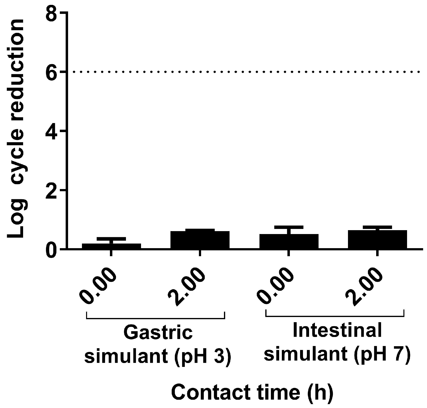
We also analyzed whether L. plantarum CNTA 628 is able to resist gastric and intestinal conditions. In this in vitro assay, we observed that our strain exhibited resistance to simulated gastric fluid, as it retained viability after 2 h, and the viable counts showed a decrease of less than 1 log cycle with respect to the initial cell concentration (Figure 2). Notably, the probiotic exhibited strong survival ability after 2 h of exposure to simulated intestinal fluid, maintaining a cell population of 7 log CFU/mL, which indicates its resistance to both simulated gastric and intestinal fluids.
Then, we evaluated the potential resistance to antibiotics for L. plantarum CNTA 628 by the broth microdilution method. L. plantarum CNTA 628 was susceptible to all antibiotics tested (Table 1).
Moreover, the SCFAs produced by L. plantarum CNTA 628 in different culture media were analyzed using GC-MS (Table 2). Thus, L. plantarum CNTA 628 was capable of producing acetic acid (in culture media with FOS and without adding a carbon source). The SCFAs propionic, butyric, valeric, caproic, and isobutyric acids were not quantified in any medium.
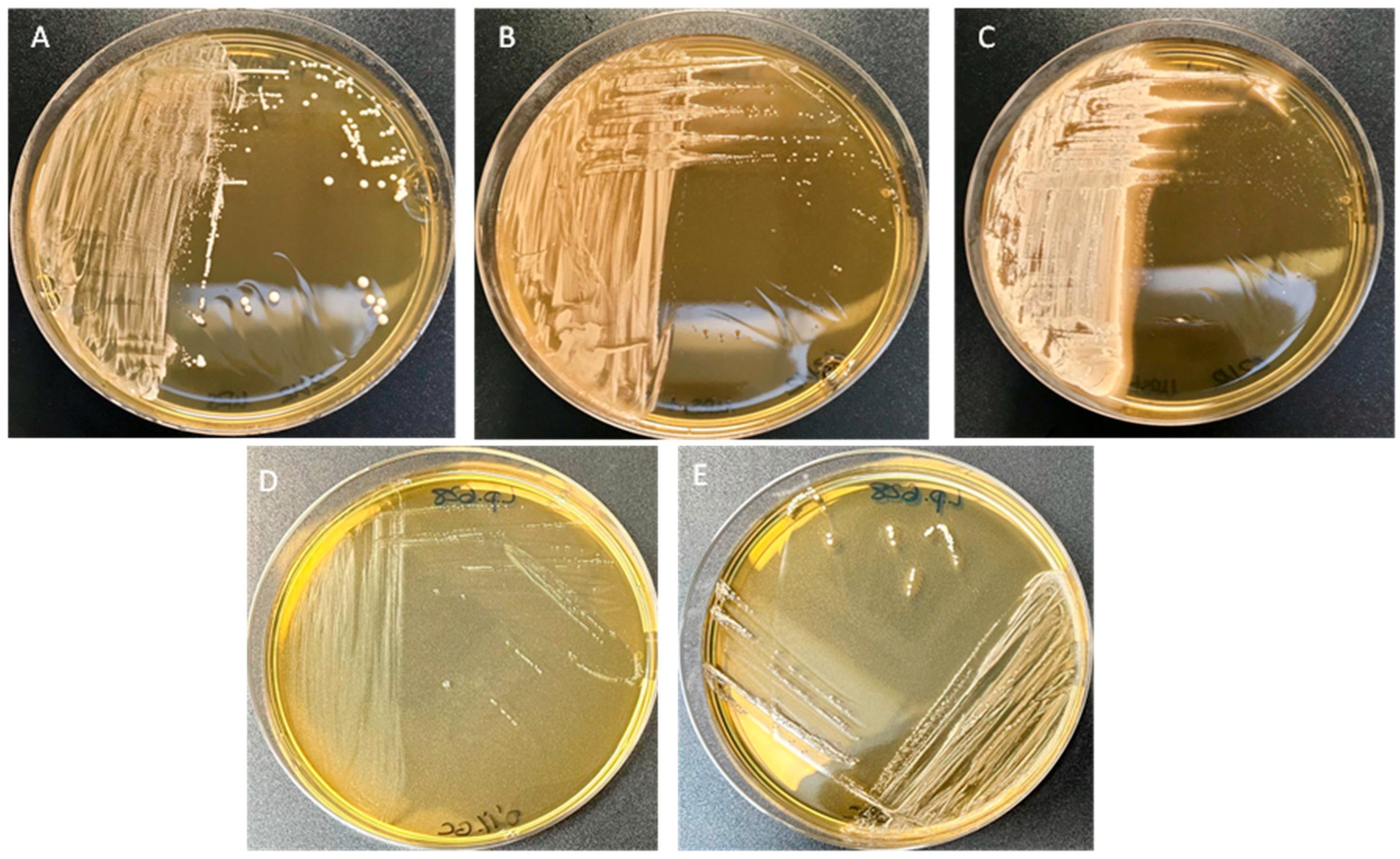
Other probiotic properties were evaluated for L. plantarum CNTA 628, including the ability to produce β-galactosidase throughout an in vitro test. This analysis demonstrated that L. plantarum CNTA 628 is able to break down the disaccharide lactose into glucose and galactose (Figure S1). Furthermore, we determined the bile salt hydrolase (BSH) activity of this strain using a qualitative direct plate assay. Visible halos surrounding the colonies and white precipitates with the colonies were observed when L. plantarum CNTA 628 was cultured on an MRS agar plate supplemented with tauro-conjugated bile salts. However, this strain produced similar colony types on plates with or without glyco-conjugated bile salts (Figure 3).
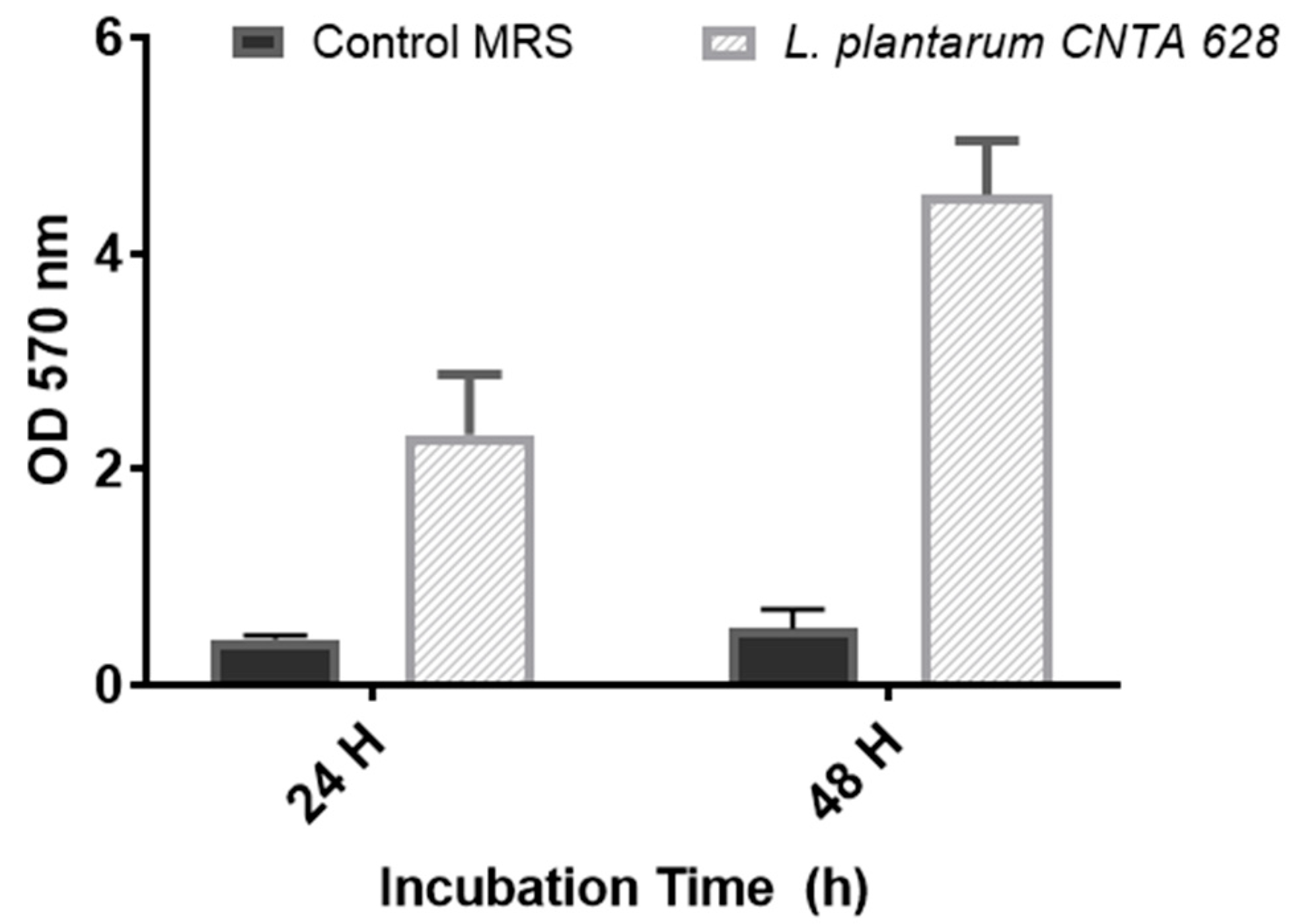
The potential biofilm-forming capability of L. plantarum CNTA 628 was evaluated in vitro. This strain displayed a very strong biofilm-forming capacity on an abiotic surface (Figure 4), with absorbance values higher than 0.5, ranging from 2.32 ± 0.55 (24 h) to 4.54 ± 0.50 (48 h).
To sum up, the probiotic strain L. plantarum CNTA 628 displays tolerance to simulated gastric and intestinal environments in vitro, exhibits susceptibility to antibiotics, demonstrates β-galactosidase and bile salt hydrolysate enzymatic activities, forms biofilms, and is able to produce acetic acid as a metabolic SCFA.

3.2. L. plantarum CNTA 628 Competes with Pathogenic Bacteria and Exhibits Immunomodulatory Capacity In Vitro

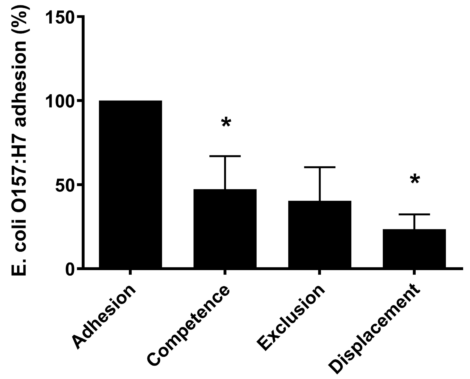
In our study, a possible effect of L. plantarum on modifying the adhesion of E. coli O157:H7 onto Caco-2 cells was addressed. Thus, the individual adhesion of the bacteria (L. plantarum CNTA 628 and E. coli O157:H7) to Caco-2 cells was determined, resulting in about 2.4 ±1.6; 2.5 ±1.5 and 3.7 ± 0.9, respectively. The percentage of adhesion of E. coli to Caco-2 cells (3.7 ± 0.9) was used to normalize the results of E. coli adhesion in competence, displacement and exclusion assays (Figure 5).
For the cell/bacteria 1:10 ratio, the ability of L. plantarum to reduce E. coli adhesion on Caco-2 cells was also investigated. When L. plantarum is added prior to the pathogenic bacteria (exclusion), E. coli adhesion is not affected. However, when L. plantarum is added at the same time as E. coli (competence), its adhesion is reduced by 53%. Furthermore, L. plantarum was capable of significantly reducing the adhesion of E. coli by 77% in displacement assays. These results indicate that our L. plantarum strain is capable of competing with and displacing E. coli on the cellular model Caco-2.
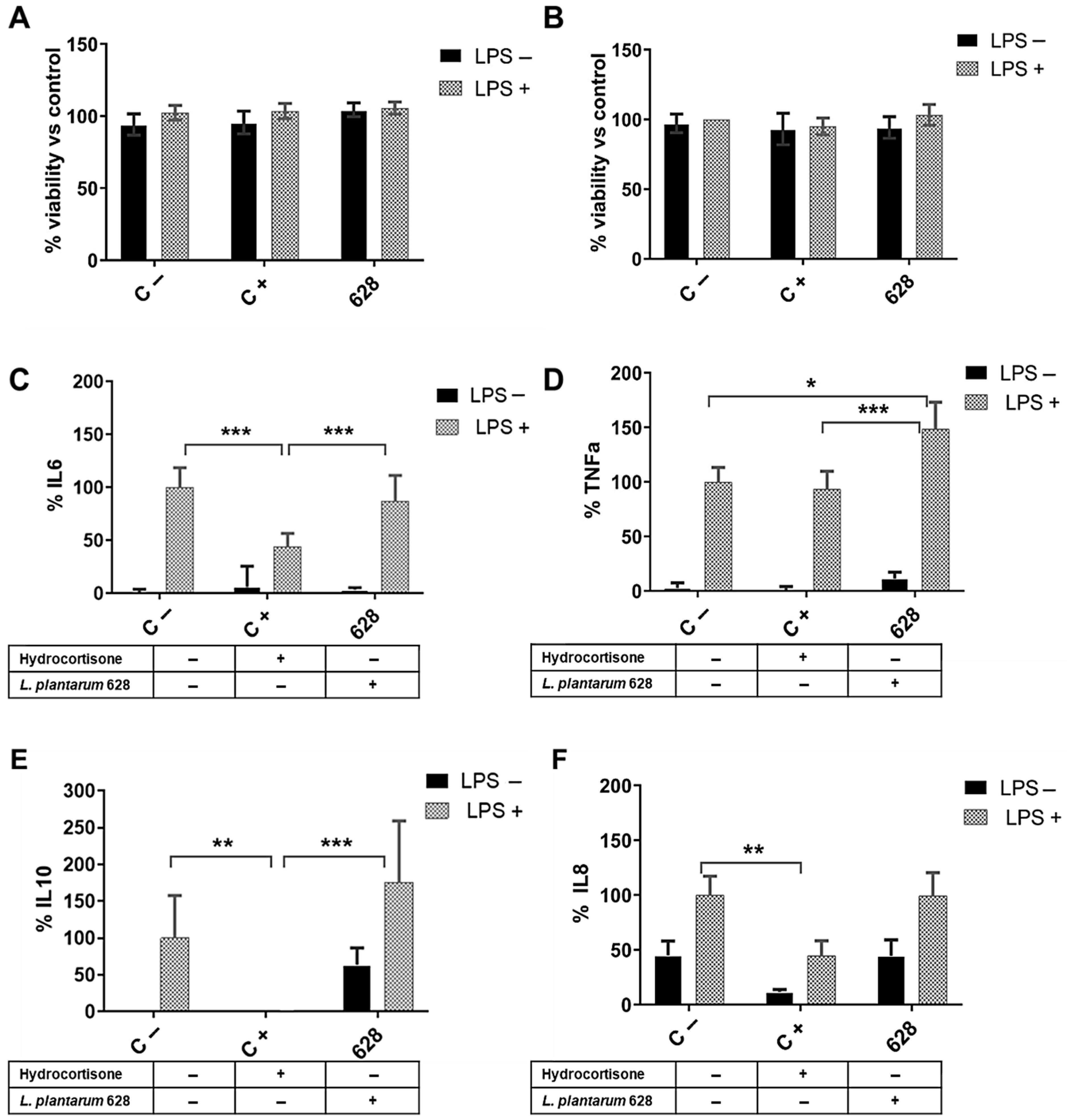
On the other hand, we also investigated the in vitro potential immunomodulatory activity of L. plantarum CNTA 628 directly on the immune system using RAW 264.7 cells and under an intestinal scenario using HT-29 cells. For a correct assessment of cytokine production, we first determined the potential cytotoxic effect of the probiotic strains on both RAW 264.7 and HT-29 cell lines, maintaining the same conditions as for the cytokine assay quantification. Macrophage cell line RAW 264.7 did not show a decrease in cell viability under the anti-inflammatory control hydrocortisone or L. plantarum CNTA 628 (Figure 6A). In a similar manner, HT-29 cell viability was unaffected by the presence of the lactic bacteria (Figure 6B). Once we demonstrated the absence of the cytotoxicity of the cells when cultured in the presence of L. plantarum, we determined the immunomodulatory activities of the probiotic. In RAW 264.7 cells, L. plantarum did not significantly alter the secretion of IL-6 (Figure 6C) or TNFa (Figure 6D) under basal conditions. However, when cells were stimulated with LPS, L. plantarum significantly increased TNF-α secretion. Additionally, we analyzed the levels of proinflammatory cytokine IL-10. Under basal conditions, L. plantarum drastically increased IL-10 production (Figure 6E). Interestingly, under LPS stimulation, L. plantarum did not significantly modify IL-10 secretion compared to only LPS control cells (Figure 6E).
Finally, in order to investigate whether L. plantarum would have an impact on intestinal cells, IL-8 was quantified in HT-29 cells. Without LPS stimulation, IL-8 secretion was not affected after L. plantarum exposure (Figure 6F). In a similar manner, in LPS induced- cells, L. plantarum pre-treatment did not induce any changes in IL-8 production compared to the control LPS-stimulated cells (Figure 6F).
In summary, L. plantarum significantly increased TNF- α secretion after exposure to LPS, but it did not show any effect on IL-10, IL-6 or IL-8 production under these conditions.

3.3. L. plantarum CNTA 628 Lowers Lipid Storage and Reduces Oxidative Stress and Senescence in C. elegans, Contributing to Lifespan Extension

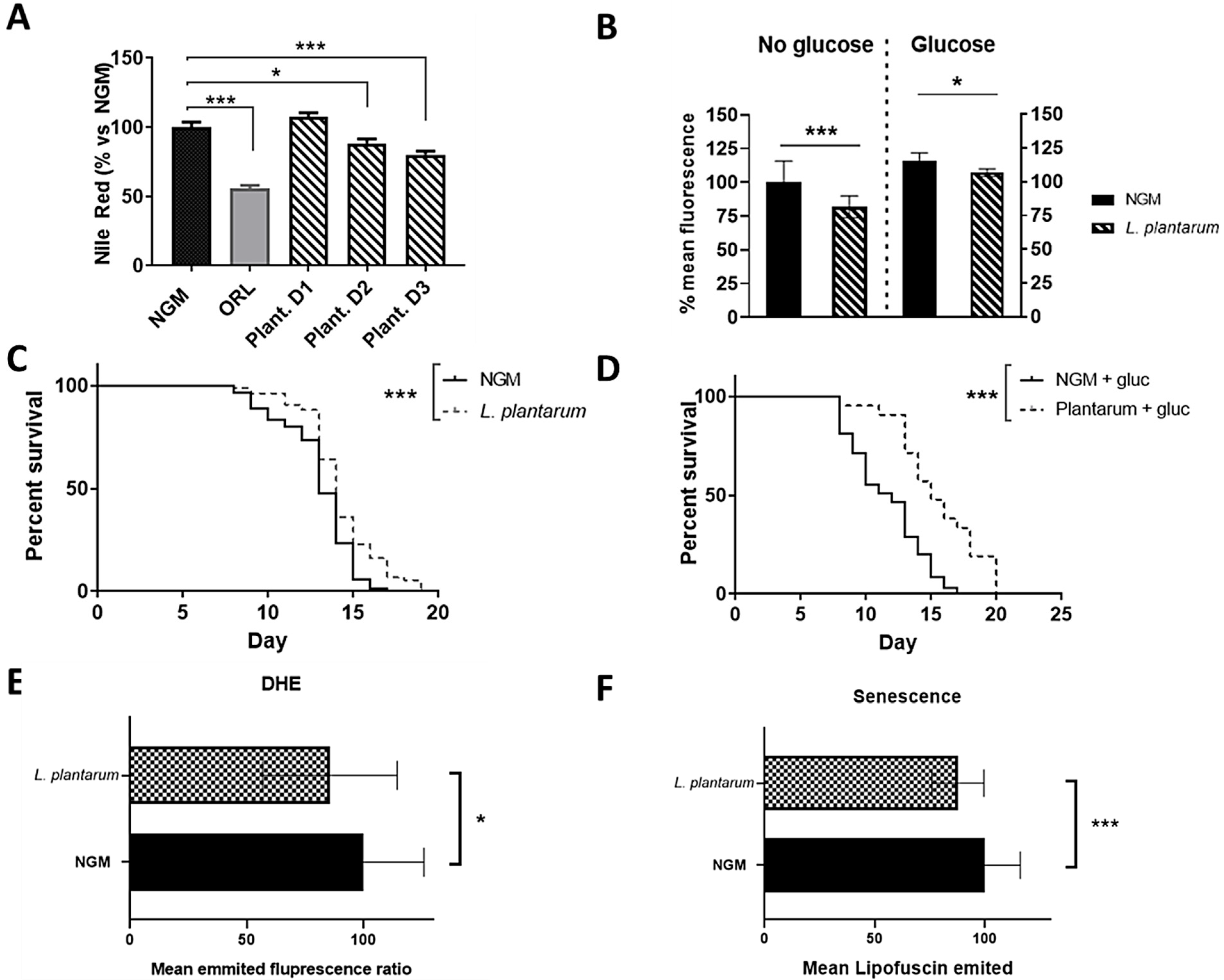
In this study, it was observed that treatment with different doses of L. plantarum CNTA 628 induced a total lipid reduction effect in C. elegans under normal (NGM media) conditions (Figure 7A), quantified by the Nile red staining method. Moreover, this effect was also achieved when grown under high-glucose (10 mM)-loaded media conditions (Figure 7B). Thus, L. plantarum 628 (1.3 × 10⁵ CFU/mL) was able to reduce fat accumulation by 18.19% compared to NGM control worms, while it induced a reduction of 7.81% in glucose-loaded media. In both conditions, orlistat (1.5 mg/mL) was used as a positive control, which caused a reduction in the fat percentage of approximately 40%.
Interestingly, supplementation with L. plantarum CNTA 628 also resulted in a significant extension in the lifespan of the nematodes (p < 0.001), both in NGM (Figure 7C) and glucose-loaded NGM (MGNg) media (Figure 7D). In fact, the median survival of the nematodes in the group treated with L. plantarum CNTA 628 increased by a day compared to the control NGM group (from 13 days in NGM group to 14 days in probiotic-treated worms). In the case of NGMg media, control worms also exhibited a reduced lifespan, with a median survival of 12 days in comparison to the 13 days of the control NGM group. However, the probiotic supplementation counteracted this lifespan-shortening effect induced by the glucose as the median survival for this group was 15 days (p < 0.001).
To ensure that the reduction in the total accumulated lipids and the extended lifespan effects were not attributed to any potential adverse effects of the probiotic on the worms, we investigated the effect of L. plantarum supplementation on the C. elegans larvae development (Figure S2). This developmental study demonstrated that worms treated with L. plantarum CNTA 628 (at the dose of 3 × 10⁵ CFU/mL) were able to reproduce normally, with egg-laying and larval development at L1 stages being evident, with no differences in the time of appearance in comparison to the NGM control plates. Hence, we demonstrated how both L. plantarum CNTA 628 can reduce fat accumulation in C. elegans without visibly affecting their development. Thus, we also evidenced that supplementation with L. plantarum CNTA 628 reduced ROS accumulation (measured as the mean emitted fluorescence of DHE) by 14.47% compared to the control NGMg group (Figure 7E). Additionally, the probiotic caused a reduction of 12.2% of the mean emitted fluorescence by the auto-fluorescent protein lipofuscin compared to the control NGMg group (Figure 7F). Both conditions were studied under high-glucose conditions to ensure the physiological need of triggering an oxidative stress response and senescence, as described in previous works [36].
Finally, we also evidenced that supplementation with L. plantarum CNTA 628 reduced ROS accumulation (measured as the mean emitted fluorescence of DHE) by 14,47% compared to the control NGMg group (Figure 7E). Additionally, the probiotic caused a reduction of 12.2% of the mean emitted fluorescence by the auto-fluorescent protein lipofuscin compared to the control NGMg group (Figure 7F).

3.4. L. plantarum CNTA 628 Influences the Transcription of Essential Metabolic Genes

We investigated the activity of L. plantarum CNTA 628 modulating the expression of key metabolic genes in C. elegans by qPCR, focusing on pathways related to lipid and glucose metabolism. The expression levels of different genes were analyzed under both normal and high-glucose conditions.
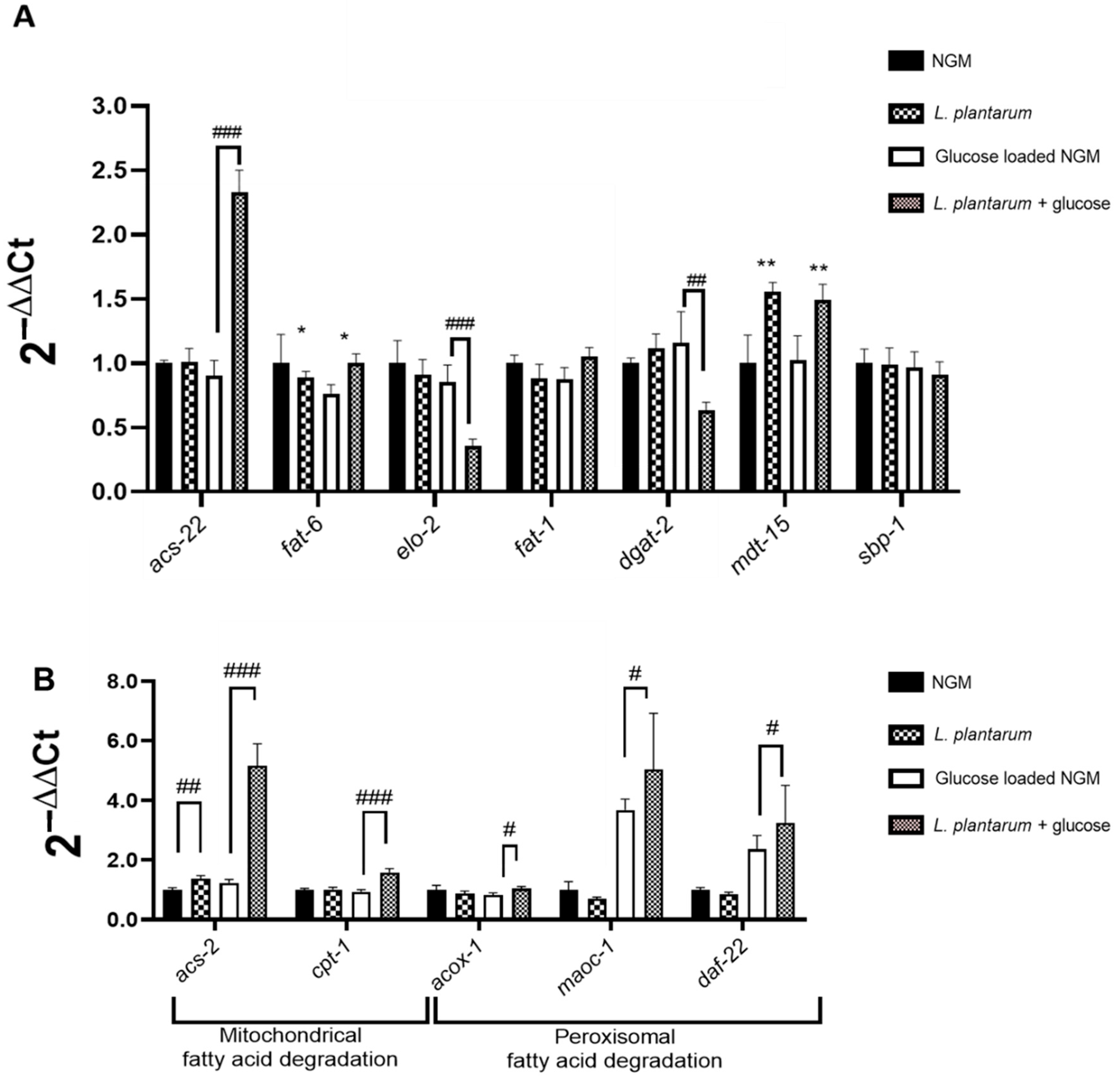
No significant effects were observed in the gene expression of daf-2 (the ortholog of insulin/insulin-like growth factor receptor IGFR1) and daf-16 (the ortholog of human FOXO) after probiotic supplementation. Regarding the FA biosynthesis pathways (Figure 8A), no differences were observed in the expression of omega-3 fatty acid desaturase fat-1 and sterol regulatory element binding protein sbp-1. However, the n-3 fatty acid desaturase fat-6, and the elongase elo-2 were significantly downregulated by the probiotic, while the expression of diacylglycerol acyltransferase dgat-2 appeared to be downregulated under high-glucose conditions. On the other hand, long-chain fatty acyl-CoA synthetase-coding gene acs-22, appeared to be highly upregulated by L. plantarum CNTA 628 in high-glucose conditions (Figure 8A).
Additionally, the C. elegans mdt-15 gene, homologous to human MED15, was upregulated in both NGM and NGMg conditions (Figure 8A), together with acs-2 (Figure 8B). This overexpression was also accompanied by the upregulation of the mitochondrial Carnitine Palmitoyl Transferase cpt-1 and the peroxisomal B-oxidation genes acox-1, daf-22 and maoc-1 in the glucose-loaded conditions (Figure 8B).

4. Discussion

Lacto-fermentative probiotics like Lactobacilli or Bifidobacteria have been shown to alleviate the negative consequences of metabolic syndrome-related diseases, including obesity and type-2 diabetes [41]. Despite the known benefits of certain lactic acid bacteria strains, a complete in vitro characterization is necessary to determine the potential application of novel bacterial species for their use as probiotics. In our work, we investigated the probiotic properties of the strain L. plantarum CNTA 628, and its potential health-promoting activities in vivo. Before assessing the probiotic properties of L. plantarum CNTA 628 in vitro, we verified the strain’s species identity. The ANI index in this study confirmed that the strain is part of the Lactiplantibacillus plantarum species, with values exceeding 96%.
A probiotic bacterium needs to survive the gastrointestinal tract to reach the colon in sufficient numbers. For health benefits, it must reach the ileum at a minimum concentration of 6 log CFU/g or CFU/mL [42]. In our study, L. plantarum CNTA 628 showed strong survival capacity, maintaining a cell population of 7 log CFU/mL in simulated gastric and intestinal fluids.
On the other hand, microbial feed additives must not contribute to the existing pool of antimicrobial resistance (AMR) genes in the gut bacterial population or promote the spread of AMR [29]. For this reason, we investigated the potential resistance of L. plantarum CNTA 628 to antibiotics according to the breakpoints recommended by the European Food Safety Authority. This analysis demonstrated the susceptibility of this L. plantarum strain to the full spectrum of antibiotics, demonstrating its suitability for use as a microbial feed additive [29].
Lactic acid bacteria (LAB) are a diverse group of microorganisms that have been extensively studied because of their prominent role in food fermentation and human health [43]. In this line, we observed that L. plantarum CNTA 628 was capable of producing acetic acid, a short-chain fatty acid with multiple positive effects to maintain human wellbeing [44]. Furthermore, we also demonstrated the ability of this strain to produce β-galactosidase. The presence of the β-galactosidase (β-gal) enzyme in these microorganisms is especially important for their use in dairy product production and for enhancing lactose metabolism in the gut as an energy source.
The ability of Lactobacillus strains to form biofilms has been previously identified and presents various benefits upon consumption. The biofilm-forming capability of lactobacillus strains extends beyond mere colonization, offering a spectrum of benefits that positively impact gastrointestinal health and overall wellbeing [45]. On the other hand, it has also been observed that probiotic species (for example, intestinal probiotic L. salivarius W24) could be cariogenic (promote the formation of bacterial biofilms on teeth) under specific growth conditions. This indicates that it is very important to make a correct selection of the probiotic to be used, which must be able to compete with and inhibit biofilm-forming pathogens according to the area of action [46]. Moreover, it has been described that several probiotics produce bile salt hydrolase (BSH), which helps to reduce serum cholesterol [47]. In our study, we also demonstrated a biofilm-forming capability and the capability to hydrolyze tauro-conjugated bile salts of L. plantarum CNTA 628, two probiotic properties with great applications.
In mammals, the main protection against pathogenic infections is the intestinal barrier, mainly composed of epithelial cells with specific characteristics. However, these cells alone are not sufficient, as the resident gut microbiota play a crucial role in preserving the integrity of the intestinal barrier. In this context, lactic acid bacteria have been extensively studied in the recent years for their capacity of “fighting” pathogens like Salmonella, Escherichia coli and Clostridium [48]. It has been speculated that alterations in the microbiome lead to the development of conditions such as inflammatory bowel disease (IBD). In fact, IBD patients display an increased proportion of Enterobacteriaceae genus and a decreased concentration of bacteria from the phylum Firmicutes [49]. Moreover, patients suffering from Crohn’s disease also have an anomalous amount of adherent invasive Escherichia coli. Caco-2 cells have been widely used as a model for the intestinal barrier because they develop the characteristics of a mature enterocyte [50]. Previous works showed that L. plantarum was able to compete and displace E. coli O157:H7, indicating that Lactobacilli might share binding sites with pathogenic bacteria, therefore being capable of excluding or displacing them. Such an effect is probably strain-specific, dependent on the different molecules and patterns expressed on the bacterial wall of each strain. Indeed, Patel et al. demonstrated that L. plantarum 299V expresses GAPDH on its surface, facilitating the adhesion and colonization of the intestinal lining [51]. Additionally, lactic acid bacteria can compete for attachment sites on intestinal epithelial cells (IECs), glycoproteins, or plasminogen within the extracellular matrix [52]. Altogether, it suggests that L plantarum strains possess great potential as treatment for proinflammatory intestinal diseases. Some mice studies have shown that only the surface proteins from L. plantarum MTCC 5690, are capable of neutralizing the histopathological damages in a pathological mouse model [53]. Finally, L. plantarum has been characterized to produce a bacteriocin, plantaricin, which is capable of inhibiting the growth of pathogenic bacteria like E. coli or Listeria monocytogenes [54]. Following this trend, here, we demonstrated that our probiotic strain L. plantarum CNTA 628 is capable of competing and displacing E. coli O157:H7 to Caco-2 cells.
Lactobacillus genus has been extensively studied for modulating immune responses. The cell wall of lactic bacteria contains certain molecules that interact with immune cells in the intestine and potentially activate them [55,56]. Such activation has been suggested to be beneficial for inhibiting viral infections. Other studies have reported that a specific strain of L. plantarum enhances the immune system in an immunocompromised mouse model [57]. Moreover, additional beneficial roles of L. plantarum have been identified, like the ameliorating lipidic profile of patients and decreasing atherosclerosis [58]. In fact, L. plantarum decreases cholesterol and reactive protein C in older patients [59]. Interestingly, three different strains of L. plantarum have been shown to exert diverse effects on modulating cytokine production in mouse splenocytes [60]. For this reason, we investigated the potential immunomodulatory activity of L. plantarum CNTA 628 in RAW 264.7 and HT-29 cell lines. Once we evidenced the lack of cytotoxicity in both cell lines after exposure to hydrocortisone and L. plantarum CNTA 628, we analyzed the potential activity of the probiotic modulating the LPS-induced cytokine production. Although no effect was observed on IL-10 and IL-6 production, L. plantarum CNTA 628 significantly increased the TNF- α secretion after LPS treatment in RAQ 264.7 cells, which would indicate the activation of the immune system through a specific pathway. No effect was observed in IL-8 production, contrary to other works where the ability of L. plantarum to decrease IL-8 production in intestinal cells has been reported for particular strains [61,62], suggesting that cytokine modulation varies by strain.
Overall, the in vitro assessment demonstrated that L. plantarum CNTA 628 shows great probiotic potential with strain-specific effects, suggesting that the composition of each strain directly affects immunomodulation in human cells. This phenomenon reinforces the potential of designing L. plantarum strains for different patient profiles.
In the same line, previous studies have reported that the health-promoting activities of a probiotic bacteria are strain-specific. Therefore, we conducted a functional evaluation of the potential metabolic effects of supplementation with L. plantarum CNTA 628. C. elegans has become a widely used in vivo model in a variety of research areas, including obesity, thanks to its anatomical simplicity, its short life cycle, its capability to self-reproduce and its approximate 65% genetic ontology with humans [63]. The similar metabolic pathways and their regulation have allowed researchers to characterize and describe complex processes such as the regulation of lipid storage, aging, or oxidative stress responses [63].
Regarding lipid metabolism, in both humans and C. elegans fatty acids (FAs) are the essential building blocks that constitute storage lipids (TAGs), membrane lipids (phospholipids and sphingolipids), and signaling lipids (fatty acyl amides, eicosanoids, and others). The FAs can be synthesized from de novo or derived from dietary fatty acids obtained from the E. coli bacteria on which they feed. Together with the synthesis of FAs, the catabolic processes under which C. elegans use stored lipids for energy production and the signaling properties of the involved molecules are well described. Also, the metabolism of FAs is linked to the uptake, processing and utilization of glucose and carbohydrate pathways [64,65,66]. Furthermore, glucose-supplemented media offer a dependable method to mimic diabetic or energy imbalance conditions, enabling the evaluation of parameters such as excessive fat accumulation, oxidative damage, cellular aging, and longevity in C. elegans [67]. In this context, the addition of drugs or bioactive compounds that disrupt their biologic processes can be studied and described as a starting point to assess their potential on humans [39,68]. Finally, this nematode model has been widely used in probiotic studies as a robust in vivo platform for rapidly identifying microorganisms with possible anti-inflammatory, anti-obesity, and beneficial health properties [22,69].
As previously described, we demonstrated that treatment with different doses of L. plantarum CNTA 628 induced a total lipid reduction effect in C. elegans under normal (NGM media) and high-glucose (10 mM)-loaded (NGMg media) conditions (Figure 7), suggesting a fat-reducing activity of this probiotic strain. Different works have described the modulation of fat storage by different probiotic strains. In a study by Patricia Martorell et al. [70], a total of 38 bacterial strains were tested for probiotic properties in C. elegans. The effect of some bifidobacterium and lactobacillus strains on fat reduction was evaluated by the same Nile red staining and measurement of the emitted fluorescence. An alternative to this technique was used by Gu et al. [71] in a study with Lactobacillus pentosus MJM60383, in which the inhibition of lipid accumulation in C. elegans was assessed by staining triglycerides with Oil red O (ORO). It is noteworthy that different works have studied fat accumulation in C. elegans and how it can be modified by probiotic strains including L. plantarum [16] or Pediococcus acidilactici [36,72]. Recently, the anti-obesity properties of another lactic acid bacteria, Latilactobacillus sakei CNTA 173, was also demonstrated using this in vivo model [27]. Interestingly, the administration of L. plantarum CNTA 628 not only reduced lipid accumulation but also significantly extended the lifespan of C. elegans. Indeed, the probiotic reversed the detrimental impact of glucose on longevity (Figure 8) [73]. All these effects were independent of an effect over worm development, as was demonstrated by the presence of egg-laying and larval development at L1 stages in all supplemented and non-supplemented plates. Hence, we demonstrated how L. plantarum CNTA 628 can reduce fat accumulation in C. elegans without visibly affecting their development. This initial result led us to a more in-depth study of other health-benefiting effects of this probiotic.
Previous studies have shown that certain probiotic strains are able to reduce the accumulation of reactive oxygen species (ROS) and the aging-related protein, lipofuscin [16,36,74]. Oxidative stress has been associated with the alteration of regulatory factors of mitochondrial activity, which in turn mediate the concentration of inflammatory markers associated with the development of new adipocytes, lipogenesis and, lastly, obesity [75]. These impairments in metabolic health are also closely related to a major risk factor in metabolic syndrome-related diseases, senescence. This factor is characterized by a growing chronic, low inflammatory profile that is also associated with the disfunction of the adipose tissue. Aging is associated with obesity, being one of the main risk factors for the development of this disease and its comorbidities. The use of C. elegans as an animal model to study senescence and the causes and consequences of its early development has been widely approved [63,76]. Some of the studies conducted in the field of probiotic supplementation have already stated that specific strains of L. plantarum can modulate the accumulation of senescence-related markers and increase the worms’ lifespan [16,76]. In our study, supplementation with L. plantarum CNTA 628 reduced ROS accumulation compared to the control NGMg group but also decreased the content in auto-fluorescent protein lipofuscin. Both conditions were studied under high-glucose conditions to ensure the physiological need of triggering an oxidative stress response and senescence, as described in previous works [36]. Putting these results together, we can hypothesize that L. plantarum CNTA 628 supplementation is able to reduce lipofuscin accumulation and enhance the oxidative stress response, confirming the health-promoting effects of this probiotic strain in vivo.
Due to the highly conserved gene ontology of C. elegans compared with humans, it is considered a reliable model to evaluate the mechanisms behind the observed changes in physiological parameters [77]. Since previous works have described the effects that probiotics can have on the expression of genes involved in energy metabolism [22], we aimed to identify the underlying mechanisms of the phenotypical changes induced by this specific probiotic strain through gene expression analyses. No significant changes were detected in the expression of daf-2 and daf-16, two central genes involved in glucose metabolism, following probiotic treatment, suggesting that the mechanism under which L. plantarum CNTA 628 promotes its health-promoting effects does not involve the IIS signaling pathway. A similar result was observed in FA biosynthesis-related genes fat-1 and sbp-1.
However, genes encoding for n-3 fatty acid desaturase fat-6 and elongase elo-2 were significantly downregulated by the probiotic, while the expression of diacylglycerol acyltransferase dgat-2 was downregulated under high-glucose conditions. On the other hand, long-chain fatty acyl-CoA synthetase-coding gene acs-22, the ortholog of human FATP1, appeared to be highly upregulated by L. plantarum CNTA 628 in high-glucose conditions (Figure 8). The role of the acyl transferase dgat-2 and acs-22 in C. elegans is crucial for the loading stages of FAs onto the endoplasmic reticulum and both have been associated with a higher accumulation of TAGs [78]. Nevertheless, acs-22 is also involved in the modulation of intestinal permeability as described in other works [79], which could explain the fact that its upregulation is not associated with a higher lipid accumulation in the worms but is related to a better functioning of the intestine absorption. Thus, the reduction in fat content caused by L. plantarum CNTA 628 could also be explained by the direct regulation of the de novo synthesis of the FA pathway in which fat-6, elo-2 and dgat-2 are involved. This effect appears to be more noticeable under high-glucose conditions due to the effect of the triacylglycerol synthesis pathway in which dgat-2 plays a crucial role in the final steps.
Interestingly, mdt-15 (the ortholog of human MED15) was upregulated in both NGM and NGMg conditions. The role of mdt-15, a co-activator of the nuclear hormone receptor nhr-49, has been previously associated with an increased lifespan in this animal model [80]. Besides being associated with lifespan, the complex mdt-15/nhr-49 also appears to be part of the regulatory system that ensures homeostasis regarding energy metabolism or the oxidative stress response [81]. Besides the mentioned implications, the mdt-15/nhr-49 system is parallel to the Peroxisome Proliferator-Activated Receptor (PPAR) family proteins in mammals, which are key regulators of fat, cholesterol, and glucose homeostasis [82]. The expression of nhr-49 has been associated with the fasting response in the worms [83] and with the positive regulation of some of the β-oxidation pathway genes through regulating the expression of acyl-CoA synthetase acs-2 [83]. Therefore, the overexpression of mdt-15 caused by L. plantarum CNTA 628 in C. elegans metabolism led to a more in-depth analysis of the expression levels of genes involved in the catabolic β-oxidation pathway.
In these subsequent analyses, we observed that both peroxisomal and mitochondrial β-oxidation-related genes were upregulated in worms treated with L. plantarum CNTA 628, in comparison with the corresponding controls. Interestingly, the expression of the nhr-49-regulated gene acyl-CoA synthetase catalyzing acs-2, which catalyzes the conversion of a fatty acid to acyl-CoA for further degradation, was upregulated both under normal and high-glucose conditions. This overexpression was also accompanied by the upregulation of the mitochondrial Carnitine Palmitoyl Transferase cpt-1 and the peroxisomal B-oxidation genes acox-1, daf-22 and maoc-1, in the glucose-loaded conditions. This gene expression profile is similar to other lactic bacteria described in previous studies. For example, Gu et al. [71] described how Lactobacillus pentosus MJM60383 is able to upregulate the expression of both nhr-49 and acs-2, therefore modulating the FA degradation pathways and justifying the observed reduction in the lipid content in the worms. Likewise, L. sakei CNTA 173 supplementation effectively diminished fat storage by modulating the expression of acox-1 and maoc-1, both genes involved in fatty acid beta-oxidation [27].
Finally, it is worth noting that, despite the promising physiological effects observed with this probiotic, the primary limitation of our study lies in the simplicity of the C. elegans model. Its distinct physiology, immune system, and microbiota differ significantly to those of mammals, which may limit the translational relevance of the results. Additionally, the probiotic dosage and administration route in this model often do not reflect physiologically meaningful exposures in humans or other animals. Although C. elegans is a powerful tool for uncovering strain-specific molecular mechanisms, its short lifespan and biological simplicity constrain its ability to model the complex interactions seen in higher organisms. Therefore, before advancing to human trials, further studies using murine models of obesity are needed to confirm the effectiveness of L. plantarum CNTA 628.

5. Conclusions

In summary, our work proposes a new bacterial strain, Lactiplantibacillus plantarum CNTA 628, as a probiotic candidate with potential application in the prevention and/or amelioration of metabolic syndrome-related disturbances. The probiotic cleared the preliminary in vitro test regarding important properties attributed to probiotics for human consumption, including an optimal growth rate, susceptibility throughout the entire set of antibiotics assessed, good adaptations towards simulated gastric environment, BHS and β-galactosidase activities, and the ability to produce acetic acid and generate biofilms in vitro. Furthermore, the probiotic competed with and displaced pathogenic bacteria E. coli O157:H7 in a cell adhesion assay and exerted immunomodulatory properties, modulating the production of TNF-α. The functional evaluation demonstrated that supplementation with L. plantarum CNTA 628 induced a reduction in total lipid content, ROS production and senescence markers in C. elegans. These activities were accompanied by an increased lifespan in both normal and high-glucose conditions, without disrupting the normal development of the worm. Gene expression analyses demonstrated that these health-promoting effects were mediated by the modulation of the lipid metabolic pathways involved in energy balance and homeostasis, upregulating both mitochondrial and peroxisomal key β-oxidation-related genes and downregulating triglyceride biosynthesis. Overall, our in vitro and in vivo work sets out a starting point for further research involving the beneficial properties of the probiotic L. plantarum CNTA 628 in the context of obesity and metabolic syndrome.

6. Patents

The works reported in this manuscript have resulted in the registration of the patent EP25382355.3, entitled Lactiplantibacillus plantarum strain and uses thereof.

Supplementary Materials

The following supporting information can be downloaded at https://www.mdpi.com/article/10.3390/app15148007/s1: Table S1: Strains closest to Lactiplantibacillus plantarum CNTA 628 according to the ANI index. Table S2: ANI values when comparing the Lactiplantibacillus sp. CNTA628 genome with all available genomes for the type strain of Lactiplantibacillus plantarum. Table S3: Growth parameters of L. plantarum CNTA 628. Figure S1: L. plantarum CNTA 628 displays BSH activity. Figure S2: L. plantarum CNTA 628 does not affect C. elegans development.

Author Contributions

Conceptualization, I.G., L.V.-V., F.I.M. and P.A.; Methodology, L.V.-V., M.L.-Y., N.L.-G. and A.S.-V.; Software, N.L.-G.; Validation, I.G., R.V., N.L.-G. and A.S.-V.; Formal Analysis, L.V.-V., M.L.-Y. and A.S.-V.; Investigation, I.G., M.L.-Y. and P.A.; Resources, R.V.; Data Curation, L.V.-V., N.L.-G. and P.A.; Writing—Original Draft, I.G., L.V.-V., F.I.M. and P.A.; Writing—Review and Editing, F.I.M.; Supervision, R.V. and F.I.M.; Project Administration, R.V.; Funding Acquisition, R.V. and P.A. All authors have read and agreed to the published version of the manuscript.

Funding

This research was funded by Gobierno de Navarra: 0011-1383-2020-000010 [PC173-174 PREDISMET], 0011-1383-2022-000000 [PC128-129 PARABIOTICS], 0011-1383-2024-000000 [PC24-PARABIOTICS-2-007-006] and CIBERobn (CB12/03/30002).

Institutional Review Board Statement

Not applicable.

Informed Consent Statement

Not applicable.

Data Availability Statement

The data presented in this study are available on request from the corresponding author due to privacy reasons.

Acknowledgments

The authors thank Viscofan S.A and Laboratorios CINFA, S.A. for the financial support of the Centre for Nutrition Research, the University of Navarra.

Conflicts of Interest

I.G., L.V.-V., R.V., F.I.M. and P.A. are authors of the patent EP25382355.3, entitled Lactiplantibacillus plantarum strain and uses thereof. The rest of the authors declare no conflicts of interest.

Abbreviations

The following abbreviations are used in this manuscript:
NGMNematode Growth Medium
NGMgNematode Growth Medium with Glucose (10 mM)
CFUColony-Forming Unit
DHEDihydroethidium

References

  1. Gomes, A.C.; Hoffmann, C.; Mota, J.F. The Human Gut Microbiota: Metabolism and Perspective in Obesity. Gut Microbes 2018, 9, 308. [Google Scholar] [CrossRef] [PubMed]
  2. Bäckhed, F.; Ding, H.; Wang, T.; Hooper, L.V.; Gou, Y.K.; Nagy, A.; Semenkovich, C.F.; Gordon, J.I. The Gut Microbiota as an Environmental Factor That Regulates Fat Storage. Proc. Natl. Acad. Sci. USA 2004, 101, 15718–15723. [Google Scholar] [CrossRef] [PubMed]
  3. Castaner, O.; Goday, A.; Park, Y.M.; Lee, S.H.; Magkos, F.; Shiow, S.A.T.E.; Schröder, H. The Gut Microbiome Profile in Obesity: A Systematic Review. Int. J. Endocrinol. 2018, 2018, 4095789. [Google Scholar] [CrossRef] [PubMed]
  4. Gérard, P. Gut Microbiota and Obesity. Cell. Mol. Life Sci. 2015, 73, 147–162. [Google Scholar] [CrossRef] [PubMed]
  5. Zhou, Z.; Sun, B.; Yu, D.; Zhu, C. Gut Microbiota: An Important Player in Type 2 Diabetes Mellitus. Front. Cell. Infect. Microbiol. 2022, 12, 834485. [Google Scholar] [CrossRef] [PubMed]
  6. Sánchez, B.; Delgado, S.; Blanco-Míguez, A.; Lourenço, A.; Gueimonde, M.; Margolles, A. Probiotics, Gut Microbiota, and Their Influence on Host Health and Disease. Mol. Nutr. Food Res. 2017, 61, 1600240. [Google Scholar] [CrossRef] [PubMed]
  7. Abenavoli, L.; Scarpellini, E.; Colica, C.; Boccuto, L.; Salehi, B.; Sharifi-Rad, J.; Aiello, V.; Romano, B.; De Lorenzo, A.; Izzo, A.A.; et al. Gut Microbiota and Obesity: A Role for Probiotics. Nutrients 2019, 11, 2690. [Google Scholar] [CrossRef] [PubMed]
  8. Million, M.; Angelakis, E.; Paul, M.; Armougom, F.; Leibovici, L.; Raoult, D. Comparative Meta-Analysis of the Effect of Lactobacillus Species on Weight Gain in Humans and Animals. Microb. Pathog. 2012, 53, 100–108. [Google Scholar] [CrossRef] [PubMed]
  9. Zare, D.; Aryaee, H.; Mirdamadi, S.; Shirkhan, F. The Benefits and Applications of Lactobacillus plantarum in Food and Health: A Narrative Review. Iran. J. Public Health 2024, 53, 2201. [Google Scholar] [CrossRef] [PubMed]
  10. Behera, S.S.; Ray, R.C.; Zdolec, N. Lactobacillus plantarum with Functional Properties: An Approach to Increase Safety and Shelf-Life of Fermented Foods. BioMed Res. Int. 2018, 2018, 9361614. [Google Scholar] [CrossRef] [PubMed]
  11. Micioni Di Bonaventura, M.V.; Coman, M.M.; Tomassoni, D.; Micioni Di Bonaventura, E.; Botticelli, L.; Gabrielli, M.G.; Rossolini, G.M.; Di Pilato, V.; Cecchini, C.; Amedei, A.; et al. Supplementation with Lactiplantibacillus plantarum IMC 510 Modifies Microbiota Composition and Prevents Body Weight Gain Induced by Cafeteria Diet in Rats. Int. J. Mol. Sci. 2021, 22, 11171. [Google Scholar] [CrossRef] [PubMed]
  12. Jacouton, E.; Mondot, S.; Langella, P.; Bermúdez-Humarán, L.G. Impact of Oral Administration of Lactiplantibacillus plantarum Strain CNCM I−4459 on Obesity Induced by High-Fat Diet in Mice. Bioengineering 2023, 10, 1151. [Google Scholar] [CrossRef] [PubMed]
  13. Yoshitake, R.; Hirose, Y.; Murosaki, S.; Matsuzaki, G. Heat-Killed Lactobacillus plantarum L-137 Attenuates Obesity and Associated Metabolic Abnormalities in C57BL/6 J Mice on a High-Fat Diet. Biosci. Microbiota Food Health 2021, 40, 84–91. [Google Scholar] [CrossRef] [PubMed]
  14. Ma, Y.; Fei, Y.; Han, X.; Liu, G.; Fang, J. Lactobacillus plantarum Alleviates Obesity by Altering the Composition of the Gut Microbiota in High-Fat Diet-Fed Mice. Front. Nutr. 2022, 9, 947367. [Google Scholar] [CrossRef] [PubMed]
  15. Kumar, A.; Kumar, V.; Pramanik, J.; Rustagi, S.; Prajapati, B.; Jebreen, A.; Pande, R. Lactiplantibacillus plantarum as a Complementary Approach for Diabetes Treatment and Management. Curr. Nutr. Rep. 2025, 14, 72. [Google Scholar] [CrossRef] [PubMed]
  16. Kumar, A.; Joishy, T.; Das, S.; Kalita, M.C.; Mukherjee, A.K.; Khan, M.R. A Potential Probiotic Lactobacillus plantarum JBC5 Improves Longevity and Healthy Aging by Modulating Antioxidative, Innate Immunity and Serotonin-Signaling Pathways in Caenorhabditis elegans. Antioxidants 2022, 11, 268. [Google Scholar] [CrossRef] [PubMed]
  17. Zhao, N.; Liu, J.M.; Yang, F.E.; Ji, X.M.; Li, C.Y.; Lv, S.W.; Wang, S. A Novel Mediation Strategy of DSS-Induced Colitis in Mice Based on an Iron-Enriched Probiotic and in Vivo Bioluminescence Tracing. J. Agric. Food Chem. 2020, 68, 12028–12038. [Google Scholar] [CrossRef] [PubMed]
  18. Chen, H.; Zhao, H.; Qi, X.; Sun, Y.; Ma, Y.; Li, Q. Lactobacillus plantarum HF02 Alleviates Lipid Accumulation and Intestinal Microbiota Dysbiosis in High-Fat Diet-Induced Obese Mice. J. Sci. Food Agric. 2023, 103, 4625–4637. [Google Scholar] [CrossRef] [PubMed]
  19. Li, C.P.; Chen, C.C.; Hsiao, Y.; Kao, C.H.; Chen, C.C.; Yang, H.J.; Tsai, R.Y. The Role of Lactobacillus plantarum in Reducing Obesity and Inflammation: A Meta-Analysis. Int. J. Mol. Sci. 2024, 25, 7608. [Google Scholar] [CrossRef] [PubMed]
  20. Palacios, T.; Vitetta, L.; Coulson, S.; Madigan, C.D.; Lam, Y.Y.; Manuel, R.; Briskey, D.; Hendy, C.; Kim, J.N.; Ishoey, T.; et al. Targeting the Intestinal Microbiota to Prevent Type 2 Diabetes and Enhance the Effect of Metformin on Glycaemia: A Randomised Controlled Pilot Study. Nutrients 2020, 12, 2041. [Google Scholar] [CrossRef] [PubMed]
  21. Plaza-Diaz, J.; Ruiz-Ojeda, F.J.; Gil-Campos, M.; Gil, A. Mechanisms of Action of Probiotics. Adv. Nutr. 2019, 10, S49–S66. [Google Scholar] [CrossRef] [PubMed]
  22. Goyache, I.; Yavorov-Dayliev, D.; Milagro, F.I.; Aranaz, P. Caenorhabditis elegans as a Screening Model for Probiotics with Properties against Metabolic Syndrome. Int. J. Mol. Sci. 2024, 25, 1321. [Google Scholar] [CrossRef] [PubMed]
  23. Kim, S.; Lee, Y.R.; Yang, H.; Park, C.H.; Yun, C.S.; Jang, B.C.; Hong, Y.; Park, D.S. Potential Probiotic Lactiplantibacillus plantarum DS1800 Extends Lifespan and Enhances Stress Resistance in Caenorhabditis elegans Model. Front. Physiol. 2024, 15, 1476096. [Google Scholar] [CrossRef] [PubMed]
  24. Marquez, A.; Andrada, E.; Russo, M.; Bolondi, M.L.; Fabersani, E.; Medina, R.; Gauffin-Cano, P. Characterization of Autochthonous Lactobacilli from Goat Dairy Products with Probiotic Potential for Metabolic Diseases. Heliyon 2022, 8, e10462. [Google Scholar] [CrossRef] [PubMed]
  25. Tjørve, K.M.C.; Tjørve, E. The Use of Gompertz Models in Growth Analyses, and New Gompertz-Model Approach: An Addition to the Unified-Richards Family. PLoS ONE 2017, 12, e0178691. [Google Scholar] [CrossRef] [PubMed]
  26. Brodkorb, A.; Egger, L.; Alminger, M.; Alvito, P.; Assunção, R.; Ballance, S.; Bohn, T.; Bourlieu-Lacanal, C.; Boutrou, R.; Carrière, F.; et al. INFOGEST Static in Vitro Simulation of Gastrointestinal Food Digestion. Nat. Protoc. 2019, 14, 991–1014. [Google Scholar] [CrossRef] [PubMed]
  27. Goyache, I.; Valdés-Varela, L.; Virto, R.; López-Yoldi, M.; López-Giral, N.; Sánchez-Vicente, A.; Milagro, F.I.; Aranaz, P. Anti-Obesity Properties of a Novel Probiotic Strain of Latilactobacillus sakei CNTA 173 in Caenorhabditis elegans. Int. J. Mol. Sci. 2025, 26, 3286. [Google Scholar] [CrossRef] [PubMed]
  28. ISO 10932:2010; Milk and Milk Products—Determination of the Minimal Inhibitory Concentration (MIC) of Antibiotics Applicable to Bifidobacteria and Non-Enterococcal Lactic Acid Bacteria (LAB). International Organization for Standardization: Geneva, Switzerland, 2010. Available online: https://www.iso.org/standard/46434.html (accessed on 21 January 2025).
  29. Rychen, G.; Aquilina, G.; Azimonti, G.; Bampidis, V.; de Lourdes Bastos, M.; Bories, G.; Chesson, A.; Cocconcelli, P.S.; Flachowsky, G.; Gropp, J.; et al. Guidance on the Characterisation of Microorganisms Used as Feed Additives or as Production Organisms. EFSA J. 2018, 16, e05206. [Google Scholar] [CrossRef] [PubMed]
  30. Valdés-Varela, L.; Hernández-Barranco, A.M.; Ruas-Madiedo, P.; Gueimonde, M. Effect of Bifidobacterium upon Clostridium difficile Growth and Toxicity When Co-Cultured in Different Prebiotic Substrates. Front. Microbiol. 2016, 7, 738. [Google Scholar] [CrossRef] [PubMed]
  31. dos Santos, K.M.O.; Vieira, A.D.S.; Buriti, F.C.A.; do Nascimento, J.C.F.; de Melo, M.E.S.; Bruno, L.M.; de Fátima Borges, M.; Rocha, C.R.C.; de Souza Lopes, A.C.; de Melo Franco, B.D.G.; et al. Artisanal Coalho Cheeses as Source of Beneficial Lactobacillus plantarum and Lactobacillus rhamnosus Strains. Dairy Sci. Technol. 2015, 95, 209–230. [Google Scholar] [CrossRef]
  32. Dashkevicz, M.P.; Feighner, S.D. Development of a Differential Medium for Bile Salt Hydrolase-Active Lactobacillus spp. Appl. Environ. Microbiol. 1989, 55, 11–16. [Google Scholar] [CrossRef] [PubMed]
  33. Kubota, H.; Senda, S.; Nomura, N.; Tokuda, H.; Uchiyama, H. Biofilm Formation by Lactic Acid Bacteria and Resistance to Environmental Stress. J. Biosci. Bioeng. 2008, 106, 381–386. [Google Scholar] [CrossRef] [PubMed]
  34. Gagnon, M.; Kheadr, E.E.; le Blay, G.; Fliss, I. In Vitro Inhibition of Escherichia coli O157:H7 by Bifidobacterial Strains of Human Origin. Int. J. Food Microbiol. 2004, 92, 69–78. [Google Scholar] [CrossRef] [PubMed]
  35. Repetto, G.; del Peso, A.; Zurita, J.L. Neutral Red Uptake Assay for the Estimation of Cell Viability/Cytotoxicity. Nat. Protoc. 2008, 3, 1125–1131. [Google Scholar] [CrossRef] [PubMed]
  36. Yavorov-Dayliev, D.; Milagro, F.I.; Ayo, J.; Oneca, M.; Aranaz, P. Pediococcus acidilactici CECT9879 (PA1c) Counteracts the Effect of a High-Glucose Exposure in C. elegans by Affecting the Insulin Signaling Pathway (IIS). Int. J. Mol. Sci. 2022, 23, 2689. [Google Scholar] [CrossRef] [PubMed]
  37. Escorcia, W.; Ruter, D.L.; Nhan, J.; Curran, S.P. Quantification of Lipid Abundance and Evaluation of Lipid Distribution in Caenorhabditis elegans by Nile Red and Oil Red O Staining. J. Vis. Exp. 2018, 2018, 57352. [Google Scholar] [CrossRef]
  38. Navarro-Herrera, D.; Aranaz, P.; Eder-Azanza, L.; Zabala, M.; Hurtado, C.; Romo-Hualde, A.; Martínez, J.A.; González-Navarro, C.J.; Vizmanos, J.L. Dihomo-Gamma-Linolenic Acid Induces Fat Loss in C. elegans in an Omega-3-Independent Manner by Promoting Peroxisomal Fatty Acid β-Oxidation. Food Funct. 2018, 9, 1621–1637. [Google Scholar] [CrossRef] [PubMed]
  39. Wu, H.; Taki, F.A.; Zhang, Y.; Dobbins, D.L.; Pan, X. Evaluation and Identification of Reliable Reference Genes for Toxicological Study in Caenorhabditis elegans. Mol. Biol. Rep. 2014, 41, 3445–3455. [Google Scholar] [CrossRef] [PubMed]
  40. Livak, K.J.; Schmittgen, T.D. Analysis of Relative Gene Expression Data Using Real-Time Quantitative PCR and the 2−ΔΔCT Method. Methods 2001, 25, 402–408. [Google Scholar] [CrossRef] [PubMed]
  41. Oudat, Q.; Okour, A. The Role of Probiotics in Modulating Gut Microbiota and Metabolic Health for Weight Management: A Mini Review. Acta Microbiol. Hell. 2025, 70, 5. [Google Scholar] [CrossRef]
  42. da Silva, M.N.; Tagliapietra, B.L.; Flores, V.D.A.; Richards, N.S.P.d.S. In Vitro Test to Evaluate Survival in the Gastrointestinal Tract of Commercial Probiotics. Curr. Res. Food Sci. 2021, 4, 320. [Google Scholar] [CrossRef] [PubMed]
  43. Fara, A.; Sabater, C.; Palacios, J.; Requena, T.; Montilla, A.; Zárate, G. Prebiotic Galactooligosaccharides Production from Lactose and Lactulose by: Lactobacillus delbrueckii subsp. bulgaricus CRL450. Food Funct. 2020, 11, 5875–5886. [Google Scholar] [CrossRef] [PubMed]
  44. Caetano, M.A.F.; Castelucci, P. Role of Short Chain Fatty Acids in Gut Health and Possible Therapeutic Approaches in Inflammatory Bowel Diseases. World J. Clin. Cases 2022, 10, 9985. [Google Scholar] [CrossRef] [PubMed]
  45. Salas-Jara, M.J.; Ilabaca, A.; Vega, M.; García, A. Biofilm Forming Lactobacillus: New Challenges for the Development of Probiotics. Microorganisms 2016, 4, 35. [Google Scholar] [CrossRef] [PubMed]
  46. Vuotto, C.; Longo, F.; Donelli, G. Probiotics to Counteract Biofilm-Associated Infections: Promising and Conflicting Data. Int. J. Oral Sci. 2014, 6, 189–194. [Google Scholar] [CrossRef] [PubMed]
  47. Geng, W.; Long, S.L.; Chang, Y.J.; Saxton, A.M.; Joyce, S.A.; Lin, J. Evaluation of Bile Salt Hydrolase Inhibitor Efficacy for Modulating Host Bile Profile and Physiology Using a Chicken Model System. Sci. Rep. 2020, 10, 4941. [Google Scholar] [CrossRef] [PubMed]
  48. Hirano, T.; Nakase, H. The Multifaceted Effects of Gut Microbiota on the Immune System of the Intestinal Mucosa. Immuno 2021, 1, 583–594. [Google Scholar] [CrossRef]
  49. Kostic, A.D.; Xavier, R.J.; Gevers, D. The Microbiome in Inflammatory Bowel Disease: Current Status and the Future Ahead. Gastroenterology 2014, 146, 1489–1499. [Google Scholar] [CrossRef] [PubMed]
  50. Pinto, M.; Robine-Leon, S.; Appay, M.-D.; Kedinger, M.; Triadou, N.; Dussaulx, E.; Lacroix, B.; Simon-Assmann, P.; Haffen, K.; Fogh, J.; et al. Enterocyte-like Differentiation and Polarization of the Human Colon Carcinoma Cell Line Caco-2 in Culture. Biol. Cell 1983, 47, 323–330. [Google Scholar] [CrossRef]
  51. Patel, D.K.; Shah, K.R.; Pappachan, A.; Gupta, S.; Singh, D.D. Cloning, Expression and Characterization of a Mucin-Binding GAPDH from Lactobacillus acidophilus. Int. J. Biol. Macromol. 2016, 91, 338–346. [Google Scholar] [CrossRef] [PubMed]
  52. Celebioglu, H.U.; Svensson, B. Dietary Nutrients, Proteomes, and Adhesion of Probiotic Lactobacilli to Mucin and Host Epithelial Cells. Microorganisms 2018, 6, 90. [Google Scholar] [CrossRef] [PubMed]
  53. Chandhni, P.R.; Pradhan, D.; Sowmya, K.; Gupta, S.; Kadyan, S.; Choudhary, R.; Gupta, A.; Gulati, G.; Mallappa, R.H.; Kaushik, J.K.; et al. Ameliorative Effect of Surface Proteins of Probiotic Lactobacilli in Colitis Mouse Models. Front. Microbiol. 2021, 12, 679773. [Google Scholar] [CrossRef] [PubMed]
  54. Man, L.L.; Xiang, D.J. Characterization of a Broad Spectrum Bacteriocin Produced by Lactobacillus plantarum MXG-68 from Inner Mongolia Traditional Fermented Koumiss. Folia Microbiol. 2019, 64, 821–834. [Google Scholar] [CrossRef] [PubMed]
  55. Kageyama, Y.; Nishizaki, Y.; Aida, K.; Yayama, K.; Ebisui, T.; Akiyama, T.; Nakamura, T. Lactobacillus plantarum Induces Innate Cytokine Responses That Potentially Provide a Protective Benefit against COVID-19: A Single-Arm, Double-Blind, Prospective Trial Combined with an in Vitro Cytokine Response Assay. Exp. Ther. Med. 2022, 23, 20. [Google Scholar] [CrossRef] [PubMed]
  56. Mizuno, H.; Arce, L.; Tomotsune, K.; Albarracin, L.; Funabashi, R.; Vera, D.; Islam, M.A.; Vizoso-Pinto, M.G.; Takahashi, H.; Sasaki, Y.; et al. Lipoteichoic Acid Is Involved in the Ability of the Immunobiotic Strain Lactobacillus plantarum CRL1506 to Modulate the Intestinal Antiviral Innate Immunity Triggered by TLR3 Activation. Front. Immunol. 2020, 11, 571. [Google Scholar] [CrossRef] [PubMed]
  57. Kim, K.J.; Paik, H.D.; Kim, J.Y. Immune-Enhancing Effects of Lactobacillus plantarum 200655 Isolated from Korean Kimchi in a Cyclophosphamide-Induced Immunocompromised Mouse Model. J. Microbiol. Biotechnol. 2021, 31, 726–732. [Google Scholar] [CrossRef] [PubMed]
  58. Zhao, L.; Shen, Y.; Wang, Y.; Wang, L.; Zhang, L.; Zhao, Z.; Li, S. Lactobacillus plantarum S9 Alleviates Lipid Profile, Insulin Resistance, and Inflammation in High-Fat Diet-Induced Metabolic Syndrome Rats. Sci. Rep. 2022, 12, 15490. [Google Scholar] [CrossRef] [PubMed]
  59. Gallego, M.B.; Mazo, J.E.; Sánchez, M.M.; Carre, M.P.; Codina, A.F.; Brugué, S.A.; Sierra, M.A.B.; Castellana, J.C. Consumption of the Probiotic Lactobacillus planctarum CECT 7315/7316 Improves General Health in the Elderly Subjects. Nutr. Hosp. 2011, 26, 642–645. [Google Scholar]
  60. Lee, Y.D.; Hong, Y.F.; Jeon, B.; Jung, B.J.; Chung, D.K.; Kim, H. Differential Cytokine Regulatory Effect of Three Lactobacillus Strains Isolated from Fermented Foods. J. Microbiol. Biotechnol. 2016, 26, 1517–1526. [Google Scholar] [CrossRef] [PubMed]
  61. Puertollano, E.; Puertollano, M.A.; Cruz-Chamorro, L.; de Cienfuegos, G.Á.; Ruiz-Bravo, A.; de Pablo, M.A. Orally Administered Lactobacillus plantarum Reduces Pro-Inflammatory Interleukin Secretion in Sera from Listeria Monocytogenes Infected Mice. Br. J. Nutr. 2008, 99, 819–825. [Google Scholar] [CrossRef] [PubMed]
  62. Ko, J.S.; Yang, H.R.; Chang, J.Y.; Seo, J.K. Lactobacillus plantarum Inhibits Epithelial Barrier Dysfunction and Interlukin-8 Secretion Induced by Tumor Necrosis Factor-α. World J. Gastroenterol. 2007, 13, 1962–1965. [Google Scholar] [CrossRef] [PubMed]
  63. Shen, P.; Yue, Y.; Park, Y. A Living Model for Obesity and Aging Research: Caenorhabditis elegans. Crit. Rev. Food Sci. Nutr. 2018, 58, 741–754. [Google Scholar] [CrossRef] [PubMed]
  64. Watts, J.L. Fat Synthesis and Adiposity Regulation in Caenorhabditis elegans. Trends Endocrinol. Metab. 2009, 20, 58–65. [Google Scholar] [CrossRef] [PubMed]
  65. Watts, J.L.; Ristow, M. Lipid and Carbohydrate Metabolism in Caenorhabditis elegans. Genetics 2017, 207, 413–446. [Google Scholar] [CrossRef] [PubMed]
  66. Perez, M.A.; Watts, J.L. Worms, Fat, and Death Caenorhabditis elegans Lipid Metabolites Regulate Cell Death. Metabolites 2021, 11, 125. [Google Scholar] [CrossRef] [PubMed]
  67. Franco-Juárez, B.; Gómez-Manzo, S.; Hernández-Ochoa, B.; Cárdenas-Rodríguez, N.; Arreguin-Espinosa, R.; de la Cruz, V.P.; Ortega-Cuellar, D. Effects of High Dietary Carbohydrate and Lipid Intake on the Lifespan of C. elegans. Cells 2021, 10, 2359. [Google Scholar] [CrossRef] [PubMed]
  68. Shen, P.; Yue, Y.; Zheng, J.; Park, Y. Caenorhabditis elegans: A Convenient In Vivo Model for Assessing the Impact of Food Bioactive Compounds on Obesity, Aging, and Alzheimer’s Disease. Annu. Rev. Food Sci. Technol. 2018, 9, 1–22. [Google Scholar] [CrossRef] [PubMed]
  69. Park, M.R.; Yun, H.S.; Son, S.J.; Oh, S.; Kim, Y. Short Communication: Development of a Direct in Vivo Screening Model to Identify Potential Probiotic Bacteria Using Caenorhabditis elegans. J. Dairy Sci. 2014, 97, 6828–6834. [Google Scholar] [CrossRef] [PubMed]
  70. Martorell, P.; Llopis, S.; González, N.; Chenoll, E.; López-Carreras, N.; Aleixandre, A.; Chen, Y.; Karoly, E.D.; Ramón, D.; Genovés, S. Probiotic Strain Bifidobacterium animalis subsp. lactis CECT 8145 Reduces Fat Content and Modulates Lipid Metabolism and Antioxidant Response in Caenorhabditis elegans. J. Agric. Food Chem. 2016, 64, 3462–3472. [Google Scholar] [CrossRef] [PubMed]
  71. Gu, M.; Werlinger, P.; Cho, J.H.; Jang, N.; Choi, S.S.; Suh, J.W.; Cheng, J. Lactobacillus pentosus MJM60383 Inhibits Lipid Accumulation in Caenorhabditis elegans Induced by Enterobacter Cloacae and Glucose. Int. J. Mol. Sci. 2022, 24, 280. [Google Scholar] [CrossRef] [PubMed]
  72. Barathikannan, K.; Chelliah, R.; Elahi, F.; Tyagi, A.; Selvakumar, V.; Agastian, P.; Arasu, M.V.; Oh, D.H. Anti-Obesity Efficacy of Pediococcus acidilactici MNL5 in Caenorhabditis elegans Gut Model. Int. J. Mol. Sci. 2022, 23, 1276. [Google Scholar] [CrossRef] [PubMed]
  73. Lee, S.J.; Murphy, C.T.; Kenyon, C. Glucose Shortens the Life Span of C. elegans by Downregulating DAF-16/FOXO Activity and Aquaporin Gene Expression. Cell Metab. 2009, 10, 379–391. [Google Scholar] [CrossRef] [PubMed]
  74. Kumar, S.; Praneet, N.S.; Suchiang, K. Lactobacillus brevis MTCC 1750 Enhances Oxidative Stress Resistance and Lifespan Extension with Improved Physiological and Functional Capacity in Caenorhabditis elegans via the DAF-16 Pathway. Free Radic. Res. 2022, 56, 555–571. [Google Scholar] [CrossRef] [PubMed]
  75. Pérez-Torres, I.; Castrejón-Téllez, V.; Soto, M.E.; Rubio-Ruiz, M.E.; Manzano-Pech, L.; Guarner-Lans, V. Oxidative Stress, Plant Natural Antioxidants, and Obesity. Int. J. Mol. Sci. 2021, 22, 1786. [Google Scholar] [CrossRef] [PubMed]
  76. Pompa, L.; Montanari, A.; Tomassini, A.; Bianchi, M.M.; Aureli, W.; Miccheli, A.; Uccelletti, D.; Schifano, E. In Vitro Probiotic Properties and In Vivo Anti-Ageing Effects of Lactoplantibacillus plantarum PFA2018AU Strain Isolated from Carrots on Caenorhabditis elegans. Microorganisms 2023, 11, 1087. [Google Scholar] [CrossRef] [PubMed]
  77. An, L.; Fu, X.; Chen, J.; Ma, J. Application of Caenorhabditis elegans in Lipid Metabolism Research. Int. J. Mol. Sci. 2023, 24, 1173. [Google Scholar] [CrossRef] [PubMed]
  78. Xu, N.; Zhang, S.O.; Cole, R.A.; McKinney, S.A.; Guo, F.; Haas, J.T.; Bobba, S.; Farese, R.V.; Mak, H.Y. The FATP1-DGAT2 Complex Facilitates Lipid Droplet Expansion at the ER-Lipid Droplet Interface. J. Cell Biol. 2012, 198, 895–911. [Google Scholar] [CrossRef] [PubMed]
  79. Qu, M.; Xu, K.; Li, Y.; Wong, G.; Wang, D. Using Acs-22 Mutant Caenorhabditis elegans to Detect the Toxicity of Nanopolystyrene Particles. Sci. Total Environ. 2018, 643, 119–126. [Google Scholar] [CrossRef] [PubMed]
  80. Brandstädt, S.; Schmeisser, K.; Zarse, K.; Ristow, M. Lipid-Lowering Fibrates Extend C. elegans Lifespan in a NHR-49/PPARalpha-Dependent Manner. Aging 2013, 5, 270–275. [Google Scholar] [CrossRef] [PubMed]
  81. Hu, Q.; D’Amora, D.R.; MacNeil, L.T.; Walhout, A.J.M.; Kubiseski, T.J. The Caenorhabditis elegans Oxidative Stress Response Requires the NHR-49 Transcription Factor. G3 Genes Genomes Genet. 2018, 8, 3857–3863. [Google Scholar] [CrossRef] [PubMed]
  82. Gissendanner, C.R.; Crossgrove, K.; Kraus, K.A.; Maina, C.V.; Sluder, A.E. Expression and Function of Conserved Nuclear Receptor Genes in Caenorhabditis elegans. Dev. Biol. 2004, 266, 399–416. [Google Scholar] [CrossRef] [PubMed]
  83. Van Gilst, M.R.; Hadjivassiliou, H.; Yamamoto, K.R. A Caenorhabditis elegans Nutrient Response System Partially Dependent on Nuclear Receptor NHR-49. Proc. Natl. Acad. Sci. USA 2005, 102, 13496–13501. [Google Scholar] [CrossRef] [PubMed]
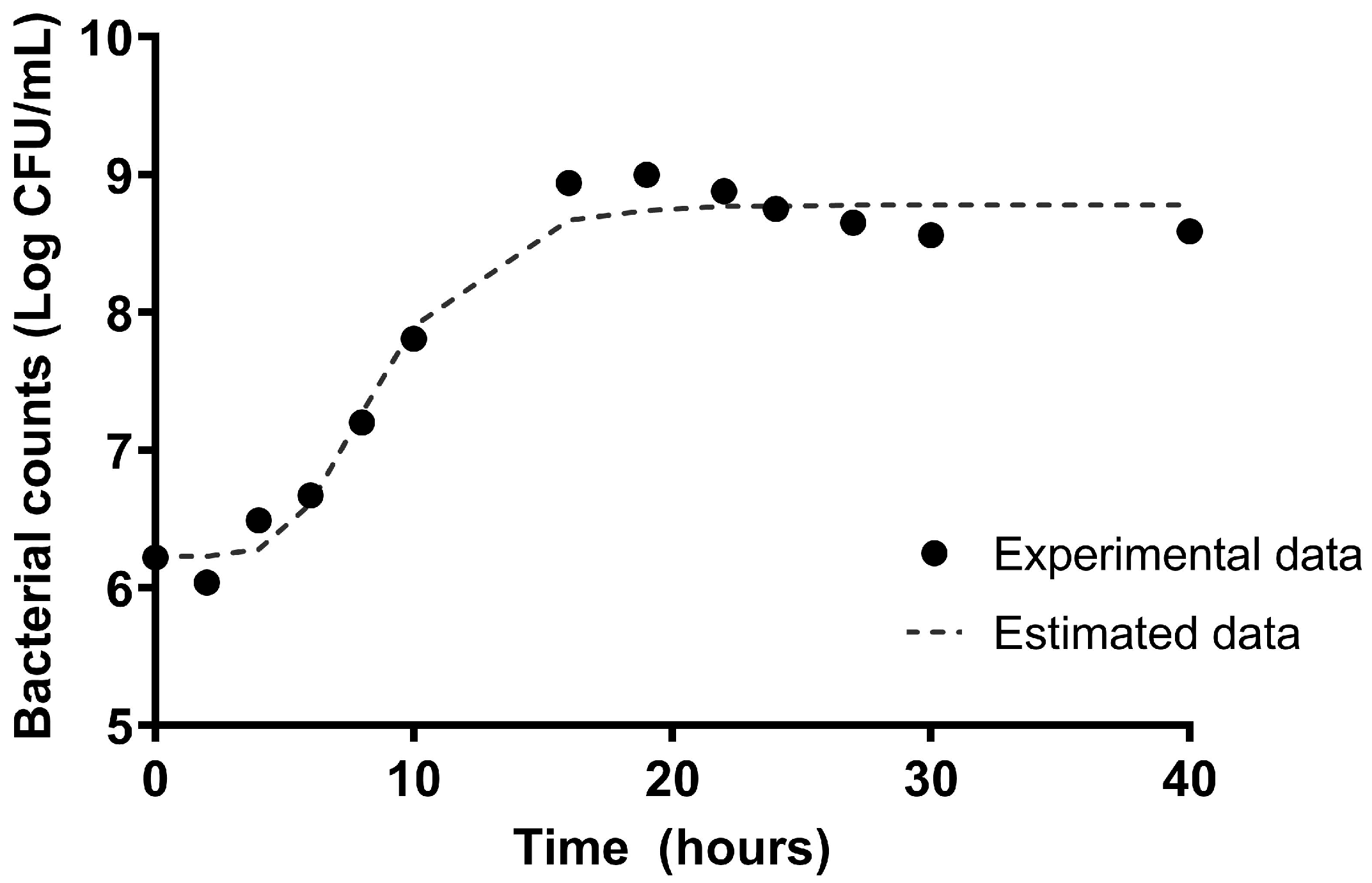
Figure 1. Growth curve of L. plantarum CNTA 628 through the incubation time at 37 °C in MRS broth. Experimental values and estimation with the Gompertz model.
Applsci 15 08007 g001
Figure 2. Tolerance of L. plantarum CNTA 628 to gastric and intestinal fluids.
Applsci 15 08007 g002
Figure 3. Morphology of Lactobacillus plantarum CNTA 628 colonies in agar plates containing MRS (A) or MRS supplemented with 0.5% sodium salt of taurocholic (B), taurodeoxycholic (C), glycocholic (D) or glycodeoxycholic acids (E).
Applsci 15 08007 g003
Figure 4. L. plantarum CNTA 628 exhibited the property of biofilm formation. The data are presented as mean ± SD. Three independent experiments were performed, with three replicates per assay.
Applsci 15 08007 g004
Figure 5. Results of adhesion of E. coli to Caco-2 cells individually, in competition with L. plantarum, exclusion displacement (E. coli/L. plantarum). Data represented as mean ± standard deviation. Total of 4 independent assays with 3 wells per assay was performed. * indicates means that are significantly different (p < 0.05) from the control, data for adhesion of E. coli alone.
Applsci 15 08007 g005
Figure 6. Cell viability in neutral red assays of RAW 264.7 (A) and HT-29 (B) after exposure to L. plantarum in untreated (black) or LPS-stimulated (gray) cells. (C) Quantification of IL-6 production in RAW 264.7 cells. (D) Quantification of TNFα production in RAW 264.7 cells. (E) Quantification of IL-10 production in RAW 264.7 cells. (F) Quantification of IL-8 production in HT-29 cells. Results are expressed as mean normalized to negative control ± SD. Three independent experiments with three technical replicates each were performed. ANOVA with Dunnet’s comparisons, * p ≤ 0.05, ** p ≤ 0.01, *** p ≤ 0.001.
Applsci 15 08007 g006
Figure 7. L. plantarum enhances health indicators and decreases lipid accumulation in C. elegans. (A) A fluorescence microscope analysis of worms stained with Nile red in NGM media, supplemented with three different dosages of the probiotic (D1: 3 × 10⁷ UFCs: D2: 3 × 10⁶ UFCs, D3: 3 × 10⁵ UFCs (mL) vs. a control group). (B) Nile red of L. plantarum (D3)-treated and untreated worms in NGM and glucose-loaded (10 mM) NGM. (C,D) L. plantarum (3 × 10⁵ UFCs/mL) increases the lifespan of C. elegans in NGM (C) or NGMg (D) media. (E) Lipofuscin (aging pigment) accumulation on L. plantarum-treated worms (3 × 10⁵ UFCs/mL), relative to NGM controls, grown in NGMg plates. (F) The quantification of ROS production was performed by measuring the average fluorescence emitted by the ROS-binding dye DHE in L. plantarum-treated worms (3 × 10⁵ UFCs/mL) against the NGM media control group in NGMg plates. The results are repressed as the mean ± standard deviation relative to the control group. Significance refers to the effect of L. plantarum with respect to control worms in NGM or in a glucose-loaded (10 mM) NGM (* p < 0.05; *** p < 0.001).
Applsci 15 08007 g007
Figure 8. Gene expression levels of lipid metabolism-related genes in normal and high-glucose conditions. (A) Lipid biosynthesis-related genes (B) Lipid catabolism-related genes. The results are expressed as the fold difference expression levels of each gene in L. plantarum 628-treated worms (3 × 10⁵ UFCs/mL) compared to the control, calculated with the 2−∆∆Ct method. ANOVA 2 × 2 (* p < 0.05; ** p < 0.01) followed by Tukey’s multiple comparison test were used when an interaction among effects (probiotic/glucose) was found (# p < 0.05; ## p < 0.01; ### p < 0.001; * p < 0.05; ** p < 0.01).
Applsci 15 08007 g008
Table 1. Antibiotic resistance profile of L. plantarum CNTA 628.
Antibiotic MIC Values (mg/L)
GmKmSmTcClCmAmEm
L. plantarum CNTA 6280.52–16-320.063–12–40.5–10.032–0.5
Gm, gentamycin (MIC cut-off: ≤16); Km, kanamycin (MIC cut-off: ≤64); Sm, streptomycin (MIC cut-off: ≤64); Tc, tetracycline (MIC cut-off: ≤32); Cl, clindamycin (MIC cut-off: ≤1); Cm, chloramphenicol (MIC cut-off: ≤4); Am, ampicillin (MIC cut-off: ≤4); Em, erythromycin (MIC cut-off: ≤1).
Table 2. Measurements of SCFA production (µg/mL) by L. plantarum CNTA 628.
MediumAcetic Acid
Defined medium without adding carbon source523.15 ± 54.57
Defined medium with glucose (2%)not detected
Defined medium with Synergy 1 (2%)500.50 ± 54.11
Defined medium with P95 (2%)866.26 ± 62.21
Disclaimer/Publisher’s Note: The statements, opinions and data contained in all publications are solely those of the individual author(s) and contributor(s) and not of MDPI and/or the editor(s). MDPI and/or the editor(s) disclaim responsibility for any injury to people or property resulting from any ideas, methods, instructions or products referred to in the content.