Article

Green Macroalgae Biomass Upcycling as a Sustainable Resource for Value-Added Applications

by
Ana Terra de Medeiros Felipe
1,
Alliny Samara Lopes de Lima
1,
Emanuelle Maria de Oliveira Paiva
1,
Roberto Bruno Lucena da Cunha
2,
Addison Ribeiro de Almeida
2,
Francisco Ayrton Senna Domingos Pinheiro
2,
Leandro De Santis Ferreira
2,
Marcia Regina da Silva Pedrini
1,
Katia Nicolau Matsui
1 and
Roberta Targino Hoskin
1,*
1
Graduate Program in Chemical Engineering, Federal University of Rio Grande do Norte (UFRN), Natal 59078-970, RN, Brazil
2
Department of Pharmacy, Federal University of Rio Grande do Norte (UFRN), Natal 59078-970, RN, Brazil
*
Author to whom correspondence should be addressed.
Appl. Sci. 2025, 15(14), 7927; https://doi.org/10.3390/app15147927
Submission received: 12 June 2025 / Revised: 7 July 2025 / Accepted: 14 July 2025 / Published: 16 July 2025
(This article belongs to the Special Issue Advanced Food Processing Technologies and Approaches)

Abstract

As the global demand for eco-friendly food ingredients grows, marine macroalgae emerge as a valuable resource for multiple applications using a circular bioeconomy approach. In this study, green macroalgae Ulva flexuosa, naturally accumulated in aquaculture ponds as a residual biomass (by-product) of shrimp and oyster farming, were investigated regarding their bioactivity, chemical composition, and antioxidant properties. The use of aquaculture by-products as raw materials not only reduces waste accumulation but also makes better use of natural resources and adds value to underutilized biomass, contributing to sustainable production systems. For this, a comprehensive approach including the evaluation of its composition and environmentally friendly extraction of bioactive compounds was conducted and discussed. Green macroalgae exhibited high fiber (37.63% dry weight, DW) and mineral (30.45% DW) contents. Among the identified compounds, palmitic acid and linoleic acid (ω-6) were identified in the highest concentrations. Pigment analysis revealed a high concentration of chlorophylls (73.95 mg/g) and carotenoids (17.75 mg/g). To evaluate the bioactivity of Ulva flexuosa, ultrasound-assisted solid–liquid extraction was performed using water, ethanol, and methanol. Methanolic extracts showed the highest flavonoid content (59.33 mg QE/100 g), while aqueous extracts had the highest total phenolic content (41.50 mg GAE/100 g). Ethanolic and methanolic extracts had the most potent DPPH scavenging activity, whereas aqueous and ethanolic extracts performed best at the ABTS assay. Overall, we show the upcycling of Ulva flexuosa, an underexplored aquaculture by-product, as a sustainable and sensible strategy for multiple value-added applications.

1. Introduction

Macroalgae are a key component in developing innovative and eco-friendly solutions to global food challenges. Compared to terrestrial plants, macroalgae cultivation has a relatively lower environmental impact, requiring minimal land for their growth and significantly lower amounts of water [1]. Moreover, algae are multitrophic systems, contributing to bioremediation activities [1,2], and in some cases, cultivation may not be required, as they can naturally occur as secondary biomass streams. Additionally, the extraction process is less impactful [2]. Life cycle assessment (LCA) studies have shown that macroalgae cultivation emits fewer greenhouse gases, consumes less freshwater, and results in lower eutrophication potential compared to conventional agricultural crops [3,4,5,6].
Marine macroalgae are a promising reservoir of bioactive metabolites [7,8]. Their nutrient-dense composition, including dietary fibers, minerals, essential fatty acids, and compounds with antioxidant properties [9,10,11,12], makes them valuable candidates for the development of functional foods and supplements [13,14,15]. Examples of algal-based products include seaweed-enriched pasta and fortified vegetable creams. Additionally, hydrocolloids extracted from algae are widely used in the food industry as thickening and stabilizing agents. Macroalgae have also been incorporated into infant formulas and specialized clinical diets due to their nutritional and functional properties [16,17,18]. However, their biochemical composition is highly diverse and dynamic, influenced by species diversity, seasonality, temperature, and geographic location. These factors significantly affect their nutritional and functional properties, particularly the concentration of minerals, pigments, and polysaccharides [19].
Recent studies have highlighted the potential applications of unique marine algae bioactive compounds [1,20]. Among these, chlorophylls and carotenoids from green algae have drawn attention due to their antioxidant capacity, anti-inflammatory, and health-relevant properties, making them valuable resources for food applications and natural pigment markets [21,22,23,24]. To enhance the extraction of compounds of interest, emerging extraction technologies have been developed to optimize recovery, thus enabling the use of these resources to develop innovative functional foods and value-added products [25]. Ultrasound-assisted extraction (UAE) has proven to be an effective technique, as it uses cavitation to optimize the extraction of polyphenols and other phytochemicals. UAE offers several advantages over conventional techniques, including reduced extraction time, lower solvent usage, and enhanced mass transfer, resulting in improved yields of bioactive compounds [26,27]. However, the process needs to be carefully designed to guarantee efficiency and avoid degradation of sensitive molecules [26,28]. Additionally, the choice of solvent plays a critical role in the performance of extraction by selectively targeting compounds based on their polarity, thereby determining the efficiency, selectivity, and yield of the extraction process [1].
Despite the increasing global interest in macroalgae, studies on species from Northeast Brazil remain limited [29,30,31]. Understanding the biochemical profile of locally grown Ulva flexuosa, particularly when sourced as aquaculture by-products, is essential for optimizing its utilization, reducing biomass waste, and ensuring resource sustainability within a circular bioeconomy framework. Therefore, this study aimed to identify and characterize the Ulva flexuosa (Chlorophyta) biomass collected from aquaculture ponds in Northeastern Brazil. The chemical composition analysis, fatty acid profiling, and pigment quantification were conducted and discussed. Additionally, ultrasound-assisted solid–liquid extraction was evaluated using different solvents to assess the bioactive compound content and antioxidant properties of Ulva flexuosa extracts. These findings contribute to the exploration of sustainable macroalgae-derived resources, reinforcing their role in the bioeconomy and future industrial applications.

2. Materials and Methods

2.1. Materials and Reagents

The gallic acid was purchased from Êxodo Scientific® (West Palm Beach, FL, USA). The Folin–Ciocalteu reagent, 2,2′-azino-bis (3-ethylbenzothiazoline-6-sulfonic acid) (ABTS), (±)-6-hydroxy-2,5,7,8-tetramethylchroman-2-carboxylic acid (Trolox), 2,2-diphenyl-1-picrylhydrazyl (DPPH), and the phenolic compound standard quercetin were all purchased from Sigma-Aldrich (São Paulo, Brazil). All other reagents used were of analytical grade and without further purification.

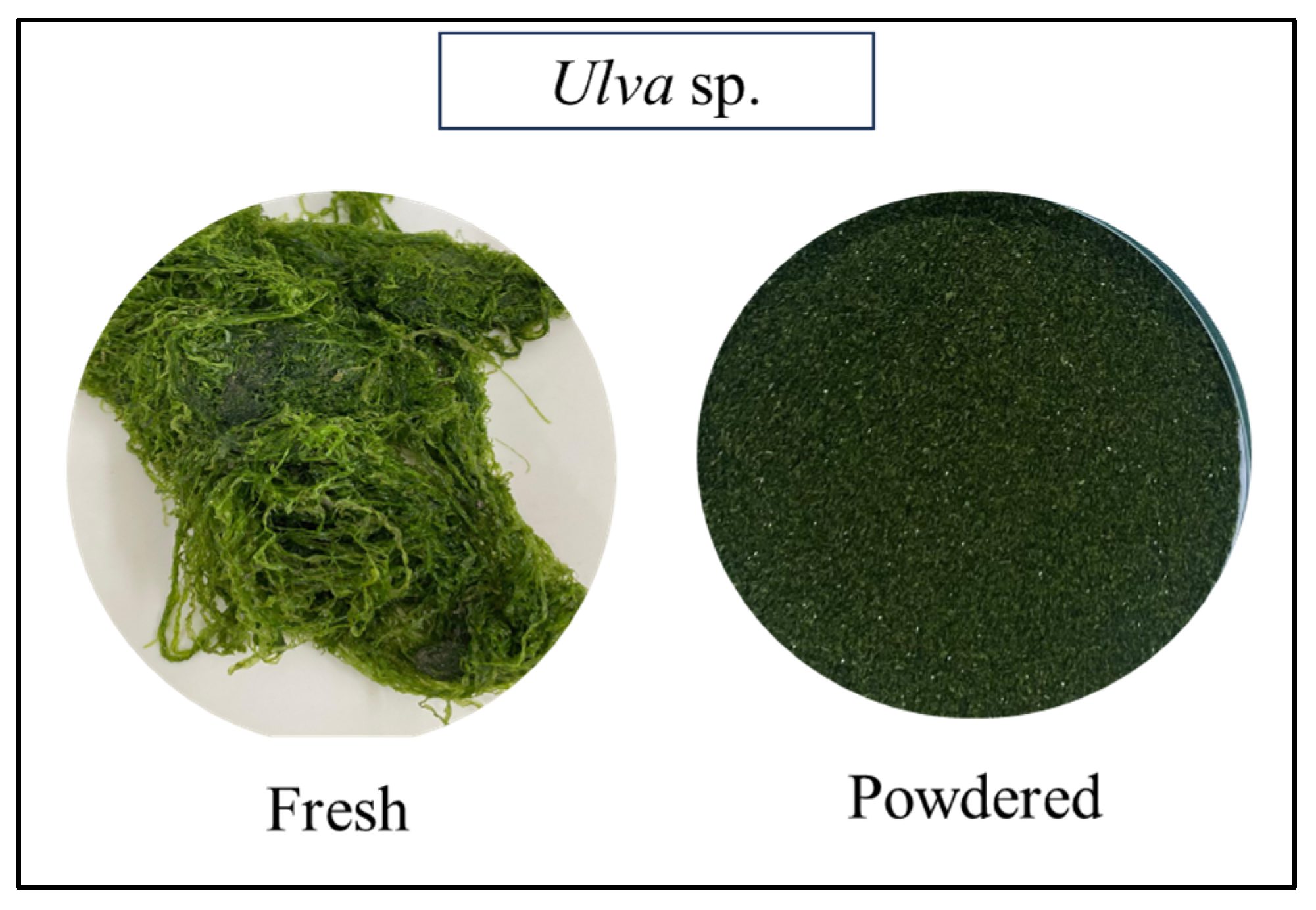
2.2. Collection of Green Macroalgae Ulva sp.

The green algae were sourced by Primar Aquacultura, an organic shrimp and oyster farm located in Tibau do Sul, RN, Brazil (Latitude: 6°13′30″ S, Longitude: 35°08′20″ W). The algae were collected between October 2022 and January 2023, before recapping the culture tanks after an average cultivation cycle of 55 to 60 days. This study used green macroalgae that naturally proliferate in aquaculture ponds as a secondary biomass stream (by-product) during the cultivation of shrimp and oysters. The ponds had an average salinity of 33 ppt in October and 28 ppt in January and an average temperature of 27.5 °C in October and 29 °C in January. The green algae were identified to the species level as Ulva flexuosa (Department of Oceanography and Limnology, Federal University of Rio Grande do Norte-UFRN, Brazil). The green macroalgae collected in this study were taxonomically identified based on morphological characteristics visible to the naked eye and under a stereomicroscope equipped with an ocular micrometer. Diagnostic features such as thallus shape, blade margins, cell arrangement, and rhizoidal structures were evaluated. Identification was conducted using specialized taxonomic keys for green macroalgae. Specifically, the samples were identified as Ulva flexuosa using the dichotomous key provided by Barata [32]. Figure 1 shows the geographic map where the green macroalgae were collected.
Collected algae samples were first rinsed with tap water to remove epiphytes, sand, salt, impurities, and solid particles. Then, they were washed with distilled water, drained, and dried in a forced-air circulation oven (Lucadema, model Luca-B2/221, Sao Jose do Rio Preto, SP, Brazil) at 40 °C for 48 h following [33,34] with modifications. After that, the material was ground in an electric blender (Philips Walita, model RI7625, Varginha, MG, Brazil) and sieved into a fine powder. The resulting macroalgae powder was stored in polyethylene bags at −18 °C, protected from light, until further analysis. Fresh and powdered macroalgae are shown in Figure 2.

2.3. Physicochemical Analysis

2.3.1. Chemical Composition

The chemical composition of oven-dried Ulva sp. powder (UODP) including moisture, crude protein, crude fiber, ether extract and total ash was determined in accordance to the Association of Official Analytical Chemists (AOAC) official methods: moisture (925.10), crude protein by Kjeldahl (978.04), crude fiber (962.09), ether extract (920.39), and total ash (942.05) [35].
Moisture content was assessed by drying approximately 5 g of sample in a lab oven at 105 °C until constant weight. The crude protein content was determined by the Kjeldahl method (acid and alkaline digestion, followed by filtration and drying), using a nitrogen-to-protein conversion factor of 6.25. Ether extract (crude fat) was quantified using Soxhlet extraction with petroleum ether as the solvent. Total ash content was determined by incinerating the sample in a muffle furnace at 550 °C for 6 h. All results were expressed as percentages on a dry weight basis [35].

2.3.2. Fatty Acids Profile

The macroalgae analysis was carried out to evaluate the fatty acid content after the esterification reaction using Gas Chromatography–Mass Spectrometry (GC-MS) (Agilent, model GC 8860, Santa Clara, CA, USA). To prepare the extract, 40 mg of UODP was transferred to a test tube and subjected to esterification. Esterification started with adding 2.5 mL of NaOH solution (0.5 N) in methanol, followed by heating in a water bath at 70 °C for 15 min. After cooling, 7.5 mL of the esterification reagent (4% HCl solution in methanol) was added, and the solution was returned to the water bath at 70 °C for 10 min. Subsequently, the samples were extracted with 2 mL of heptane and a 20% saturated NaCl solution. The apolar phase containing methyl ester compounds was analyzed by GC-MS.
The GC-MS analysis was performed in splitless mode with an injection volume of 1 µL using the equipment of Agilent (GC model 8860 and MS model 5977B) and the column HP-5MS (30 m × 0.25 mm × 0.25 µm). The temperature gradient began with the oven at 75 °C, and the initial temperature was maintained for 5 min. Then, it increased to a rate of 6 °C/min up to 290 °C and remained constant for 20 min. The injector temperature and transfer line were 250 °C and 280 °C, respectively, and the ion source temperature was 230 °C. The data were acquired in scanning mode (m/z 50–750), and the cutting time of the solvent was fixed at 5 min. The substances were identified according to comparison with the standard mass spectra using the National Institute of Standards and Technology (NIST) 17 collection library and confirmed by Retention Index (RI). A standard linear alkanes (C8-C40) solution was prepared and analyzed under identical GC-MS conditions to calculate the RI of the compounds. Fatty acid measurements by GC-MS were performed in duplicate.

2.4. Pigments Analysis (Chlorophylls and Carotenoids)

Pigment extraction was performed using 1 g of UODP with 50 mL of acetone, kept at 4 °C for 24 h, and then centrifuged (Daiki, DT4500, Osaka, Japan). Absorbance of the supernatants was measured on a spectrophotometer (Biospectro, model SP-22, Curitiba, PR, Brazil) at 452.5, 630, 645, 663, and 664 nm.
The chlorophyll a (chl. a), chlorophyll b (chl. b), total chlorophylls, chlorophyll c1 + c2, and carotenoids were calculated using the Equations (1)–(5) described by Palaniyappan et al. [1].
Chlorophyll a (mg/g) = (12.7 × A663) − (2.69 × A645),
Chlorophyll b (mg/g) = (22.9 × A645) − (4.68 × A663),
Total chlorophyll (mg/g) = (20.2 × A645) + (8.02 × A663),
Chlorophylls c1 + c2 (mg/g) = (24.36 × A630) − (3.73 × A664),
Carotenoids (mg/g) = (4.2 × A452.5) − (0.0264 × chl. a) + (0.426 × chl. b)

2.5. Extraction of Oven-Dried Powder of Ulva sp.

The extracts were prepared following Correia et al. [36] and Kumar et al. [37] with modifications as shown in Figure 3. For performance comparison, three different solvents were used in this study: water, ethanol, and methanol. A w/v ratio of 1:100 g/mL (UODP/solvent) was applied for all experiments. All extraction protocols were conducted using ultrasound-assisted solid–liquid extraction (UAE). Briefly, UAE (Altsonic, Clean 3IA 3 L, North Salem, NY, USA) was conducted for 30 min at 40 kHz and 100 W, followed by continuous magnetic stirring (SP Labor, SP-10209/A, Presidente Prudente, SP, Brazil) at room temperature (25 °C) for 2 h. After that, the material was filtered under vacuum (Solab, model SL 61, Recife, PE, Brazil) using filter paper (Whatman n° 1) and stored under refrigeration (4 °C) until further analyses.

2.6. Polyphenols

2.6.1. Total Phenolic Content (TPC)

It was determined using a modified Folin–Ciocalteu method [38]. The analysis was performed by a mixture of 50 µL of distilled water, 25 µL of sample, and 25 µL of Folin–Ciocalteu reagent with water (1:1 v/v) added with 100 µL of 7.5% (w/v) sodium bicarbonate solution, in a 96-well microplate. The microplates were incubated for 90 min at room temperature, protected from light. The absorbance was measured at 765 nm using a microplate reader (Biochrom Ltd., Asys UVM340, Waterbeach, UK). A standard curve was built using gallic acid (6.5–250 mg/L), and results were expressed as milligrams of gallic acid equivalents per gram (mg GAE/100 g).

2.6.2. Total Flavonoid Content (TFC)

It was analyzed by the spectrophotometric method of Palaniyappan et al. [1]. For this, extracts (0.2 mL) were added to tubes containing 4.8 mL distilled water, followed by the addition of 0.3 mL of 5%sodium nitrite (NaNO2) solution and vigorous vortex homogenization. After 5 min, 0.3 mL of 10%AlCl3 solution was added, followed by 2 mL of 1 M NaOH, and filled with distilled water to 10 mL. A standard curve was built with quercetin, and results were expressed as milligrams of quercetin equivalents per gram (mg QE/100 g). The absorbance was measured at 414 nm in a spectrophotometer (Biospectro, model SP-22, Curitiba, PR, Brazil).

2.7. DPPH Radical Scavenging Activity

The DPPH radical scavenging activity was determined following Bobo-García et al. [39]. A 180 µL aliquot of 80% methanolic DPPH solution (150 µM) was added to 20 µL of the samples in a 96-well microplate. The mixtures were then incubated at room temperature in the dark for 40 min, and the absorbance was measured at 515 nm using a microplate reader (Biochrom Ltd., Asys UVM340, Waterbeach, UK). A control consisting of all reagents except the algae extracts was run in parallel. Results (%) were calculated by Equation (6):
% of DPPH inhibition = ((Ac − As)/Ac) × 100,
where Ac is the absorbance of the blank control, and As is the absorbance of the samples at 515 nm.

2.8. ABTS Radical Scavenging Activity

The ABTS+ radical scavenging capacity was determined following Rufino et al. [40]. For this, an aqueous solution of ABTS (7 mM) was transferred to a 96-well microplate containing potassium persulfate (140 mM) in a ratio of 5.0 mL to 88 µL, respectively. The mixture was incubated in the dark at room temperature for 16 h. Subsequently, the solution was diluted with ethanol until reaching an absorbance of 0.8 at 734 nm. For sample analysis, 280 µL of the ABTS+ solution was added to 20 µL of the sample in a 96-well microplate. After a 6-min reaction in low light conditions, the absorbance was measured at 734 nm using a microplate reader (Biochrom Ltd., Asys UVM340, Waterbeach, UK). A standard curve was constructed using varying concentrations of Trolox, and the results are expressed as μmol of Trolox equivalent per gram (μmol TE/g). The absorbance of a blank control (without sample) was used to adjust the initial radical concentration.

2.9. Statistical Analysis

All experiments were conducted in triplicate, unless noted. The results were expressed as mean ± standard deviation (SD). The statistical analysis comparing the means was carried out with software Jamovi 2.6.2 and Statistica 12.0, where the variance method (one-way ANOVA) was applied, followed by Tukey’s post hoc test. The level of statistical significance for differences between means was 5% (p < 0.05).

3. Results and Discussion

3.1. Physicochemical Analysis

3.1.1. Chemical Composition

Understanding the chemical composition of Ulva sp. (Table 1) is essential for evaluating its biological and nutritional properties and identifying specific industrial applications for it [1]. This analysis guides the efficient use of natural resources for food, health, and biotechnology applications [41,42].
Results highlight high crude fiber and total ash concentrations in the dried green macroalgae. The high fiber content reveals an abundance of polysaccharide compounds, while ash content reflects a significant concentration of minerals. Similar findings were reported by Limiñana et al. [43], who observed 40% of insoluble carbohydrates in Ulva sp., while Mandalka et al. [33] showed 22.1% for the green algae Codium isthmocladum collected in Brazil. Moreover, the total ash content of 30% is consistent with previous reports [1,34,44].
Our findings agree with the low fat content previously reported for green algae [44]. Comparable results were shown for the ether extract of Caulerpa racemosa (1.8%) by Palaniyappan et al. [1] and for Ulva sp. (3%) by Limiñana et al. [43]. Indeed, fat content levels, including fatty acid profiles, vary with species, season, geography, and growth conditions [33].
The protein content of UODP (3.39%) is higher than the 2.46% reported by Lourenço et al. [45] for Ulva lactuca (formally Ulva fasciata). However, literature values for protein content in Ulva sp. vary widely, with results reaching up to 47% [34]. Such variations in protein content can be attributed to nitrogen availability at the cultivation site. Studies comparing land-based and sea-based cultivation have reported green algae with protein content ranging from 5% to 20% [33,43], as it can reach up to 47% in red seaweeds [33].
Overall, environmental conditions (water temperature, salinity, depth, and pollution) and processing methods (collection, cleaning, drying, and analytical techniques) play a significant role in the variations in chemical composition of macroalgae species [33,43,45].

3.1.2. Fatty Acid Profile

Five fatty acids were identified with retention times (RT) ranging from 26.60 to 29.75 min and RI from 1920 to 2121 (Table 2). The analysis revealed the presence of four fatty acids in the form of methyl esters: palmitic acid (hexadecanoic acid, C16:0), heptadecanoic acid (C17:0), linoleic acid (9,12-octadecanoic acid, C18:2, also known as omega-6), and stearic acid (C18:0).
The fatty acids identified have significant bioactive potential through bioactive lipid mediators [50,51]. Palmitic acid, a saturated fatty acid, is known for its role in cellular membrane structure and energy metabolism [52]. Heptadecanoic acid has been linked to potential cardioprotective effects and metabolic regulation [53]. Linoleic acid, a polyunsaturated omega-6 fatty acid, is well-documented for its anti-inflammatory properties and ability to improve cardiovascular health by modulating lipid profiles [50,51]. Stearic acid is a saturated fatty acid generally regarded as neutral in its effects on cholesterol levels. Due to its emollient properties, it may also promote skin health [54].
Researchers [55] analyzed the chemical composition of aqueous-ethanolic extracts of Ulva lactuca (as Ulva fasciata) and identified palmitic acid as the main component. A similar profile was observed for UODP, which had a high content of saturated fatty acids, predominantly palmitic acid (C16:0), accounting for 71.62% of the total area. This finding shows that Ulva flexuosa is a potential candidate for biofuels and bioenergy production. The production of biofuels requires substantial quantities of fatty acids, especially C16 and C18, which offer considerable oxidative and thermal stability [56,57]. Taken altogether, it underscores the bioactive potential of Ulva flexuosa, particularly due to its marine lipids, which are rich in long-chain polyunsaturated fatty acids (PUFAs) and monounsaturated fatty acids. This type of lipid is nutritionally important, as humans are unable to endogenously synthesize some of the essential PUFAs, such as omega-6 fatty acids [33,58,59,60].

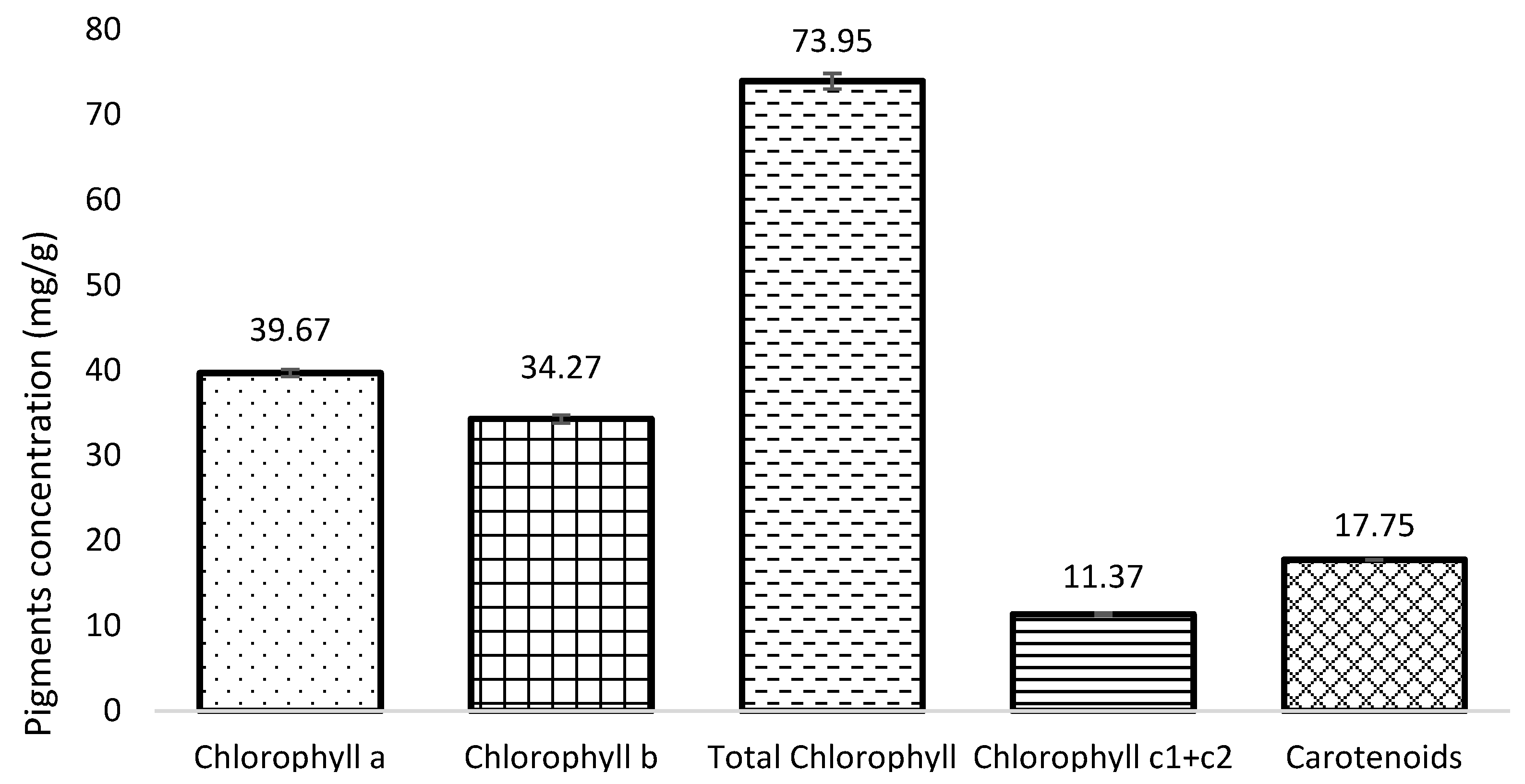
3.2. Pigment Content (Chlorophylls and Carotenoids)

UODP is highly concentrated in chlorophylls and carotenoids (Figure 4). Chlorophyll a had the highest content (39.67 ± 0.436 mg/g), followed by chlorophyll b (34.27 ± 0.487 mg/g) and total carotenoids (17.75 ± 0.038 mg/g). Chlorophyll c1 + c2 was detected in smaller amounts (11.37 ± 0.141 mg/g) and plays an important role in underwater photosynthetic processes. Chlorophyll b acts as an accessory pigment, expanding the range of light absorption. Carotenoids not only assist in photosynthesis but also exhibit significant antioxidant properties, protecting cells from oxidative damage. These compounds contribute to algae’s ability to thrive in dynamic aquatic environments by mitigating stress caused by light and reactive oxygen species [61,62].
Lower concentrations of chlorophyll a (0.92 mg/g) and carotenoids (0.20 mg/g) were reported before for Ulva lactuca [63]. Similarly, Fabrowska et al. [64] observed lower contents of chlorophyll a (6.8 mg/g), chlorophyll b (4.1 mg/g), and total carotenoids (1.3 mg/g) in Ulva flexuosa extracts. The concentrations of pigments obtained from Ulva flexuosa in this study are notably high when compared to values reported in the literature for Ulva species. Differences between studies are justified by environmental conditions, species-specific pigment composition, and different extraction methodologies [63]. These levels are competitive for commercial applications in food and cosmetic industries, where natural pigments are increasingly valued for their functional and visual properties [65,66,67,68].

3.3. Phenolic Compounds

Phenolic acids and flavonoids, though less studied in green algae compared to other phyla, have been identified in various forms such as rutin, quercetin, and catechin derivatives. The production and diversity of these compounds are influenced by stress factors, which enhance defensive responses and compound complexity [69,70].
In this study, significant variation in phenolic and flavonoid content was observed across different solvent extracts (Table 3), reflecting different affinities of solvents based on the polarity and solubility of extracted compounds. Due to its high polarity and hydrogen-bonding capacity, water demonstrated higher efficiency in extracting total phenolics [71], while methanol showed better performance on flavonoid extraction. This finding suggests the presence of non-flavonoid phenolics in aqueous extracts and the potential for flavonoid enrichment using algae extracts obtained with alcoholic solvents [72].
A TPC of 55.61 mg GAE/100 g was reported for methanolic extracts of Ulva lactuca [63]. Similarly, higher results have been shown in the literature, such as by Uribe et al. [73] who found approximately 90 mg GAE/100 g using a methanolic extraction for Ulva species and by [27] who reported 74 mg GAE/100 g in Ulva rigida.
For TFC, methanol was the most effective extractor among the investigated solvents (p < 0.05). This is consistent with Palaniyappan et al. and Yap et al. [1,74], who highlighted methanol’s capacity to extract flavonoids of varying polarities. Likewise, Vinuganesh et al. [75] studied the green seaweed Chaetomorpha sp. and reported lower TFC (approximately 20 mg QE/100 g, fresh weight) in ethanolic extracts.
The variations in TPC and TFC emphasize the influence of solvent polarity, extraction conditions, and matrix effects, and their correlation suggests that phenolic compounds other than flavonoids are present in the aqueous extracts. Additionally, these differences can be attributed to biotic and abiotic factors, such as species-specific and environmental influences on phytochemical profiles [70]. This study demonstrates the importance of selecting appropriate solvents to optimize the extraction of specific bioactive compounds of interest.

3.4. Antioxidant Activity (DPPH and ABTS Assays)

Antioxidant activity in Ulva extracts, as assessed by DPPH and ABTS assays, showed solvent-dependent profiles, which is indicative of different classes of active compounds found in the extracts (Table 4).
Overall, methanolic and ethanolic extracts showed higher radical scavenging activity in DPPH assays, consistent with their superior flavonoid content. Indeed, water-based extraction demonstrated a low antioxidant capacity (1.66 ± 0.001%), while ethanol (16.42 ± 1.52%) and methanol (21.24 ± 2.35%) extracts showed higher activities (Table 4). The similarity between ETA and MET indicates that both solvents effectively extract antioxidants, possibly due to their moderate polarity, which is compatible with phenolic compounds found in the sample.
These findings are consistent with those reported by Palaniyappan et al. [1] for Caulerpa sp. extracts, where methanol showed approximately 25% DPPH inhibition, slightly better than ethanolic extracts. Additionally, Tanna et al. [76] reported DPPH results ranging from 5% to 20% for methanolic (70%) extracts of Ulva sp. at concentrations four times higher than those used in this study, suggesting a lower antioxidant potential. The results also align with Nguyen et al. [77], who observed approximately 10% inhibition of DPPH radical scavenging activity in ethanolic extracts for Caulerpa lentillifera.
In contrast, ABTS activity was more pronounced in ethanolic extracts (9.39 ± 1.57). These significant differences (p < 0.05) suggest solvent-specific efficiency, with ethanol showing broader solubility for antioxidant compounds. Different from our results, Arcos Limiñana et al. [43] reported that ethanolic (70%) extracts of Ulva sp. yielded lower antioxidant activity in the ABTS assay, with approximately 4 μmol TE/g. Similarly, Pangestuti et al. [78] found ABTS values for water extracts of Ulva lactuca ranging from 0.15 to 8.14 (mg ascorbic acid equivalents/g), depending on extraction conditions. These findings align with the 7.00 μmol TE/g reported here for water-based extraction.
The differences in the results obtained by the DPPH and ABTS methods can be attributed to distinct antioxidant-related mechanisms involved in each assay. The DPPH method is based on electron transfer and estimates the ability of an antioxidant to reduce an oxidant that undergoes a color change during the reduction process. In contrast, the ABTS method involves both electron transfer and hydrogen atom transfer [79,80].
For all extracts, ABTS values were higher than in DPPH analysis, showing that the ABTS method detects a broader or different set of compounds compared to the DPPH assay [43]. Overall, the TFC results are associated with the antioxidant activity observed in the DPPH assay, following the descending order of methanol > ethanol > water. This suggests that flavonoids play a major role in the antioxidant activity of the extracts. However, the differing trends between TPC and antioxidant activity indicate that compounds other than phenolics, such as non-phenolic pigments, fatty acids, and polysaccharides, might contribute significantly to the antioxidant effects observed in the extracts [71,81].
Moreover, solubility plays a role in the observed differences between DPPH and ABTS results [82]. For instance, aqueous extract, although showing lower DPPH antioxidant capacity, may contain polar compounds such as polysaccharides, amino acids, and some phenolic acids, which are more soluble in water but may have limited radical scavenging activity [83]. The low antioxidant activities observed in the aqueous extract, despite its high total phenolic content, may be explained by the predominance of water-soluble macromolecules such as polysaccharides. In particular, Ulva species are known to be rich in ulvan, and such compounds may not exhibit strong radical scavenging capacity detectable in DPPH or ABTS assays [27,84].
In contrast, the ethanolic and methanolic extracts, which showed higher antioxidant activity, likely contain a broader range of moderately polar compounds, including flavonoids, tannins, and other phenolic compounds, which are known for their potent radical scavenging properties [85,86]. Ethanol had superior efficiency in the ABTS assay, possibly due to its ability to solubilize a wider spectrum of antioxidant compounds, including those with intermediate polarity, such as certain terpenoids and phytosterols, which may not be as effectively extracted by methanol or water. These findings agree with previous studies indicating that solvent polarity is a key factor in the extraction efficiency of bioactive compounds, directly influencing their antioxidant potential [87,88,89].

4. Conclusions

This study investigated the potential of green macroalgae Ulva flexuosa aquaculture by-product biomass as a sustainable and valuable natural resource. This work contributes to a better understanding of macroalgae biochemical composition and bioactive properties and provides insights for optimizing cultivation and extraction strategies, with particular emphasis on species from Northeast Brazil.
Our findings revealed significant concentrations of chlorophylls and carotenoids in the green macroalgae biomass, as well as their notable antioxidant capacity. When the bioactivity of different Ulva flexuosa extracts was investigated, a clear influence of the type of solvent used was observed. While methanol and ethanol extracts had higher flavonoid concentration and better performance on antioxidant assays, water extracts had higher total phenolic concentration.
Taking altogether, our findings highlight the potential of Ulva flexuosa as a source of essential nutrients, such as minerals, fibers, essential fatty acids, and bioactive compounds for multiple applications. However, one important limitation is that the bioactivity was determined using in vitro assays, which may not fully reflect their effectiveness or safety for in vivo systems. Further research is needed on isolating and characterizing individual compounds and evaluating their potential applications in food, pharmaceutical, and cosmetic formulations. Moreover, the investigation of heavy metals and microbial contaminants is warranted for safety purposes. It is also worth noting that, while ethanol is considered a safe and sustainable solvent for food applications, methanol is not approved for such use due to its toxicity. Therefore, future research should consider the use of food-grade and environmentally friendly solvents. Lastly, future studies should explore sensory acceptance and compliance with food regulations to support the effective incorporation of Ulva flexuosa-based ingredients into products. By advancing knowledge in this field, we will be able to address food challenges and contribute to sustainable marine resource utilization, which aligns with global efforts to develop high-quality alternative products using environmentally friendly practices.

Author Contributions

Conceptualization, A.T.d.M.F., K.N.M., M.R.d.S.P. and R.T.H.; methodology, A.T.d.M.F.; formal analysis, A.T.d.M.F., A.S.L.d.L., E.M.d.O.P., L.D.S.F., R.B.L.d.C., A.R.d.A. and F.A.S.D.P.; data curation, A.T.d.M.F. and A.S.L.d.L.; writing—original draft preparation, A.T.d.M.F.; writing—review and editing, A.T.d.M.F., K.N.M. and R.T.H.; supervision, M.R.d.S.P., K.N.M. and R.T.H. All authors have read and agreed to the published version of the manuscript.

Funding

This study was financed in part by the Coordenação de Aperfeiçoamento de Pessoal de Nível Superior—Brasil (CAPES, Brazil)—Finance Code 001 (process 88887.912319/2023-00).

Institutional Review Board Statement

Not applicable.

Informed Consent Statement

Not applicable.

Data Availability Statement

The raw data supporting the conclusions of this article will be made available by the authors on request.

Acknowledgments

The authors are grateful to UFRN and LEA (Food Engineering Laboratory) for supporting this research. The authors would like to thank the company PRIMAR AQUACULTURE for the kind donation of the green algae biomass used in this study.

Conflicts of Interest

The authors declare no conflicts of interest.

Abbreviations

The following abbreviations are used in this manuscript:
MICMinimum Inhibitory Concentration
ABTS2,2′-azino-bis(3-ethylbenzothiazoline-6-sulfonic acid)
DPPH2,2-diphenyl-1-picrylhydrazyl
PBSPhosphate-Buffered Saline
DOLDepartment of Oceanography and Limnology
UFRNFederal University of Rio Grande do Norte
UODPUlva sp. Oven-Dried Powder
AOACAssociation of Official Analytical Chemists
GC-MSGas Chromatography–Mass Spectrometry
NISTNational Institute of Standards and Technology
RIRetention Index
TPCTotal Phenolic Content
GAEGallic acid Equivalents
TFCTotal Flavonoids Content
QEQuercetin Equivalents
TETrolox Equivalents
CFUColony-Forming Units
SDStandard Deviation
DWDry Weight
RTRetention Time
PUFAPolyunsaturated Fatty Acids
METMethanolic Extract
ETAEthanolic Extract
WATAqueous Extract
NDNot Detected
CAPESCoordination for Personal Improvement of Higher Education
LEAFood Engineering Laboratory

References

  1. Palaniyappan, S.; Sridhar, A.; Kari, Z.A.; Téllez-Isaías, G.; Ramasamy, T. Evaluation of Phytochemical Screening, Pigment Content, In Vitro Antioxidant, Antibacterial Potential and GC-MS Metabolite Profiling of Green Seaweed Caulerpa racemosa. Mar. Drugs 2023, 21, 278. [Google Scholar] [CrossRef] [PubMed]
  2. Zhou, Y.; Yang, H.; Hu, H.; Liu, Y.; Mao, Y.; Zhou, H.; Xu, X.; Zhang, F. Bioremediation potential of the macroalga Gracilaria lemaneiformis (Rhodophyta) integrated into fed fish culture in coastal waters of north China. Aquaculture 2006, 252, 264–276. [Google Scholar] [CrossRef]
  3. Aitken, D.; Bulboa, C.; Godoy-Faundez, A.; Turrion-Gomez, J.L.; Antizar-Ladislao, B. Life cycle assessment of macroalgae cultivation and processing for biofuel production. J. Clean. Prod. 2014, 75, 45–56. [Google Scholar] [CrossRef]
  4. Pantis, A.; Nikoloudakis, C.; Tsoutsos, T. A Critical Review of Macroalgae Exploitation Pathways Implemented under the Scope of Life Cycle Assessment. ChemEngineering 2024, 8, 74. [Google Scholar] [CrossRef]
  5. Parsons, S.; Allen, M.J.; Abeln, F.; McManus, M.; Chuck, C.J. Sustainability and life cycle assessment (LCA) of macroalgae-derived single cell oils. J. Clean. Prod. 2019, 232, 1272–1281. [Google Scholar] [CrossRef]
  6. Hashemi, M.; Mirmohamadsadeghi, S.; Khoshnevisan, B.; Galán-Martín, Á.; Denayer, J.F.M.; Karimi, K. Life cycle assessment of bioenergy and value-added biochemical production from Nizimudinia zanardini brown macroalgae. Sci. Total Environ. 2025, 976, 179225. [Google Scholar] [CrossRef] [PubMed]
  7. Carpena, M.; Pereira, C.S.G.P.; Silva, A.; Barciela, P.; Jorge, A.O.S.; Perez-Vazquez, A.; Pereira, A.G.; Barreira, J.C.M.; Oliveira, M.B.P.P.; Prieto, M.A. Metabolite Profiling of Macroalgae: Biosynthesis and Beneficial Biological Properties of Active Compounds. Mar. Drugs 2024, 22, 478. [Google Scholar] [CrossRef] [PubMed]
  8. Narayanankutty, A.; Famurewa, A.C.; Oprea, E. Natural Bioactive Compounds and Human Health. Molecules 2024, 29, 3372. [Google Scholar] [CrossRef] [PubMed]
  9. Minicante, S.A.; Bongiorni, L.; De Lazzari, A. Bio-Based Products from Mediterranean Seaweeds: Italian Opportunities and Challenges for a Sustainable Blue Economy. Sustainability 2022, 14, 5634. [Google Scholar] [CrossRef]
  10. Francezon, N.; Tremblay, A.; Mouget, J.-L.; Pasetto, P.; Beaulieu, L. Algae as a Source of Natural Flavors in Innovative Foods. J. Agric. Food Chem. 2021, 69, 11753–11772. [Google Scholar] [CrossRef] [PubMed]
  11. Lopes, D.; Rey, F.; Leal, M.C.; Lillebø, A.I.; Calado, R.; Domingues, M.R. Bioactivities of Lipid Extracts and Complex Lipids from Seaweeds: Current Knowledge and Future Prospects. Mar. Drugs 2021, 19, 686. [Google Scholar] [CrossRef] [PubMed]
  12. Mouritsen, O.G.; Rhatigan, P.; Pérez-Lloréns, J.L. World cuisine of seaweeds: Science meets gastronomy. Int. J. Gastron. Food Sci. 2018, 14, 55–65. [Google Scholar] [CrossRef]
  13. El-Shafei, R.; Hegazy, H.; Acharya, B. A Review of Antiviral and Antioxidant Activity of Bioactive Metabolite of Macroalgae within an Optimized Extraction Method. Energies 2021, 14, 3092. [Google Scholar] [CrossRef]
  14. Yordi, E.G.; Martínez, A.P.; Radice, M.; Scalvenzi, L.; Abreu-Naranjo, R.; Uriarte, E.; Santana, L.; Matos, M.J. Seaweeds as Source of Bioactive Pigments with Neuroprotective and/or Anti-Neurodegenerative Activities: Astaxanthin and Fucoxanthin. Mar. Drugs 2024, 22, 327. [Google Scholar] [CrossRef] [PubMed]
  15. Qiu, Y.; Chen, Z.; Zhu, Y.; Wen, J.; Wen, Y.; Liu, Y.; Chen, W.; Zhao, C. Green algal polysaccharides and derivatives as potential therapeutics for metabolic diseases. Food Biosci. 2024, 62, 105310. [Google Scholar] [CrossRef]
  16. Boukid, F.; Castellari, M. Algae as Nutritional and Functional Food Sources. Foods 2023, 12, 122. [Google Scholar] [CrossRef] [PubMed]
  17. Wells, M.L.; Potin, P.; Craigie, J.S.; Raven, J.A.; Merchant, S.S.; Helliwell, K.E.; Smith, A.G.; Camire, M.E.; Brawley, S.H. Algae as nutritional and functional food sources: Revisiting our understanding. J. Appl. Phycol. 2017, 29, 949–982. [Google Scholar] [CrossRef] [PubMed]
  18. Devaprakash, M.; Thirumalaivasan, R.; Sivakumar, N.; Shyam Kumar, R.; Ponmurugan, K. Nutraceuticals and Functional Foods from Algae: Formulation and Health Benefits. In Value Added Products from Bioalgae Based Biorefineries: Opportunities and Challenges; Arya, S.K., Khatri, M., Singh, G., Eds.; Springer Nature: Singapore, 2024; pp. 289–341. ISBN 978-981-97-1662-3. [Google Scholar]
  19. El-Beltagi, H.S.; Mohamed, A.A.; Mohamed, H.I.; Ramadan, K.M.A.; Barqawi, A.A.; Mansour, A.T. Phytochemical and Potential Properties of Seaweeds and Their Recent Applications: A Review. Mar. Drugs 2022, 20, 342. [Google Scholar] [CrossRef] [PubMed]
  20. Mutavski, Z.; Jerković, I.; Nikolić, N.Ć.; Radman, S.; Flanjak, I.; Aladić, K.; Šubarić, D.; Vulić, J.; Jokić, S. Comprehensive Phytochemical Profiling of Ulva lactuca from the Adriatic Sea. Int. J. Mol. Sci. 2024, 25, 11711. [Google Scholar] [CrossRef] [PubMed]
  21. Esim, N.; Dawar, P.; Arslan, N.P.; Orak, T.; Doymus, M.; Azad, F.; Ortucu, S.; Albayrak, S.; Taskin, M. Natural metabolites with antioxidant activity from micro-and macro-algae. Food Biosci. 2024, 62, 105089. [Google Scholar] [CrossRef]
  22. Vinosha, M.; Palanisamy, S.; Jeneeta, S.; Rajasekar, P.; Marudhupandi, T.; Karthikeyan, M.; Mohandoss, S.; You, S.; Prabhu, N.M. Antibacterial, anticancer and antioxidant effects of sulfated galactan from Halymenia dilatata: In vitro and in vivo analysis. Food Biosci. 2024, 60, 104420. [Google Scholar] [CrossRef]
  23. Zammuto, V.; Rizzo, M.G.; Spanò, A.; Spagnuolo, D.; Di Martino, A.; Morabito, M.; Manghisi, A.; Genovese, G.; Guglielmino, S.; Calabrese, G.; et al. Effects of crude polysaccharides from marine macroalgae on the adhesion and biofilm formation of Pseudomonas aeruginosa and Staphylococcus aureus. Algal Res. 2022, 63, 102646. [Google Scholar] [CrossRef]
  24. Patel, A.K.; Albarico, F.P.J.B.; Perumal, P.K.; Vadrale, A.P.; Nian, C.T.; Chau, H.T.B.; Anwar, C.; Wani, H.M.U.D.; Pal, A.; Saini, R.; et al. Algae as an emerging source of bioactive pigments. Bioresour. Technol. 2022, 351, 126910. [Google Scholar] [CrossRef] [PubMed]
  25. Donn, P.; Prieto, M.A.; Mejuto, J.C.; Cao, H.; Simal-Gandara, J. Functional foods based on the recovery of bioactive ingredients from food and algae by-products by emerging extraction technologies and 3D printing. Food Biosci. 2022, 49, 101853. [Google Scholar] [CrossRef]
  26. Monroy-García, I.N.; Torres-Romero, S.; Castro-Ochoa, L.D.; Mendoza-Acosta, A.; Viveros-Valdez, E.; Ayala-Zavala, F. Bioactive Compounds from Marine Macroalgae: A Natural Defense Against Oxidative Stress-Related Diseases. Stresses 2025, 5, 22. [Google Scholar] [CrossRef]
  27. Silva, A.; Silva, S.A.; Carpena, M.; Garcia-Oliveira, P.; Gullón, P.; Barroso, M.F.; Prieto, M.A.; Simal-Gandara, J. Macroalgae as a Source of Valuable Antimicrobial Compounds: Extraction and Applications. Antibiotics 2020, 9, 642. [Google Scholar] [CrossRef] [PubMed]
  28. Golshany, H.; Yu, Q.; Fan, L. Comparative extraction and antioxidant potential of bioactive compounds from Fucus vesiculosus: Kinetic modeling and UPLC-Q-TOF-MS phenolic profiling. Food Biosci. 2024, 57, 103575. [Google Scholar] [CrossRef]
  29. Carvalho, A.; Müller, L.; Rosas, V.; Tesser, M.B.; Ventura-Lima, J.; Turan, G.; Pias, M.; Poersch, L.H. Evaluation of the Inclusion of the Seaweed Ulva lactuca Produced in an Integrated System with Biofloc in the Diet of Juvenile Tilapia Oreochromis niloticus. Appl. Sci. 2025, 15, 6410. [Google Scholar] [CrossRef]
  30. Marques, M.L.M.; Presa, F.B.; Viana, R.L.S.; Costa, M.S.S.P.; Amorim, M.O.R.; Bellan, D.L.; Alves, M.G.C.F.; Costa, L.S.; Trindade, E.S.; Rocha, H.A.O. Anti-Thrombin, Anti-Adhesive, Anti-Migratory, and Anti-Proliferative Activities of Sulfated Galactans from the Tropical Green Seaweed, Udotea flabellum. Mar. Drugs 2019, 17, 5. [Google Scholar] [CrossRef] [PubMed]
  31. Carneiro, V.A.R.; Martins, N.T.; da Silva, S.L.A.; de Barros-Barreto, M.B.; Pereira, S.B.; Cassano, V. Revealing the diversity of the genus Ulva (Ulvales, Chlorophyta) in southeastern Brazil, with a description of Ulva kanagawae sp. nov. Phycologia 2023, 62, 407–420. [Google Scholar] [CrossRef]
  32. Barata, D. Clorofíceas Marinhas Bentônicas do Estado do Espírito Santo. Master’s Thesis, Instituto de Botânica da Secretaria de Estado do Meio Ambiente, São Paulo, Brazil, 2004; 210p. Available online: https://smastr16.blob.core.windows.net/pgibt/2013/09/Diogina_Barata_MS.pdf (accessed on 13 June 2025).
  33. Mandalka, A.; Cavalcanti, M.I.L.G.; Harb, T.B.; Fujii, M.T.; Eisner, P.; Schweiggert-Weisz, U.; Chow, F. Nutritional Composition of Beach-Cast Marine Algae from the Brazilian Coast: Added Value for Algal Biomass Considered as Waste. Foods 2022, 11, 1201. [Google Scholar] [CrossRef] [PubMed]
  34. Pappou, S.; Dardavila, M.M.; Savvidou, M.G.; Louli, V.; Magoulas, K.; Voutsas, E. Extraction of Bioactive Compounds from Ulva lactuca. Appl. Sci. 2022, 12, 2117. [Google Scholar] [CrossRef]
  35. Horwitz, W. Official Methods of Analysis of AOAC International; AOAC International: Gaithersburg, MD, USA, 2000; ISBN 978-0-935584-67-7. [Google Scholar]
  36. Correia, R.; Grace, M.H.; Esposito, D.; Lila, M.A. Wild blueberry polyphenol-protein food ingredients produced by three drying methods: Comparative physico-chemical properties, phytochemical content, and stability during storage. Food Chem. 2017, 235, 76–85. [Google Scholar] [CrossRef] [PubMed]
  37. Kumar, Y.; Singhal, S.; Tarafdar, A.; Pharande, A.; Ganesan, M.; Badgujar, P.C. Ultrasound assisted extraction of selected edible macroalgae: Effect on antioxidant activity and quantitative assessment of polyphenols by liquid chromatography with tandem mass spectrometry (LC-MS/MS). Algal Res. 2020, 52, 102114. [Google Scholar] [CrossRef]
  38. Singleton, V.L.; Orthofer, R.; Lamuela-Raventós, R.M. Analysis of total phenols and other oxidation substrates and antioxidants by means of folin-ciocalteu reagent. In Methods in Enzymology; Elsevier: New York, NY, USA, 1999; Volume 299, pp. 152–178. ISBN 978-0-12-182200-2. [Google Scholar]
  39. Bobo-García, G.; Davidov-Pardo, G.; Arroqui, C.; Vírseda, P.; Marín-Arroyo, M.R.; Navarro, M. Intra-laboratory validation of microplate methods for total phenolic content and antioxidant activity on polyphenolic extracts, and comparison with conventional spectrophotometric methods. J. Sci. Food Agric. 2015, 95, 204–209. [Google Scholar] [CrossRef] [PubMed]
  40. Rufino, M.; Alves, R.; Brito, E.; Morais, S.; Sampaio, C.; Pérez-Jiménez, J. Metodologia Científica: Determinacão da atividade antioxidante total em frutas pela captura do radical ABTS+. In Embrapa Agroindústria Tropical Comunicado Técnico #128; Embrapa: Brasília, Brazil, 2007. [Google Scholar]
  41. Chen, Q.; Pan, X.-D.; Huang, B.-F.; Han, J.-L. Distribution of metals and metalloids in dried seaweeds and health risk to population in southeastern China. Sci. Rep. 2018, 8, 3578. [Google Scholar] [CrossRef] [PubMed]
  42. Xu, J.; Liao, W.; Liu, Y.; Guo, Y.; Jiang, S.; Zhao, C. An overview on the nutritional and bioactive components of green seaweeds. Food Prod. Process. Nutr. 2023, 5, 18. [Google Scholar] [CrossRef] [PubMed]
  43. Limiñana, V.A.; Benoist, T.; Sempere, S.A.; Pérez, S.E.M.; Moya, M.S.P. Chemical composition of sustainable Mediterranean macroalgae obtained from land-based and sea-based aquaculture systems. Food Biosci. 2023, 54, 102902. [Google Scholar] [CrossRef]
  44. Rodrigues, D.; Freitas, A.C.; Pereira, L.; Rocha-Santos, T.A.P.; Vasconcelos, M.W.; Roriz, M.; Rodríguez-Alcalá, L.M.; Gomes, A.M.P.; Duarte, A.C. Chemical composition of red, brown and green macroalgae from Buarcos bay in Central West Coast of Portugal. Food Chem. 2015, 183, 197–207. [Google Scholar] [CrossRef] [PubMed]
  45. Lourenço, S.O.; Barbarino, E.; De-Paula, J.C.; Otávio, L.; Pereira, S.; Marquez, U.M.L. Amino acid composition, protein content and calculation of nitrogen-to-protein conversion factors for 19 tropical seaweeds. Phycol. Res. 2002, 50, 233–241. [Google Scholar] [CrossRef]
  46. Rostad, C.E.; Pereira, W.E. Kovats and lee retention indices determined by gas chromatography/mass spectrometry for organic compounds of environmental interest. J. High Resolut. Chromatogr. 1986, 9, 328–334. [Google Scholar] [CrossRef]
  47. Kotb, S.S.; Ayoub, I.M.; El-Moghazy, S.A.; Singab, A.N.B. Profiling the Lipophilic Fractions of Pithecellobium dulce Bark and Leaves Using GC/MS and Evaluation of Their Antioxidant, Antimicrobial and Cytotoxic Activities. Chem. Biodivers. 2020, 17, e2000048. [Google Scholar] [CrossRef] [PubMed]
  48. De Sousa, R.L.; Silva, S.G.; Costa, J.M.; Da Costa, W.A.; Maia, A.A.B.; De Oliveira, M.S.; De Andrade, E.H.A. Chemical profile of manually extracted andiroba oil (Carapa guianensis Aubl., Meliaceae) from Mamangal community, located in Igarapé-Miri, Pará, Brazil. Sci. Plena 2021, 17, 127201. [Google Scholar] [CrossRef]
  49. Daneshvand, B.; Ara, K.M.; Raofie, F. Comparison of supercritical fluid extraction and ultrasound-assisted extraction of fatty acids from quince (Cydonia oblonga Miller) seed using response surface methodology and central composite design. J. Chromatogr. A 2012, 1252, 1–7. [Google Scholar] [CrossRef] [PubMed]
  50. Simopoulos, A.P. The importance of the omega-6/omega-3 fatty acid ratio in cardiovascular disease and other chronic diseases. Exp. Biol. Med. 2008, 233, 674–688. [Google Scholar] [CrossRef] [PubMed]
  51. Calder, P.C. n-3 polyunsaturated fatty acids, inflammation, and inflammatory diseases. Am. J. Clin. Nutr. 2006, 83, 1505S–1519S. [Google Scholar] [CrossRef] [PubMed]
  52. Riley, T.M.; Sapp, P.A.; Kris-Etherton, P.M.; Petersen, K.S. Effects of saturated fatty acid consumption on lipoprotein (a): A systematic review and meta-analysis of randomized controlled trials. Am. J. Clin. Nutr. 2024, 120, 619–629. [Google Scholar] [CrossRef] [PubMed]
  53. Jenkins, B.; West, J.A.; Koulman, A. A Review of Odd-Chain Fatty Acid Metabolism and the Role of Pentadecanoic Acid (C15:0) and Heptadecanoic Acid (C17:0) in Health and Disease. Molecules 2015, 20, 2425–2444. [Google Scholar] [CrossRef] [PubMed]
  54. Grundy, S.M. Influence of stearic acid on cholesterol metabolism relative to other long-chain fatty acids. Am. J. Clin. Nutr. 1994, 60, 986S–990S. [Google Scholar] [CrossRef] [PubMed]
  55. Rodeiro, I.; Olguín, S.; Santes, R.; Herrera, J.A.; Pérez, C.L.; Mangas, R.; Hernández, Y.; Fernández, G.; Hernández, I.; Hernández-Ojeda, S.; et al. Gas Chromatography-Mass Spectrometry Analysis of Ulva fasciata (Green Seaweed) Extract and Evaluation of Its Cytoprotective and Antigenotoxic Effects. Evid. Based Complement. Alternat. Med. 2015, 2015, 520598. [Google Scholar] [CrossRef] [PubMed]
  56. Caamaño, E.; Loperena, L.; Hinzpeter, I.; Pradel, P.; Gordillo, F.; Corsini, G.; Tello, M.; Lavín, P.; González, A.R. Isolation and molecular characterization of Thraustochytrium strain isolated from Antarctic Peninsula and its biotechnological potential in the production of fatty acids. Braz. J. Microbiol. 2017, 48, 671–679. [Google Scholar] [CrossRef] [PubMed]
  57. Jeong, G.-T.; Park, D.-H. Optimization of lipid extraction from marine green macro-algae as biofuel resources. Korean J. Chem. Eng. 2015, 32, 2463–2467. [Google Scholar] [CrossRef]
  58. Yang, B.; Zhou, Y.; Wu, M.; Li, X.; Mai, K.; Ai, Q. ω-6 Polyunsaturated fatty acids (linoleic acid) activate both autophagy and antioxidation in a synergistic feedback loop via TOR-dependent and TOR-independent signaling pathways. Cell Death Dis. 2020, 11, 607. [Google Scholar] [CrossRef] [PubMed]
  59. Oppedisano, F.; Macrì, R.; Gliozzi, M.; Musolino, V.; Carresi, C.; Maiuolo, J.; Bosco, F.; Nucera, S.; Zito, M.C.; Guarnieri, L.; et al. The Anti-Inflammatory and Antioxidant Properties of n-3 PUFAs: Their Role in Cardiovascular Protection. Biomedicines 2020, 8, 306. [Google Scholar] [CrossRef] [PubMed]
  60. Vázquez, J.A.; Meduíña, A.; Durán, A.I.; Nogueira, M.; Fernández-Compás, A.; Pérez-Martín, R.I.; Rodríguez-Amado, I. Production of Valuable Compounds and Bioactive Metabolites from By-Products of Fish Discards Using Chemical Processing, Enzymatic Hydrolysis, and Bacterial Fermentation. Mar. Drugs 2019, 17, 139. [Google Scholar] [CrossRef] [PubMed]
  61. Kurniawan, R.; Nurkolis, F.; Taslim, N.A.; Subali, D.; Surya, R.; Gunawan, W.B.; Alisaputra, D.; Mayulu, N.; Salindeho, N.; Kim, B. Carotenoids Composition of Green Algae Caulerpa racemosa and Their Antidiabetic, Anti-Obesity, Antioxidant, and Anti-Inflammatory Properties. Molecules 2023, 28, 3267. [Google Scholar] [CrossRef] [PubMed]
  62. Tjandrawinata, R.R.; Nurkolis, F. A Comparative Analysis on Impact of Extraction Methods on Carotenoids Composition, Antioxidants, Antidiabetes, and Antiobesity Properties in Seagrass Enhalus acoroides: In Silico and In Vitro Study. Mar. Drugs 2024, 22, 365. [Google Scholar] [CrossRef] [PubMed]
  63. Nunes, N.; Ferraz, S.; Valente, S.; Barreto, M.C.; Pinheiro de Carvalho, M.A.A. Biochemical composition, nutritional value, and antioxidant properties of seven seaweed species from the Madeira Archipelago. J. Appl. Phycol. 2017, 29, 2427–2437. [Google Scholar] [CrossRef]
  64. Fabrowska, J.; Messyasz, B.; Szyling, J.; Walkowiak, J.; Łęska, B. Isolation of chlorophylls and carotenoids from freshwater algae using different extraction methods. Phycol. Res. 2018, 66, 52–57. [Google Scholar] [CrossRef]
  65. Ahmad, N.; Mounsef, J.R.; Lteif, R. Pigment production by Scenedesmus dimorphus using different low-cost and alternative culture media. J. Chem. Technol. Biotechnol. 2021, 97, 287–294. [Google Scholar] [CrossRef]
  66. Sun, H.; Wang, Y.; He, Y.; Liu, B.; Mou, H.; Chen, F.; Yang, S. Microalgae-Derived Pigments for the Food Industry. Mar. Drugs 2023, 21, 82. [Google Scholar] [CrossRef] [PubMed]
  67. Gateau, H.; Solymosi, K.; Marchand, J.; Schoefs, B. Carotenoids of Microalgae Used in Food Industry and Medicine. Available online: https://core.ac.uk/reader/95356110?utm_source=linkout (accessed on 1 July 2025).
  68. Saddiqa, A.; Faisal, Z.; Akram, N.; Afzaal, M.; Saeed, F.; Ahmed, A.; Almudaihim, A.; Touqeer, M.; Ahmed, F.; Asghar, A.; et al. Algal pigments: Therapeutic potential and food applications. Food Sci. Nutr. 2024, 12, 6956–6969. [Google Scholar] [CrossRef] [PubMed]
  69. Rahman, M.M.; Rahaman, M.S.; Islam, M.R.; Rahman, F.; Mithi, F.M.; Alqahtani, T.; Almikhlafi, M.A.; Alghamdi, S.Q.; Alruwaili, A.S.; Hossain, M.S.; et al. Role of Phenolic Compounds in Human Disease: Current Knowledge and Future Prospects. Molecules 2022, 27, 233. [Google Scholar] [CrossRef] [PubMed]
  70. Lomartire, S.; Cotas, J.; Pacheco, D.; Marques, J.C.; Pereira, L.; Gonçalves, A.M.M. Environmental Impact on Seaweed Phenolic Production and Activity: An Important Step for Compound Exploitation. Mar. Drugs 2021, 19, 245. [Google Scholar] [CrossRef] [PubMed]
  71. Tavakoli, S.; Kharaziha, M.; Nemati, S.; Kalateh, A. Nanocomposite hydrogel based on carrageenan-coated starch/cellulose nanofibers as a hemorrhage control material. Carbohydr. Polym. 2021, 251, 117013. [Google Scholar] [CrossRef] [PubMed]
  72. Nawaz, H.; Shad, M.A.; Rehman, N.; Andaleeb, H.; Ullah, N. Effect of solvent polarity on extraction yield and antioxidant properties of phytochemicals from bean (Phaseolus vulgaris) seeds. Braz. J. Pharm. Sci. 2020, 56, e17129. [Google Scholar] [CrossRef]
  73. Uribe, E.; Vega-Gálvez, A.; García, V.; Pastén, A.; López, J.; Goñi, G. Effect of different drying methods on phytochemical content and amino acid and fatty acid profiles of the green seaweed, Ulva spp. J. Appl. Phycol. 2019, 31, 1967–1979. [Google Scholar] [CrossRef]
  74. Yap, W.-F.; Tay, V.; Tan, S.-H.; Yow, Y.-Y.; Chew, J. Decoding Antioxidant and Antibacterial Potentials of Malaysian Green Seaweeds: Caulerpa racemosa and Caulerpa lentillifera. Antibiotics 2019, 8, 152. [Google Scholar] [CrossRef] [PubMed]
  75. Vinuganesh, A.; Kumar, A.; Korany, S.M.; Alsherif, E.A.; Selim, S.; Prakash, S.; Beemster, G.T.S.; AbdElgawad, H. Seasonal Changes in the Biochemical Constituents of Green Seaweed Chaetomorpha antennina from Covelong, India. Biomolecules 2022, 12, 1475. [Google Scholar] [CrossRef] [PubMed]
  76. Tanna, B.; Choudhary, B.; Mishra, A. Metabolite profiling, antioxidant, scavenging and anti-proliferative activities of selected tropical green seaweeds reveal the nutraceutical potential of Caulerpa spp. Algal Res. 2018, 36, 96–105. [Google Scholar] [CrossRef]
  77. Nguyen, V.T.; Ueng, J.-P.; Tsai, G.-J. Proximate Composition, Total Phenolic Content, and Antioxidant Activity of Seagrape (Caulerpa lentillifera). J. Food Sci. 2011, 76, C950–C958. [Google Scholar] [CrossRef] [PubMed]
  78. Pangestuti, R.; Haq, M.; Rahmadi, P.; Chun, B.-S. Nutritional Value and Biofunctionalities of Two Edible Green Seaweeds (Ulva lactuca and Caulerpa racemosa) from Indonesia by Subcritical Water Hydrolysis. Mar. Drugs 2021, 19, 578. [Google Scholar] [CrossRef] [PubMed]
  79. Prior, R.L. Oxygen radical absorbance capacity (ORAC): New horizons in relating dietary antioxidants/bioactives and health benefits. J. Funct. Foods 2015, 18, 797–810. [Google Scholar] [CrossRef]
  80. Gülçin, İ. Antioxidant activity of food constituents: An overview. Arch. Toxicol. 2012, 86, 345–391. [Google Scholar] [CrossRef] [PubMed]
  81. Nazarudin, M.F.; Yasin, I.S.M.; Mazli, N.A.I.N.; Saadi, A.R.; Azizee, M.H.S.; Nooraini, M.A.; Saad, N.; Ferdous, U.T.; Fakhrulddin, I.M. Preliminary screening of antioxidant and cytotoxic potential of green seaweed, Halimeda opuntia (Linnaeus) Lamouroux. Saudi J. Biol. Sci. 2022, 29, 2698–2705. [Google Scholar] [CrossRef] [PubMed]
  82. Shalaby, E.A.; Shanab, S.M.M. Comparison of DPPH and ABTS assays for determining antioxidant potential of water and methanol extracts of Spirulina platensis. Indian J. Geo-Mar. Sci. 2013, 42, 556–564. [Google Scholar]
  83. Mokrani, A.; Madani, K. Effect of solvent, time and temperature on the extraction of phenolic compounds and antioxidant capacity of peach (Prunus persica L.) fruit. Sep. Purif. Technol. 2016, 162, 68–76. [Google Scholar] [CrossRef]
  84. Qi, H.; Zhao, T.; Zhang, Q.; Li, Z.; Zhao, Z.; Xing, R. Antioxidant activity of different molecular weight sulfated polysaccharides from Ulva pertusa Kjellm (Chlorophyta). J. Appl. Phycol. 2005, 17, 527–534. [Google Scholar] [CrossRef]
  85. Dawood, D.H.; Elmongy, M.S.; Negm, A.; Taher, M.A. Extraction and chemical characterization of novel water-soluble polysaccharides from two palm species and their antioxidant and antitumor activities. Egypt. J. Basic Appl. Sci. 2020, 7, 141–158. [Google Scholar] [CrossRef]
  86. Formagio, A.S.N.; Volobuff, C.R.F.; Santiago, M.; Cardoso, C.A.L.; Vieira, M.D.C.; Pereira, Z.V. Evaluation of Antioxidant Activity, Total Flavonoids, Tannins and Phenolic Compounds in Psychotria Leaf Extracts. Antioxidants 2014, 3, 745–757. [Google Scholar] [CrossRef] [PubMed]
  87. Gulcin, İ. Antioxidants and antioxidant methods: An updated overview. Arch. Toxicol. 2020, 94, 651–715. [Google Scholar] [CrossRef] [PubMed]
  88. Chew, Y.L.; Lim, Y.Y.; Omar, M.; Khoo, K.S. Antioxidant activity of three edible seaweeds from two areas in South East Asia. LWT—Food Sci. Technol. 2008, 41, 1067–1072. [Google Scholar] [CrossRef]
  89. Yuan, Y.V.; Bone, D.E.; Carrington, M.F. Antioxidant activity of dulse (Palmaria palmata) extract evaluated in vitro. Food Chem. 2005, 91, 485–494. [Google Scholar] [CrossRef]
Figure 1. Geographical location of the green macroalgae (Ulva flexuosa) collection site.
Applsci 15 07927 g001
Figure 2. Fresh (left) and oven-dried green algae Ulva flexuosa (UODP, right) used in this study.
Applsci 15 07927 g002
Figure 3. Extraction protocol of oven-dried green algae Ulva flexuosa (UODP), consisting of ultrasound-assisted solid–liquid extraction.
Applsci 15 07927 g003
Figure 4. Detected pigments (chlorophylls and carotenoids) in oven-dried green algae Ulva sp. (UODP).
Applsci 15 07927 g004
Table 1. Chemical composition (%) of oven-dried green algae Ulva sp. (UODP).
CompoundsResults (% DW)
Moisture14.77 ± 0.20
Crude protein3.39 ± 0.31
Crude fiber37.63 ± 6.84
Fat1.74 ± 0.11
Ash30.45 ± 1.19
Values are expressed as mean ± standard deviation. DW: dry weight.
Table 2. Identified fatty acids in oven-dried green algae Ulva sp. (UODP).
PeakCompound NameRT (min)Area (%)RIReference
1Palmitic acid (C16:0) methyl ester 26.6071.6219201926 1
2Heptadecanoic acid (C17:0) methyl ester28.211.2920202026 2
3Undec-10-enoic acid tetradecyl ester 28.851.532061-
4Linoleic acid (C18:2) methyl ester29.3715.5420952096 3
5Stearic acid (C18:0) methyl ester 29.7510.0121212126 4
1 [46]; 2 [47]; 3 [48]; 4 [49]. RT: retention time; RI: retention index.
Table 3. Total phenolic (TPC) and total flavonoid (TFC) contents in aqueous (WAT), ethanolic (ETA), and methanolic extracts (MET) derived from oven-dried green algae Ulva sp. (UODP).
SamplesTPC (mg GAE/100 g)TFC (mg QE/100 g)
WAT41.50 ± 0.06 b1.77 ± 0.002 a
ETA6.14 ± 0.03 a31.50 ± 0.03 b
MET7.83 ± 0.02 a59.33 ± 0.10 c
Values are expressed as mean ± standard deviation. Different letters in the same column indicate significantly different values (p < 0.05). GAE: gallic acid equivalents; QE: quercetin equivalents.
Table 4. Antioxidant activity of aqueous (WAT), ethanolic (ETA), and methanolic (MET) extracts derived from oven-dried green algae Ulva sp. (UODP).
SamplesDPPH (% of Inhibition)ABTS (μmol TE/g)
WAT1.66 ± 0.001 b7.00 ± 1.50 a
ETA16.42 ± 1.52 a9.39 ± 1.57 b
MET21.24 ± 2.35 a3.57 ± 0.73 c
Values are expressed as mean ± standard deviation. Different letters in the same column indicate significantly different values (p < 0.05). DPPH: (2,2-diphenyl-1-picrylhydrazyl) radicals. ABTS: (2,2′-azino-bis 3-ethylbenzothiazoline-6-sulfonic acid); TE: Trolox (6-hydroxy-2,5,7,8-tetramethylchroman-2-carboxylic acid) equivalents.
Disclaimer/Publisher’s Note: The statements, opinions and data contained in all publications are solely those of the individual author(s) and contributor(s) and not of MDPI and/or the editor(s). MDPI and/or the editor(s) disclaim responsibility for any injury to people or property resulting from any ideas, methods, instructions or products referred to in the content.

