Review

Structural Connectivity of the Substantia Nigra: A Comprehensive Review of Diffusion Imaging and Tractography Studies

1
Faculty of Biomedical Engineering, Czech Technical University in Prague, 272 01 Kladno, Czech Republic
2
Department of Neurology and Center of Clinical Neuroscience, First Faculty of Medicine, Charles University and General University Hospital in Prague, 128 21 Prague, Czech Republic
*
Author to whom correspondence should be addressed.
Appl. Sci. 2025, 15(14), 7902; https://doi.org/10.3390/app15147902
Submission received: 22 May 2025 / Revised: 10 July 2025 / Accepted: 11 July 2025 / Published: 15 July 2025
(This article belongs to the Special Issue Brain Functional Connectivity: Prediction, Dynamics, and Modeling)

Abstract

The substantia nigra (SN) has historically been regarded as a pivotal element of the brain’s motor circuits, notably within the context of the nigrostriatal pathway and Parkinson’s disease. However, recent advancements in neuroimaging techniques, particularly tractography, have facilitated the delineation of its anatomical projections. These techniques have revealed the involvement of the SN in a more extensive array of functional networks encompassing cognitive, emotional, and motivational domains. This paper reviews the current knowledge on the structural connectivity of the SN in humans based on diffusion tensor imaging and tractography. It summarizes the main projection pathways, including classical and newly described connections, such as the direct SN pars compacta connections to the thalamus, cortico–neural inputs, and connections to limbic regions and the hippocampus. Furthermore, the text delves into the distinctions between the SN pars compacta and SN pars reticulata subregions, exploring their parcellation based on connectivity. The paper demonstrates that the SN is a functionally diversified nucleus, the implications of which are significant for the understanding of both motor and neuropsychiatric disorders. The present study addresses the paucity of comprehensive treatment in this area and provides a framework for further research on dopaminergic circuits.

1. Introduction

Diffusion tensor imaging (DTI) is a diffusion-weighted magnetic resonance imaging (MRI) technique that quantifies the diffusivity, its direction, and anisotropy of water diffusion in voxels [1,2,3,4,5,6]. DTI can therefore be used for the reconstruction of the primary fiber direction within the white matter [7,8,9,10]. The calculation of parameters such as fractional anisotropy, axial, radial, and mean diffusivity [11,12,13,14] enables further description of microstructural properties of white matter tracts within the voxel, implying their potential damage or reorganization [1,13,15,16,17,18]. Fractional anisotropy (FA) quantifies the degree of directional dependence of water diffusion in biological tissue by measuring the variance of the diffusion tensor’s eigenvalues normalized to its trace [5,12,14,19]. Mean diffusivity (MD) is defined as the arithmetic mean of the diffusion tensor’s eigenvalues, providing an index of the overall magnitude of water diffusion independent of its directional bias [12,14,19].
Building on these principles, tractography may be used to reconstruct entire white matter tracts [2,19,20,21]. In deterministic tractography, the algorithm invariably follows paths of greatest diffusivity creating streamlines, theoretically reconstructing the underlying tracts [3,11,19,20]. Conversely, probabilistic tractography randomly samples the direction according to a distribution function and generates a multitude of possible trajectories [3,8,19,20,21]. However, tractography has limitations. It cannot determine the direction of the connection (afferent or efferent) [12,22,23], it has certain restrictions when it comes to investigating white matter (WM) areas with various fiber orientations and numerous associative fiber tracts [24,25], and can produce both false-positive and false-negative associations, complicating the interpretation of results [26,27].
The substantia nigra (SN) and the ventral tegmental area (VTA) are two key dopaminergic midbrain structures that play a crucial role in basal ganglia (BG) circuits [28,29,30,31,32]. The SN primarily projects to the putamen via the nigrostriatal pathway (connections from the SN pars compacta to the dorsal striatum) [28,33,34,35,36] (see Figure 1A). The VTA, in contrast, has diverse neuronal populations that project to various forebrain regions, including the nucleus accumbens and amygdala [29,37,38,39,40,41]. The SN has traditionally been divided into the dopaminergic part (the pars compacta (SNc)) and the GABAergic part (the pars reticulata (SNr)) [29,31,42,43,44,45]. The SNc projects to the striatum in order to modulate movement, while the SNr receives inputs from the striatum and the subthalamic nucleus, and sends outputs to the thalamus (nucleus ventralis anterior and lateralis) [28,34,46,47] (see Figure 1B). Tractography has facilitated non-invasive mapping of connections [48,49,50,51,52]. Recent studies have confirmed the presence of connections between the SN and various cortical areas, with the prefrontal, motor, and somatosensory cortices being of note [7,34,53]. The present findings suggest that the SN is not merely an isolated component of the basal ganglia but rather is involved in broader brain networks [7,34,36,54,55,56]. This is of crucial importance for understanding the pathophysiology of neurological and psychiatric disorders involving the SN [7,34].
However, despite the growing number of studies focusing on individual SN connections, a comprehensive review that systematically maps individual pathways, their anatomy, functional significance, and clinical implications is lacking. The extant literature is characterized by a preponderance of isolated descriptions of individual connections. The objective of this review is to provide an overview of the current knowledge on SN structural connectivity, with a particular emphasis on its functionally specialized projections. The text synthesizes both conventionally delineated pathways and recently identified connections, encompassing their topography, fiber types, participation in functional circuits, and pertinence to clinical neurology and neuropsychiatry. Furthermore, the differences between the SNc and SNr subregions are considered, alongside their connectivity diversity and the potential for parcellation by functional domains.
The objective of this study is twofold: firstly, to address a significant gap in the existing literature by providing an overview of the subject matter; and secondly, to offer a systematic knowledge of the structural connectivity of the SN and its functional relevance in the human brain.

2. Materials and Methods

This review was designed in accordance with the PRISMA 2020 recommendations for systematic reviews, considering its adapted application to short review studies. The objective of this study was to provide a comprehensive overview of the current state-of-the-art and novel findings concerning the utilization of DTI and tractography for the investigation of SN connectivity in humans. A systematic search for relevant literature was performed in March 2025 in the PubMed, Web of Science, and ScienceDirect databases. Furthermore, the SciSpace tool was utilized, and the outputs were subjected to a rigorous process of validation to ensure their relevance and appropriateness for inclusion. The temporal scope of the search covered articles published up to March 2025, with the aim of including recent developments in the field.
The following were included in the review: studies conducted on human subjects (including healthy volunteers and clinical populations), studies using DTI or tractography methods to investigate SN connectivity, and articles published in any language. Studies performed in animal models, post-mortem studies (ex vivo imaging), articles that did not make a relevant contribution to the topic of connectivity, and abstracts without available full text were excluded from the analysis.
A single reviewer conducted the screening and selection process for the articles. The initial phase of this process involved searching for articles based on keywords (substantia nigra, tractography, connection) and evaluating the titles and abstracts. Following this preliminary assessment, a comprehensive full-text examination was performed.
In the first step, a total of 150 records were identified, from the following databases: PubMed (n = 38), Web of Science (n = 28), ScienceDirect (n = 67) and SciSpace (n = 17). Following the removal of duplicates (30 records), 120 articles were included in the preliminary screening.
Following an evaluation of the titles and abstracts, 75 records were excluded based on the established criteria listed above. The primary reasons for exclusion included a lack of focus on SN, an absence of DTI methods, and overall irrelevance to the subject matter.
A comprehensive full-text screening process was conducted on the remaining 45 articles, resulting in the exclusion of further 13 studies, primarily due to their emphasis on animal models, post-mortem data, or an indirect relevance to the subject matter.
The remaining 32 studies were selected for the final review. In consideration of the nature of the review, it was deemed inappropriate to apply a formal tool for the purpose of assessing the quality of the included studies. The primary objective of this study was to produce a qualitative map of the predominant trends and novel methodological approaches in the domain of SN connectivity, utilizing DTI.

3. Results

3.1. Connection with the Striatum

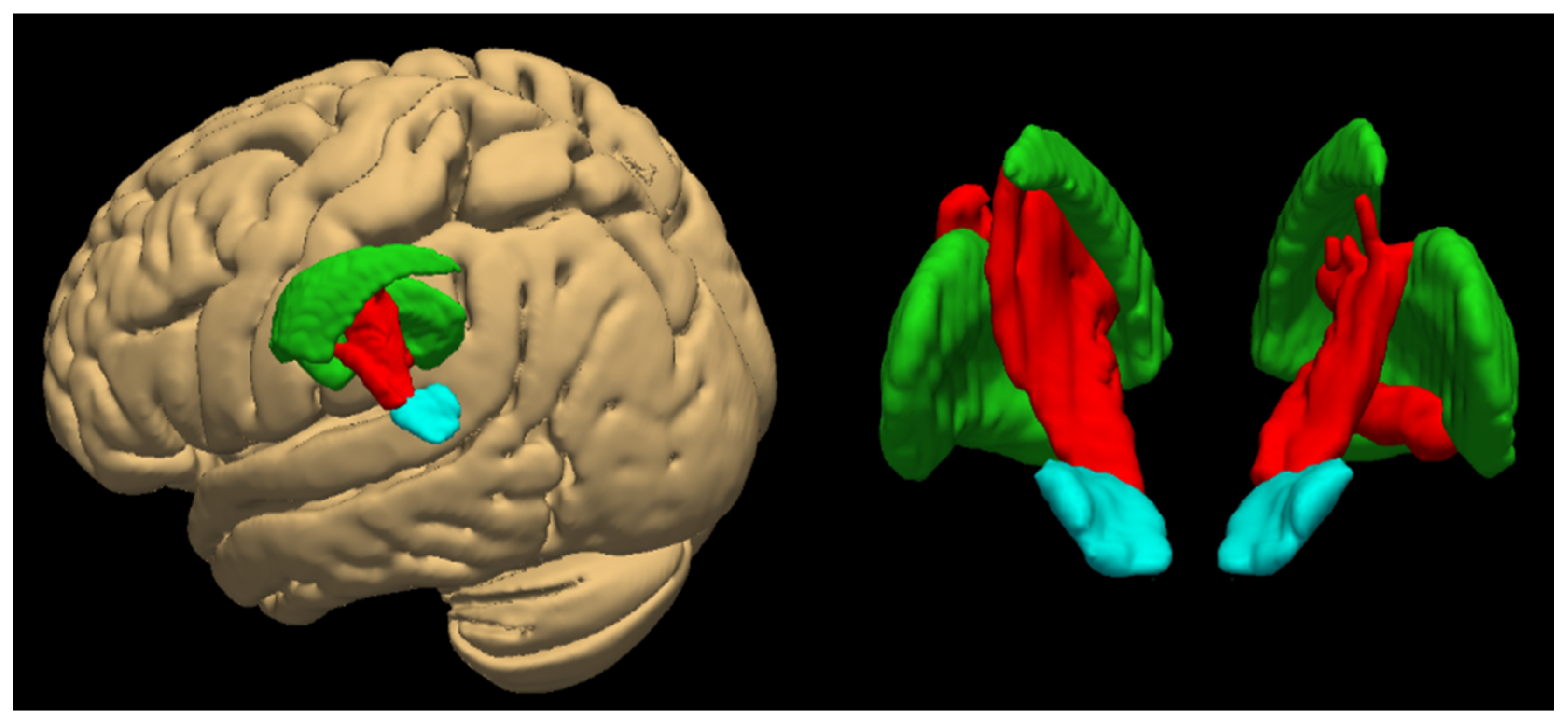
The tractography reliably identifies the classical pathway from the SNc to the striatum [35,57,58,59] (see Figure 2). In a considerable number of studies, nigrostriatal fibers have been repeatedly visualized and exhibited different diffusion properties in both healthy subjects and patients with PD [11,14,60,61].
In the multicenter Parkinson’s Progression Markers Initiative study, 50 patients with PD and 27 controls exhibited systematic abnormalities in the diffusion parameters of the nigrostriatal tract, including reduced fractional anisotropy (FA) and changes in radial/axial diffusivity, indicative of loss of fiber integrity [62]. Furthermore, these changes were found to be associated with the severity of motor symptoms and a decline in striatal dopaminergic marker uptake on nuclear medicine methods, thereby substantiating the functional significance of the pathway damage. Tractography has been demonstrated to quantify the integrity of nigrostriatal connections [62] with the caveat that this is closely related to motor deficits. In a study with 21 PD patients, Tan et al. (2015) utilized deterministic DTI tractography to reveal a substantial decrease in FA and streamline count within the nigrostriatal pathway when compared to a control group [11]. Magnetic resonance imaging alterations are indicative of dopaminergic fiber degeneration. Furthermore, an association was observed between streamline count and motor impairment scores, thereby substantiating the clinical significance of connectivity metrics. Current 7T MRI techniques facilitate more detailed imaging of SNs and their projections. However, the increased resolution of these techniques poses a significant challenge in terms of standardization. A study by Shim and Baek (2022) revealed discrepancies in detected fiber counts between 3T and 7T systems, attributable to variations in acquisition parameters [63].

3.2. Connection with the Thalamus

The main basal ganglia output pathway originates in the SNr and the inner segment of the globus pallidus (GPi) and leads to the thalamus. These pathways are referred to as the nigrothalamic and pallidothalamic pathways, respectively [64]. D.H. Kwon et al. (2021) [64] utilized super-resolution track-density imaging (TDI) on 7T MRI to generate three-dimensional maps of the nigrothalamic and pallidothalamic projections and to identify the SNr substructures in detail. The study demonstrates that SNr projects directly to disparate thalamic nuclei. Two such pathways have been identified: the lateral pathway, which originates in the SN, leading through the reticular nucleus and internal capsule and to the ventral anterior nucleus (VA) and ventral lateral posterior nucleus (VLp); and the medial pathway, which originates in the caudal SNr, traverses the SNc, and terminates at the ventromedial nucleus (VM) of the thalamus. As demonstrated in the latter work, the superior and inferior parietal lobes provide parallel but topographically distinct outputs to the thalamus.
A recent finding revealed the existence of a direct connection between SNc dopaminergic neurons and the thalamus. The conventional model proposed by DeLong (1990) posited that the SNc exerts its influence on the thalamus solely indirectly, through the intermediary of the striatum and subsequent downstream circuits [28]. As demonstrated in the study by Cirillo et al. (2025) [28], the authors conducted a selective monitoring of connections through the SNc, thereby ascertaining that, on average, approximately 12% of all streamlines passing through the SNc proceed directly to the thalamus. This nigrothalamic pathway was reproducibly identified in all young healthy subjects in the study and formed a separate bundle not involving other structures (i.e., it was not a branch to the striatum, cortex, or cerebellum). This constitutes quantitative in vivo evidence of a direct SNc link to the thalamus in humans. This finding extends the established model of basal ganglia circuits, indicating that, in addition to the expected inhibitory pathway from the SNr, there is also modulation of the thalamus by dopamine from the SNc. This represents a significant advancement in our understanding of basal ganglia circuits. The authors illustrated the nigrothalamic system in schematic representation as previously seen in Figure 1B [28].
In addition to the direct connections of SNr and SNc to the thalamus, a recent study using generalized q-sampling imaging (GQI) tractography has allowed the complex involvement of the ansa lenticularis (AL) to be described. Li et al. (2022) [65] reconstructed four subcomponents of the AL, one of which, the globus pallidus–substantia nigra (GPSN), demonstrates a direct anatomical connection between these structures. This connection indicates the presence of additional, more precisely organized circuits that integrate basal ganglia output activity not only to the thalamus but also in the opposite direction towards the SN.

3.3. Connection with the Cortex

Until recently, the relationship between the cerebral cortex and SN was believed to be only indirect, with connections via cortex–striatum–SNr or cortex–subthalamic nucleus–SNr circuits. However, several studies have revealed direct projections from the cortex to the SN, the so-called cortico–nigral pathway.
Cacciola et al. (2016) conducted a study that utilized probabilistic tractography to identify pathways connecting broad regions of the cerebral cortex to the SN [34]. The most prominent connections were observed to be with the prefrontal cortex and with sensorimotor regions, including the gyrus precentralis, postcentralis, and superior parietal lobule. These findings expanded the network of SN connections to include direct cortical connection, a finding that is supported by a study by H.G. Kwon and Jang (2014) [7], which also suggested that the SN has high connectivity with cortical areas. In healthy subjects, the SN demonstrated connections (>70% probability) with virtually all cortical areas tested. The authors observed the highest SN connectivity to areas of the frontal, parietal, and occipital lobes, to the primary motor and somatosensory cortex, and to the cerebellum. The presence of these pathways in humans has contributed to the clarification of certain ambiguities, such as the reasons for the alterations in SN activity during specific cognitive tasks or sensory inputs [34].

3.4. Connection with Limbic Structures

3.4.1. Mesolimbic Projection of the SNc/Ventral Tegmental Area (VTA)

The midbrain dopaminergic system is traditionally divided into the nigrostriatal pathway (SNc → dorsal striatum) and the mesolimbic pathway, which leads from the VTA to limbic structures (ventral striatum, amygdala, prefrontal cortex). However, it appears that the SNc itself, particularly its dorsal region adjacent to the VTA, also contributes to limbic innervation [28]. Consequently, SN involvement in the limbic system is predominantly through projections to the ventral striatum and limbic cortex. The ventral striatum exerts its influence on circuits associated with reward and emotion. As demonstrated by Zhang et al. (2017) [66], the orbitofrontal cortex and the anterior cingulum receive dopaminergic inputs from the SNc and the VTA. In turn, these regions send fibers (directly or indirectly) back to the aforementioned regions, modulating the activity of dopaminergic neurons.

3.4.2. Connection to the Hippocampus

In addition to connections with the striatum and cortex, the SN/VTA also appears to be structurally connected to the hippocampus via specific fibers identified by probabilistic tractography. Carmichael et al. (2021) provided a concise overview of the literature on this subject, concluding that SN/VTA projection neurons contribute only marginally to the formation of the hippocampus [47]. Nonetheless, indirect interactions have been demonstrated. For instance, the hippocampus, via the subiculum, exerts influence on the VTA/SNc, and conversely, dopamine from the midbrain modulates hippocampal function, which is important for novelty and memory consolidation [47]. A study by Elliott et al. (2022) [67] demonstrated that the degree of SN/VTA–hippocampus connectivity exhibited a correlation with performance on a task requiring motivated memory encoding. That is to say, the study revealed that learning was influenced by the expectation of reward or punishment. Conversely, SN/VTA connectivity to the striatum was not significant in this cognitive context. The results of this study indicate that individual dopaminergic mesencephalic projections are not only anatomically distinct, but also exhibit functional specialization, in this case to influence memory processes [67].

3.4.3. Functional Diversity of Limbic Connections

A study by García-Gomar et al. (2022) [68] utilized 7T probabilistic tractography to create a detailed structural brainstem connectome. The authors distinguished two parts of the substantia nigra—SN1 (corresponding to the SNr) and SN2 (SNc)—and described their structural connectivity with the brainstem nuclei, cortex and limbic regions. While the first part (SN1) exhibited predominant connections with motor areas, the second part (SN2) demonstrated direct connectivity with the amygdala, the ventral tegmental area, the periaqueductal grey, the reticular formation, and other nuclei implicated in emotion regulation, stress response, and sleep. The results provide support for the hypothesis that the SNc is not only part of the motor and reward circuits, but also an integral node of broader limbic circuits that may be relevant to mood disorders, anxiety, REM atonia, or attention disorders.

3.4.4. Projection to the Nucleus Accumbens

A significant finding of the research conducted by MacNiven et al. (2020) [69] is the identification of a specific projection from the midbrain (including the SN and VTA) to the NAc that passes through the lateral hypothalamus. This projection corresponds to the classical description of the inferior portion of the medial forebrain bundle. Utilizing diffusion parameters (FA and inverse mean diffusivity (MD)), the authors demonstrated that the diminished structural integrity of this pathway exhibited a robust correlation with heightened impulsivity in healthy subjects. This connectivity was also significantly reduced in patients with stimulant use disorder. Consequently, the results of the latter study demonstrate the pivotal function of SN/VTA–ventral striatum dopaminergic projections in the modulation of motivated behavior and impulsivity. These findings extend the classical concept of the dopaminergic system and confirm the functional diversity of the different subregions of the SN.

3.5. Regional Subdivisions of the Substantia Nigra

The SNc and SNr exhibit marked differences in neurochemistry, connectivity, and function. The SNc contains dopaminergic neurons whose axons form ascending projections (i.e., nigrostriatal and mesolimbic) that modulate the activity of striatal circuits [28]. In contrast, the SNr predominantly consists of GABAergic neurons that function as output cells of the basal ganglia. These neurons are analogous to GPi, in that they send inhibitory signals to the motor and associative nuclei of the thalamus and to the brainstem [64].
Tractography has enabled the differentiation of distinct functional subcomponents of the SN based on their connectivity.
Menke et al. (2010) [70] performed DTI segmentation of the SN and divided it into two parts, corresponding to the SNc and SNr. The inner segment (SNc) demonstrated predominant connections with the dorsal striatum (putamen, caudate), globus pallidus, anterior nuclei of the thalamus, and via thalamic pathways with the prefrontal cortex. In contrast, the outer segment (SNr) has been shown to be predominantly connected with the posterior and ventral thalamus, the dorsal striatum, and pathways directed to the premotor and primary motor cortex.
A complementary approach to the segmentation of dopaminergic structures is connectivity-driven parcellation, a method which Basile et al. (2020) [71] applied to the SNc and the adjacent VTA. Utilizing probabilistic tractography and clustering analysis, the researchers categorized the dopaminergic midbrain into three functionally distinct domains: the limbic, prefrontal, and sensorimotor domains. These domains exhibit topographical overlap with the dorsoventral gradient. This approach is consistent with the established functional subdivisions of dopamine neurons in primates and underscores the significant heterogeneity of the SNc/VTA in humans.
Another study by Chowdhury et al. (2013) [72] divided the SN according to connections to the striatum. This revealed that the dorsomedial SN preferentially innervates the ventral striatum (NAc), whereas the ventrolateral SN connects to the dorsal striatum (putamen and caudate).
In addition to the classical division between dorsal and ventral SN projections to the striatum, a recent study using probabilistic tractography demonstrated increased structural connectivity between the VTA and the SN and NAc in individuals who carry the fat mass and obesity-associated (FTO) risk gene. This connectivity was found to be significantly higher than in the control group and was also associated with higher levels of motor impulsivity. As demonstrated by the authors, the structural connection between the SN/VTA and the NAc partially mediated the relationship between genetic risk and impulsive behavior [73].
A study by Li et al. (2022) [65] utilizing GQI tractography described four subcomponents of the AL, including the GPSN branch, which is the connection between the globus pallidus and the SN. The present study demonstrated that this pathway runs laterally to the other outputs of the AL and is directed specifically towards the SN. This is evidenced by the presence of a special anatomical bundle connecting these two structures. The presence of the GPSN lends support to the hypothesis that the SN is not merely an exit point (SNr) or an entry node (SNc), but may also be involved in the integration of signals from other nuclei of the basal ganglia, including the globus pallidus.
As illustrated in Table 1, the key structural connections of the SN are outlined, including their anatomical course, functional significance, and related clinical implications.

4. Discussion

This paper provides a synthesis of the current knowledge on the DTI and tractography-derived structural connectivity of the SN and its relevance to the understanding of neurological and neuropsychiatric disorders. The tractographic reconstructions confirm that the SN is not merely part of the classical motor circuits but rather represents a complex integrative node that links motor, cognitive, and affective brain regions. This perspective extends conventional neuroanatomical frameworks and possesses extensive clinical ramifications, which are elaborated upon in subsequent sections.
The nigrostriatal pathway has received the most research attention of all SN connections. The degeneration of this pathway is the central pathophysiological mechanism of PD. As Zhang et al. (2015) [62] confirmed, an analysis of the available data indicates a close correlation between the diffusion parameters of the pathway in question and the motor deficits in question. A number of studies have indicated the possibility that DTI may assist in the early detection of Parkinson’s disease and the subsequent monitoring of its progression [12]. The utilization of DTI markers (e.g., increased diffusivity in SN) is also being explored to distinguish idiopathic PN from atypical Parkinsonian syndromes [12]. Jang and Cho (2022) [76] measured two key parameters from diffusion tensor tractography: fractional anisotropy and tract volume for both the ipsilesional nigrostriatal tract and the corticospinal tract. The results showed that the tract volume of the ipsilesional nigrostriatal tract was strongly positively correlated with the Motricity Index score, which is a measure of motor function in patients who suffered putaminal hemorrhage. Similarly, both the fractional anisotropy and tract volume of the ipsilesional corticospinal tract had strong positive correlations with the Motricity Index score. This means that as the integrity and fiber amount (volume) of these tracts increased, the motor function of the patients improved. Wende et al. (2021) [77] used a tractography technique that extracts directional diffusion information in brain white matter using diffusion tensor imaging (DTI). Tractography reconstructs the corticospinal tract (CST), a major nerve fiber bundle responsible for motion control. This study used an automated workflow to extract the FA (fractional anisotropy) values within the reconstructed CST volume while ensuring that anatomically unrealistic fibers and noise were minimized. The study found that the mean FA in the CST was around 0.4406 and identified a lower cutoff of 0.15 as a reliable threshold for neurosurgical tractography, with infiltrated or compressed CSTs requiring consideration of an even lower cutoff (around 0.1) to accurately depict affected fibers [77]. In addition to diagnostics, structural connectivity of the SN is also studied in the context of therapy; for example, changes in diffusion parameters following the deployment of dopaminergic therapy or in the context of deep brain stimulation may reflect circuit remodeling. The capacity to quantify degenerative changes in the SN–striatum circuit furnishes objective markers for clinical assessment of patient status and treatment efficacy. This renders DTI and other advanced methods a valuable tool not only for research but potentially for clinical use.
While degeneration of the nigrostriatal pathway leads to the classical motor symptoms of PN, abnormalities in other SN pathways, particularly in SNr–thalamic wiring, may underlie the complex motor phenotypes of other disorders. For instance, in cases of dystonia, creation of a lesion (i.e., pallidotomy) or high-frequency stimulation of the GPi have been observed to result in the alleviation of symptoms, which may be attributable to the reorganization of the connections between the GPi, SNr, and thalamus. It has been hypothesized that alterations in the connectivity of these circuits may affect the transmission of inhibitory signals to the thalamus and subsequently to the cortex, with the potential to affect motor output [7,34]. The results of this study underscore the significance of tractographic imaging not only of classical pathways but also of output connections, which may serve as a target for therapeutic intervention in a range of motor disorders. Lin et al. (2023) [78] explored the microstructural changes in the SN and its tracts to the dorsal striatum and dorsolateral prefrontal cortex in PD patients using diffusion MRI and histopathology. The research involved post-mortem analysis of PD and control donors, revealing increased MD and FA in specific SN tracts in PD compared to controls. These microstructural alterations were associated with dopaminergic degeneration and Lewy neurite pathology, suggesting that diffusion MRI can effectively capture these changes and potentially aid in understanding motor and cognitive impairments in these disorders.
In addition to motor function, the SN also affects a range of behavioral and cognitive functions, as evidenced by impulsivity disorders, addictions, and mood disorders. Impulsivity manifests in diverse forms within the context of the basal ganglia, encompassing motor impulsiveness, characterized by an inability to inhibit action, and decision-making impulsivity, marked by a propensity to seek reward irrespective of risk. A review of the extant literature confirms that dopaminergic inputs from the SN/VTA to limbic and prefrontal regions are significantly involved in modulating memory, learning, and impulsivity [66]. Research by Elliott et al. (2022) [67] demonstrated that structural connectivity between the SN/VTA and the hippocampus is associated with the capacity for memory concerning motivation (e.g., potential reward). Therefore, the SN is hypothesized to function as a central node in the modulation of motivated behavior and learning. Recent research by Liu et al. (2024) [75] utilizing 7T DTI revealed a substantial decrease in connectivity between the SN and GP in patients diagnosed with major depressive disorder (MDD). It is hypothesized that this pathway constitutes a motor component of the BG output system; however, its disruption in MDD suggests the possibility of a regulatory role in motivation, reward, and emotional state. Correlations with insomnia scores and age of onset provide further evidence for the functional importance of this pathway in non-motor symptoms of affective disorders. Dopaminergic projections from the SN/VTA to the ventral striatum have been demonstrated to play a critical role in the regulation of impulsivity and motivated behavior. The research conducted by MacNiven et al. (2020) [69] and Edwin Thanarajah et al. (2023) [73] demonstrated a robust correlation between the structural integrity of these pathways (e.g., the medial forebrain bundle) and measures of impulsiveness. The study further suggested that these pathways can be subject to developmental modulation by genetic factors, such as polymorphisms in the FTO. It has been demonstrated that individuals with higher levels of motor impulsivity, and patients diagnosed with stimulant use disorder, exhibit increased levels of connectivity within these circuits. This finding serves to substantiate their significance in the context of the neurobiology of addiction and behavioral disorders. This provides a novel basis for research, establishing a link between neurodevelopment, genetics, and imaging with a view to comprehending the brain circuits underpinning behavior.
The identification of the direct dopaminergic SNc–thalamus pathway [28] offers novel insights into the non-motor symptoms of PD, including hallucinations, REM sleep disturbances, and cognitive dysfunction. The hypothesis that the direct influence of thalamic dopamine from the SNc may modulate large-scale thalamo–cortical networks could provide a theoretical framework for explaining some non-motor symptoms in PD and other neuropsychiatric conditions. Research by Antal et al. (2014) [79] highlighted that the SN has a dual role in influencing thalamic activity through both inhibitory and excitatory outputs. This dual mechanism suggests a complex synaptic connection that allows the basal ganglia to modulate thalamic activity in a subtle manner, which is essential for various motor and cognitive functions. A study by Oishi et al. (2020) utilizing ex vivo diffusion tensor imaging and tractography identified fiber connections between the substantia nigra and thalamus within the human subthalamic area, to map these pathways [80]. The significance of this direct pathway remains to be fully elucidated; however, its existence serves to reinforce the role of the SN as a central integrator of both motor and cognitive functions, as well as sensory functions.
The evidence reviewed above illustrates that distinct components of the SN network are selectively affected in different pathological conditions, with specific disruptions correlating with clinical symptoms. For instance, degeneration of the nigrostriatal pathway is closely linked to the motor deficits of PD [12,62,70], whereas reduced connectivity between the SN and globus pallidus or limbic structures has been observed in patients with MDD and addiction [67,69,75]. Altered connectivity between the SN and the thalamus or prefrontal cortex may further contribute to cognitive dysfunction and impulsivity [69,73]. In this context, structural connectivity mapping of SN circuits may hold promise for advancing individualized diagnostic and therapeutic strategies. Subject-specific tractography can identify patterns of circuit disruption that underlie motor, affective, or cognitive impairments, potentially guiding personalized interventions. For instance, mapping the integrity of pathways such as the medial forebrain bundle or SN and thalamus projections may inform deep brain stimulation target selection or stratify patients for pharmacological therapies. Integrating diffusion imaging with genetic, behavioral, and clinical markers may further enhance the precision of diagnosis and treatment planning in disorders involving SN dysfunction [67,78].
Building on this expanded network view, the imaging of SN connectivity using advanced tractography represents a promising tool not only for research but potentially for clinical practice. In addition to its association with PD, there is an emerging body of evidence that suggests these pathways may also play a role in a wide range of neuropsychiatric conditions. Future research should aim to validate the use of connectivity markers against longitudinal data, integrating structural and functional methods, and further subdividing individual SN subregions. It is imperative that particular emphasis is placed on the standardization of methodology and the development of applications in the field of personalized medicine.
Considering the complex connectivity of the SN and its role in various pathologies, it is important to take into account additional brain regions that may act as potential nodes within SN-related networks. One such structure is the zona incerta (ZI), a subthalamic area that has attracted increasing interest due to its anatomical and functional connections with components of the SN [81,82]. Ossowska et al. (2020) [83] reported, based on both preclinical findings and therapeutic outcomes in humans, that the ZI maintains widespread anatomical connectivity with the SNr and SNc. They suggested that deep brain stimulation targeting the ZI may exert its therapeutic effects in Parkinson’s disease by restoring the function of a broader SN–ZI–thalamus–striatum network. Supporting this notion, recent findings by Londei et al. (2024) revealed that neurons within the ZI exhibit functional connectivity with the SNr, thalamic nuclei, and the caudatoputamen, forming recurrent loops that may contribute to motor and non-motor regulation [84].

5. Conclusions

This paper reviews the current knowledge on SN connectivity and its importance for motor, cognitive, and emotional brain functions. Evidence from the extant literature suggests that the SN is a structurally and functionally diversified node that is connected not only to the striatum but also to the thalamus, hippocampus, limbic regions, and brainstem. In the future, the development of sophisticated brain pathway imaging techniques has the potential to facilitate the identification of biomarkers. These biomarkers may then be used to facilitate early diagnosis, to monitor treatment response, and to develop personalized therapeutic strategies.

Funding

This research was funded by the National Institute for Neurological Research (Programme EXCELES, ID Project No. LX22NPO5107)–Financed by the European Union–Next Generation EU, and was supported by the Grant Agency of the Czech Technical University in Prague, grant No. SGS25/187/OHK4/3T/17.

Conflicts of Interest

The authors declare no conflicts of interest.

Abbreviations

The following abbreviations are used in this manuscript:
ALAnsa lenticularis
BDBasal ganglia
CCaudate
DTIDiffusion tractography imaging
FAFractional anisotropy
GPeExternal globus pallidus
GPiInternal globus pallidus
ILIntralaminar nuclei
LPLatero posterior nucleus
MDMedio dorsal nucleus
MDMean diffusivity
MDDMajor depressive disorder
MRIMagnetic resonance imaging
NAcNucleus accumbens
PDParkinson’s disease
PuPutamen
SNSubstantia nigra
SNcSubstantia nigra pars compacta
SNrSubstantia nigra pars reticulata
STNSubthalamic nucleus
TTesla
VAVentral anterior nucleus
VLpVentral lateral posterior nucleus
VMVentromedial nucleus
VTAArea tegmentalis ventralis
ZIZona incerta

References

  1. Alexander, D.C.; Dyrby, T.B.; Nilsson, M.; Zhang, H. Imaging brain microstructure with diffusion MRI: Practicality and applications. NMR Biomed. 2019, 32, e3841. [Google Scholar] [CrossRef]
  2. McMaster, E.M.; Newlin, N.R.; Cho, C.; Rudravaram, G.; Saunders, A.M.; Krishnan, A.R.; Remedios, L.W.; Kim, M.E.; Xu, H.; Schilling, K.G.; et al. Sensitivity of quantitative diffusion MRI tractography and microstructure to anisotropic spatial sampling. arXiv 2024, arXiv:2409.18255. [Google Scholar] [CrossRef]
  3. Smith, R.E.; Calamante, F.; Connelly, A. Mapping connectomes with diffusion MRI: Deterministic or probabilistic tractography? Magn. Reson. Med. 2020, 83, 787–790. [Google Scholar] [CrossRef] [PubMed]
  4. Mormina, E.; Arrigo, A.; Calamuneri, A.; Granata, F.; Quartarone, A.; Ghilardi, M.F.; Inglese, M.; Di Rocco, A.; Milardi, D.; Anastasi, G.P.; et al. Diffusion tensor imaging parameters’ changes of cerebellar hemispheres in Parkinson’s disease. Neuroradiology 2015, 57, 327–334. [Google Scholar] [CrossRef]
  5. Reid, A.T.; Camilleri, J.A.; Hoffstaedter, F.; Eickhoff, S.B. Tract-specific statistics based on diffusion-weighted probabilistic tractography. Commun. Biol. 2022, 5, 138. [Google Scholar] [CrossRef] [PubMed]
  6. Qu, Y. DTI Tractography. TNS 2024, 44, 102–110. [Google Scholar] [CrossRef]
  7. Kwon, H.G.; Jang, S.H. Differences in neural connectivity between the substantia nigra and ventral tegmental area in the human brain. Front. Hum. Neurosci. 2014, 8, 41. [Google Scholar] [CrossRef]
  8. Carrozzi, A.; Gramegna, L.L.; Sighinolfi, G.; Zoli, M.; Mazzatenta, D.; Testa, C.; Lodi, R.; Tonon, C.; Manners, D.N. Methods of diffusion MRI tractography for localization of the anterior optic pathway: A systematic review of validated methods. NeuroImage Clin. 2023, 39, 103494. [Google Scholar] [CrossRef]
  9. Shepherd, T.M.; Hoch, M.J. MRI-Visible Anatomy of the Brainstem. Neuroimaging Clin. N. Am. 2022, 32, 553–564. [Google Scholar] [CrossRef]
  10. Xu, C.; Neuroth, T.; Fujiwara, T.; Liang, R.; Ma, K.-L. A Predictive Visual Analytics System for Studying Neurodegenerative Disease Based on DTI Fiber Tracts. IEEE Trans. Visual. Comput. Graph. 2023, 29, 2020–2035. [Google Scholar] [CrossRef]
  11. Tan, W.-Q.; Yeoh, C.-S.; Rumpel, H.; Nadkarni, N.; Lye, W.-K.; Tan, E.-K.; Chan, L.-L. Deterministic Tractography of the Nigrostriatal-Nigropallidal Pathway in Parkinson’s Disease. Sci. Rep. 2015, 5, 17283. [Google Scholar] [CrossRef] [PubMed]
  12. Zhang, Y.; Burock, M.A. Diffusion Tensor Imaging in Parkinson’s Disease and Parkinsonian Syndrome: A Systematic Review. Front. Neurol. 2020, 11, 531993. [Google Scholar] [CrossRef] [PubMed]
  13. Figley, C.R.; Uddin, M.N.; Wong, K.; Kornelsen, J.; Puig, J.; Figley, T.D. Potential Pitfalls of Using Fractional Anisotropy, Axial Diffusivity, and Radial Diffusivity as Biomarkers of Cerebral White Matter Microstructure. Front. Neurosci. 2022, 15, 799576. [Google Scholar] [CrossRef]
  14. Bergamino, M.; Keeling, E.G.; Mishra, V.R.; Stokes, A.M.; Walsh, R.R. Assessing White Matter Pathology in Early-Stage Parkinson Disease Using Diffusion MRI: A Systematic Review. Front. Neurol. 2020, 11, 314. [Google Scholar] [CrossRef]
  15. Wang, J.; Zhang, F.; Zhao, C.; Zeng, Q.; He, J.; O’DOnnell, L.J.; Feng, Y. Investigation of local white matter abnormality in Parkinson’s disease by using an automatic fiber tract parcellation. Behav. Brain Res. 2020, 394, 112805. [Google Scholar] [CrossRef]
  16. Li, Y.; Yang, F.G.; Nguyen, C.T.; Cooper, S.R.; LaHue, S.C.; Venugopal, S.; Mukherjee, P. Independent component analysis of DTI reveals multivariate microstructural correlations of white matter in the human brain. Hum. Brain Mapp. 2012, 33, 1431–1451. [Google Scholar] [CrossRef]
  17. Kuchling, J.; Brandt, A.U.; Paul, F.; Scheel, M. Diffusion tensor imaging for multilevel assessment of the visual pathway: Possibilities for personalized outcome prediction in autoimmune disorders of the central nervous system. EPMA J. 2017, 8, 279–294. [Google Scholar] [CrossRef]
  18. Pieper, C.C.; Konrad, C.; Sommer, J.; Teismann, I.; Schiffbauer, H. Structural changes of central white matter tracts in Kennedy’s disease—A diffusion tensor imaging and voxel-based morphometry study. Acta Neurol. Scand. 2013, 127, 323–328. [Google Scholar] [CrossRef]
  19. Yang, J.Y.-M.; Yeh, C.-H.; Poupon, C.; Calamante, F. Diffusion MRI tractography for neurosurgery: The basics, current state, technical reliability and challenges. Phys. Med. Biol. 2021, 66, 15TR01. [Google Scholar] [CrossRef] [PubMed]
  20. Theisen, F.; Leda, R.; Pozorski, V.; Oh, J.M.; Adluru, N.; Wong, R.; Okonkwo, O.; Dean, D.C.; Bendlin, B.B.; Johnson, S.C.; et al. Evaluation of striatonigral connectivity using probabilistic tractography in Parkinson’s disease. Neuroimage Clin. 2017, 16, 557–563. [Google Scholar] [CrossRef]
  21. Sarwar, T.; Ramamohanarao, K.; Zalesky, A. Mapping connectomes with diffusion MRI: Deterministic or probabilistic tractography? Magn. Reson. Med. 2019, 81, 1368–1384. [Google Scholar] [CrossRef]
  22. Jbabdi, S.; Johansen-Berg, H. Tractography: Where Do We Go from Here? Brain Connect. 2011, 1, 169–183. [Google Scholar] [CrossRef] [PubMed]
  23. Behrens, T.; Woolrich, M.; Jenkinson, M.; Johansen-Berg, H.; Nunes, R.; Clare, S.; Matthews, P.; Brady, J.; Smith, S. Characterization and propagation of uncertainty in diffusion-weighted MR imaging. Magn. Reson. Med. 2003, 50, 1077–1088. [Google Scholar] [CrossRef] [PubMed]
  24. Behrens, T.E.J.; Berg, H.J.; Jbabdi, S.; Rushworth, M.F.S.; Woolrich, M.W. Probabilistic diffusion tractography with multiple fibre orientations: What can we gain? NeuroImage 2007, 34, 144–155. [Google Scholar] [CrossRef]
  25. Jones, D.K.; Cercignani, M. Twenty-five pitfalls in the analysis of diffusion MRI data. NMR Biomed. 2010, 23, 803–820. [Google Scholar] [CrossRef]
  26. Schilling, K.G.; Nath, V.; Hansen, C.; Parvathaneni, P.; Blaber, J.; Gao, Y.; Neher, P.; Aydogan, D.B.; Shi, Y.; Ocampo-Pineda, M.; et al. Limits to anatomical accuracy of diffusion tractography using modern approaches. NeuroImage 2019, 185, 1–11. [Google Scholar] [CrossRef]
  27. Thomas, C.; Ye, F.Q.; Irfanoglu, M.O.; Modi, P.; Saleem, K.S.; Leopold, D.A.; Pierpaoli, C. Anatomical accuracy of brain connections derived from diffusion MRI tractography is inherently limited. Proc. Natl. Acad. Sci. USA 2014, 111, 16574–16579. [Google Scholar] [CrossRef]
  28. Cirillo, G.; Caiazzo, G.; Franza, F.; Cirillo, M.; Papa, M.; Esposito, F. Evidence for direct dopaminergic connections between substantia nigra pars compacta and thalamus in young healthy humans. Front. Neural Circuits 2025, 18, 1522421. [Google Scholar] [CrossRef]
  29. Beier, K.T.; Steinberg, E.E.; DeLoach, K.E.; Xie, S.; Miyamichi, K.; Schwarz, L.; Gao, X.J.; Kremer, E.J.; Malenka, R.C.; Luo, L. Circuit Architecture of VTA Dopamine Neurons Revealed by Systematic Input–Output Mapping. Cell 2015, 162, 622–634. [Google Scholar] [CrossRef]
  30. Miller, D.R.; Guenther, D.T.; Maurer, A.P.; Hansen, C.A.; Zalesky, A.; Khoshbouei, H. Dopamine Transporter Is a Master Regulator of Dopaminergic Neural Network Connectivity. J. Neurosci. 2021, 41, 5453–5470. [Google Scholar] [CrossRef]
  31. Groenewegen, H.J. The basal ganglia and motor control. Neural Plast. 2003, 10, 107–120. [Google Scholar] [CrossRef]
  32. Ring, H.; Serra-Mestres, J. Neuropsychiatry of the basal ganglia. J. Neurol. Neurosurg. Psychiatry 2002, 72, 12–21. [Google Scholar] [CrossRef]
  33. Harris, J.P.; Burrell, J.C.; Struzyna, L.A.; Chen, H.I.; Serruya, M.D.; Wolf, J.A.; Duda, J.E.; Cullen, D.K. Emerging regenerative medicine and tissue engineering strategies for Parkinson’s disease. npj Park. Dis. 2020, 6, 4. [Google Scholar] [CrossRef]
  34. Cacciola, A.; Milardi, D.; Anastasi, G.P.; Basile, G.A.; Ciolli, P.; Irrera, M.; Cutroneo, G.; Bruschetta, D.; Rizzo, G.; Mondello, S.; et al. A Direct Cortico-Nigral Pathway as Revealed by Constrained Spherical Deconvolution Tractography in Humans. Front. Hum. Neurosci. 2016, 10, 374. [Google Scholar] [CrossRef] [PubMed]
  35. Seo, J.-P.; Koo, D.-K. Aging of the Nigrostriatal Tract in the Human Brain: A Diffusion Tensor Imaging Study. Medicina 2021, 57, 994. [Google Scholar] [CrossRef] [PubMed]
  36. Basso, V.; Döbrössy, M.D.; Thompson, L.H.; Kirik, D.; Fuller, H.R.; Gates, M.A. State of the Art in Sub-Phenotyping Midbrain Dopamine Neurons. Biology 2024, 13, 690. [Google Scholar] [CrossRef] [PubMed]
  37. Morales, M.; Margolis, E.B. Ventral tegmental area: Cellular heterogeneity, connectivity and behaviour. Nat. Rev. Neurosci. 2017, 18, 73–85. [Google Scholar] [CrossRef]
  38. Breton, J.M.; Charbit, A.R.; Snyder, B.J.; Fong, P.T.K.; Dias, E.V.; Himmels, P.; Lock, H.; Margolis, E.B. Relative contributions and mapping of ventral tegmental area dopamine and GABA neurons by projection target in the rat. J. Comp. Neurol. 2019, 527, 916–941. [Google Scholar] [CrossRef]
  39. Taylor, S.R.; Badurek, S.; Dileone, R.J.; Nashmi, R.; Minichiello, L.; Picciotto, M.R. GABAergic and glutamatergic efferents of the mouse ventral tegmental area. J. Comp. Neurol. 2014, 522, 3308–3334. [Google Scholar] [CrossRef]
  40. Park, J.-Y.; Park, S.Y.; Kwon, H.; Song, Y.; Yun, B.; Lee, Y.; Cho, Y.; Joo, A.; Han, P.-L. A Group of Descending Glutamatergic Neurons Activated by Stress in Corticolimbic Regions Project to the Nucleus Accumbens. Exp. Neurobiol. 2018, 27, 387–396. [Google Scholar] [CrossRef]
  41. Hou, G.; Hao, M.; Duan, J.; Han, M.-H. The Formation and Function of the VTA Dopamine System. Int. J. Mol. Sci. 2024, 25, 3875. [Google Scholar] [CrossRef] [PubMed]
  42. Obeso, J.A.; Rodríguez-Oroz, M.C.; Benitez-Temino, B.; Blesa, F.J.; Guridi, J.; Marin, C.; Rodriguez, M. Functional organization of the basal ganglia: Therapeutic implications for Parkinson’s disease. Mov. Disord. 2008, 23, S548–S559. [Google Scholar] [CrossRef] [PubMed]
  43. Zhou, F.-M.; Lee, C.R. Intrinsic and integrative properties of substantia nigra pars reticulata neurons. Neuroscience 2011, 198, 69–94. [Google Scholar] [CrossRef] [PubMed]
  44. Saitoh, K.; Isa, T.; Takakusaki, K. Nigral GABAergic inhibition upon mesencephalic dopaminergic cell groups in rats. Eur. J. Neurosci. 2004, 19, 2399–2409. [Google Scholar] [CrossRef]
  45. Ma, S.; Zhong, H.; Liu, X.; Wang, L. Spatial Distribution of Neurons Expressing Single, Double, and Triple Molecular Characteristics of Glutamatergic, Dopaminergic, or GABAergic Neurons in the Mouse Ventral Tegmental Area. J. Mol. Neurosci. 2023, 73, 345–362. [Google Scholar] [CrossRef]
  46. Carmichael, K.; Sullivan, B.; Lopez, E.; Sun, L.; Cai, H. Diverse midbrain dopaminergic neuron subtypes and implications for complex clinical symptoms of Parkinson’s disease. Ageing Neur. Dis. 2021, 1, 4. [Google Scholar] [CrossRef]
  47. Voorn, P.; Vanderschuren, L.J.M.J.; Groenewegen, H.J.; Robbins, T.W.; Pennartz, C.M.A. Putting a spin on the dorsal–ventral divide of the striatum. Trends Neurosci. 2004, 27, 468–474. [Google Scholar] [CrossRef]
  48. Bingham, C.S.; Petersen, M.V.; Parent, M.; McIntyre, C.C. Evolving characterization of the human hyperdirect pathway. Brain Struct. Funct. 2023, 228, 353–365. [Google Scholar] [CrossRef]
  49. Maier-Hein, K.H.; Neher, P.F.; Houde, J.-C.; Côté, M.-A.; Garyfallidis, E.; Zhong, J.; Chamberland, M.; Yeh, F.-C.; Lin, Y.-C.; Ji, Q.; et al. The challenge of mapping the human connectome based on diffusion tractography. Nat. Commun. 2017, 8, 1349. [Google Scholar] [CrossRef]
  50. Lazar, M. Mapping brain anatomical connectivity using white matter tractography. NMR Biomed. 2010, 23, 821–835. [Google Scholar] [CrossRef]
  51. Behrens, T.E.J.; Johansen-Berg, H.; Woolrich, M.W.; Smith, S.M.; Wheeler-Kingshott, C.A.M.; A Boulby, P.; Barker, G.J.; Sillery, E.L.; Sheehan, K.; Ciccarelli, O.; et al. Non-invasive mapping of connections between human thalamus and cortex using diffusion imaging. Nat. Neurosci. 2003, 6, 750–757. [Google Scholar] [CrossRef]
  52. Leergaard, T.B.; Hilgetag, C.C.; Sporns, O. Mapping the Connectome: Multi-Level Analysis of Brain Connectivity. Front. Neurosci. 2012, 6, 14. [Google Scholar] [CrossRef] [PubMed]
  53. Gerfen, C.R.; Staines, W.A.; Fibiger, H.C.; Arbuthnott, G.W. Crossed connections of the substantia nigra in the rat. J. Comp. Neurol. 1982, 207, 283–303. [Google Scholar] [CrossRef] [PubMed]
  54. Cacciola, A.; Calamuneri, A.; Milardi, D.; Mormina, E.; Chillemi, G.; Marino, S.; Naro, A.; Rizzo, G.; Anastasi, G.; Quartarone, A. A Connectomic Analysis of the Human Basal Ganglia Network. Front. Neuroanat. 2017, 11, 85. [Google Scholar] [CrossRef] [PubMed]
  55. Quartarone, A.; Cacciola, A.; Milardi, D.; Ghilardi, M.F.; Calamuneri, A.; Chillemi, G.; Anastasi, G.; Rothwell, J. New insights into cortico-basal-cerebellar connectome: Clinical and physiological considerations. Brain 2020, 143, 396–406. [Google Scholar] [CrossRef]
  56. Kawahata, I.; Finkelstein, D.I.; Fukunaga, K. Dopamine D1–D5 Receptors in Brain Nuclei: Implications for Health and Disease. Receptors 2024, 3, 155–181. [Google Scholar] [CrossRef]
  57. Burke, R.E.; O’Malley, K. Axon degeneration in Parkinson’s disease. Exp. Neurol. 2013, 246, 72–83. [Google Scholar] [CrossRef]
  58. Cheng, H.-C.; Ulane, C.M.; Burke, R.E. Clinical progression in Parkinson disease and the neurobiology of axons. Ann. Neurol. 2010, 67, 715–725. [Google Scholar] [CrossRef]
  59. Andica, C.; Kamagata, K.; Hatano, T.; Okuzumi, A.; Saito, A.; Nakazawa, M.; Ueda, R.; Motoi, Y.; Kamiya, K.; Suzuki, M.; et al. Neurite orientation dispersion and density imaging of the nigrostriatal pathway in Parkinson’s disease: Retrograde degeneration observed by tract-profile analysis. Park. Relat. Disord. 2018, 51, 55–60. [Google Scholar] [CrossRef]
  60. Uhr, L.; Tsolaki, E.; Pouratian, N. Diffusion tensor imaging correlates of depressive symptoms in Parkinson disease. J. Comp. Neurol. 2022, 530, 1729–1738. [Google Scholar] [CrossRef]
  61. Moghaddam, H.S.; Dolatshahi, M.; Mohebi, F.; Aarabi, M.H. Structural white matter alterations as compensatory mechanisms in Parkinson’s disease: A systematic review of diffusion tensor imaging studies. J. Neurosci. Res. 2020, 98, 1398–1416. [Google Scholar] [CrossRef]
  62. Zhang, Y.; Wu, I.-W.; Ba, S.B.; Coffey, C.S.; Foster, E.; Mendick, S.; Seibyl, J.; Schuff, N. Diffusion Tensor Imaging of the Nigrostriatal Fibers in Parkinson’s Disease. Mov. Disord. 2015, 30, 1229–1236. [Google Scholar] [CrossRef]
  63. Shim, J.-H.; Baek, H.-M. White Matter Connectivity between Structures of the Basal Ganglia using 3T and 7T. Neuroscience 2022, 483, 32–39. [Google Scholar] [CrossRef]
  64. Kwon, D.-H.; Paek, S.H.; Kim, Y.-B.; Lee, H.; Cho, Z.-H. In vivo 3D Reconstruction of the Human Pallidothalamic and Nigrothalamic Pathways with Super-Resolution 7T MR Track Density Imaging and Fiber Tractography. Front. Neuroanat. 2021, 15, 739576. [Google Scholar] [CrossRef] [PubMed]
  65. Li, M.; Ribas, E.C.; Zhang, Z.; Wu, X.; Wang, X.; Liu, X.; Liang, J.; Chen, G.; Li, M. Tractography of the ansa lenticularis in the human brain. Clin. Anat. 2022, 35, 269–279. [Google Scholar] [CrossRef] [PubMed]
  66. Zhang, Y.; Larcher, K.M.-H.; Misic, B.; Dagher, A. Anatomical and functional organization of the human substantia nigra and its connections. eLife 2017, 6, e26653. [Google Scholar] [CrossRef]
  67. Elliott, B.L.; D’Ardenne, K.; Murty, V.P.; Brewer, G.A.; McClure, S.M. Midbrain-Hippocampus Structural Connectivity Selectively Predicts Motivated Memory Encoding. J. Neurosci. 2022, 42, 9426–9434. [Google Scholar] [CrossRef] [PubMed]
  68. García-Gomar, M.G.; Singh, K.; Cauzzo, S.; Bianciardi, M. In vivo structural connectome of arousal and motor brainstem nuclei by 7 Tesla and 3 Tesla MRI. Hum. Brain Mapp. 2022, 43, 4397–4421. [Google Scholar] [CrossRef]
  69. MacNiven, K.H.; Leong, J.K.; Knutson, B. Medial forebrain bundle structure is linked to human impulsivity. Sci. Adv. 2020, 6, eaba4788. [Google Scholar] [CrossRef]
  70. Menke, R.A.; Jbabdi, S.; Miller, K.L.; Matthews, P.M.; Zarei, M. Connectivity-based segmentation of the substantia nigra in human and its implications in Parkinson’s disease. NeuroImage 2010, 52, 1175–1180. [Google Scholar] [CrossRef]
  71. Basile, G.A.; Bramanti, A.; Bertino, S.; Cutroneo, G.; Bruno, A.; Tisano, A.; Paladina, G.; Milardi, D.; Anastasi, G. Structural Connectivity-Based Parcellation of the Dopaminergic Midbrain in Healthy Subjects and Schizophrenic Patients. Medicina 2020, 56, 686. [Google Scholar] [CrossRef] [PubMed]
  72. Chowdhury, R.; Lambert, C.; Dolan, R.J.; Duezel, E. Parcellation of the human substantia nigra based on anatomical connectivity to the striatum. Neuroimage 2013, 81, 191–198. [Google Scholar] [CrossRef] [PubMed]
  73. Thanarajah, S.E.; Hanssen, R.; Melzer, C.; Tittgemeyer, M. Increased meso-striatal connectivity mediates trait impulsivity in FTO variant carriers. Front. Endocrinol. 2023, 14, 1130203. [Google Scholar] [CrossRef] [PubMed]
  74. Funk, A.T.; Hassan, A.A.O.; Brüggemann, N.; Sharma, N.; Breiter, H.C.; Blood, A.J.; Waugh, J.L. In humans, striato-pallido-thalamic projections are largely segregated by their origin in either the striosome-like or matrix-like compartments. Front. Neurosci. 2023, 17, 1178473. [Google Scholar] [CrossRef]
  75. Liu, W.; Heij, J.; Liu, S.; Liebrand, L.; Caan, M.; van der Zwaag, W.; Veltman, D.J.; Lu, L.; Aghajani, M.; van Wingen, G. Structural connectivity of dopaminergic pathways in major depressive disorder: An ultra-high resolution 7-Tesla diffusion MRI study. Eur. Neuropsychopharmacol. 2024, 89, 58–70. [Google Scholar] [CrossRef]
  76. Jang, S.H.; Cho, M.J. Relationship of the Nigrostriatal Tract with the Motor Function and the Corticospinal Tract in Chronic Hemiparetic Stroke Patients: A Diffusion Tensor Imaging Study. Healthcare 2022, 10, 731. [Google Scholar] [CrossRef]
  77. Wende, T.; Kasper, J.; Wilhelmy, F.; Dietel, E.; Hamerla, G.; Scherlach, C.; Meixensberger, J.; Fehrenbach, M.K. Assessment of a Reliable Fractional Anisotropy Cutoff in Tractography of the Corticospinal Tract for Neurosurgical Patients. Brain Sci. 2021, 11, 650. [Google Scholar] [CrossRef]
  78. Lin, C.; Knoop, L.E.; Frigerio, I.; Bol, J.G.; Rozemuller, A.J.; Berendse, H.W.; Pouwels, P.J.; van de Berg, W.D.; Jonkman, L.E. Nigral Pathology Contributes to Microstructural Integrity of Striatal and Frontal Tracts in Parkinson’s Disease. Mov. Disord. 2023, 38, 1655–1667. [Google Scholar] [CrossRef]
  79. Antal, M.; Beneduce, B.M.; Regehr, W.G. The Substantia Nigra Conveys Target-Dependent Excitatory and Inhibitory Outputs from the Basal Ganglia to the Thalamus. J. Neurosci. 2014, 34, 8032–8042. [Google Scholar] [CrossRef]
  80. Oishi, K.; Mori, S.; Troncoso, J.C.; Lenz, F.A. Mapping tracts in the human subthalamic area by 11.7T ex vivo diffusion tensor imaging. Brain Struct. Funct. 2020, 225, 1293–1312. [Google Scholar] [CrossRef]
  81. Shi, Y.; Zhang, J.; Xiu, M.; Xie, R.; Liu, Y.; Xie, J.; Shi, L. The zona incerta system: Involvement in Parkinson’s disease. Exp. Neurol. 2024, 382, 114992. [Google Scholar] [CrossRef]
  82. Wu, F.-L.; Chen, S.-H.; Li, J.-N.; Zhao, L.-J.; Wu, X.-M.; Hong, J.; Zhu, K.-H.; Sun, H.-X.; Shi, S.-J.; Mao, E.; et al. Projections from the Rostral Zona Incerta to the Thalamic Paraventricular Nucleus Mediate Nociceptive Neurotransmission in Mice. Metabolites 2023, 13, 226. [Google Scholar] [CrossRef]
  83. Ossowska, K. Zona incerta as a therapeutic target in Parkinson’s disease. J. Neurol. 2020, 267, 591–606. [Google Scholar] [CrossRef]
  84. Londei, F.; Arena, G.; Ferrucci, L.; Russo, E.; Ceccarelli, F.; Genovesio, A. Connecting the dots in the zona incerta: A study of neural assemblies and motifs of inter-area coordination in mice. iScience 2024, 27, 108761. [Google Scholar] [CrossRef]
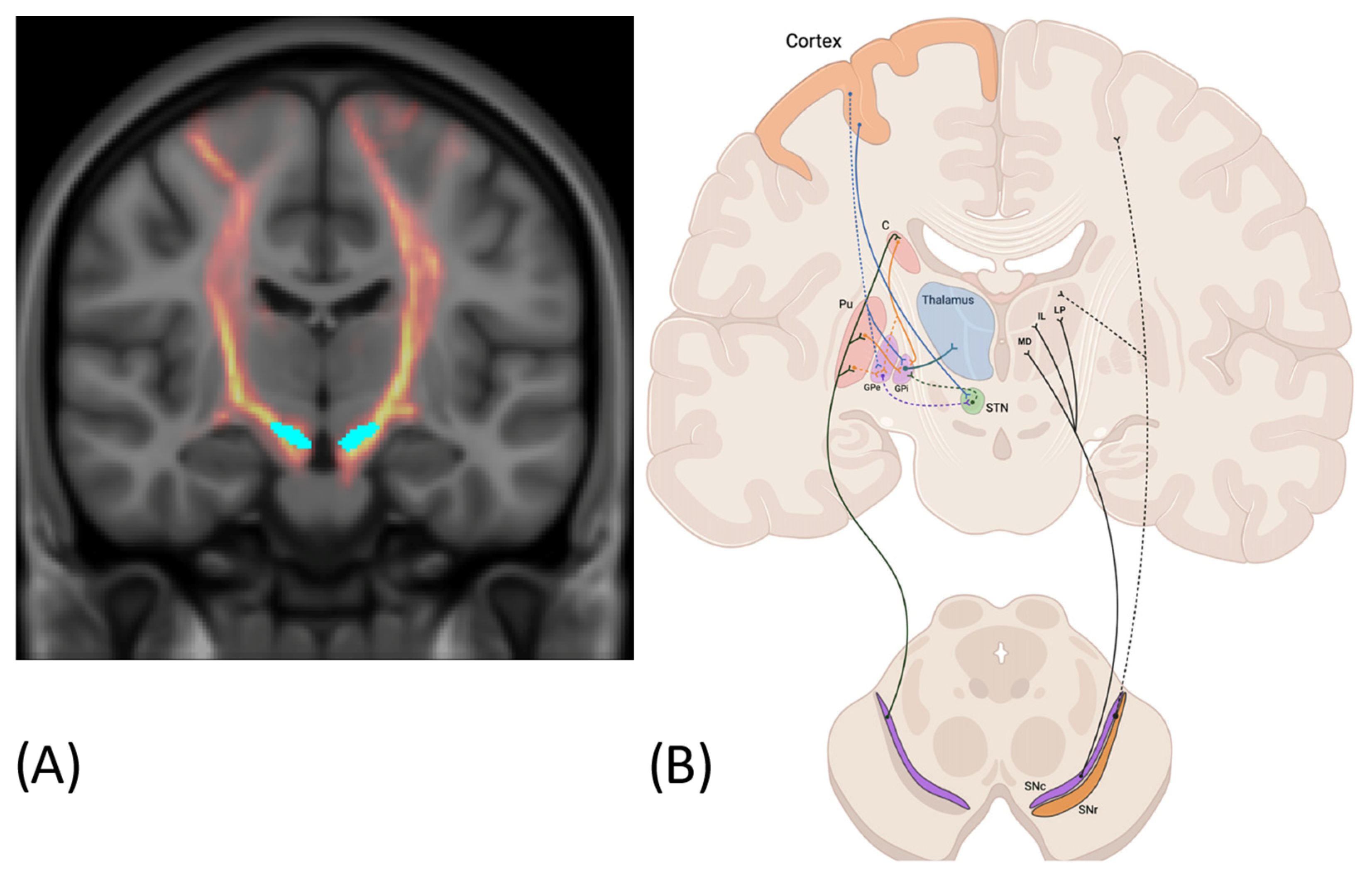
Figure 1. (A) Tractographic visualization of fiber pathways originating from the substantia nigra, derived from real diffusion MRI data. (B) Schematic representation of the nigrothalamic system. Reproduced from Cirillo et al., 2025 [28]. Efferent connections of the SNc to the dorsal striatum (caudate and putamen) (left) and to thalamic nuclei (MD, IL, LP) (right), together with nigro–cortical fibers (dashed black line). Direct, indirect and hyperdirect pathways are represented on the left side. C, caudate; Pu, putamen; MD, medio dorsal nucleus; IL, intralaminar nuclei; LP, latero posterior nucleus; GPe, external globus pallidus; GPi, internal globus pallidus; STN, subthalamic nucleus; SNc, substantia nigra pars compacta; SNr, substantia nigra pars reticulata.
Applsci 15 07902 g001
Figure 2. Nigrostriatal connectivity visualized using diffusion MRI. The substantia nigra pars compacta (SNc) are shown in cyan, the dopaminergic projection pathway in red, and the striatum in green.
Applsci 15 07902 g002
Table 1. Connections of the substantia nigra.
PathwayConnectionSignificance for NeurologyStudy
NigrostriatalSNc → dorsal striatum (putamen, caudate)A key pathway for motor control; its degeneration leads to the motor symptoms of PD[11,12,14,57,59,60,61,62,63,70]
NigrothalamicSNr → VA, VL, VM thalamic nuclei Influence motor and associative cortical circuits, important for motor and cognitive function, involved in dystonia pathophysiology[28,64,70]
SNc → thalamus Dopaminergic connections from the SNc directly to the thalamusPossible dopamine influence on thalamocortical circuits; potential mechanism for non-motor symptoms of PD[28]
Cortico–NigralDirect projections from the cortex (prefrontal, sensorimotor areas) → SNCognitive and sensory modulation of dopamine neurons; SN responds to cortical inputs[7,34,74]
SN–ventral striatum (mesolimbic pathway)SNc/VTA → NAc and orbitofrontal cortexRegulation of reward, emotion, and impulsiveness; key to addictions and behavioral disorders[28,66,69,72,73]
SN–hippocampusSN/VTA ↔ hippocampus (direct and indirect connections)Modulation of motivation, novelty, and learning; relevance to stress; major depressive disorder (MDD)[47,67]
SN–amygdala, limbic cortexSN2 (part of SNc) → amygdala, cingulum, orbitofrontal regionEmotion regulation; involved in mood disorders, anxiety, REM atonia[68]
SN–GPi/ansa lenticularis (GPSN)Globus pallidus → SN (via specific GPSN branch)Possible feedback effect of pallidum on SN; integration of BG outputs and feedback[65]
SNc–subcortical circuits (reticular formation)SNc ↔ brainstem, VTA, PAG, hypothalamusInvolved in regulation of sleep and autonomic functions; important in MDD, stress, and sleep disorders[68,75]
Disclaimer/Publisher’s Note: The statements, opinions and data contained in all publications are solely those of the individual author(s) and contributor(s) and not of MDPI and/or the editor(s). MDPI and/or the editor(s) disclaim responsibility for any injury to people or property resulting from any ideas, methods, instructions or products referred to in the content.