Article

Gallium-Containing Bioactive Glasses: Their Influence on Ion Release and the Bioactivity of Resulting Glass Polyalkenoate Cements

by
Lana Margaret Placek
1,
Danielle Lee Perry
1,
Mark Robert Towler
2 and
Anthony William Wren
1,*
1
Inamori School of Engineering, Alfred University, Alfred, NY 14802, USA
2
Chemical and Biochemical Engineering, Missouri University of Science and Technology, Rolla, MO 65409, USA
*
Author to whom correspondence should be addressed.
Appl. Sci. 2025, 15(14), 7756; https://doi.org/10.3390/app15147756
Submission received: 21 May 2025 / Revised: 8 July 2025 / Accepted: 9 July 2025 / Published: 10 July 2025
(This article belongs to the Special Issue Novel Ceramic Materials: Processes, Properties and Applications)

Abstract

A series of glasses (0.48SiO2 − [0.40-x]ZnO − 0.12CaO-xGa2O3, x = 0, 0.8, 0.16) was developed to formulate a series of Ga-containing glass polyalkenoate cements (GPCs). The solubility of GPCs was tested using DI water, and it was found that the sample containing the highest mol% of Ga, LGa-2, had the most Ga ion release. The GPCs were incubated in SBF, and SEM/EDS analysis revealed that the at% of P increased, while the at% of Si decreased, highlighting the CaP precipitation on the GPC surface. The at% of Ga also decreased, reinforcing the Ga release from the GPC. Cellular testing against fibroblasts and osteoblasts showed that a concentration of 25 mg/mL of the liquid extracts from the LGa-2 GPC had increased cell viability compared to other concentrations and GPCs tested. Antibacterial studies against E. coli and S. epidermidis demonstrated inhibition zones around the GPCs, highlighting their effectiveness in the elimination of bacteria on contact.

1. Introduction

Glass polyalkenoate cements (GPCs) were developed in the early 1970s and are typically used in restorative dentistry for luting and lining applications [1,2]. They have been candidates for use in skeletal cementation due to their excellent bone-contact biocompatibility, ability to adhere to surgical metals and bone, and lack of volumetric shrinkage and heat evolution [3]. GPCs are composite materials that set via an acid/base reaction between an ion-leachable glass (SiO2-AlO2-CaO, SiO2-AlO2-CaF) and a polyalkenoic acid, usually polyacrylic acid (PAA). Upon mixing, an acid–base reaction occurs where metal cations are released from the glass (e.g., Al3+, Ca2+) and form a polyacid salt with carboxylate (COO) groups from the PAA chains. These metal cations cross-link the polyacrylate chains, forming a cement of residual glass particles and a surrounding siliceous hydrogel embedded in a polysalt matrix [4,5,6,7]. GPCs are tailored explicitly for applications in contact with hard biological materials such as tooth enamel and dentin, which consist predominantly of tightly packed hydroxyapatite (HA) crystals that form a microporous structure. When in contact with HA, GPCs can create a direct chemical bond, which is achieved through ionic exchange at the GPC/HA interface [8]. Polyacrylic acid chains can enter the molecular surface of HA, replacing a concentration of calcium and phosphate ions [9].
Due to their therapeutic effects in dentistry, GPCs have been considered for orthopedic applications such as bone cementation and maxillofacial and cranial surgery due to their strong chemical bond to hard tissues [10,11,12]. However, the presence of Al3+ within the glass phase contraindicates the use of GPCs in bone; this can be addressed by modifying the glass composition to include more biocompatible ions such as Sr2+, Cu2+, and Zn2+ [13,14,15,16,17,18,19] in place of Al. Al3+ release (from the glass phase) in humans is associated with poor local bone mineralization and collagen synthesis, neurotoxicity (one fatal case) [20,21,22], and in vitro toxicity, which manifests as mineralization defects when tested in animal models [23]. Despite the Al3+ concerns, conventional GPCs have been proven to exhibit exceptional biocompatibility in the oral cavity [24]. Modifying their composition is required for use close to physiological fluids. GPCs are highly versatile materials as numerous methods can be utilized to modify their chemistry, which include altering the glass composition, changing the molecular weight of the PAA used, varying the concentration of PAA used during the cement forming process, and modifying the powder-to-liquid ratio (P:L ratio) [25,26,27]. Changing any or each of these characteristics will significantly affect the materials’ properties, i.e., the setting chemistry, solubility, mechanical properties, and bioactivity.
For this work, the glass composition was altered to contain more biocompatible components for use in close contact with physiological fluids. Using bioactive glasses as the glass component for these materials is advantageous, as these materials have been heavily studied and have seen commercial success and use in dentistry, skeletal tissue repair, and wound healing [28,29,30,31,32,33,34]. However, there are specific requirements for GPC formation, as the glass composition needs to incorporate ions capable of forming bonds with the COO groups on the PAA chains. The glass phase developed for this research is a silicate-based glass (SiO2-ZnO-CaO). The divalent cations (Ca2+, Zn2+) were incorporated into the glass phase as they are known to be released and form cross-links with the polyacrylic acid, thereby promoting the GPC setting reaction [35]. In addition, these are ions that have a positive therapeutic effect close to growing bone tissue and are known to diffuse between the GPC and host bone tissue. Low SiO2 is a well-recognized characteristic of bioactive glasses that facilitates degradation in a hydrated environment [36]. Zn2+ can impart positive therapeutic effects on bone metabolism [37,38], while Ca2+ is interchangeable within mineralized tissues, thereby facilitating bone bonding properties. However, there exists a hierarchy when incorporating ions within a GPC, and Al3+ is known to form the strongest bond during the setting reaction (M = Al3+ > Cu2+ > Zn2+ > Ca2+ > Mg2+) [4]. Divalent cations can link two polyanionic chains together, thereby forming a bond with a high ionic strength. These bonds form as the materials cure and are responsible for their unique physical and mechanical properties [10]. The availability of the unbound ions facilitates ion release and the biological properties associated with these materials.
The focus of this study was to elucidate the bioactive properties of gallium (Ga3+) when incorporated within a SiO2-ZnO-CaO glass phase prior to forming a series of Ga-glass polyalkenoate cements (Ga-GPCs). Ga was incorporated at 0.08 and 0.16 mol% additions at the expense of Zn2+. Ga3+ is reported to have numerous beneficial effects in vivo, including antimicrobial properties [39,40,41,42], anti-inflammatory [43], and anti-cancerous effects [44,45,46]. Previous work has demonstrated the efficacy of Ga3+ against opportunistic pathogenic bacteria such as Pseudomonas aeruginosa (P. aeruginosa), which can cause airway infections in cystic fibrosis patients [39,40,41,46], and opportunistic fungi such as Cryptococcus neoformans, which can cause life-threatening diseases in immunocompromised patients [47]. Olakanmi et al. investigated the effects of both gallium nitrate and transferrin-gallium on the bacterial species Mycobacterium tuberculosis and Mycobacterium avium, discovering that these two gallium compounds acted to inhibit the iron-dependent growth of the bacteria [48]. A study conducted by Kaneko et al. investigated the effects of gallium nitrate on P. aeruginosa pneumonia in mice. This investigation revealed that the gallium-containing compounds inhibited the growth of P. aeruginosa and also prevented the formation of biofilm. This study also showed that in the case of P. aeruginosa, gallium nitrate again interfered with iron uptake, resulting in the death of both planktonic and biofilm bacteria in vitro [41]. Ga3+ has also been shown to increase bone calcium levels by inhibiting resorption [49,50,51]. The mechanism for this activity involves the recruitment of Ga3+ to bone tissue and the suppression of osteoclastic differentiation markers (TRAP, CTR, CTK, and RANK) [50]. Metabolic studies with bone cancer patients also support this observation; patients who were given a gallium nitrate supplement showed a decreased concentration of calcium in the urine, suggesting improved mineral retention and utilization [52].
Ga has also been studied for its ability to treat cancer; the absorption of radioactive gallium by malignant cells led to research being conducted by the National Cancer Institute (NCI), in which they investigated the ability of salts of group IIIa metals to inhibit the growth of malignant cells in mice and rats [53]. Out of the various compounds tested, gallium nitrate (Ga(NO3)3) exhibited the ability to suppress the growth of subcutaneously implanted tumors, while also exhibiting the least toxicity of the compounds investigated. Since these abilities of gallium nitrate were discovered, many clinical trials have been conducted in an attempt to identify which forms of cancer this compound is effective against. Gallium nitrate has been proven effective in the treatment of non-Hodgkin lymphoma [54], bladder cancer [55], multiple myeloma [56], and metastatic urothelial carcinoma [57], in addition to being investigated for treating cancer-related hypercalcemia [58,59].

2. Materials and Methods

2.1. Glass Synthesis

Three glass compositions (Lcon, LGa-1, and LGa-2) were formulated for this study, with the principal aim being to investigate any property changes resulting from the addition of Ga to the glass phase of the GPC. The Control glass (Lcon) was a Ga-free CaO-ZnO-SiO2 glass; LGa-1 and LGa-2 contain incremental concentrations (0.08, 0.16 mol%) of Ga at the expense of zinc (Zn) as presented in Table 1. Glasses were prepared by weighing out appropriate amounts of analytical-grade reagents (Fisher Scientific, Pittsburgh, PA, USA) and ball milling (1 h). The mix was then oven-dried (100 °C, 1 h) and fired (1500 °C, 1 h) in a platinum crucible and shock quenched into water. The resulting frit was dried, ground, and sieved to retrieve a glass powder with a maximum particle size of 45 μm.

2.2. GPC Sample Preparation

Ga-GPCs were prepared by thoroughly mixing each of the glass powders (<45 μm) with E11 polyacrylic acid (PAA—Mw 210,000, <90 μm, Advanced Healthcare Limited, Kent, UK) and distilled water on a glass plate. The cements were formulated in a P:L ratio of 2:1.5 with 50 wt% additions of PAA. Complete mixing was undertaken within 20 s.

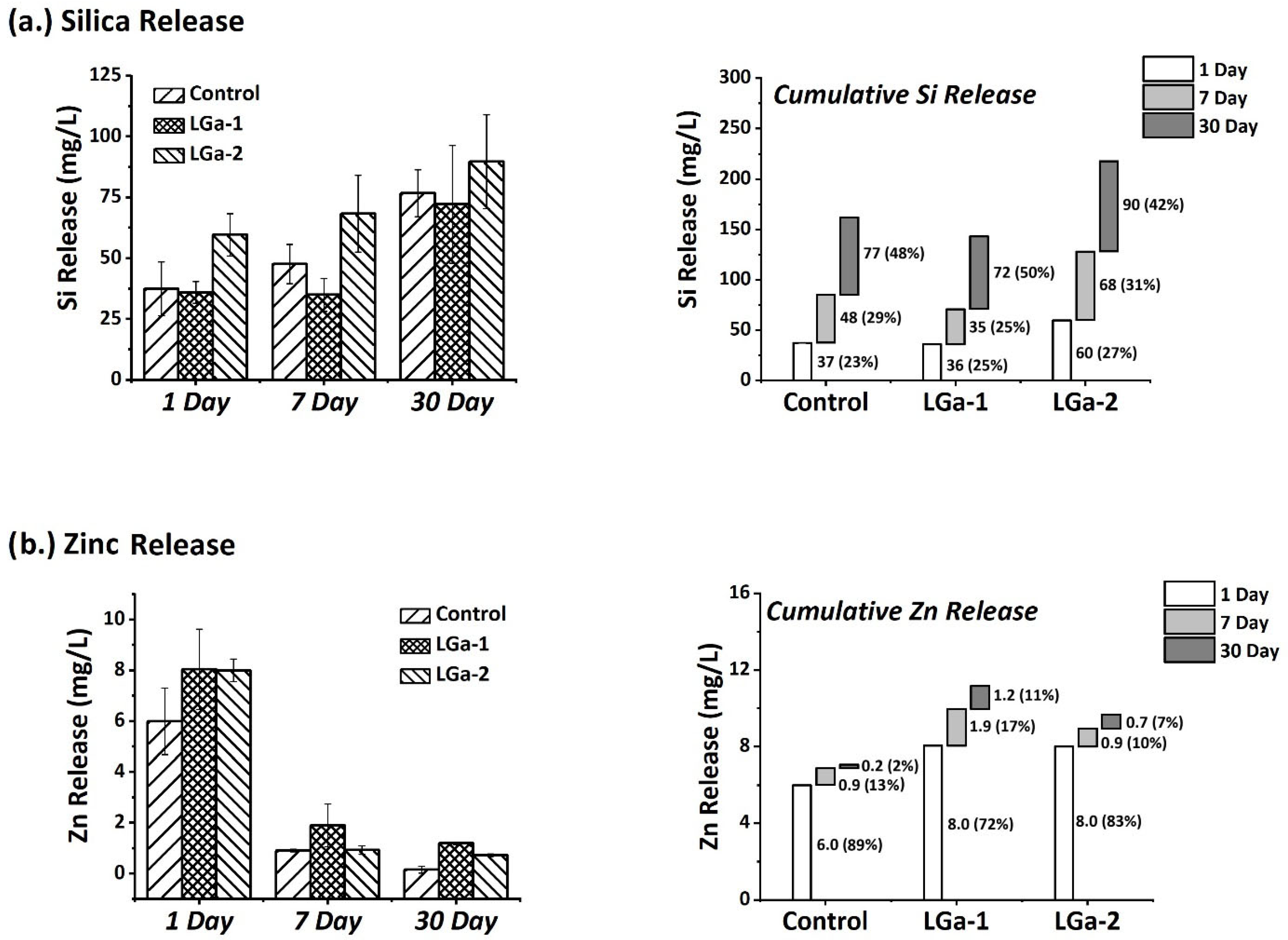
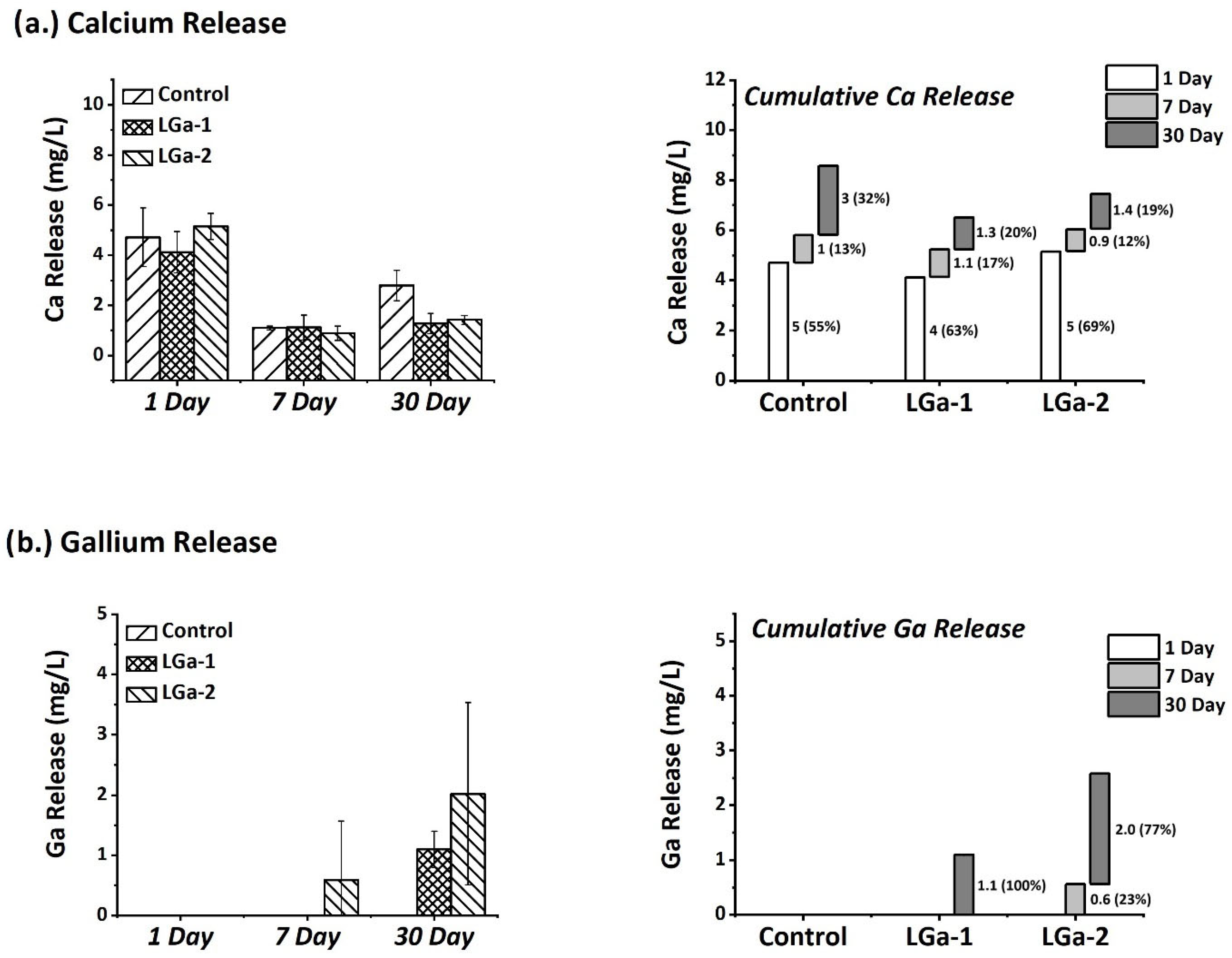
2.3. Ion-Release Profiles
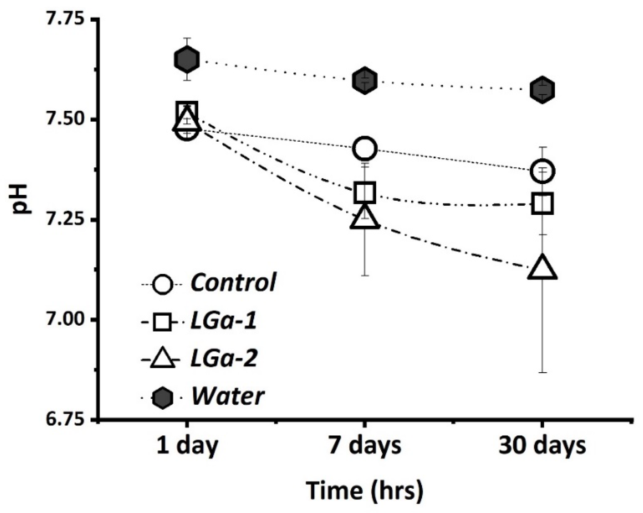
Each GPC (Control, LGa-1, and LGa-2, where n = 3) was immersed in sterile de-ionized H2O and placed on an oscillating platform at 37 °C for 1, 7, and 30 days. The ion-release profile of each glass was measured using Inductively Coupled Plasma Optical Emission spectroscopy (ICP–OES) on a Perkin–Elmer Optima 5300 UV (Perkin–Elmer, Waltham, MA, USA). ICP–OES calibration standards for Ca, Si, Zn, and Ga ions were prepared from a stock solution on a gravimetric basis. Three target calibration standards were prepared for each ion, and de-ionized water was used as a control.

2.4. pH Analysis

Changes in solution pH were monitored using a Corning 430 pH meter. Prior to testing, the pH meter was calibrated using pH buffer solutions of 4.00 ± 0.02 and 7.00 ± 0.02 (Fisher Scientific, Pittsburgh, PA, USA). Sample solutions were prepared by exposing GPC discs (where n = 3) in sterile de-ionized H2O as described in the method for ion-release testing. Measurements were recorded at 1, 7, and 30 days.

2.5. Preparation of Ion Release and Cell Culture Extracts

Approximately 50 g of each glass (Control, LGa-1, and LGa-2, where n = 3) was sterilized using γ-irradiation at 25 kGray (Isotron Ltd., Mayo, Ireland) prior to forming cements. Tissue culture water (Sigma Aldrich, Dublin, Ireland) was selected as the solvent for preparing extracts. The volume of extract was determined using Equation (1).
V s = S a 10
Vs = volume of extract used.
Sa = exposed surface area of the cement disc.
GPC Samples (n = 3, ϕ6 mm × 2 mm) were aseptically immersed in appropriate volumes of sterile tissue culture water and agitated at 37 °C ± 2 °C for 1, 7, and 30 days for ion release, pH analysis, and cytotoxicity testing. For cytotoxicity testing, 100 µL aliquots (n = 3) of each extract were removed after each time period.

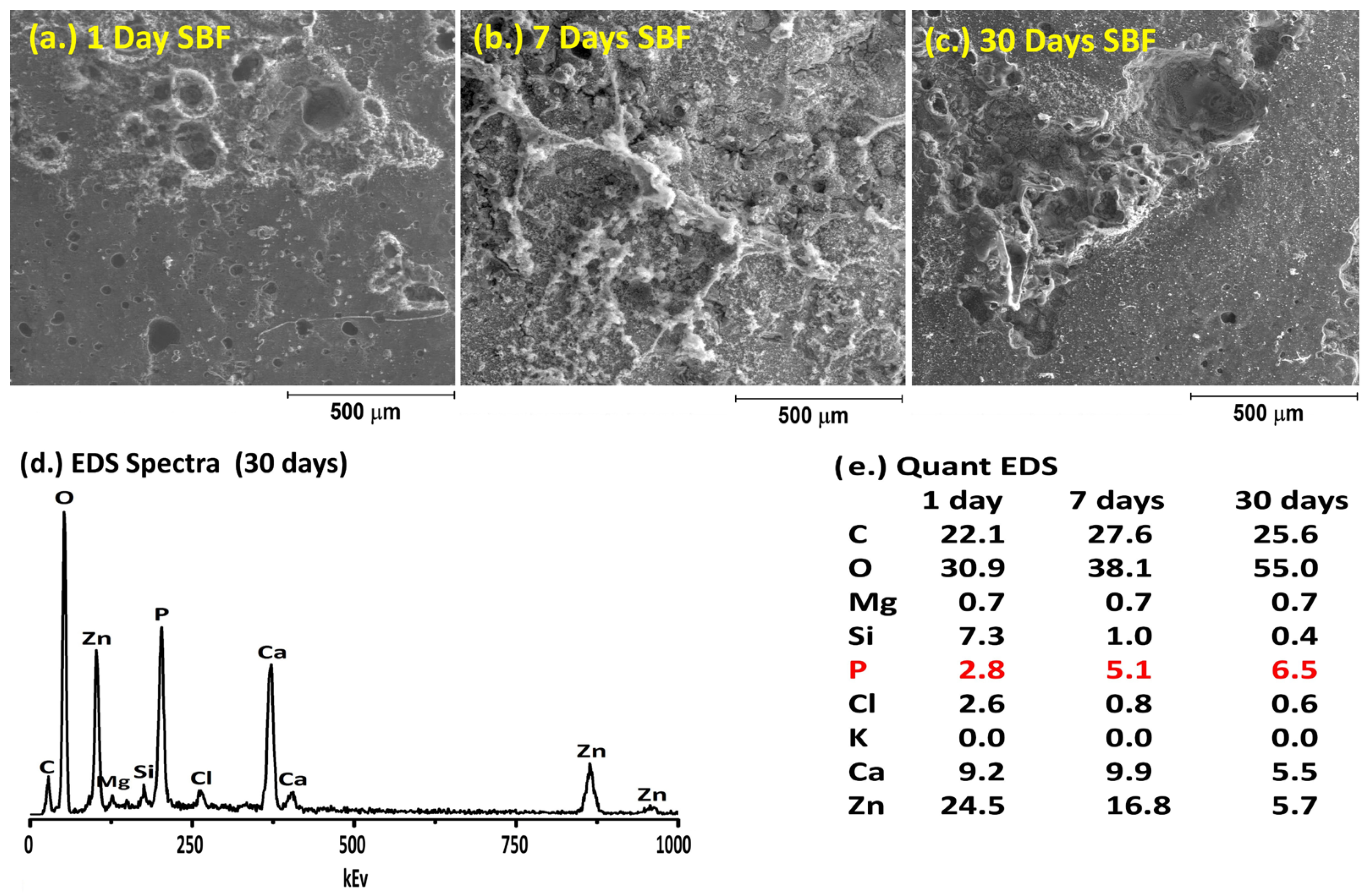
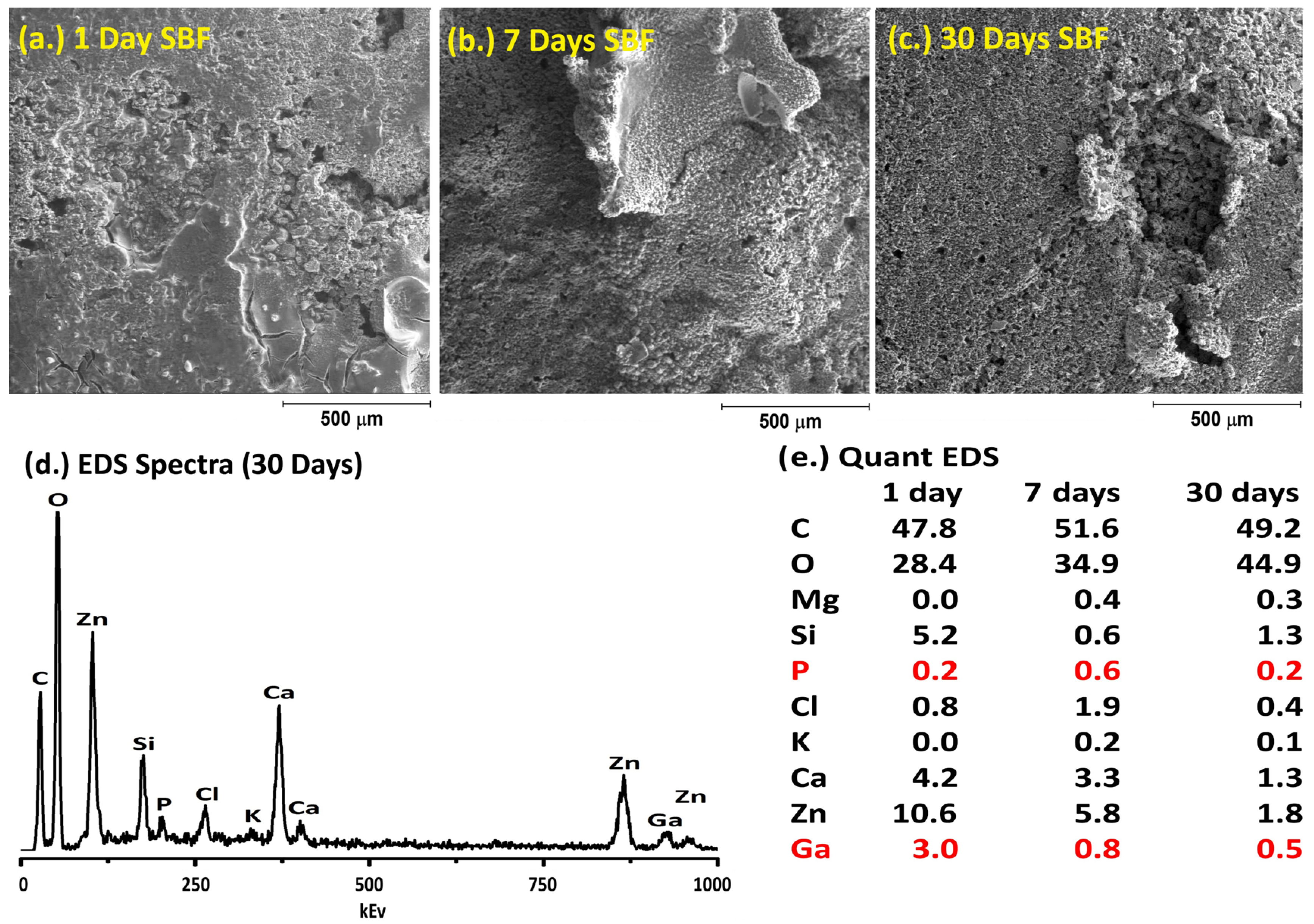
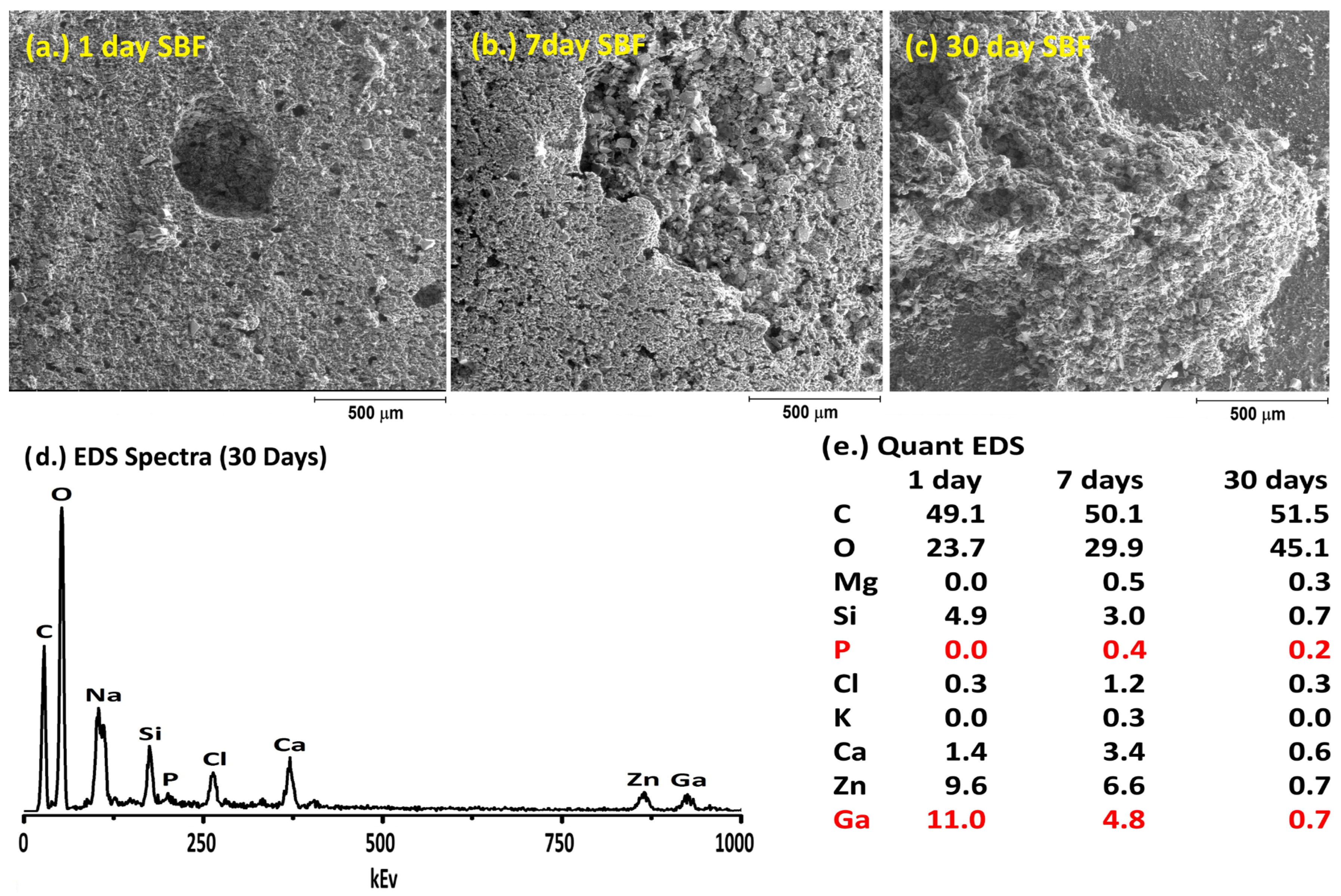
2.6. Simulated Body Fluid Trial

Simulated body fluid (SBF) was produced in accordance with the procedure outlined by Kokubo et al. [23]. The reagents were dissolved in order, from reagent 1 to 9, in 500 mL of purified water using a magnetic stirrer. The solution was maintained at 36.5 °C. An amount of 1 M-HCl was titrated to adjust the pH of the SBF to 7.4. Purified water was then used to adjust the volume of the solution up to 1 L. GPC discs (n = 2, ϕ6 mm × 2 mm) were immersed in concentrations of SBF as determined by Equation (1) and were subsequently stored for 1, 7, and 30 days in an incubator at 37 °C. A JOEL JSM-840 scanning electron microscope (JOEL, Peabody, MA, USA) equipped with a Princeton Gamma Tech (PGT) Energy Dispersive X-ray (EDX) system was used to obtain secondary electron images and carry out chemical analysis of the surface of the cement discs. All EDX spectra were collected at 20 kV, using a beam current of 0.26 nA. Quantitative EDX converted the collected spectra into concentration data by using standard reference spectra obtained from pure elements under similar operating parameters.

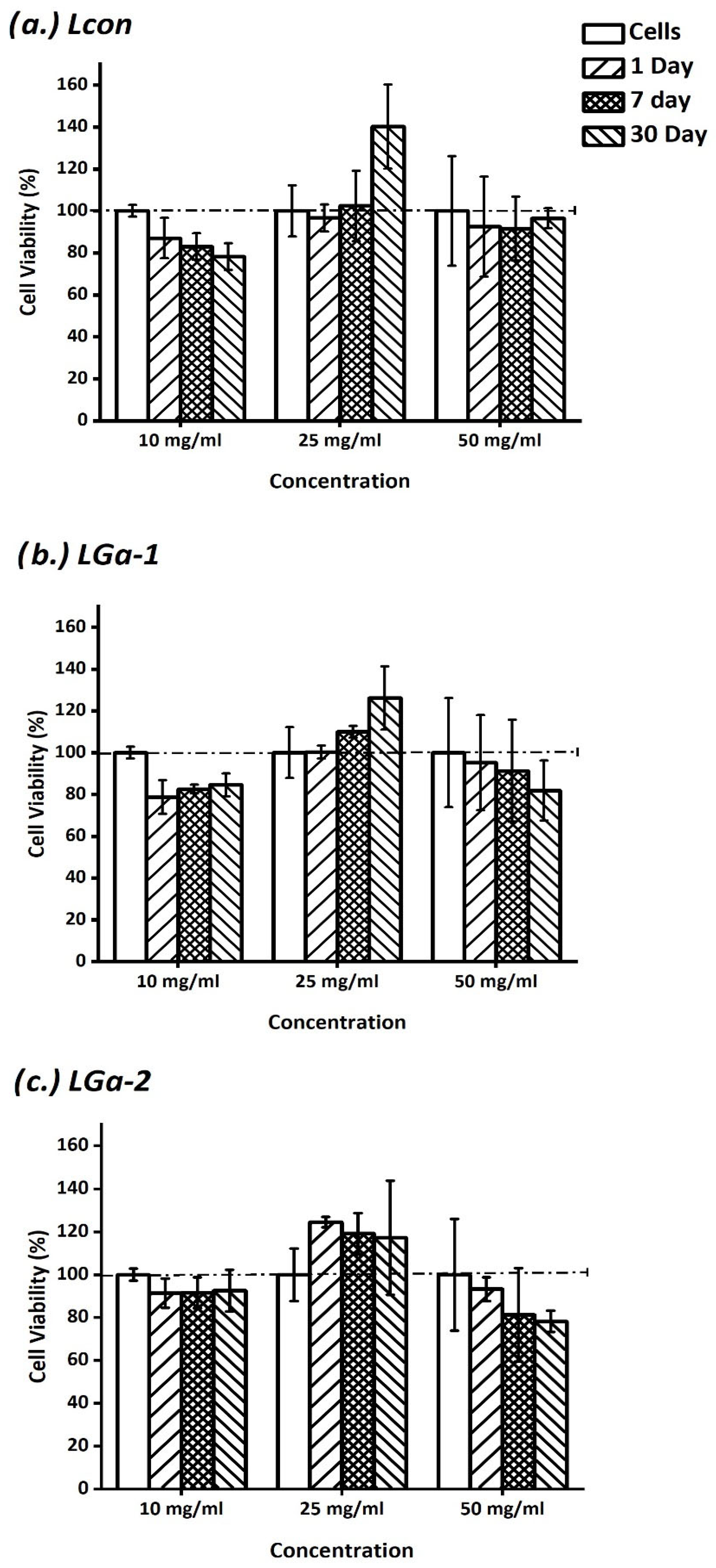
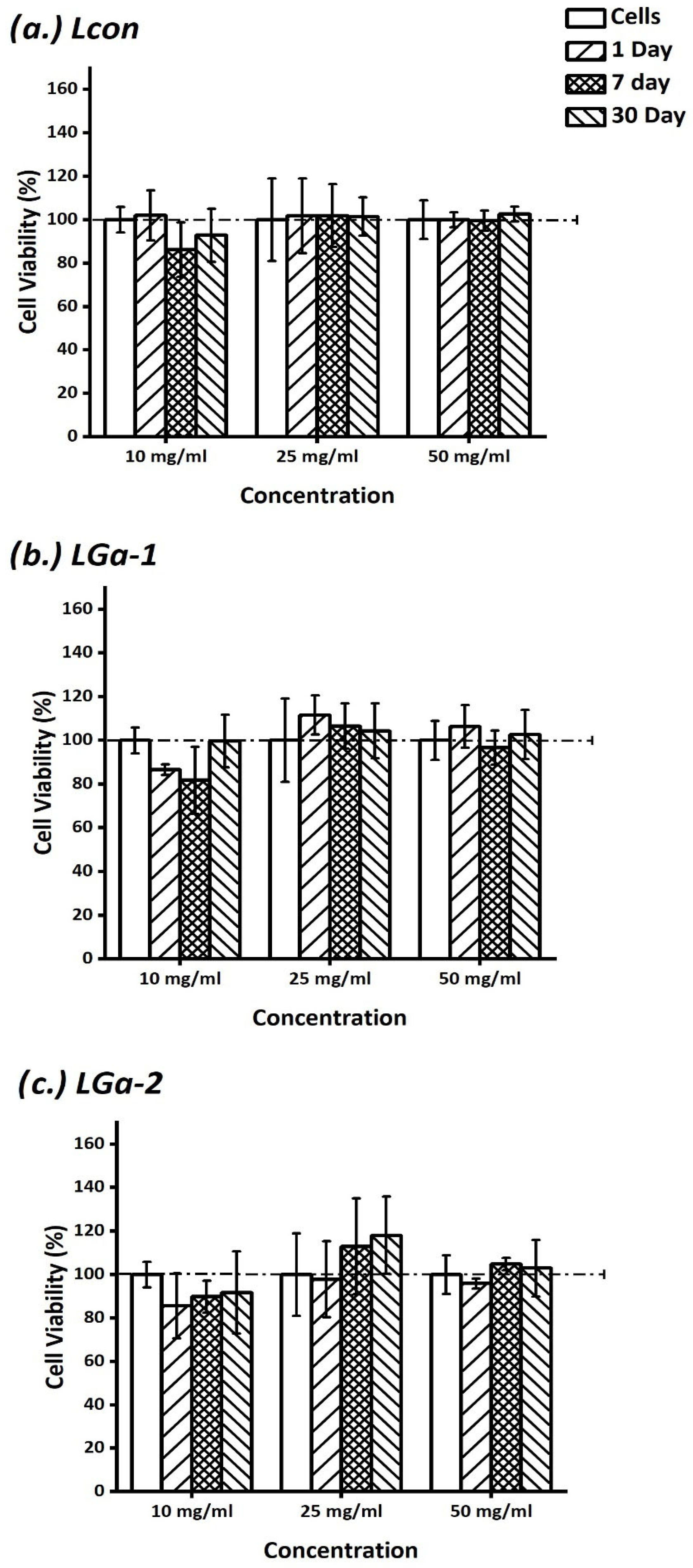
2.7. In Vitro Assessment of Cement Extracts

The established fibroblast cell line L-929 (ATCC CCL 1, NCTC clone 929) and osteoblast MC 3T3 (ATCC CRL-2593) were used in this study, as required by ISO10993 part 5 [34,60]. Cells were maintained on a regular feeding regimen in a cell culture incubator at 37 °C/5% CO2/95% air atmosphere. Cells were seeded into 24-well plates at a density of 10,000 cells per well and incubated for 24 h prior to testing with both extracts and cement discs. The culture media used was M199 media (Fisher Scientific) supplemented with 10% fetal bovine serum (Fisher Scientific) and 1% (2 mM) L-glutamine (Fisher Scientific). The cytotoxicity of the GPC extracts was evaluated using the methyl thiazolyl tetrazolium (MTT) assay in 24-well plates. Aliquots (100 µL) of undiluted sample were added into wells containing each cell line in culture medium (1 mL) in triplicate over 1, 7, and 30 days. Each of the prepared plates was incubated for 24 h at 37 °C/5% CO2. The MTT assay was then added in an amount equal to 10% of the culture medium volume/well. The cultures were then re-incubated for a further 2 h (37 °C/5% CO2). Next, the cultures were removed from the incubator, and the resultant formazan crystals were dissolved by adding an amount of MTT solubilization solution (10% Triton x-100 in acidic isopropanol, 0.1 N HCl) equal to the original culture medium volume. Once the crystals were fully dissolved, the absorbance was measured at a wavelength of 570 nm. Aliquots (100 µL) of tissue culture water were used as controls, and cells were assumed to have metabolic activities of 100%.

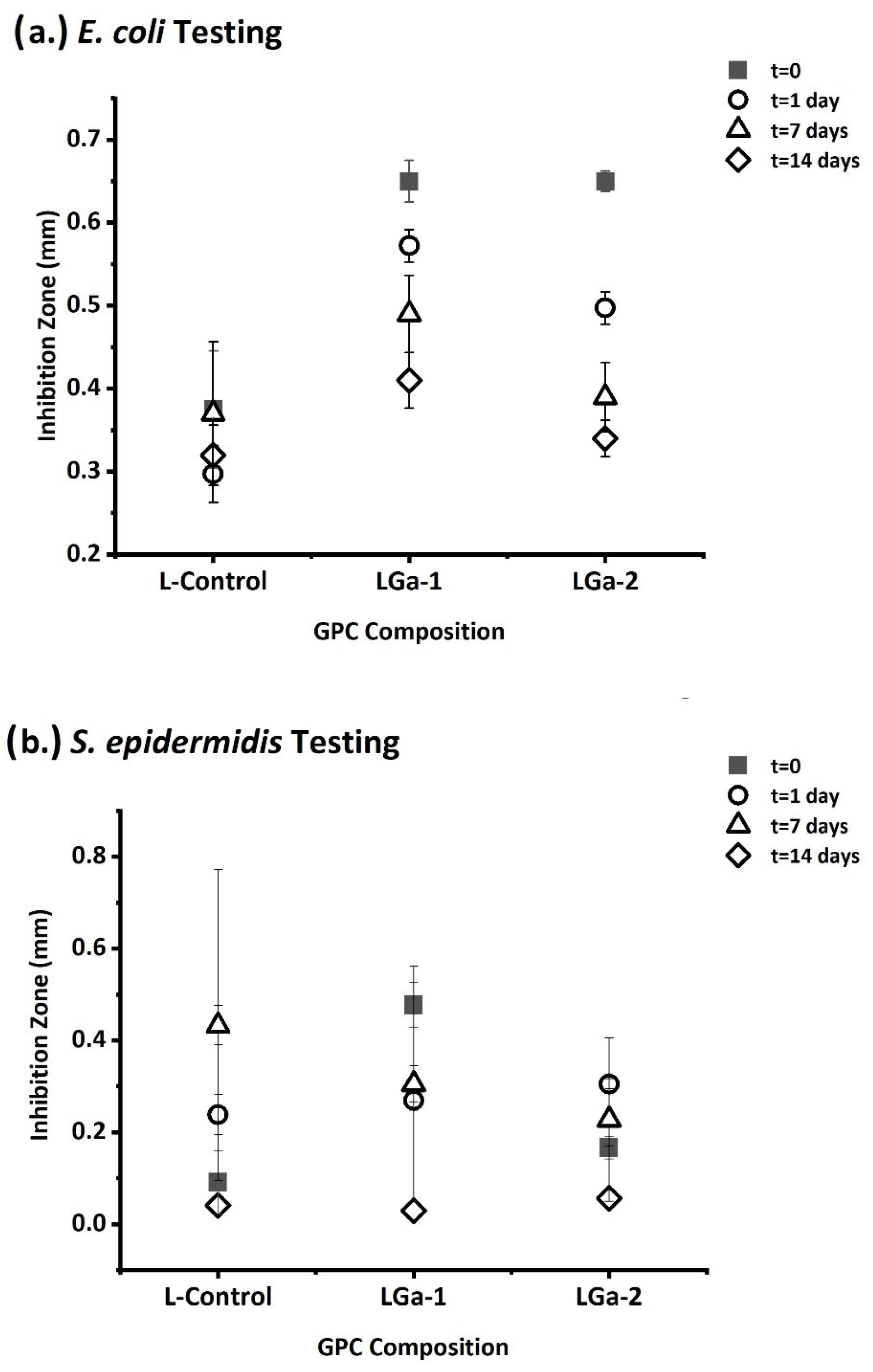
2.8. Antibacterial Evaluation

The antibacterial activity of the Ga-GPCs (ϕ6 mm × 2 mm, where n = 3) was evaluated using E. coli strain ATCC 8739 (LB agar and broth) and S. epidermidis strain ATCC 14990 (BHI agar and broth). Each cement sample (Control, LGa-1, and LGa-2) was placed in inoculated plates, and the plates were cultured for 24 h at 37 °C. Each microbe was initially grown aerobically in liquid broth at 37 °C for 24 h. Preparation of the agar disc diffusion plates involved seeding agar plates with a sterile swab dipped in a 1/50 dilution of the appropriate 24 h culture of bacteria. The agar diffusion test was performed under standard laboratory sterile conditions in a fumigation hood using sterile swabs for the inoculation of bacteria. Digital calipers were used to measure zones of inhibition, where each sample was analyzed in triplicate, and mean zone sizes ± standard deviations were calculated. Inhibition zone sizes were computed using Equation (2).
I n h i b i t i o n   Z o n e   ( m m ) = H a l o D i s c 2

2.9. Statistical Analysis

One-way analysis of variance (ANOVA) was employed to compare the changes in fibroblast and osteoblast cell viability with those of the growing unmodified cell populations. Comparison of relevant means was performed using the post hoc Bonferroni test. Differences between groups were deemed significant when p ≤ 0.05.

3. Results and Discussion

The authors have previously reported on the preliminary physical properties of the Ga-GPCs [61]. To summarize, the starting glasses were all characterized and were determined to be amorphous with a particle size range of approximately 7–13 μm, with individual mean particle sizes of 9.3 μm (Control), 9.8 μm (LGa-1), and 9.3 μm (LGa-2) [61]. Utilizing a similar particle size for the glass phase of each GPC is critical, as this significantly influences the dissolution rate during cement curing. This contributes to the fact that any differences in cement properties will be due to the inclusion of Ga, and not due to physical differences in the glass particles. Previous studies by the authors [61] have described the physical properties, i.e., working (Tw) and setting (Ts) times and mechanical properties (both compressive and biaxial flexural strength), and Ga-addition to the glass phase of these GPCs has had a positive influence on the rheological properties, while the mechanical properties were reduced. The effect of Ga on the glass structure was also described using X-ray photoelectron spectroscopy and MAS-NMR, which determined that Ga assumes a network-modifying role in these glasses [61].
The current work elucidates the ion-release profiles and the resulting bioactivity of this cement series. Simulated body fluid (SBF) testing, cell culture analysis, and antimicrobial activity have been investigated in this study. The ion-release data are presented in Figure 1a,b, which present the release of silica (Si) and zinc (Zn), respectively. Also, the rates of ion release are presented for each cement, and the potential cumulative release is determined by time.
Si release (Figure 1a) was 37–77 mg/L over 1–30 days for the Control cement, where the highest release rate was experienced for the 30-day sample (48%). A similar trend was observed with LGa-1 and LGa-2. The LGa-1 Si release ranged from 36–72 mg/L over 1–30 days, LGa-2 ranged from 60–90 mg/L over 1–30 days, and both Ga-GPCs experienced the highest release rates after 30 days, with LGa-1 at 72 mg/L (50%) and LGa-2 at 90 mg/L (42%). Zn release is also presented in Figure 1b, and the release rates were much lower compared to Si. In each case, the majority of the Zn release occurred after 1 day: Control at 6 mg/L (89%), LGa-1 at 8 mg/L (72%), and LGa-2 at 8 mg/L (83%). Zn release was found to significantly reduce over time, where the 30-day release rates range from 2 to 11% for the Control, LGa-1, and LGa-2. Calcium (Ca) and gallium (Ga) ion release is presented in Figure 2a,b.
Ca release (Figure 2a) was highest in the Control GPC and ranged from 4 to 5 mg/L over 1–30 days. The release of Ca followed a similar trend to Zn, where the release rates were low and the trend showed that the majority of Ca release was experienced after 1 day for each cement. The Control released 5 mg/L (55%), LGa-1 released 4 mg/L (63%), and LGa-2 released 5 mg/L (69%). Ga release is presented in Figure 2b and was released at lower rates than each of the other ions tested. No Ga was detected from the Control GPCs at any time and was only detected from LGa-1 and LGa-2 after the extended time periods. LGa-1 released 1.1 mg/L (100%) of its Ga only after 30 days of incubation, while LGa-2 was found to release 0.6 mg/L (23%) of the Ga concentration, and at 30 days, 2.0 mg/L (77%) was released. Other studies conducted on Ga have also seen high release rates of Ga over a similar period [62]. The solution pH was also recorded at each time interval, and the data are presented in Figure 3.
From Figure 3, it is evident that there was a slight reduction in solution pH recorded, which is attributed, more specifically, to the increase in Ga concentration within the glass. The Control glass experienced a minor reduction in pH over 1–30 days, reducing from 7.4 to 7.3. LGa-1 was reduced from 7.5 to 7.2, and LGa-2 was found to be reduced from 7.4 to 7.1.
SBF testing was conducted to determine the potential for bioactivity, as SBF represents the ionic concentration of human blood plasma and is widely regarded as a precursor to in vivo bioactivity. The precipitation of CaP minerals on material surfaces when immersed in SBF is typically regarded as a positive indicator of bioactivity and bone bonding in vivo. Each of the GPCs was incubated in SBF for 1, 7, and 30 days, and the Control SBF results are presented in Figure 4.
Figure 4a–c shows the SEM images taken over 1–30 days, while Figure 4d presents the EDS spectra, and Figure 4e shows the quantitative EDS data. The SBF testing on the Control GPC was found to encourage the precipitation of minerals on the GPC’s surface. Each of the ions present in the Control glass (Si, Ca, Zn) used to formulate the GPC was detected, in addition to ions present in SBF (Mg, Cl, K, P). The quantitative EDS data show that the concentration of P increased with respect to time over 1, 7, and 30 days at 2.8, 5.8, and 6.5 at%. The Zn concentrations were found to reduce over time from 24.5 to 5.7 at% over 1–30 days. Additionally, a strong indicator of CaP precipitation is the reduction in Si concentrations over time, where Si is reduced from 7.3 to 0.4 at%. This is a strong indicator that, as CaP minerals are precipitating on the surface, the underlying Si from the GPC is being masked. The LGa-1 SBF data is presented in Figure 5.
Although there are CaP mineral deposits evident on the surface of the material, the concentration was not as high as that of the Control GPC. Additionally, a much lower concentration of P was found on the LGa-1 GPC surface (0.2–0.6 at%). The concentrations of Zn (10.6–1.8 at%), Si (5.2–1.3 at%), and Ca (4.2–1.3 at%) were also found to decrease, while Ga concentrations were found to increase, 3.0–0.8 at%. LGa-2 GPC (Figure 6) was found to follow the same trend, where P levels were low, ranging from 0 to 0.4 at%. Si levels also reduced, from 4.9 to 0.7 at%; Zn levels reduced, from 9.6 to 0.7 at%; and Ca levels reduced, from 3.4 to 0.6 at%. Ga levels were initially high at 11.0 at% but reduced to 0.7 after 30 days of incubation in SBF.
Cytocompatibility testing was conducted in fibroblast cells (Figure 7) and osteoblasts (Figure 8). Liquid extracts were tested from GPC incubation after 1-, 7-, and 30-day incubation, and these cells were then incubated with different concentrations of extract (10 mg/mL, 25 mg/mL, and 50 mg/mL) to determine the approximate cytotoxicity limits. Testing of the GPC series in fibroblasts is presented in Figure 7a–c for the Control, LGa-1, and LGa-2 GPCs.
Regarding the Control GPCs (Figure 7a), cell cytocompatibility was reduced compared to the control cells at each time period, i.e., 1 day (87%) and 7 days (82%), and reached significance at 30 days (78%, p = 1.000) for the 10 mg/mL concentration. The higher concentrations of 25 mg/mL and 50 mg/mL showed no significant difference when compared to the control cell population, with the exception of 25 mg/mL at 30 days (140%), where there was a significant increase in cell viability compared to the control cells (p = 1.000). The LGa-1 GPC is presented in Figure 7b, and a similar trend was observed where the 10 mg/mL was significantly reduced at each time period, 1 day (86%, p = 1.000), 7 days (82%, p = 1.000), and 30 days (78%, p = 1.000) compared to the control cells. At a concentration of 25 mg/mL there was an increase in cell viability at 7 days (110%, p = 0.000) and 30 days (126%, p = 0.000), and at 50 mg/mL, the cell viability reduced compared to the control cells at each time period, 1 day (95%, p = 0.000), 7 days (91%, p = 0.000), and 30 days (81%, p = 0.000). The LGa-2 GPC is presented in Figure 7c and also experiences a similar trend to that of the other cements. There was a slight reduction in cell viability for the 10 mg/mL at 1 day (91%), 7 days (91%), and 30 days (92%) compared to the control cells, but at the 25 mg/mL concentrations, increases in cell viability were observed at each time period, 1 day (124%, p = 1.000), 7 days (119%, p = 0.000), and 30 days (117%, p = 0.000). The 50 mg/mL concentration was found to show reductions in cell viability compared to the control cells at 1 day (93%), 7 days (81%), and 30 days (78%). One common aspect for each of the GPCs is that the 25 mg/mL concentration resulted in better cell viability in each case. Still, this effect was more prominent in the LGa-2 GPC, as the increase in cell viability was evident at each time period tested. The 10 mg/mL and 50 mg/mL concentrations did not present the same effects.
Cell testing was also conducted in osteoblasts at analogous concentrations at each time period, and the data are presented in Figure 8.
The Control GPC is presented in Figure 8a, and in each case, there was no significant difference among the concentrations tested, i.e., 10 mg/mL, 25 mg/mL, and 50 mg/mL. There were slight reductions at 7 days (86%) and 30 days (92%). However, this difference was not deemed significant compared to the control cells. The LGa-1 GPC’s cell viability is presented in Figure 8b. It is evident that the 10 mg/mL resulted in a reduction at 1 day (86%, p = 1.000), and increases in cell viability were observed at 25 mg/mL for each time period, 1 day (111%), 7 days (106%), and 30 days (104%), but these increases were not deemed significant compared to the control cells, p = 0.000. The 50 mg/mL concentrations showed increased viability at 1 day (106%), but these effects were not significant when compared to the control cells. The LGa-2 GPC data is presented in Figure 8c and shows initial reductions in cell viability after 1–7–30 days (85%, 89%, and 91%); however, the most evident increases in viability were associated with 25 mg/mL after 7 days (113%, p = 0.000) and 30 days (118%, p = 0.000). This trend is similar to that observed with the fibroblast (Figure 7) testing, where the overall highest increase in viability was observed with the LGa-2 GPC, with a concentration of 25 mg/mL.
Antibacterial testing was conducted in both E. coli and S. epidermidis bacteria, and the data are presented in Figure 9a,b, respectively.
GPCs were initially tested after setting (t = 0), and also when removed from incubating in sterile DI water after 1, 7, and 14 days (t = 1, t = 7, and t = 14). Testing in E. coli is presented in Figure 9a, and it is evident that the Ga-GPCs (LGa-1, LGa-2) typically resulted in greater inhibition zones than the Control GPC. The inhibition zones ranged from 0.3 to 0.4 mm when incubated from t = 0–14, whereas the LGa-1 GPC ranged from 0.4 to 0.7 mm, and the LGa-2 GPC ranged from 0.3 to 0.7 mm. The most significant inhibition was observed with the LGa-1 (0.7 mm) and LGa-2 (0.7 mm) GPC at the time period t = 0. Testing in S. epidermidis is presented in Figure 9b over the same time periods. The Control, LGa-1, and LGa-2 GPC performed similarly and ranged from 0.1 to 0.4 mm. Similar results were seen in Gao et al. [63], where Ga-containing alloys showed improved antibacterial properties against E. coli and S. epidermidis bacteria after 3 days compared to Ga-free alloys. Although each of the cement inhibition zones measured in this study was relatively small, it is highly beneficial to have materials eliminate bacteria on contact.

4. Conclusions

This study was conducted to determine the effect of gallium (Ga) on the solubility and bioactivity of a series of novel silicate-based glasses (SiO2-ZnO-CaO) used for synthesizing Ga-containing glass polyalkenoate cements (GPCs), i.e., LGa-1 and LGa-2. Some of the key findings associated with this work are related to the materials’ solubility. Ion-release profiles depicted the release rates of Si, Zn, Ca, and Ga over 30 days, with LGa-2 having the most Si and Ga release over 30 days. The pH values associated with these ion-release profiles show decreased values from 1 to 30 days for every sample tested. When the GPCs were in contact with SBF, CaP precipitates were seen on the surface of the cement using SEM. This was supported through EDS, where an increased at% of P was detected as well as decreased Si at%, an indicator of CaP precipitation. Ga at% was also shown to decrease from 1 to 30 days, highlighting its dissolution from the GPC into the SBF medium. Cellular testing against fibroblast and osteoblast cells revealed that a concentration of 25 mg/mL of the liquid extracts from the LGa-2 GPC increased cell viability compared to the other concentrations and those of the Control and LGa-1. The antibacterial testing against E. coli and S. epidermidis showed that GPCs at t = 0 produced larger inhibition zones for each GPC against E. coli and the LGa-1 against S. epidermidis compared to the GPCs that were soaked in SBF prior to inoculation. This reinforces the release of Ga from the GPC in SBF and explains why, prior to incubation, the GPC has stronger antibacterial properties. Although the inhibition zones are smaller after incubation in SBF, the GPCs still possess the ability to eliminate bacteria on contact, which is beneficial for dentistry and orthopedics.

Author Contributions

Conceptualization, A.W.W. and M.R.T.; methodology, L.M.P.; validation, A.W.W., D.L.P. and M.R.T.; formal analysis, L.M.P. and A.W.W.; data curation, A.W.W. and L.M.P.; writing—original draft preparation, A.W.W. and D.L.P.; writing—review and editing, A.W.W., D.L.P. and M.R.T.; visualization, A.W.W. and D.L.P.; supervision, A.W.W. and M.R.T.; project administration, A.W.W. and M.R.T. All authors have read and agreed to the published version of the manuscript.

Funding

This research received no external funding.

Institutional Review Board Statement

Not applicable.

Informed Consent Statement

Not applicable.

Data Availability Statement

The original contributions presented in this study are included in the article. Further inquiries can be directed to the corresponding author.

Conflicts of Interest

The authors declare no conflicts of interest.

Abbreviations

The following abbreviations are used in this manuscript:
GPCGlass Polyalkenoate Cement
SEMScanning Electron Microscopy
EDSEnergy Dispersive Spectroscopy
CaPCalcium Phosphate
SBFSimulated Body Fluid
ICP-OESInductively Coupled Plasma Optical Emission Spectroscopy
PAAPolyacrylic Acid
HAHydroxyapatite
COO-Carboxylate

References

  1. Cho, S.; Cheng, A.C. A review of glass ionomer restorations in the primary dentition. J. Can. Dent. Assoc. 1999, 65, 491–495. [Google Scholar] [PubMed]
  2. DeBruyne, M.A.A.; DeMoor, R.J.G. The use of glass ionomer cements in both conventional and surgical endodontics. Int. Endod. J. 2004, 37, 91–104. [Google Scholar] [CrossRef] [PubMed]
  3. Szoradi, G.T.; Feier, A.M.; Zuh, S.G.; Russu, O.M.; Pop, T.S. Polymethyl Methacrylate bone cement polymerization induced thermal necrosis at the cement–bone interface: A narrative review. Appl. Sci. 2024, 14, 11651. [Google Scholar] [CrossRef]
  4. Hill, R. Glass Ionomer Polyalkenoate Cements and related materials: Past, present and future. Br. Dent. J. 2022, 232, 653–657. [Google Scholar] [CrossRef]
  5. Nicholson, J.W.; Sidhu, S.K.; Czarnecka, B. Can glass polyalkenoate (glass ionomer) dental cements be considered bioactive? A review. Heliyon 2024, 10, e25239. [Google Scholar] [CrossRef]
  6. Makanjuola, J.; Deb, S. Chemically activated glass ionomer cements as bioactive materilas in dentistry: A review. Prosthesis 2023, 5, 327–345. [Google Scholar] [CrossRef]
  7. Kim, H.-J.; Bae, H.E.; Lee, J.-E.; Park, I.-S.; Kim, H.-G.; Kwon, J.; Kim, D.-S. Effect of bioactive glass incorporation into glass ionomer cement on demineralized dentin. Sci. Rep. 2021, 11, 7016. [Google Scholar] [CrossRef]
  8. Carter, D.H.; Sloan, P.; Brook, I.M.; Hatton, P.V. Role of exchanged ions in the integration of ionomeric (glass polyalkenoate) bone substitutes. Biomaterials 1997, 18, 459–466. [Google Scholar] [CrossRef]
  9. Billington, R.W.; Williams, J.A.; Pearson, G.J. Ion processes in glass ionomer cements. J. Dent. 2006, 34, 544–555. [Google Scholar] [CrossRef]
  10. Akinmade, A.; Nicholson, J. Glass-ionomer cements as adhesives. J. Mater. Sci. Mater. Med. 1993, 4, 95–101. [Google Scholar] [CrossRef]
  11. Böker, K.O.; Richter, K.; Jäckle, K.; Taheri, S.; Grunwald, I.; Borcherding, K.; von Byern, J.; Hartwig, A.; Wildemann, B.; Schilling, A.F. Current State of Bone Adhesives—Necessities and Hurdles. Materials 2019, 12, 3975. [Google Scholar] [CrossRef] [PubMed]
  12. Hatton, P.V.; Kearns, V.R.; Brook, I.M. Bone–cement fixation: Glass–ionomer cements. J. Rep. Tech. 2008, 252–263. [Google Scholar]
  13. Boyd, D.; Clarkin, O.M.; Wren, A.W.; Towler, M.R. Zinc-based glass polyalkenoate cements with improved setting times and mechanical properties. Acta Biomater. 2008, 4, 425–431. [Google Scholar] [CrossRef] [PubMed]
  14. Boyd, D.; Watts, S.; Hill, R.; Wren, A.W.; Clarkin, O.M.; Towler, M.R. The role of Sr2+ on the structure and reactivity of SrO-CaO-ZnO-SiO2 ionomer glasses. J. Mater. Sci. Mater. Med. 2008, 19, 953–957. [Google Scholar] [CrossRef]
  15. Wren, A.W.; Kidari, A.; Cummins, N.M.; Towler, M.R. A Spectroscopic investigation into the setting and mechanical properties of titanium containing glass ionomer cements. J. Mater. Sci. Mater. Med. 2010, 21, 2355–2364. [Google Scholar] [CrossRef]
  16. Deb, S.; Nicholson, J.W. The effect of Strontium oxide in glass ionomer cements. J. Mater. Sci. Mater. Med. 1999, 10, 471–474. [Google Scholar] [CrossRef]
  17. Lewis, G.; Towler, M.R.; Boyd, D.; German, M.J.; Wren, A.W.; Clarkin, O.M.; Yates, A. Evaluation of two novel aluminum-free, zinc-based glass polyalkenoate cements as alternatives to PMMA bone cement for use in vertebroplasty and balloon kyphoplasty. J. Mater. Sci. Mater. Med. 2010, 21, 59–66. [Google Scholar] [CrossRef]
  18. Mokhtari, S.; Skelly, K.D.; Krull, E.A.; Coughlan, A.; Mellott, N.P.; Gong, Y.; Borges, R.; Wren, A.W. Copper-containing glass polyalkenoate cements based on SiO2–ZnO–CaO–SrO–P2O5 glasses: Glass characterization, physical and antibacterial properties. J. Mater. Sci. 2017, 52, 8886–8903. [Google Scholar] [CrossRef]
  19. Rico, H.; Villa, L.F. Zinc, a new coherent therapy for osteoporosis. Calcif. Tissue Int. 2000, 67, 422–423. [Google Scholar] [CrossRef]
  20. Hoang-Xuan, K.; Perrotte, P.; Dubas, F.; Philippon, J.; Poisson, F.M. Myoclonic encephalopathy after exposure to aluminium. Lancet 1996, 34, 910–911. [Google Scholar] [CrossRef]
  21. Reusche, E.; Pilz, P.; Oberascher, G.; Linder, B.; Egensperger, R.; Gloeckner, K.; Trinka, E.; Iglseder, B. Subacute fatal aluminium encephalopathy after reconstructive otoneurosurgery: A Case Report. Hum. Pathol. 2001, 32, 1136–1139. [Google Scholar] [CrossRef] [PubMed]
  22. Bush, A. Aluminium and Alzheimers Disease. Alzheimer’s Aust. 2004, 1–5. [Google Scholar]
  23. Firling, C.E.; Hill, T.A.; Severson, A.R. Aluminium toxicity perturbs long bone calcification in the embryonic chick. Arch. Toxicol. 1999, 73, 359–366. [Google Scholar] [CrossRef] [PubMed]
  24. Hatton, P.V.; Hurrell-Gillingham, K.; Brook, I.M. Biocompatability of glass ionomer bone cements. J. Dent. 2006, 34, 598–601. [Google Scholar] [CrossRef]
  25. Smith, D.C. Development of glass-ionomer cement systems. Biomaterials 1998, 19, 467–478. [Google Scholar] [CrossRef]
  26. Griffin, S.; Hill, R. Influence of poly(acrylic acid) molar mass on the fracture properties of glass polyalkenoate cements. J. Mater. Sci. 1998, 33, 5383–5396. [Google Scholar] [CrossRef]
  27. Moshaverinia, A.; Roohpour, N.; Chee, W.W.; Schricker, S.R. A review of powder modifications in conventional glass-ionomer dental cements. J. Mater. Chem. 2011, 21, 1319–1328. [Google Scholar] [CrossRef]
  28. Brown, R.F.; Day, D.E.; Day, T.E.; Jung, S.; Rahaman, M.N.; Fu, Q. Growth and differentiation of osteoblastic cells on 13-93 bioactive glass fibers and scaffolds. Acta Biomater. 2008, 4, 387–396. [Google Scholar] [CrossRef]
  29. Chen, Q.-Z.; Rezwan, K.; Françon, V.; Armitage, D.; Nazhat, S.N.; Jones, F.H.; Boccaccini, A.R. Surface functionalization of Bioglass®-derived porous scaffolds. Acta Biomater. 2007, 3, 551–562. [Google Scholar] [CrossRef]
  30. Chen, Q.Z.; Thompson, I.D.; Boccaccini, A.R. 45S5 Bioglass®-derived glass-ceramic scaffolds for bone tissue engineering. Biomaterials 2006, 27, 2414–2425. [Google Scholar] [CrossRef]
  31. Hench, L.L. The story of Bioglass. J. Mater. Sci. Mater. Med. 2006, 17, 967–978. [Google Scholar] [CrossRef] [PubMed]
  32. Karimi, A.Z.; Rezabeigi, E.; Drew, R.A. Glass ionomer cements with enhanced mechanical and remineralizing properties containing 45S5 bioglass-ceramic particles. J. Mech. Behav. Biomed. Mater. 2019, 97, 396–405. [Google Scholar] [CrossRef] [PubMed]
  33. Zhao, S.; Li, L.; Wang, H.; Zhang, Y.; Cheng, X.; Zhou, N.; Rahaman, M.N.; Liu, Z.; Huang, W.; Zhang, C. Wound dressings composed of copper-doped borate bioactive glass microfibers stimulate angiogenesis and heal full-thickness skin defects in a rodent model. Biomaterials 2015, 53, 379–391. [Google Scholar] [CrossRef] [PubMed]
  34. Yli-Urpo, H.; Lassila, L.V.; Närhi, T.; Vallittu, P.K. Compressive strength and surface characterization of glass ionomer cements modified by particles of bioactive glass. Dent. Mater. 2005, 21, 201–209. [Google Scholar] [CrossRef]
  35. Hatton, P.V.; Hurrell-Gillingham, K.; Reaney, I.M.; Miller, C.A.; Crawford, A. Devitrification of ionomer glass and its effect on the in vitro biocompatability of glass ionomer cements. Biomaterials 2003, 24, 3153–3160. [Google Scholar]
  36. Kovarik, R.E.; Haubenreich, J.E.; Gore, D. Glass ionomer cements: A review of composition, chemistry, and biocompatibility as a dental and medical implant material. J. Long-Term Eff. Med. Implant. 2005, 15, 655–671. [Google Scholar] [CrossRef]
  37. Du, R.L.; Chang, J.; Ni, S.Y.; Zhai, W.Y.; Wang, J.Y. Characterization and in vitro bioactivity of zinc-containing bioactive glass and glass-ceramics. J. Biomater. Appl. 2006, 20, 341–360. [Google Scholar] [CrossRef]
  38. Yamaguchi, M.; Ma, Z.J. Role of endogenous zinc in the enhancement of bone protein synthesis associated with bone growth of newborn rats. J. Bone Miner. Metab. 2001, 19, 38–44. [Google Scholar]
  39. Coleman, M.; Kuskie, K.; Liu, M.; Chaffin, K.; Libal, M.; Giguère, S.; Bernstein, L.; Cohen, N. In vitro antimicrobial activity of gallium maltolate against virulent Rhodococcus equi. Vet. Microbiol. 2010, 146, 175–178. [Google Scholar] [CrossRef]
  40. da Silva, J.G.; Azzolini, L.S.; Wardell, S.M.S.V.; Wardell, J.L.; Beraldo, H. Increasing the antibacterial activity of gallium(III) against Pseudomonas aeruginosa upon coordination to pyridine-derived thiosemicarbazones. Polyhedron 2009, 28, 2301–2305. [Google Scholar] [CrossRef]
  41. Kaneko, Y.; Thoendel, M.; Olakanmi, O.; Britigan, B.E.; Singh, P.K. The transition metal gallium disrupts Pseudomonas aeruginosa iron metabolism and has antimicrobial and antibiofilm activity. J. Clin. Investig. 2007, 117, 877–888. [Google Scholar] [CrossRef] [PubMed]
  42. Valappil, S.P.; Ready, D.; Abou Neel, E.A.; Pickup, D.M.; O’Dell, L.A.; Chrzanowski, W.; Pratten, J.; Newport, R.J.; Smith, M.E.; Wilson, M.; et al. Controlled delivery of antimicrobial gallium ions from phosphate-based glasses. Acta Biomater. 2009, 5, 1198–1210. [Google Scholar] [CrossRef] [PubMed]
  43. Chitambar, C.R. Medical applications and toxicities of gallium compounds. Int. J. Environ. Res. Public Health 2010, 7, 2337–2361. [Google Scholar] [CrossRef] [PubMed]
  44. DeVita, V.; Hellman, S.; Rosenberg, S. Important advances in oncology. Anticancer Drugs 1991, 2, 419. [Google Scholar] [CrossRef]
  45. Collery, P.; Keppler, B.; Madoulet, C.; Desoize, B. Gallium in cancer treatment. Crit. Rev. Oncol. Hematol. 2002, 42, 283–296. [Google Scholar] [CrossRef]
  46. Ortega, R.; Suda, A.; Devès, G. Nuclear microprobe imaging of gallium nitrate in cancer cells. Nucl. Instrum. Methods Phys. Res. Sect. B Beam Interact. Mater. At. 2003, 210, 364–367. [Google Scholar] [CrossRef]
  47. de Oliveira Bastos, T.; Soares, B.M.; Cisalpino, P.S.; Mendes, I.C.; dos Santos, R.G.; Beraldo, H. Coordination to gallium(III) strongly enhances the potency of 2-pyridineformamide thiosemicarbazones against Cryptococcus opportunistic fungi. Microbiol. Res. 2010, 165, 573–577. [Google Scholar] [CrossRef]
  48. Olakanmi, O.; Britigan, B.E.; Schlesinger, L.S. Gallium disrupts iron metabolism of mycobacteria residing within human macrophages. Infect. Immun. 2000, 68, 5619–5627. [Google Scholar] [CrossRef]
  49. Bockman, R.S.; Boskey, A.L.; Blumenthal, N.C.; Alcock, N.W.; Warrell, R.P. Gallium increases bone calcium and crystallite perfection of hydroxyapatite. Calcif. Tissue Int. 1986, 39, 376–381. [Google Scholar] [CrossRef]
  50. Verron, E.; Masson, M.; Khoshniat, S.; Duplomb, L.; Wittrant, Y.; Baudhuin, M.; Badran, Z.; Bujoli, B.; Janvier, P.; Scimeca, J.-C.; et al. Gallium modulates osteoclastic bone resorption in vitro without affecting osteoblasts. Br. J. Pharmacol. 2010, 159, 1681–1692. [Google Scholar] [CrossRef]
  51. Bockman, R.; Guidon, P.; Pan, L.; Salvatori, R.; Kawaguchi, A. Gallium nitrate increases type I collagen and fibronectin mRNA and collagen protein levels in bone and Fibroblast cells. J. Cell. Biochem. 1993, 52, 396–403. [Google Scholar] [CrossRef] [PubMed]
  52. Warrell, R.P.; Isaacs, M.; Coonley, C.J.; Alcook, N.W.; Bockman, R.S. Metabolic effects of gallium nitrate administration by prolonged infusion. Can. Treat. Rep. 1985, 69, 653–655. [Google Scholar]
  53. Hart, M.M.; Adamson, R.H. Antitumor activity and toxicity of salts of inorganic group IIIa metals: Aluminum, Gallium, Indium, and Thallium. Proc. Natl. Acad. Sci. USA 1971, 68, 1623–1626. [Google Scholar] [CrossRef] [PubMed]
  54. Pro, B.; Bociek, R.G.; Chitambar, C.R.; Gregory, S.A.; Leonard, J.P.; Smith, S.; Novick, S. Phase 2 Multicenter trial of gallium nitrate in patients with advanced non-Hodgkin’s Lymphoma. Blood 2004, 104, 2487. [Google Scholar] [CrossRef]
  55. Crawford, E.D.; Saiers, J.H.; Baker, L.H.; Costanzi, J.H.; Bukowski, R.M. Gallium nitrate in advanced bladdercarcinoma: Southwest oncology group study. Urology 1991, 38, 355–357. [Google Scholar] [CrossRef]
  56. Warrell, R.P., Jr.; Lovett, D.; Dilmanian, F.A.; Schneider, R.; Heelan, R.T. Low-dose gallium ntrate for prevention of osteolysis in myeloma: Results of a pilot randomized study. J. Clin. Oncol. 1993, 11, 2443–2450. [Google Scholar] [CrossRef]
  57. Einhorn, L.H.; Roth, B.J.; Ansari, R.; Dreicer, R.; Gonin, R.; Loehrer, P.J. Phase II trial of vinblastine, Ifosfamide, and gallium combination chemotherapy in metastatic urothelial carcinoma. J. Clin. Oncol. 1994, 12, 2271–2276. [Google Scholar] [CrossRef]
  58. Warrell, R.P., Jr.; Murphy, W.K.; Schulman, P.; O’Dwyer, P.J.; Heller, G. A randomized double-blind study of gallium nitrate compared with Etidronate for acute control of cancer-related hypercalcemia. J. Clin. Oncol. 1991, 9, 1467–1475. [Google Scholar] [CrossRef]
  59. Cvitkovic, F.; Armand, J.P.; Tubiana-Hulin, M.; Rossi, J.F.; Warrell, R.P., Jr. Randomized, double-blind, phase II trial of gallium nitrate compared with Pamidronate for acute control of cancer-related hypercalcemia. Can. Treat. Rep. 2006, 12, 47–53. [Google Scholar] [CrossRef]
  60. ISO10993; Biological Evaluation of Medical Devices. British Standards Institution: London, UK, 2003.
  61. Wren, A.W.; Coughlan, A.; Placek, L.; Towler, M.R. Gallium containing glass polyalkenoate anti-cancerous bone cements: Glass characterization and physical properties. J. Mater. Sci. Mater. Med. 2012, 23, 1823–1833. [Google Scholar] [CrossRef]
  62. Rahimnejad Yazdi, A.; Torkan, L.; Stone, W.; Towler, M.R. The impact of gallium content on degradation, bioactivity, and antibacterial potency of zinc borate bioactive glass. J. Biomed. Mater. Res. Part B Appl. Biomater. 2018, 106, 367–376. [Google Scholar] [CrossRef]
  63. Gao, Z.; Song, M.; Liu, R.L.; Shen, Y.; Ward, L.; Cole, I.; Chen, X.B.; Liu, X. Improving in vitro and in vivo antibacterial functionality of Mg alloys through micro-alloying with Sr and Ga. Mater. Sci. Eng. C Mater. Biol. Appl. 2019, 104, 109926. [Google Scholar] [CrossRef]
Figure 1. Ion-release profiles of Ga-GPCs considering (a) silica and (b) zinc in addition to the rate of Si and Zn ion release with respect to time.
Applsci 15 07756 g001
Figure 2. Ion-release profiles of Ga-GPCs considering (a) calcium and (b) gallium in addition to the rate of Ca and Ga ion release with respect to time.
Applsci 15 07756 g002
Figure 3. The pH of Ga-GPCs over 1, 7, and 30 days immersed in de-ionized water.
Applsci 15 07756 g003
Figure 4. Control cements immersed in simulated body fluid for 1, 7, and 30 days (ac) with 30-day EDS (d) and quantitative 1-, 7-, and 30-day EDS (e).
Applsci 15 07756 g004
Figure 5. LGa-1 cements immersed in simulated body fluid for 1, 7, and 30 days (ac) with 30-day EDS (d) and quantitative 1-, 7-, and 30-day EDS (e).
Applsci 15 07756 g005
Figure 6. LGa-2 cements immersed in simulated body fluid for 1, 7, and 30 days (ac) with 30-day EDS (d) and quantitative 1-, 7-, and 30-day EDX (e).
Applsci 15 07756 g006
Figure 7. Cytotoxicity testing of (a) Lcon, (b) LGa-1, and (c) LGa-2 in L929 mouse fibroblasts at three different concentrations, 10 mg/mL, 25 mg/mL, and 50 mg/mL.
Applsci 15 07756 g007
Figure 8. Cytotoxicity testing of (a) Lcon, (b) LGa-1, and (c) LGa-2 in MC 3T3 osteoblasts at three different concentrations, 10 mg/mL, 25 mg/mL, and 50 mg/mL.
Applsci 15 07756 g008
Figure 9. Antibacterial testing of Ga-GPCs in (a) E. coli and (b) S. epidermidis using cements incubated in SBF for 1, 7, and 14 days.
Applsci 15 07756 g009
Table 1. Glass compositions (mol. fr.) used to formulate the Ga-GPC series.
ControlLGa-1LGa-2
SiO20.480.480.48
Ga2O30.000.080.16
ZnO0.400.320.24
CaO0.120.120.12
Disclaimer/Publisher’s Note: The statements, opinions and data contained in all publications are solely those of the individual author(s) and contributor(s) and not of MDPI and/or the editor(s). MDPI and/or the editor(s) disclaim responsibility for any injury to people or property resulting from any ideas, methods, instructions or products referred to in the content.

