Article

Isolation of a Novel Bioactive Fraction from Saffron (Crocus sativus L.) Leaf Waste: Optimized Extraction and Evaluation of Its Promising Antiproliferative and Chemoprotective Effects as a Plant-Based Antitumor Agent

by
Raúl Sánchez-Vioque
1,2,
Julio Girón-Calle
3,
Manuel Alaiz
3,
Javier Vioque-Peña
3,
Adela Mena-Morales
2,
Esteban García-Romero
2,
Lourdes Marchante-Cuevas
2 and
Gonzalo Ortiz de Elguea-Culebras
2,*
1
Institute of Human Resources for Science and Technology (INCRECYT), Science and Technology Park Foundation from Castilla—La Mancha, Paseo de la Innovación 1, 02006 Albacete, Spain
2
Regional Institute of Agri-Food and Forestry Research and Development of Castilla-La Mancha (IRIAF), N-310, 19, 13700 Tomelloso, Spain
3
Fat Institute (IG), Spanish National Research Council (CISC), Pablo de Olavide University, Ctra. de Utrera, Km. 1, 41012 Sevilla, Spain
*
Author to whom correspondence should be addressed.
Appl. Sci. 2025, 15(13), 7376; https://doi.org/10.3390/app15137376
Submission received: 12 May 2025 / Revised: 19 June 2025 / Accepted: 26 June 2025 / Published: 30 June 2025

Abstract

Featured Application

The phenolic-enriched fraction of saffron leaf demonstrates strong potential as an anticancer and chemoprotective agent, due to its antioxidant, antiproliferative and metal-chelating properties, supporting its application in complementary cancer therapies and the development of health-promoting nutraceuticals.

Abstract

Saffron spice is obtained from the flower’s stigmas through a labor-intensive process. However, other organs (particularly the leaves and tepals) are often regarded as waste. To investigate the health benefits of saffron leaf by-products, an optimized methodology was developed to obtain a phenol-enriched fraction. The main components of this fraction were identified by HPLC-DAD/ESI-MS and the antiproliferative and metal-chelating effects on colon cancer cells (Caco-2) and Fe2+ and Cu2+ ions, respectively, were evaluated. The process involved the extraction of saffron leaves with a 70% hydroalcoholic solution, followed by purification using liquid chromatography. Chemical characterization revealed the presence of several phenolic compounds, including flavonoids (kaempferol, luteolin and quercetin glycosides) as major constituents; whereas, in vitro assays revealed a strong dose-dependent inhibition of cell proliferation. Likewise, the sample exhibited significant iron- and copper-chelating activity, suggesting its potential as a natural chelator to help mitigate the carcinogenic effects of metal accumulation in humans. In summary, this study underscores the potential of the saffron leaf fraction as a promising natural and complementary chemoprotective agent in colorectal cancer. Additionally, these results underscore the value of agricultural by-products, supporting a circular bioeconomy by reducing environmental impact and promoting the sustainable use of natural resources.

1. Introduction

Saffron (Crocus sativus L.) is a highly profitable crop mainly cultivated in Iran, which accounts for 80–90% of the world’s production. Other significant producing countries include India, Morocco, Greece, Spain, Italy, China, and Afghanistan. The production of saffron spice, derived from the dried stigma of the flowers, is an exceptionally labor-intensive process which requires approximately 150,000 to 200,000 flowers to yield just 1 kg, thus resulting in its reputation as the most expensive spice globally [1]. Since only the stigmas are used, there is growing interest in exploring the potential uses of the residues resulting from saffron cultivation. Some researchers have investigated the possible uses of the floral wastes remaining after stigma removal, including the elaboration of novel food products [2] or the extraction of bioactive compounds such as natural antioxidants [3], etc. In contrast, other parts of the saffron plant with low to no commercial value have received considerably less attention. It is estimated that 1500 kg of leaves are involved in the production of each kilogram of dry stigmas [4]. This represents a substantial biomass with greater potential for innovative applications that could boost the overall sustainability and profitability of the crop. More recently, innovative approaches have emerged, such as the use of saffron leaf extracts as a reducing and stabilizing agent in the green synthesis of ZnO nanoparticles [5] and to enhance the corrosion resistance of aluminum [6].
Among the plant metabolites found in saffron leaves, phenolic compounds have been identified as particularly promising due to their antioxidant properties [7]. In addition, Sánchez-Vioque et al. (2016) [4] suggested a correlation between the observed antiproliferative effects of the leaf extracts of saffron on human colon adenocarcinoma cells (Caco-2) and its phenolic compounds; which is consistent with Araújo et al. (2011) [8] who demonstrated the chemoprotective effect of dietary polyphenols such as quercetin, rutin, myricetin, chrysin, catechins, resveratrol, or xanthohumol in colorectal cancer cell lines. This protective role, considering the potential direct exposure of the intestinal tract to novel products containing saffron extracts, underscores the necessity of exploring natural alternatives for the treatment of one of the most prevalent cancers worldwide: colorectal cancer. Furthermore, there is a significant association between iron and copper intake and colorectal cancer, mainly attributed to the generation of free radicals through Fenton’s reaction. In line with this, Fatfat et al. (2014) [9] demonstrated that chelating the intracellular copper that accumulates in colon cancer cells induces their death without affecting normal cells. Given the exploration of chelators as novel antitumor agents, the expected metal-chelating capacities of phenols from saffron leaves may also offer a promising strategy to slow down the progression of the cancer.
This research seeks to verify whether the phenolic compounds present in saffron leaves are responsible for the antiproliferative effects previously observed on Caco-2 cells, as suggested by Sánchez-Vioque et al. (2016) [4]. This study involved the development of a novel methodology for isolating a phenol-rich fraction, the identification and quantification of its main components using HPLC-DAD/ESI-MS and its evaluation through cell inhibition assays. In addition, the metal-chelating capabilities of the extract on iron and copper ions were also assessed.

2. Materials and Methods

2.1. Plant Material

Saffron plants were meticulously collected from the WSCC (World Saffron and Crocus Collection), located at the Agroforestry Research Center of Albaladejito (Cuenca, Spain; 40.0737, −2.2037), as extensively documented by Fernández et al. (2011) [10]. Cultivation was conducted under rainfed conditions, with cultural practices limited to manual weed control and without the use of chemical phytosanitary products to ensure the natural quality of the plants. The leaves were carefully collected in mid-April and the vegetal material was then dried under controlled conditions at room temperature in the dark to avoid the degradation of chemical compounds. Once dried, 200 g of the samples was finely ground using a ZM 1000 mill (0.25 mm mesh; Retsch GmbH, Düsseldorf, Germany) to achieve a uniform particle size, which is crucial for the subsequent extraction process. This meticulous preparation ensured that the phenolic compounds were preserved and available for efficient extraction, thus preparing the sample for the subsequent stages of the research.

2.2. Materials and Reagents

For the fractionation, 100 × 25 mm octadecyl-functionalized silica gel column and 65 × 20 mm silica gel 60 column (25–40 µm) were purchased from Merck KGaA (Darmstadt, Germany). For fractionation monitoring, 2,2-diphenyl-1-picrylhydrazyl (DPPH) and silica gel 60 plates (Macherey-Nagel GmbH & Co. KG, Düren, Germany) were used; whereas iron (II) chloride, 3-(2-pyridyl)-5,6-diphenyl-1,2,4-triazine-p,p-disulfonic acid monosodium salt hydrate (FerroZine™ Iron Reagent), copper(II) sulfate, pyrocatechol violet and gallic acid were employed for the chelation assays. All these reagents were acquired from Merck KGaA (Darmstadt, Germany). Phenolic standards for chemical characterization and quantification, including vitexin and isoorientin, were also obtained from Merck KGaA (Darmstadt, Germany). Finally, for the cytotoxicity testing, tissue culture media, fetal bovine serum, trypsin-EDTA solution and non-essential amino acids for cell culture, along with trypan blue and neutral red 0.33% (w/v) solution were sourced from Invitrogen-Gibco (Barcelona, Spain). All other chemicals and solvents used in the experiments were of analytical grade.

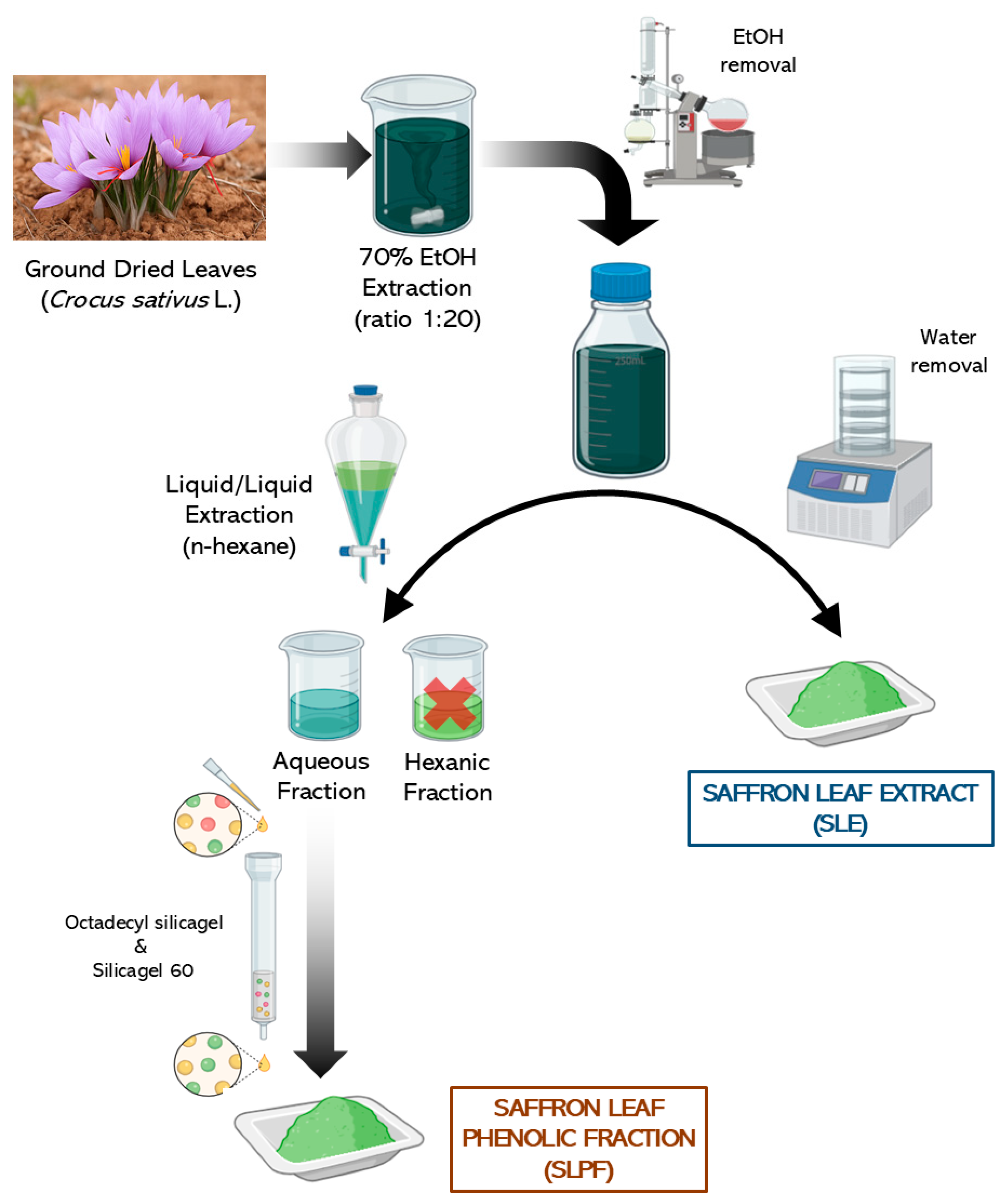
2.3. Saffron Leaf Extraction

Ground dried leaves (30 g) were extracted in 70% ethanol at a ratio of 1:20 for 24 h with continuous stirring in the dark at room temperature (Figure 1), maximizing the extraction of phenolic compounds while preventing their degradation. After the extraction period, the mixture was centrifuged at 5450× g for 30 min to separate the solid residues from the liquid extract. The resulting supernatants were then carefully filtered through a Whatman glass microfiber filter grade GF/A to remove any remaining particulate matter. The ethanol in the filtered supernatant was removed efficiently using an R-100 rotary evaporator (BÜCHI Labortechnik AG, Flawil, Switzerland) at 50 °C and 175 mbar, which helped to concentrate the extract without exposing it to high temperatures that could degrade sensitive compounds. Following the removal of ethanol, the remaining aqueous mixture was subjected to lyophilization using a LyoQuest lyophilizer (Syntegon Telstar, SLU, Barcelona, Spain). This freeze-drying process removed water from the extract, resulting in a dry saffron leaf extract (SLE) that could be stored for subsequent analyses.

2.4. Purification Protocol for Saffron Leaf Phenol-Rich Fraction

Likewise, the aqueous mixture (Figure 1) was subjected to liquid–liquid extraction with n-hexane, discarding the hexanic fraction. Then, the remaining aqueous phase was loaded onto a 100 × 25 mm octadecyl-functionalized silica gel column. Initially, the column was eluted with distilled water to remove non-phenolic compounds such as sugars, amino acids and proteins. Subsequently, methanol was used to elute the polyphenols. The methanol was evaporated using a rotary evaporator and the resulting fraction was dissolved in ethyl acetate before loading onto a 65 × 20 mm silica gel 60 column (25–40 µm). The column was first eluted with ethyl acetate to remove impurities, followed by methanol to elute phenolic compounds. After evaporating the methanol, the final extract was washed with n-hexane and diethyl ether to obtain the semi-purified saffron leaf phenolic fraction (SLPF). The sample was stored at −20 °C before chemical and biological evaluation. The purification of the polyphenols was monitored by thin layer chromatography (TLC) with silica gel 60 plates and a mobile phase of ethyl acetate–water–acetic acid–formic acid (20:5:2:2). Visualization of phenolic compounds on TLC plates was achieved by two methods: (i) spraying with a mixture of sulfuric acid–water–acetic acid (1:4:20) followed by heating on a hot plate and (ii) with 0.03% DPPH in methanol to exploit their antioxidant activity and enhance spot contrast.

2.5. Identification and Quantification of Phenols by HPLC-DAD/ESI-MS

The main phenolic compounds of the SLE and SLPF samples were accurately identified using High-Performance Liquid Chromatography coupled with Diode Array Detection and Electrospray Ionization Mass Spectrometry (HPLC-DAD/ESI-MS) in an HPLC Agilent 1100 Series equipped with an Agilent Technologies 6120 Quadrupole LC/MS (Agilent Technologies, Inc., Santa Clara, CA, USA). The samples were injected into an ACE Excel 3 SuperC18, 150 × 3 mm column (Avantor, Inc., Radnor, PA, USA) at a flow rate of 0.5 mL/min. The injection sample was 10 μL (5 mg/mL) and the analyses were performed in ESI negative ion acquisition mode and spectra were recorded in the 50–1000 m/z range. The interface conditions were as follows: fragmentation 300 V, drying gas flow 10 L/min, nebulizer pressure 40 psig, drying gas temperature 340 °C, vaporizer temperature 150 °C, capillary voltage: 4000 V, charging voltage 2000 V. Mobile phase A (0.05% formic acid in water) and mobile phase B (acetonitrile) were used in gradient for the separation of polyphenols [11]: initial 95% A, 5% B; 10 min 85% A, 15% B; 30 min 75% A, 25% B; 35 min 70% A, 30% B; 50 min 45% A, 55% B; 55 min 10% A, 90% B; 57 min 0% A, 100% B; 58 min 95% A, 5% B; 75 min 95% A, 5% B. Phenolic compounds were tentatively identified by comparing their UV–Vis and mass spectral data, as well as chromatographic retention times, against those of commercial standards and reported compounds in the literature, with additional support from ChatGpt-assisted interpretation [12]. Likewise, phenolics quantification was performed using the calibration curves of commercial aglycones in the range of 0.02–5 µg and expressed as mg/g of dry extract: apigenin (y = 2,074,864.16x − 28,892.21; R2 = 1.00), kaempferol (y = 1,781,684.79x + 1801.24; R2 = 1.00), luteolin (y = 1,430,139.3483x − 10,870.64; R2 = 1.00) and quercetin (y = 1,442,409.29x − 24,915.92; R2 = 1.00).

2.6. Cell Culture and Treatment

Caco-2 cells were cultured with 5% CO2 in Dulbecco’s modified Eagle medium (1000 mg/mL glucose, 110 mg/mL pyruvate and 580 mg/mL glutamine) supplemented with 10% fetal bovine serum, 1% non-essential amino acids, 100 µg/mL penicillin and 100 µg/mL streptomycin. Fetal bovine serum was heat-inactivated at 56 °C for 30 min. The cells were subcultured weekly using trypsin-EDTA and diluted to ensure subculturing before reaching 100% confluence. The medium was renewed once during each passage. The vital stain trypan blue was used for routine cell counting. The cells were seeded in 96-well microplates (90 µL, 2 × 10³ cells/well) and the SLPFs were added immediately afterward (10 µL) to reach a final concentration of 0.5 mg of phenolic extract/mL, 0.5% (v/v) ethanol. The saffron leaf hydroalcoholic extract (SLE) served as a reference model as reported in Sánchez-Vioque (2016) [4], while ethanol at similar concentrations served as the negative control, enabling a reliable assessment of the phenolic fraction’s specific bioactivity. The medium was removed from the 96-well plates and the cells were incubated in fresh growth medium containing the vital stain neutral red (50 µg/mL) for 30 min. The cells were then washed using PBS and absorbance was measured at 550 nm using a plate reader after adding 75 µL of 1% (v/v) acetic acid in 50% (v/v) ethanol [13]. The data are expressed as mean ± SD.

2.7. Iron- and Copper-Chelating Activities

The determination of the iron-chelating activity of the saffron samples followed the protocol described in Sánchez-Vioque et al. (2013) [14] involving the formation of the Fe2+–Ferrozine complex. This method is based on the chelation of this metal ion with ferrozine to yield a red-colored complex. In the presence of phenol-chelating agents, the formation of the complex is disrupted, resulting in a decrease in the red color of the complexes. Samples (0.5 mL) at different concentrations (10–200 mg/mL) were mixed with 2 mL 0.1 M sodium acetate buffer pH 4.9 and 50 μL 2 mM iron(II) chloride, and incubated for 30 min at room temperature. Then, 0.2 mL of 5 mM ferrozine was added and absorbance was measured at 562 nm. Similarly, copper-chelating activity was determined using pyrocatechol violet (PV) as described in Sánchez-Vioque et al. (2013) [14]. The determination of the chelating activity against Cu2+ is based on the formation of the blue-colored Cu2+-PV complex. The blue color turns yellow in the presence of chelating agents that dissociate the complex, and the chelating activity can thus be estimated by measuring the rate of color fading. Samples (0.5 mL) at different concentrations were mixed with 2 mL of 50 mM sodium acetate buffer pH 6.0 and 50 μL 5 mM CuSO4 and incubated at room temperature for 30 min. Then 50 μL 4 mM PV was added, and absorbance at 632 nm was measured. The percentage of inhibition of Ferrozine-Fe2+ and PV–Cu2+ complex formation was calculated as [(A0 − A1)/A0] × 100, where A0 was the absorbance of the negative control, and A1 was the absorbance of the samples. Distilled water and gallic acid served as negative and positive controls for the assays, respectively.

2.8. Statistical Analysis

To evaluate statistical differences in antiproliferative activity between the saffron leaf polyphenol fraction (SLPF) and the saffron leaf extract (SLE), considering both concentration (mg/mL) and exposure time (days), a one-way ANOVA followed by Tukey’s HSD post hoc test (p < 0.01) was performed using SPSS Statistics version 25 (IBM Corp., New York, NY, USA). Additionally, R version 4.4.2 was used for data processing, visualization and for performing linear multivariate and logarithmic regression analyses. These analyses initially supported the estimation of time-specific inhibition percentages (using the general multivariate model) and the calculation of inhibition over time (through logarithmic regression). Subsequently, these analyses allowed for the determination of EC50 and EC90 values (defined as the concentrations required to inhibit 50% and 90% of Caco-2 cell proliferation) through linear regression based on the dose-dependent concentration–inhibition relationship. Similarly, EC50 and EC90 values for the metal-chelating capacities (Fe2+ and Cu2+) were determined through linear regression, based on the dose–response relationship between the concentration of the tested samples and the percentage of metal ion chelation.

3. Results

3.1. Phenolic Composition of Saffron Leaf Extracts

The phenolic profile (Figure 2 and Table 1) of both extracts showcased similar chemical chromatograms, both presenting three major peaks at 20.06, 22.64, and 24.86 min.
Peak (1) likely represents the co-elution of two flavonoid tetrahexosides with respective [M−H] values of 949 and 933 m/z, whose fragmentation patterns were deeply investigated [15] (Table 1). The parent ion with m/z at 949 has been identified as a quercetin tetrahexoside, comprising quercetin + four hexoses (4 × 162 u; C6H12O6-H2O) [C15H10O7+162+162+162+162], probably the quercetin-3-O-sophotrioside-7-O-glucoside [15]. The parent ion at m/z 933 is likely a kaempferol tetrahexoside [C15H10O6+162+162+162+162], which is also consistent with the assignment of the deprotonated molecular ion at m/z 933 observed by Carmona et al. (2007) [16]. Moreover, fragments at m/z 771 [M−H−162], 609 [M−H−162−162] and 447 [M-H-162-162-162] indicated the presence of kaempferol, incorporating a sophotriose (glucose trimer) and a glucose at positions R3 and R7 [15]. While their fragmentation patterns have been meticulously identified, the UV spectra reveal discrepancies, as the detector only exhibited peaks at 272 and 324 nm, suggesting a significant abundance in the kaempferol glycoside, being the quercetin isomer limited to trace amounts.
Peak (2) displayed a quercetin-based molecule through its UV spectra with peaks at 253, 271sh and 342 nm. This suggests the presence of a quercetin (C15H10O7) glycoside. Ion at m/z 625 [C15H10O7+162+162] confirms this, pointing a quercetin dihexoside structure. Ions at m/z at 505 [M-H-120] and 445 [M-H-180], also reported by [15], indicated the presence of quercetin-3-O-sophoroside, previously identified in floral residues of saffron [1,18]. Additionally, the ion at m/z 803 was dismissed as an artifact.
Peak (3), which shows an [M−H] ion at m/z 787, has been variously assigned to a quercetin trihexoside [15] or to myricetin 3-rutinoside-7-O-glucoside [18], though both occur only in trace amounts in saffron petals. Its UV–Vis absorption, however, aligns more closely with kaempferol derivatives. Since multiple studies identify a kaempferol trihexoside as the major flavonoid in saffron extracts [4,16,18,19], we tentatively attribute peak 3 to kaempferol-3-O-glucoside-7-O-sophoroside.
Peak (4) showed UV peaks at 253 and 352 nm, characteristic of the flavonol isorhamnetin (C16H12O7). The ions at m/z 963 [C16H12O7+162+162+162+162] suggest the presence of an isorhamnetin tetrahexoside, as also supported by the ions at m/z 801 [M−H−162] and 681 [M−H−162-120], previously interpreted by Vallejo et al. (2004) [15] as major fragments of isorhamnetin-3-O-sophorotrioside-7-O-glucoside. Moreover, the ion at m/z 771 may correspond to the kaempferol-3-O-glucoside-7-O-sophoroside [C15H10O6+162+162+162] also suggested for peak (3) and present in trace amounts.
Peak (5) presented a UV spectrum that matches with a kaempferol glycoside (C15H10O6). The 609 m/z [C15H10O6+162+162] ion confirmed the presence of a kaempferol diglucoside, which is consistent with the ions 489 [M-H-120] and 429 [M-H-180] of kaempferol-3-O-sophoroside reported by Vallejo et al. (2004) [15].
Peak (6) was identified as luteolin 6-C-glucoside by comparing it to a known commercial standard, as showed the characteristic fragmentation of this C-glucosyl flavone with ions at 447 [M−H], 357 [M−H−90] and 327 m/z [M−H−120], but also to the retention time. Interestingly, another luteolin isomer, luteolin 8-C-glucoside, has also been previously spotted in saffron leaf extracts [19].
Peak (7) presented an unexpected UV spectrum similar to that of a flavanone and an MS pattern with peaks at m/z 771, 609, 429 and 181. Fragment ions at m/z 771 [C15H10O6+162+162+162], 609 [M−H−162] and 429 m/z [M−H−162−180] were in agreement with the fragmentation described by different authors for kaempferol-3-O-sophoroside-7-O-glucoside ([M−H]− at m/z 771) [1,15,16,17]. Although the mass spectrometry (MS) pattern matches this compound, the UV spectrum suggests verifying the presence of a contaminating flavanone or co-elution.
Peak (8) showed a complex mixture of components, as deduced by the different UV spectrum observed. The major and more interesting compound was identified as vitexin (431 [M−H]−, 341 [M−H−90] and 311 [M−H−120]) on the basis of the retention time and fragmentation pattern of the commercial standard.
Peak (9) presented a flavanone UV pattern, but was not definitively attributed to any possible compound. In terms of quantification (Table 1), the semi-purified fraction (SLPF) exhibited a significantly higher concentration of phenolic compounds compared to the crude hydroalcoholic extract (SLE).
The novel purification method resulted in a more than fourfold increase in total phenolic content, from 75.3 mg/g to 330.0 mg/g of dry extract, highlighting the efficiency of the purification process in concentrating these bioactive compounds. The substantial increase in phenolic compounds through this developed methodology underlines the potential of the semi-purified fraction as a rich source of phenolics. With respect to individual phenolic components, their concentrations showed considerable variation, with increases ranging from 2- to 5-fold depending on the compound, demonstrating the selective enrichment of specific compounds during the purification. In particular, kaempferol glycosides were identified as the predominant phenolic components in saffron leaf extracts. Its concentration showed a notable increase from 46 mg/g in the crude extract to 200 mg/g in the SLPF, representing a more than fourfold enrichment.

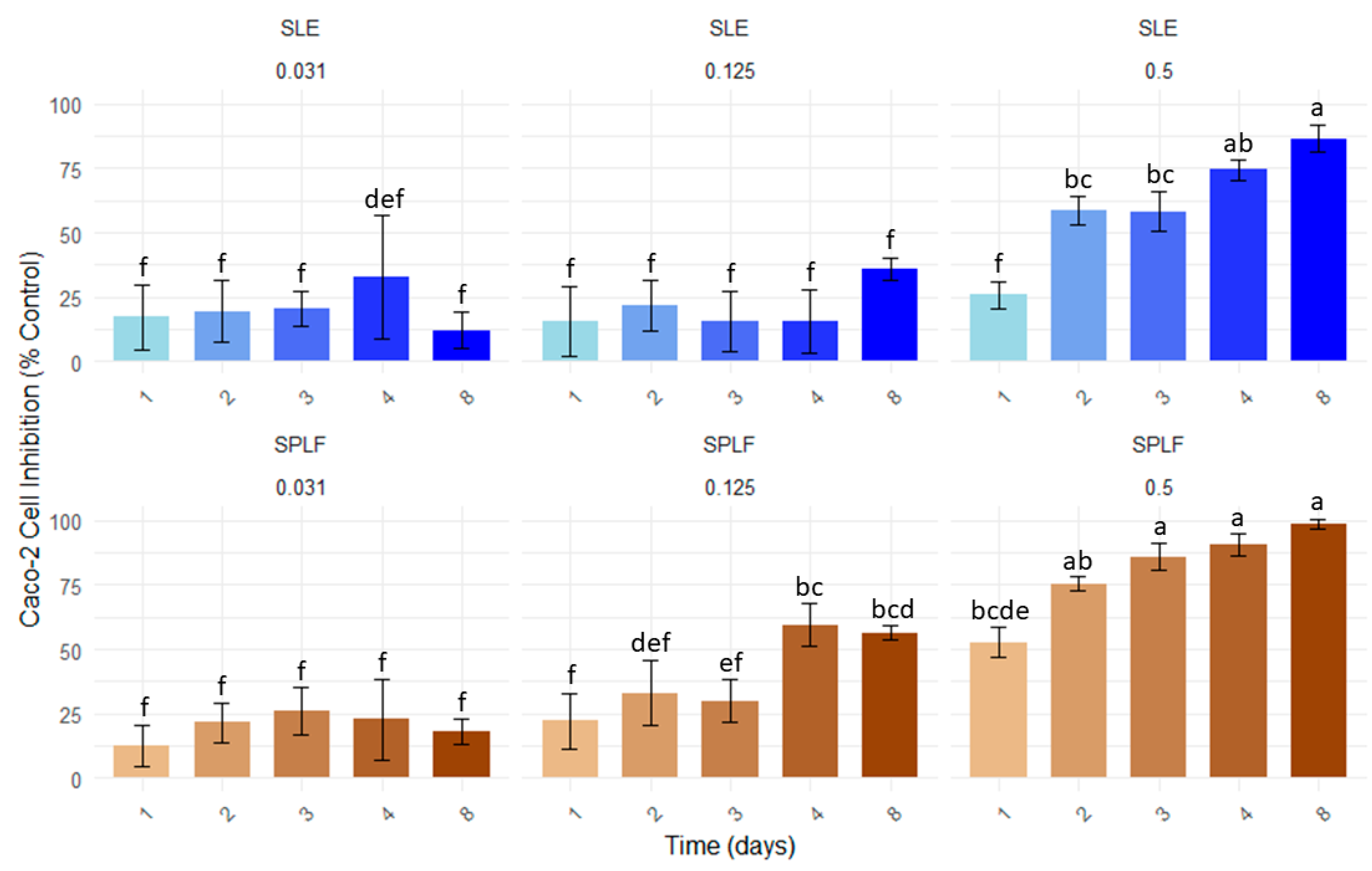
3.2. Effect of Saffron Leaf Extracts on Proliferation of Caco-2 Cells

The effect of saffron leaf phenolic fraction (SLPF) on the proliferation of Caco-2 cells was evaluated in in vitro assays. Caco-2 cells, derived from human colon carcinoma, were cultured under standard conditions to evaluate the impact of these extracts on cell growth. The results, presented in Figure 3, express the inhibition of proliferation in percentages with respect to the control (EtOH), whereas Table 2 presents the EC50 and EC90 values, allowing for a direct comparison between the phenolic fraction of saffron and the unprocessed extract.
The assayed extracts exhibited a clear dose- and time-dependent antiproliferative effect on Caco-2 cells (Figure 3), with SLPF consistently demonstrating stronger inhibitory activity than SLE, especially at intermediate concentrations. Statistical analysis revealed that SLE exerted limited cytotoxic activity on day 1, with robust effects from day 2 to day 4, especially at the highest concentration. In contrast, SLPF induced a progressive and significant inhibitory response from the first 24 h, supporting a more immediate and cumulative mechanism of action. At the concentration of 0.5 mg/mL, SLE reduced cell proliferation by nearly 90% on day 4, whilst for SLPF similar levels were achieved by day 8, suggesting a more sustained and potent inhibitory response for the SLPF. At 0.125 mg/mL, only SLPF induced a significant inhibitory effect, reinforcing the extract’s greater antiproliferative capacity. In contrast, the lowest concentration (0.031 mg/mL) resulted in a modest but constant inhibition of about 20% for both extracts, which gradually decreased over time, and which can be attributed to the chemical degradation of the compounds. This suggests baseline biological activity even at minimal doses. Notably, the control Caco-2 cells continued to proliferate steadily throughout the eight-day incubation period, confirming their viability and normal growth kinetics in the absence of treatment. This observation enhanced the development of a multivariate model (Table 2) that incorporates both concentration (mg/L) and time (days) to statistically estimate the inhibitory potential of each concentration and extract. Otherwise, a logarithmic regression analysis was performed for each sample to mathematically calculate the percentage inhibition at each concentration. Based on these data, linear regression was applied to calculate and estimate the EC50 and EC90 values, thereby quantifying the superior inhibitory potency of SLPF relative to SLE. On day 4, when only a modest statistical increase in inhibitory activity was observed, the EC50 value for SLE was calculated to be 0.31 mg/mL, while that of SLPF was 0.22 mg/mL, indicating an approximately 30% greater inhibitory effect for the phenolic fraction. Furthermore, the EC90 values were calculated to be 0.71 mg/mL for SLE and 0.52 mg/mL for SLPF, further supporting the higher efficacy of SLPF in suppressing Caco-2 cell proliferation. Similarly, the multivariate model estimated EC50 and EC90 values very similar to those obtained from the empirical logarithmic regression data considering inhibition vs. time at the different concentrations, thus improving its reliability, particularly for the SLPF (R2 = 0.82).

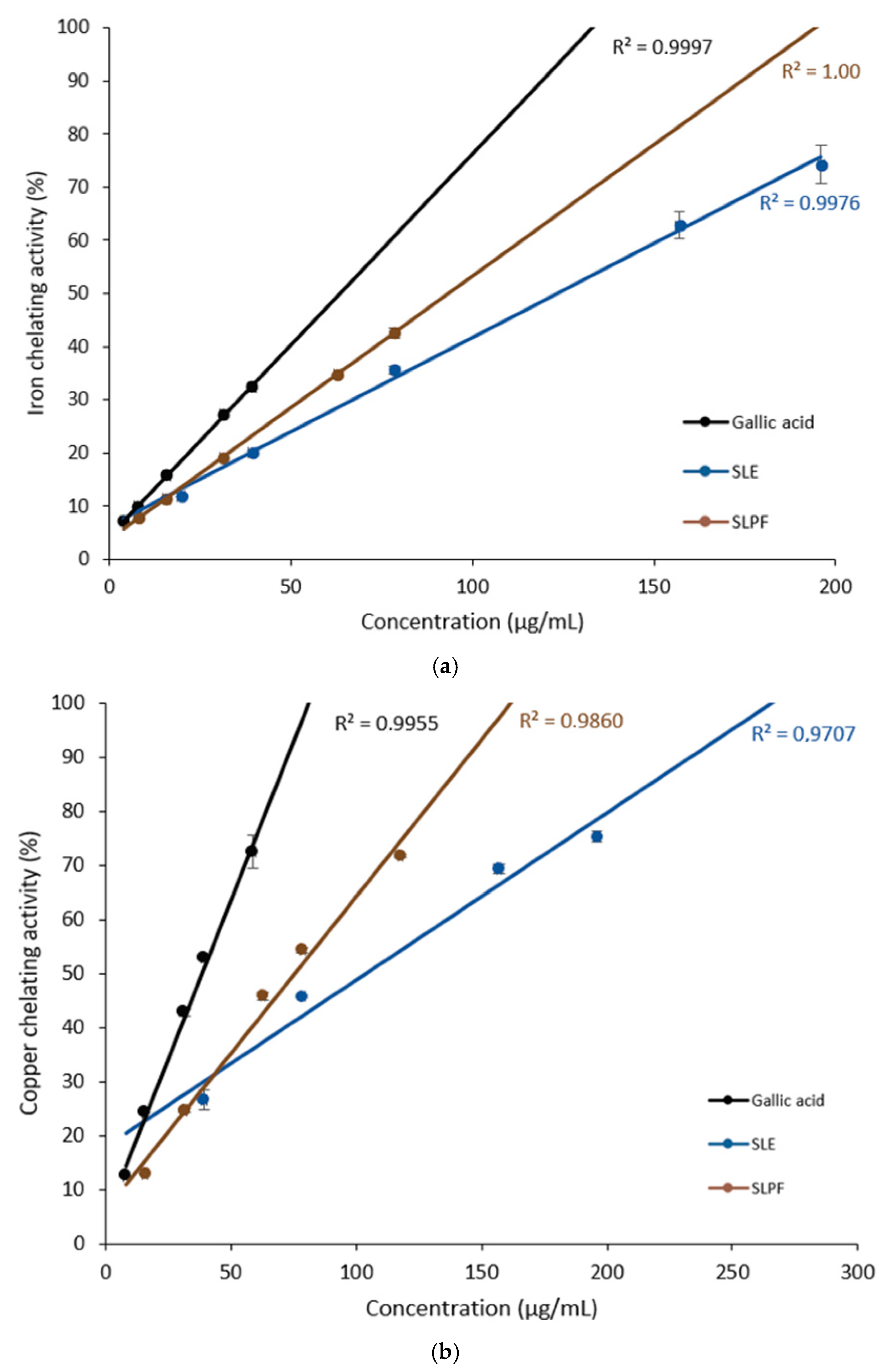
3.3. Iron- and Copper-Chelating Activities of Saffron Leaf Extracts

As shown in Figure 4a, the iron-chelating capacity of SLPF (EC50 = 93 µg/mL) was significantly higher than that of SLE (EC50 = 124 µg/mL), indicating a higher ability of SLPF to bind and sequester metal ions.
Specifically, the chelating activity of SLPF was approximately 70% of the activity observed for the positive control, gallic acid (EC50 = 64 µg/mL), suggesting that the phenolic compounds present in SLPF contribute substantially to its chelating properties by binding to metals. This high chelating efficiency is due to the presence of flavonoids and polyphenolic compounds, known for their strong affinity for metal ions. In terms of copper-chelating activity (Figure 4b), SLPF also demonstrated a stronger effect than SLE, with EC50 values of 75 µg/mL and 104 µg/mL, respectively. These EC50 values represent approximately 50% of the chelating activity observed for gallic acid (EC50 = 38 µg/mL), reinforcing the important role that polyphenolic compounds play in the sequestration of metal ions. The differences observed in the chelating capacities for iron and copper can probably be attributed to the different affinities of the different phenolic compounds for each metal ion. This disparity may be due to the unique structural characteristics of the polyphenols in SLPF and SLE, which could preferentially bind to certain metal ions based on their size, charge, and coordination chemistry.

4. Discussion

Numerous flavonoids have showcased promising chemoprotective effects on human colon cancer cell lines through multiple molecular and biochemical mechanisms of action [8]. For example, quercetin presents inhibitory effects on the growth and proliferation of several colorectal cancer cells, including Caco-2 cells. Similarly, myricetin has been shown to inhibit proliferation and induce apoptosis in colorectal cancer cells via the modulation of apoptotic and inflammatory pathways [8]. Quercetin-3-O-sophoroside along with other quercetin and kaempferol glycosides have demonstrated cytotoxic activity against P-388 murine leukemia cells [20]. Additionally, isorhamnetin glycosides have been shown to induce apoptosis in HT-29 and Caco-2 human colon cancer cells [21]. Saffron leaves are rich in flavonoid C-glycosides such as luteolin-6-C-glucoside (isoorientin) or apigenin-8-C-glucoside (vitexin), which have recently captured the attention of researchers due to their health benefits, including anticancer properties [22]. According to He et al. (2016) [23], vitexin inhibits autophagy and thereby induces apoptosis in hepatocellular carcinoma cells by activating the JNK signaling pathway. Conversely, isoorientin has been shown to induce apoptosis, decrease invasiveness and downregulate vascular endothelial growth factor in pancreatic cancer cells [24]. An intriguing aspect of flavonoid C-monoglycosides concerning colorectal cancer is their limited absorption by humans, potentially leading to the accumulation of higher concentrations in the colon compared to aglycones or flavonoid O-glycosides, which are more easily absorbed in the gastrointestinal tract [22].
Experimental evidence suggests that iron and other metals may act as carcinogenic factors involved in both the initiation and promotion of cancer. Transition metals like iron catalyze Fenton’s reaction to produce reactive oxygen species that contribute to carcinogenesis, so elevated iron levels have been documented in colorectal tumor tissues [25]. This role is particularly relevant in colorectal cancer because of the relatively high concentration of iron in feces, especially in individuals with a diet rich in red meat [8]. To address this issue, several synthetic chelators have been developed to deplete iron levels in cells for antitumor therapies, potentially increasing therapeutic benefits in the management of colorectal cancer.
The ability of phenolic compounds to chelate transition metals is well-established, dependent on the presence and arrangement of hydroxyl and carbonyl groups within the molecules. Notably, the phenolic compounds identified in saffron leaves present some of these reactive sites: kaempferol, isorhamnetin and apigenin derivatives have available the 4-carbonyl and 5-hydroxy groups, and quercetin glycosides and isoorientin 4-carbonyl and 5-hydroxy groups as well as the 3′ and 4′ dihydroxy groups. This is consistent with the significant iron- and copper-chelating activities observed in this study. The EC50 values of SLE for iron and copper chelation activities compare favorably with those of other plant by-products previously studied [14], highlighting the potential of saffron leaves as a source of chelating compounds. Notably, the chelating activity of its phenolic compounds was found to be comparable to that of well-established chelating agents, such as the hydroxybenzoic acid phenol, gallic acid. Several studies have extensively explored the iron- and copper-chelating capabilities of the aglycones present in the major flavonoids identified in saffron leaves, including quercetin, kaempferol, isorhamnetin, luteolin and apigenin. Despite variations observed due to the pH, the preferred binding sites, and the resulting stoichiometry of the formed complexes, all these flavonoids demonstrated metal-chelating activity [26]. Quercetin, one of the most abundant flavonoids in nature, has been extensively studied for its metal-chelating properties. Depending on the metal chelated, the resultant complexes may display higher [27] or lower [28] antioxidant activity compared to quercetin itself, or may have even demonstrated prooxidant activity as in copper complexes [29].
Considering that colorectal adenocarcinoma cells are subjected to high oxidative stress and accumulate copper levels up to seven times higher than the surrounding non-tumorous epithelial cells, compounds capable of chelating this metal, including phenolic compounds, are being explored as selective agents for inducing cancer cell death through a copper-dependent pro-oxidant pathway [9]. Conversely, Cu2+ serves as a potent stimulator of blood vessel proliferation, which can promote malignant angiogenesis and tumor growth [30]. Consequently, reducing copper levels through chelation may represent a therapeutic strategy through naturally occurring flavonoids.
This study not only advances the scientific understanding of the biological effects of saffron leaf polyphenols but also introduces an innovative and efficient methodology for their extraction and characterization. It thus lays the fundamental foundation for transforming agricultural waste into high-value biomedical resources, thereby promoting sustainability and circularity within the saffron industry. This insight represents a pioneering contribution to the integration of circular bioeconomy principles into saffron production, opening new avenues for the development of complementary and environmentally friendly treatments for colorectal cancer with broad biotechnological applications.
However, it is important to note that this is an initial exploratory and preliminary study with several limitations. First, the results are derived exclusively from in vitro experiments with a single colorectal cancer cell line (Caco-2), which do not reflect the biological complexity of actual tumor behavior. Second, the underlying mechanisms of action and the bioavailability of individual phenolic components remain unclear, requiring further purification, elucidation and investigation; as does the chemical variability among different saffron samples (heterogeneity, age, phenological changes, chemical biodegradability, etc.). Third, the lack of in vivo studies limits the ability to assess systemic effects, potential cytotoxic effects on healthy cells and the therapeutic relevance of the extracts. Finally, rigorous human clinical trials are essential to validate both their efficacy and safety before considering their incorporation into cancer prevention and treatment strategies.

5. Conclusions

This study demonstrates that phenolic compounds isolated from saffron leaves are in the main responsible for the antiproliferative effects on Caco-2 cells, as suggested in previous studies. Phenolic profile analysis identified several flavonoid glycosides, including those derived from the aglycones quercetin, kaempferol, isorhamnetin, luteolin and apigenin, highlighting the diverse composition of saffron leaves. In particular, the phenolic fraction of the saffron leaf (SLPF) exhibited a significantly higher concentration of these bioactive compounds compared to the crude hydroalcoholic extract (SLE), suggesting its greater potential for therapeutic applications, particularly in experimental medical treatments. Our insights reveal that SLPF exhibited more potent inhibitory effects than SLE on Caco-2 cell proliferation, one of the most recognized test models for colorectal cancer, underscoring the critical role of phenolic compounds in suppressing tumor cell growth. A key innovation of this work lies in the development of an extractive methodology and the demonstration of the dose-dependent antiproliferative capacity of SLPF, reinforcing the availability of its extract as a promising experimental agent for cancer treatment. In addition, the iron- and copper-chelating activities of the saffron leaf polyphenols emphasize their potential as chemoprotective agents, particularly in the context of colorectal cancer, where the deregulation of metal ions may promote carcinogenesis. These findings highlight the untapped potential of saffron leaves, an underutilized waste, as a valuable source of bioactive compounds with promising health promotion capacities.

Author Contributions

Conceptualization, R.S.-V. and E.G.-R.; data curation, J.V.-P.; formal analysis, M.A.; investigation, R.S.-V., J.G.-C., J.V.-P., A.M.-M., E.G.-R. and L.M.-C.; methodology, R.S.-V., J.G.-C., J.V.-P., A.M.-M., E.G.-R. and L.M.-C.; software, M.A.; supervision, J.G.-C.; validation, M.A.; writing—original draft, R.S.-V. and G.O.d.E.-C.; writing—review and editing, G.O.d.E.-C. All authors have read and agreed to the published version of the manuscript.

Funding

This research was internally funded by the Regional Institute of Agri-Food and Forestry Research and Development of Castilla-La Mancha (IRIAF).

Institutional Review Board Statement

Not applicable.

Informed Consent Statement

Not applicable.

Data Availability Statement

Data will be made available on request.

Acknowledgments

We extend our gratitude to Miguel Ángel Escolano Tercero for his valuable assistance in agricultural fieldwork and saffron collection, as well as to Noemi Cerro Ibáñez and David Prieto Blanco for their technical support in the analytical procedures. During the preparation of this manuscript, the authors utilized ChatGPT [17] to assist with grammar refinement and data validation. The authors have reviewed and edited the output and take full responsibility for the content of this publication.

Conflicts of Interest

The authors declare no conflicts of interest.

References

  1. Serrano-Díaz, J.; Sánchez, A.M.; Martínez-Tomé, M.; Winterhalter, P.; Alonso, G.L. Flavonoid Determination in the Quality Control of Floral Bioresidues from Crocus sativus L. J. Agric. Food Chem. 2014, 62, 3125–3133. [Google Scholar] [CrossRef] [PubMed]
  2. Serrano-Díaz, J.; Sánchez, A.M.; Martínez-Tomé, M.; Winterhalter, P.; Alonso, G.L. A Contribution to Nutritional Studies on Crocus sativus Flowers and Their Value as Food. J. Food Compos. Anal. 2013, 31, 101–108. [Google Scholar] [CrossRef]
  3. Serrano-Díaz, J.; Sánchez, A.M.; Maggi, L.; Martínez-Tomé, M.; García-Diz, L.; Murcia, M.A.; Alonso, G.L. Increasing the Applications of Crocus sativus Flowers as Natural Antioxidants. J. Food Sci. 2012, 77, C1162–C1168. [Google Scholar] [CrossRef]
  4. Sánchez-Vioque, R.; Santana-Méridas, O.; Polissiou, M.; Vioque, J.; Astraka, K.; Alaiz, M.; Herraiz-Peñalver, D.; Tarantilis, P.A.; Girón-Calle, J. Polyphenol Composition and In Vitro Antiproliferative Effect of Corm, Tepal and Leaf from Crocus sativus L. on Human Colon Adenocarcinoma Cells (Caco-2). J. Funct. Foods 2016, 24, 18–25. [Google Scholar] [CrossRef]
  5. Rahaiee, S.; Ranjbar, M.; Azizi, H.; Govahi, M.; Zare, M. Green Synthesis, Characterization, and Biological Activities of Saffron Leaf Extract-Mediated Zinc Oxide Nanoparticles: A Sustainable Approach to Reuse an Agricultural Waste. Appl. Organomet. Chem. 2020, 34, e5705. [Google Scholar] [CrossRef]
  6. Soror, T.Y. Saffron Extract as Environmentally Safe Corrosion Inhibitors for Aluminium Dissolution in 2M HCl Solution. Eur. Chem. Bull. 2013, 2, 191–196. [Google Scholar]
  7. Sánchez-Vioque, R.; Rodríguez-Conde, M.F.; Reina-Ureña, J.V.; Escolano-Tercero, M.A.; Herraiz-Peñalver, D.; Santana-Méridas, O. In Vitro Antioxidant and Metal Chelating Properties of Corm, Tepal and Leaf from Saffron (Crocus sativus L.). Ind. Crop Prod. 2012, 39, 149–153. [Google Scholar] [CrossRef]
  8. Araújo, J.R.; Gonçalves, P.; Martel, F. Chemopreventive Effect of Dietary Polyphenols in Colorectal Cancer Cell Lines. Nutr. Res. 2011, 31, 77–87. [Google Scholar] [CrossRef]
  9. Fatfat, M.; Merhi, R.A.; Rahal, O.; Stoyanovsky, D.A.; Zaki, A.; Haidar, H.; Kagan, V.E.; Gali-Muhtasib, H.; Machaca, K. Copper Chelation Selectively Kills Colon Cancer Cells through Redox Cycling and Generation of Reactive Oxygen Species. BMC Cancer 2014, 14, 527. [Google Scholar] [CrossRef]
  10. Fernández, J.A.; Santana, O.; Guardiola, J.L.; Molina, R.V.; Heslop-Harrison, P.; Borbely, G.; Branca, F.; Argento, S.; Maloupa, E.; Talou, T.; et al. The World Saffron and Crocus Collection: Strategies for Establishment, Management, Characterisation and Utilisation. Genet. Resour. Crop Evol. 2011, 58, 125–137. [Google Scholar] [CrossRef]
  11. Jordán, M.J.; Lax, V.; Rota, M.C.; Lorán, S.; Sotomayor, J.A. Effect of the Phenological Stage on the Chemical Composition, and Antimicrobial and Antioxidant Properties of Rosmarinus Officinalis L Essential Oil and Its Polyphenolic Extract. Ind. Crops Prod. 2013, 48, 144–152. [Google Scholar] [CrossRef]
  12. OpenAI ChatGPT-4o. Available online: https://chat.openai.com/chat (accessed on 10 April 2025).
  13. Girón-Calle, J.; Alaiz, M.; Vioque, J. Effect of Chickpea Protein Hydrolysates on Cell Proliferation and In Vitro Bioavailability. Food Res. Int. 2010, 43, 1365–1370. [Google Scholar] [CrossRef]
  14. Sánchez-Vioque, R.; Polissiou, M.; Astraka, K.; de los Mozos-Pascual, M.; Tarantilis, P.; Herraiz-Peñalver, D.; Santana-Méridas, O. Polyphenol Composition and Antioxidant and Metal Chelating Activities of the Solid Residues from the Essential Oil Industry. Ind. Crops Prod. 2013, 49, 150–159. [Google Scholar] [CrossRef]
  15. Vallejo, F.; Tomás-Barberán, F.A.; Ferreres, F. Characterisation of Flavonols in Broccoli (Brassica Oleracea L. Var. Italica) by Liquid Chromatography-UV Diode-Array Detection-Electrospray Ionisation Mass Spectrometry. J. Chromatogr. A 2004, 1054, 181–193. [Google Scholar] [CrossRef]
  16. Carmona, M.; Sánchez, A.M.; Ferreres, F.; Zalacain, A.; Tomás-Barberán, F.; Alonso, G.L. Identification of the Flavonoid Fraction in Saffron Spice by LC/DAD/MS/MS: Comparative Study of Samples from Different Geographical Origins. Food Chem. 2007, 100, 445–450. [Google Scholar] [CrossRef]
  17. Goupy, P.; Vian, M.A.; Chemat, F.; Caris-Veyrat, C. Identification and Quantification of Flavonols, Anthocyanins and Lutein Diesters in Tepals of Crocus sativus by Ultra Performance Liquid Chromatography Coupled to Diode Array and Ion Trap Mass Spectrometry Detections. Ind. Crop Prod. 2013, 44, 496–510. [Google Scholar] [CrossRef]
  18. Naim, N.; Bouymajane, A.; Oulad, Y.; Majdoub, E.; Ezrari, S.; Lahlali, R.; Tahiri, A.; Ennahli, S.; Lagan, R.; Cacciola, F.; et al. Crocus sativus L. Petal Extracts. 2023, 28, 186. [Google Scholar]
  19. Smolskaite, L.; Talou, T.; Fabre, N.; Venskutonis, P.R. Volarization of Saffron Industry by-Products: Bioactive Compounds from Leaves. In Proceedings of the 6th Baltic Conference on Food Science and Technology FOODBALT-2011, Jelgava, Latvia, 5–6 May 2011; pp. 67–72. [Google Scholar]
  20. Aisyah, L.S.; Yun, Y.F.; Herlina, T.; Julaeha, E.; Zainuddin, A.; Nurfarida, I.; Hidayat, A.T.; Supratman, U.; Shiono, Y. Flavonoid Compounds from the Leaves of Kalanchoe Prolifera and Their Cytotoxic Activity against P-388 Murine Leukimia Cells. Nat. Prod. Sci. 2017, 23, 139–145. [Google Scholar] [CrossRef]
  21. Antunes-Ricardo, M.; Moreno-García, B.E.; Gutiérrez-Uribe, J.A.; Aráiz-Hernández, D.; Alvarez, M.M.; Serna-Saldivar, S.O. Induction of Apoptosis in Colon Cancer Cells Treated with Isorhamnetin Glycosides from Opuntia Ficus-Indica Pads. Plant Foods Hum. Nutr. 2014, 69, 331–336. [Google Scholar] [CrossRef]
  22. Xiao, J.; Capanoglu, E.; Jassbi, A.R.; Miron, A. Advance on the Flavonoid C-Glycosides and Health Benefits. Crit. Rev. Food Sci. Nutr. 2016, 56, S29–S45. [Google Scholar] [CrossRef]
  23. He, M.; Min, J.W.; Kong, W.L.; He, X.H.; Li, J.X.; Peng, B.W. A Review on the Pharmacological Effects of Vitexin and Isovitexin. Fitoterapia 2016, 115, 74–85. [Google Scholar] [CrossRef] [PubMed]
  24. Ye, T.; Su, J.; Huang, C.; Yu, D.; Dai, S.; Huang, X.; Chen, B.; Zhou, M. Isoorientin Induces Apoptosis, Decreases Invasiveness, and Downregulates VEGF Secretion by Activating AMPK Signaling in Pancreatic Cancer Cells. OncoTargets Ther. 2016, 9, 7481–7492. [Google Scholar] [CrossRef]
  25. Labropoulou, E.P.; Datsis, A.; Kekelos, S.; Rogdakis, A.; Katsiki, E.; Labropoulou, C.M.; Spiliotis, J.; Christopoulou, A. The Presence and Significance of Iron in Neoplasia of the Colorectum. Tech. Coloproctol. 2004, 8, s211–s213. [Google Scholar] [CrossRef] [PubMed]
  26. Mladěnka, P.; MacÁková, K.; Filipský, T.; Zatloukalová, L.; Jahodář, L.; Bovicelli, P.; Silvestri, I.P.; Hrdina, R.; Saso, L. In Vitro Analysis of Iron Chelating Activity of Flavonoids. J. Inorg. Biochem. 2011, 105, 693–701. [Google Scholar] [CrossRef] [PubMed]
  27. Chen, W.; Sun, S.; Cao, W.; Liang, Y.; Song, J. Antioxidant Property of Quercetin-Cr(III) Complex: The Role of Cr(III) Ion. J. Mol. Struct. 2009, 918, 194–197. [Google Scholar] [CrossRef]
  28. Dehghan, G.; Khoshkam, Z. Tin(II)-Quercetin Complex: Synthesis, Spectral Characterisation and Antioxidant Activity. Food Chem. 2012, 131, 422–426. [Google Scholar] [CrossRef]
  29. Tan, J.; Wang, B.; Zhu, L. DNA Binding and Oxidative DNA Damage Induced by a Quercetin copper(II) Complex: Potential Mechanism of Its Antitumor Properties. J. Biol. Inorg. Chem. 2009, 14, 727–739. [Google Scholar] [CrossRef]
  30. Nasulewicz, A.; Mazur, A.; Opolski, A. Role of Copper in Tumour Angiogenesis—Clinical Implications. J. Trace Elem. Med. Biol. 2004, 18, 1–8. [Google Scholar] [CrossRef]
Figure 1. A schematic overview of the complete extraction and purification process, from ground saffron leaves to the final saffron leaf phenolic fraction (SLPF).
Applsci 15 07376 g001
Figure 2. HPLC-DAD chromatogram of the saffron leaf polyphenol fraction (SLPF) recorded at 280 nm, highlighting phenolic compound profiles. Peak identification (1–9) is reported in Table 1.
Applsci 15 07376 g002
Figure 3. Antiproliferative effects of saffron leaf extract (SLE; blue) and saffron leaf polyphenol fraction (SLPF; brown) on Caco-2 cell proliferation over 8 days, expressed as a percentage relative to the control (EtOH). Cells were treated with three concentrations: 0.5 mg/mL, 0.125 mg/mL and 0.031 mg/mL. Data represent mean ± SD. Different letters denote statistically significant differences between treatments and time points (Tukey’s test, p < 0.01).
Applsci 15 07376 g003
Figure 4. Iron- (a) and copper (b)-chelating activities (%) of SLE and SLPF at various concentrations (0–300 µg/mL). Data are presented as mean ± SD of three determinations.
Applsci 15 07376 g004
Table 1. Identification and quantification of phenolic compounds in saffron leaf extracts.
#PeakRt (min)UV-Vis (λmax; nm)Mass (g/mol)Fragmentation Ions
(m/z)
Tentative Identification (UV + MS Data)ReferencesSLE
(mg/g)
SLPF
(mg/g)
Δ (%)
118.68 ± 0.2272/324(a) 950
(b) 934
949(100), 933(30), 771(10), 609(5), 447(5)(a) Quercetin-3-O-sophotrioside-7-O-glucoside (tr.)
(b) Kaempferol 3-O-glucoside-7-O-sophotrioside
(c) Kaempferol 3-O-sophtrioside-7-O-glucoside
[15,16]1.425.06256.01
219.77 ± 0.3253/271sh/342626803(100), 625(90), 505(20), 445(5), 314(10)Quercetin 3-O-sophoroside[1,15,17]4.1521.71422.54
320.06 ± 0.0271/337787787(100)(a) Kaempferol-3-O-glucoside-7-O-sophoroside
(b) Quercetin trihexoside (tr.)
(c) Myricetin 3-rutinoside-7-glucoside
[4,16,18,19]25.63132.97418.71
421.89 ± 0.3253/352(a) 964
(b) 772
963(15), 801(100), 771(55), 681(10)(a) Isorhamnetin-3-O-sophotrioside-7-O-glucoside a
(b) Kaempferol-3-O-glucoside-7-O-sophoroside (tr.)
[15]1.164.09251.70
521.20 ± 0.6272/334610609(100), 489(15), 429(10), 298(10)Kaempferol 3-O-sophoroside[1,15,17]1.907.68305.26
622.64 ± 0.3254/268sh/340448447(100), 357(95), 327(90), 298(55), 133(40)Luteolin 6-C-glucoside (isoorientin)[19]; Standard15.7586.06446.50
724.86 ± 0.4284772771(100), 609(15), 429(10), 181(10)Kaempferol-3-O-sophoroside-7-O-glucoside[1,15,16,17]17.0253.44213.94
825.48 ± 0.2272/330432431(100), 341(40), 311(90), 283(55), 117(25)Apigenin-8-C-glucoside (vitexin)[19]; Standard0.762.33206.97
929.29 ± 0.7284/330-401(20), 196(30), 181(100), 153(20)Unknown-7.5316.65121.08
TOTAL75.33330.00438.07
a quantified as quercetin; sh: shoulder.
Table 2. Multivariate Model Test and Estimated Linear Regression Model (for day 4), EC50 and EC90 values ±SE (with upper and lower limits; 96% confidence intervals) for saffron extract-induced inhibition estimated from regression analyses on day 4.
SampleMultivariate
Model (R2)
Linear Regression Model (Day 4)EC50 (Day 4)EC90 (Day 4)
Estimated (R2) 1Calculated (R2) 2Estimated 1CalculatedEstimated 1Calculated 2
SLEy = 95.82C + 2.84t + 3.48
(0.67)
y = 95.83x + 14.85
(1.00)
y = 100.35x + 19.15
(0.92)
0.370.31 ± 0.02
(0.26–0.35)
0.780.71 ± 0.05
(0.60–0.81)
SLPFy = 124.82C + 3.45t + 7.43
(0.82)
y = 124.82x + 21.24
(1.00)
y = 135.33x + 19.87
(0.96)
0.230.22 ± 0.01
(0.20–0.24)
0.550.52 ± 0.03
(0.46–0.57)
Δ (%)37.8429.0329.4826.76
1 Based on the Multivariate Linear Model, 2 data was calculated using a logarithmic regression model relating time (days) to percentage of inhibition.
Disclaimer/Publisher’s Note: The statements, opinions and data contained in all publications are solely those of the individual author(s) and contributor(s) and not of MDPI and/or the editor(s). MDPI and/or the editor(s) disclaim responsibility for any injury to people or property resulting from any ideas, methods, instructions or products referred to in the content.

