Article

Acute Effects of Static Stretching on Submaximal Force Control of the Ankle

1
Graduate School of Health Sciences, Hokkaido University, Sapporo 060-0812, Japan
2
Faculty of Health Sciences, Hokkaido University, Sapporo 060-0812, Japan
3
Department of Rehabilitation, Orthopedic Hokushin Hospital, Sapporo 060-0908, Japan
*
Author to whom correspondence should be addressed.
Appl. Sci. 2025, 15(13), 7294; https://doi.org/10.3390/app15137294 (registering DOI)
Submission received: 18 May 2025 / Revised: 22 June 2025 / Accepted: 26 June 2025 / Published: 28 June 2025
(This article belongs to the Special Issue Sports Biomechanics and Injury Prevention)

Abstract

Static stretching (SS) is widely used in clinical and sports settings. However, the acute effects on neuromuscular control during dynamic tasks remain unclear. This study aimed to examine the immediate effects of SS on force control using a randomized crossover design. Seventeen healthy young males performed low-range (10–30% of maximal voluntary isometric contraction: MVIC) and high-range (40–60% MVIC) isometric force tracking tasks. In the SS condition, the ankle plantar flexors were stretched for 60 s; in the control condition, the participants remained at rest. The primary outcomes included ankle dorsiflexion range of motion (ROM), musculotendinous stiffness, and the root mean square error (RMSE) of force tracking. Compared to the control group, SS significantly increased dorsiflexion ROM and reduced musculotendinous stiffness. A significant reduction in the RMSE was observed during the force release phase when participants smoothly decreased force output in the high-range task following SS (p = 0.030, d = 0.79), but no significant changes were found during the force generation phase in the high-range task or during either phase (generation or release) in the low-range task. These findings suggest that a brief SS intervention may acutely refine the dynamic force control under high neuromuscular demands. Therefore, SS may enhance motor control in tasks that involve submaximal force modulation.

1. Introduction

Static stretching (SS) involves extending a particular muscle or muscle group to its end range and maintaining that position for a prescribed time with the goal of enhancing flexibility and increasing joint mobility. It is widely utilized in sports and rehabilitation settings [1,2,3]. In the context of sports performance, SS has traditionally been incorporated into warm-up routines to prepare the muscles for subsequent physical activity by optimizing muscle extensibility and joint mobility. Moreover, SS contributes to the prevention of acute musculoskeletal injuries, particularly in sports involving rapid or repetitive muscle lengthening [4]. However, despite its common use, the acute physiological effects of SS on neuromuscular function, particularly in relation to force production and motor control, remain under ongoing investigation.
SS is known to decrease the stiffness of the muscle–tendon complex [5] and reduce the sensitivity of muscle spindles [6]. Reduced muscle spindle sensitivity may compromise proprioceptive input and reflex activity. These SS-induced alterations, including changes in muscle–tendon compliance, length–tension relationship, and central neural drive, may modulate the nervous system to finely regulate muscle force output [5,7,8,9,10]. Taken together, these findings suggest that SS can acutely influence neuromuscular function and potentially affect the precision of force modulation.
Many daily tasks demand precise adjustment of the force output and accurate modulation of muscle force in accordance with environmental and task-specific demands is essential for motor control [11]. When such modulations are effective, it can facilitate smooth movement and lead to joint stability [12,13]. On the other hand, impaired force modulation may result in excessive or insufficient force production, leading to movement inefficiency and compromised postural control [14,15]. Maximal muscle strength is a common performance outcome in sports. However, sports such as football and basketball require rapid movement transitions and fine motor adjustments, necessitating the ability to exert force at the appropriate timing and magnitude. Thus, an impaired ability to finely adjust muscle force can lead to inefficient movement strategies, increased energy expenditure, and reduced movement accuracy or stability, which may hinder functional performance in both daily life and sports [16,17]. Conversely, appropriate modulation of muscle activity may contribute to smoother coordination, more successful task performance, and lower risk of injury. Therefore, assessment of force control at submaximal intensities is considered important.
Force control is defined as the ability to generate muscle contraction with precision and stability under static or dynamic conditions [18]. Previous studies have suggested that the SS negatively affects this ability. For instance, SS consisting of three sets of 60 s applied to the knee extensor muscles significantly decreased force steadiness at 5% and 20% maximal voluntary isometric contraction (MVIC) [19]. Similarly, five sets of 60 s of SS applied to the ankle plantar flexors resulted in a significant decline in force steadiness at 20% MVIC [20]. However, these studies primarily assessed force steadiness using isometric contractions with a constant target torque. The effects of SS on force control under dynamic conditions involving force fluctuations are unclear. In real-world movements, force is not exerted at a fixed level but is dynamically adjusted to match the demands of the tasks. Moreover, many of these real-world tasks are preceded by SS as part of warm-up or rehabilitation protocols. Therefore, understanding how SS affects the ability to modulate submaximal force under dynamic conditions is important. Understanding how SS affects submaximal force control may help clarify the mechanisms underlying SS impact on performance, thereby supporting its more effective application in sports and rehabilitation settings.
One approach to evaluating force control in dynamic conditions is the use of a force-tracking task, in which participants are required to continuously match their force output to a changing target trajectory in real time. This method allows for the assessment of not only the overall accuracy of force production but also the ability to modulate force precisely in both timing and magnitude, providing a comprehensive assessment of neuromuscular control [21,22]. Specifically, the task demands temporal precision, adjusting the force output at the correct moment to follow the target waveform, and magnitude accuracy, which involves generating the appropriate level of force to match the target amplitude. In contrast, previous studies evaluating the effects of SS on force control have primarily focused on force steadiness measured under isometric conditions with constant target torques [19,20]. This approach primarily reflects magnitude accuracy, that is, how consistently one can maintain a specified force level without accounting for the temporal aspects of force modulation. Given that SS has been shown to impair force steadiness, it is plausible that similar neuromuscular alterations may extend to dynamic tasks that require continuous adjustments in both the timing and magnitude of force. Moreover, SS has been reported to reduce the rate of force development (RFD) [23,24], which may hinder the rapid modulation of the force output required in the time-sensitive phases of dynamic tasks. A decline in RFD may compromise temporal precision, especially during the force generation or release phases that demand fast and accurate transitions in force levels.
To date, no studies have investigated the effects of SS on dynamic force control during force-tracking tasks, which require continuous and precise adjustment of submaximal force. Therefore, this study aimed to investigate the acute effects of SS of ankle plantar flexors on force control using a time-varying force-tracking task. It was hypothesized that SS would lead to a decrease in force-tracking performance, particularly in tasks that require precise modulation of the submaximal force.

2. Materials and Methods

2.1. Participants

Seventeen healthy young male participants were enrolled in the study (age: 23.5 ± 2.0 years; height: 172.6 ± 4.5 cm; body mass: 65.9 ± 6.1 kg). Individuals with musculoskeletal impairments or neurological conditions affecting the lower extremities were excluded from the study. To control for potential confounding factors, all participants were instructed to avoid strenuous physical activity, alcohol consumption, and caffeine intake for at least 24 h before testing. Female participants were excluded to minimize the effects of hormonal fluctuations related to the menstrual cycle, which can influence muscle stiffness and neuromuscular control [25,26]. All experimental sessions were conducted at a consistent time of day for each participant within a ± 1 h window, to minimize the influence of circadian variation. The testing limb was selected based on limb dominance. All participants were right-limb dominant in this study, so the measurements were performed on the right leg. Ethical approval was obtained from the Institutional Review Board of the affiliated university (approval number: 23-31). All participants were given a comprehensive explanation of the study purpose and procedures and provided written informed consent before participation. Prior to recruitment, a priori power analysis was performed using G*Power 3.1 (Heinrich Heine University, Düsseldorf, Germany) to determine the minimum sample size. Based on a two-way repeated-measures ANOVA (condition × time) with an assumed medium effect size (f = 0.25, alpha = 0.05, power = 0.80), the required sample size was estimated to be 17.

2.2. Study Design

This study employed a randomized crossover design to examine the acute effects of SS on force control of the plantar flexors. All participants completed both an SS condition (a single 60 s static stretching) and a control condition (no stretching). The order of these conditions was randomized for each participant via a random draw using a mobile application (Randomizer App, Randomness LLC, Corning, IA, USA). The two experimental sessions were separated by at least 48 h to minimize carry-over effects.
Upon arrival at the laboratory, the participants were instructed to remain seated at rest for 10 min to allow for acclimatization. They then completed two practice trials for each of the two types of force tracking tasks. Subsequently, range of motion (ROM) at ankle joint was assessed, and the peak passive resistance torque, used later in the SS protocol, was determined. Before and after the 60-s SS intervention, performance on force-tracking tasks and maximal dorsiflexion ROM were evaluated. All measurements and interventions were conducted on the dominant leg, which is defined as the leg used to kick a ball. All participants identified their right leg as their dominant leg. Laboratory room temperature was maintained at 24 ± 1 °C throughout the procedure.

2.3. Measurement of Maximum Angle of Ankle Dorsiflexion and Passive Resistance Torque

The maximum dorsiflexion angle of the ankle joint and the corresponding passive resistance torque were assessed using a Biodex System 4 dynamometer (Biodex Medical Systems, Inc., Shirley, NY, USA) in conjunction with the LabVIEW 2013 software (National Instruments, Austin, TX, USA). The measurement position was set with the hip joint at 70° and the right knee joint at full extension. The participant’s right foot was positioned at 0° of ankle dorsiflexion with the lateral malleolus carefully aligned to the rotational axis of the dynamometer. The foot was stabilized on the footplate by securing straps. An ankle joint angle of 0° was defined as the orientation in which the footplate stood perpendicular to the floor. The trunk, pelvis, and distal thigh were stabilized using straps. The assessment began with the ankle positioned at 20° of plantar flexion, followed by passive dorsiflexion at a constant angular velocity of 5°/s until the torque was reached the preset maximum passive resistance level. The maximum passive resistance torque was identified at the torque which the participant experienced the greatest tolerable stretch without pain. The participants were instructed to remain relaxed throughout the measurement period to avoid any voluntary muscle activation.

2.4. Force-Tracking Task

The force-tracking task was carried out using Biodex System 4 dynamometer. During all trials, the participants were seated with their hip fixated at 70° flexion, knee fully extended (0°), and ankle positioned neutrally. The pelvis and torso were stabilized with straps across the waist and chest (Figure 1a). Prior to performing the task, the peak torque of the right ankle plantar flexors was measured using two maximal voluntary isometric contractions (MVICs) conducted before the stretching intervention. Each contraction was sustained for 3 s with 1 min rest between trials [27]. The highest value from the two trials was recorded as the peak torque of the participant. A computer monitor was positioned 100 cm in front of the participant, displaying both the target force trajectory and the real-time force output generated by pushing against the dynamometer attachment in the plantarflexion direction at the neutral ankle position (Figure 1b). In this system, greater force exertion causes the feedback line to move upward on the screen. The participants were instructed to match their output as accurately and rapidly as possible to the target trace, but no specific guidance was provided regarding the ankle movement strategy.
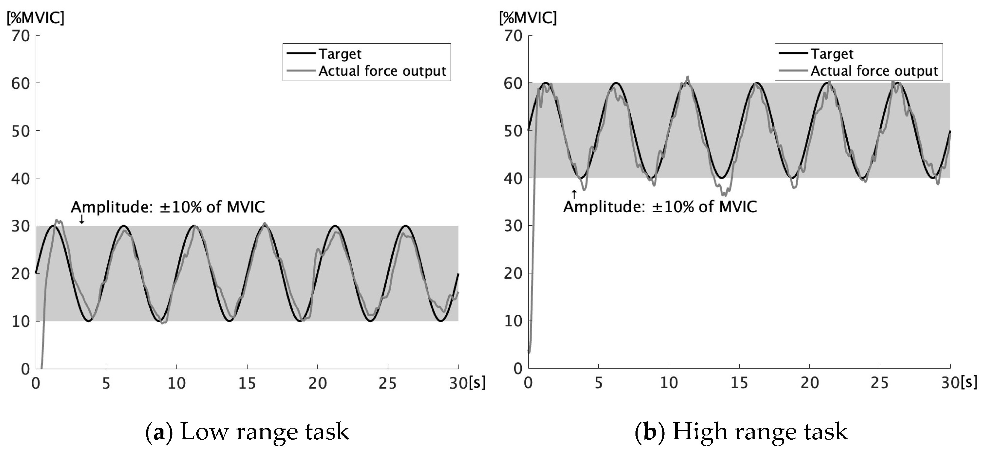
A sinusoidal pattern of the target force was generated using a custom LabVIEW program (Figure 2). Task parameters were determined based on the peak plantar flexion torque (MVIC) of each participant. Two conditions were tested: (1) a low-range task, with a frequency of 0.2 Hz (equivalent to 12.6%/s) and a force range of 10% to 30% of MVIC [22], and (2) a high-range task, also at 0.2 Hz, but spanning 40% to 60% of MVIC [21]. Both conditions shared an amplitude of 10% of MVIC. The tasks were administered in a fixed order from low to high, with adequate rest intervals between trials. The post-stretch measurements followed the same sequence as the pre-stretch measurements. Throughout the task, the monitor presented both the reference torque trajectory and the participant’s applied torque, thus offering continuous visual feedback.

2.5. Static Stretching Protocol

SS was applied to the right ankle plantar flexors using a Biodex System 4 dynamometer, with the participant positioned identically to the measurement of the maximal dorsiflexion angle and passive resistance torque. The stretch began at 20° of plantarflexion and passively progressed at an angular velocity of 5°/s until the predetermined maximal passive resistance torque was reached. Subsequently, a constant torque stretch was performed for 60 s using the torque-controlled mode of the Biodex System 4, whereby the torque level was held constant, and the joint angle was allowed to change over time. In the control condition, participants remained seated at rest in the same position without any stretching intervention.

2.6. Data Processing and Analysis

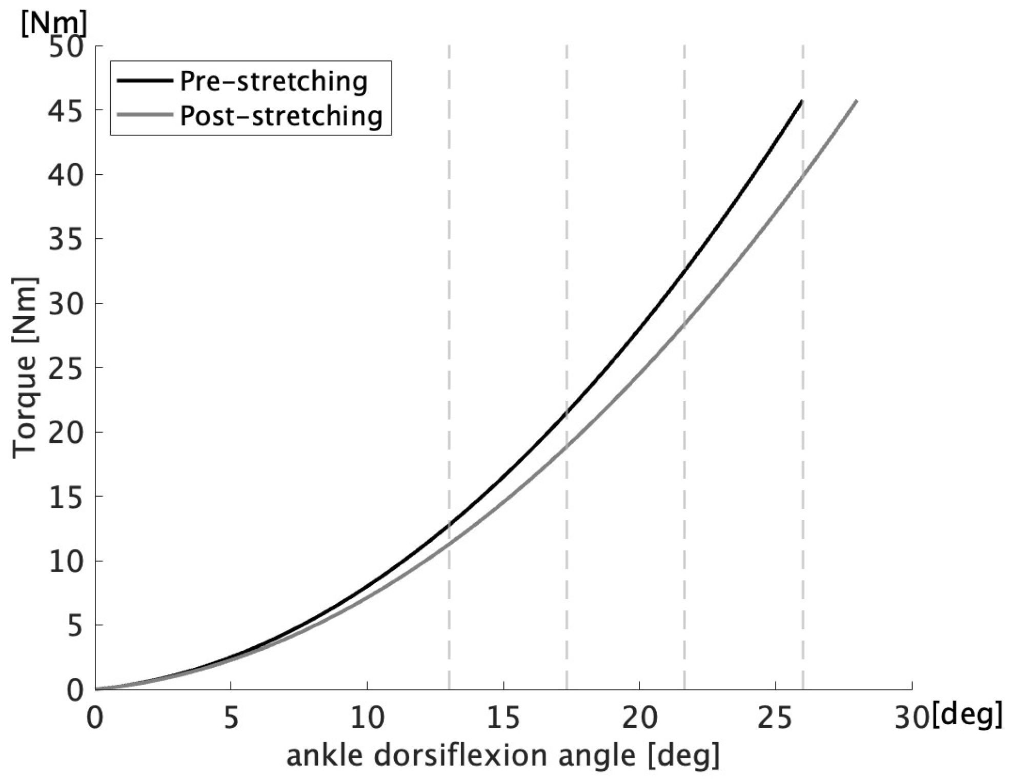
All data processing and analyses were conducted using a custom script developed in MATLAB R2024a (MathWorks Inc., Natick, MA, USA). The force signals obtained from the Biodex system were low-pass filtered using a zero-lag second-order Butterworth filter with a cutoff frequency of 10 Hz [28]. A torque–angle curve was generated based on the measured passive resistance torque and dorsiflexion angle data and fitted using a second-order polynomial regression. Musculotendinous stiffness is defined as the slope of the curve. The slope values were computed at 4° intervals across the final 13° of the range of motion, and the mean of these four points was used to represent overall stiffness [29,30]. Identical joint angle ranges were used for calculations in both intervention and control conditions (Figure 3).
For the force-tracking task, data from five cycles, excluding the initial force trajectory, were obtained for each participant. The data were analyzed separately for the force generation phase (increasing force) and the force release phase (decreasing force). The RMSE values were calculated for each of the five individual cycles, and the average of the three lowest RMSE values, indicating the highest accuracy, was used as the representative value for each condition. This “best-of” approach was adopted to minimize the influence of sporadic performance variability, such as attentional lapses or transient motor errors, and to better reflect participants’ optimal tracking performance. Similar methods have been employed in motor control research to ensure stable and representative outcomes [31]. The force tracking performance was evaluated in terms of both temporal accuracy and magnitude accuracy. The RMSE between the actual force and target force was calculated for all tasks using the following equation [32]:
RMSE = 1 n i = 1 n T i F i 2
where n is the number of data points, Ti is the target force, and Fi is the actual force.

2.7. Statistical Analysis

All statistical analyses were conducted using the SPSS software (version 26.0, IBM Corp., Armonk, NY, USA). Descriptive statistics were reported as means and standard deviations. The normality of the data distribution was assessed using the Shapiro–Wilk test. To evaluate the effects of condition (SS vs. control) and time (pre vs. post), a two-way repeated measures analysis of variance (ANOVA) was performed. When a significant main effect or interaction was observed, post hoc comparisons were conducted using Bonferroni correction to control for multiple comparisons. Effect sizes were calculated using Cohen’s d, with thresholds of 0.2, 0.5, and 0.8, indicating small, medium, and large effects, respectively [33]. Statistical significance was set at p < 0.05.

3. Results

As illustrated in Table 1, two-way repeated-measures ANOVA identified a significant time × condition interaction for the maximal ankle dorsiflexion angle (F (1,16) = 19.14, p < 0.001, partial η2 = 0.55). Post hoc tests revealed a significant increase in the dorsiflexion angle from pre- to post-intervention in the SS condition (p < 0.001, d = 1.57) whereas no significant change was observed in the control condition (p > 0.999, d = 0.50). Table 1 presents the results for the stiffness. Significant main effects were found for both time (F (1,16) = 7.71, p = 0.013, partial η2 = 0.33) and condition (F (1,16) = 12.92, p = 0.002, partial η2 = 0.45), with no significant interaction. Follow-up comparisons revealed a significant reduction in stiffness only in the SS condition (p = 0.034, d = 0.78), without notable changes in the control condition (p > 0.999, d = 0.14).
Table 2 summarizes the RMSE results from the force-tracking tasks. A significant reduction in RMSE was found in the high-range release phase under the SS condition (p = 0.030, d = 0.79), whereas no significant change in RMSE was observed in the control condition during the same phase. In addition, significant main effects of time were found in the low-range generation phase (F (1,16) = 15.47, p = 0.001, partial η2 = 0.49) and the high-range release phase (F (1,16) = 13.55, p = 0.002, partial η2 = 0.46). However, no significant main effects of condition or interaction effects were detected in any phase. All other comparisons were not statistically significant.

4. Discussion

This study aimed to investigate the acute effects of SS of the plantar flexors on force control during a dynamic force-tracking task. Notably, the force-tracking performance, as assessed by the RMSE, did not significantly change in the low-range task, but showed a significant improvement in the high-range task, particularly during the force release phase. These findings provide novel evidence that the effects of SS on neuromuscular performance may vary depending on the intensity and phase-specific demands of the task. In addition, SS led to a significant increase in ankle dorsiflexion range of motion and a decrease in musculotendinous stiffness, consistent with previous reports [5,29].
The high-range task, which requires participants to produce a larger absolute torque within the same relative range (40–60% MVIC), likely imposes greater neuromuscular demands compared to the low-range task. Specifically, the need for precise control over higher force levels may render the task more sensitive to subtle neuromechanical changes induced by the SS. Among the two force phases, the release phase appears to be particularly demanding, as it requires rapid and smooth downregulation of muscular activity, a process that is generally more difficult to control than force generation [21]. In contrast, the generation phase primarily involves straightforward force production, which may be less influenced by small changes in neuromuscular function. Taken together, the refinement observed specifically in the release phase of the high-range task suggests that this condition effectively captured acute neuromuscular changes induced by SS.
Although previous studies have consistently reported impaired force steadiness following SS during isometric tasks with constant force demands [24,25], the current results demonstrate a contrasting enhancement in isometric tasks involving dynamic force modulation, particularly during the high-range release phase. A key factor that may explain this discrepancy is the nature of force control required for each task. Traditional steadiness assessments involve maintaining a fixed level of force output, which emphasizes stability. In contrast, the force-tracking task used in this study required participants to continuously adjust their force to match a time-varying target, engaging different aspects of neuromuscular control, particularly those related to fine-tuned modulation over time.
Furthermore, the stretching duration may also contribute to divergent outcomes. Studies reporting negative effects often employed longer SS protocols (e.g., five sets of 60 s), which may have induced neuromuscular inhibition, characterized by decreased excitability of muscle spindles and α-motoneuron, as well as suppressed central pathways, which led to reduced spinal excitability revealed by attenuated H-reflex responses [34,35,36,37], and reduced force transmission efficiency due to increased compliance of the muscle–tendon unit [38]. In contrast, the present study used a single 60 s stretch, which may have been sufficient to reduce mechanical resistance and promote smoother movement without eliciting inhibitory neural responses [39]. Recent findings also suggest that 4 × 30 s SS can suppress unnecessary co-contraction without compromising the force-generating capacity [40]. The suppression of co-contraction may reduce unnecessary force from antagonist muscles, allowing the agonist to contract more efficiently, which in turn enables more precise and smoother modulation of force output. This mechanism has been supported by previous research showing that improvements in force control are accompanied by reductions in antagonist co-contraction [41]. These results indicate that both the stretching duration and modulation of co-contraction are key factors that influence the acute neuromechanical effects of static stretching on force control.
From practical perspectives, the findings of this research suggest that short-duration SS may be a useful preparatory strategy to enhance dynamic force control in tasks that require rapid modulation of submaximal force output. Significant enhancement was observed only during the release phase of the high-range task, where the RMSE showed a statistically significant reduction following SS. This change was supported by a medium effect size (Cohen’s d = 0.79), indicating a consistent effect of the intervention. However, its functional relevance should be interpreted with caution. Taken together, these results provide evidence that short-duration static stretching may modestly enhance neuromuscular control, particularly in tasks requiring precise and timely force modulation. Although the findings are limited to a single-session, laboratory-based setting, they suggest a potential foundation for future applications in athletic and rehabilitation contexts. Further research is necessary to determine whether these effects translate to real-world functional performance or long-term outcomes.
This study had several limitations that should be acknowledged. First, although a randomized crossover design was employed, the potential influence of learning or task familiarization effects cannot be entirely ruled out. Second, the sample consisted exclusively of healthy young male adults, which restricts the generalizability of the findings to other populations, such as females, older adults, or clinical groups. Third, this study focused solely on plantar flexors, and it remains unclear whether the findings can be generalized to other muscle groups or joints with different anatomical and functional properties. Fourth, the absence of direct neurophysiological assessments (e.g., EMG, reflex responses, or corticospinal excitability) limits the ability to infer the underlying mechanisms responsible for the observed changes in force control. For instance, co-contractions of antagonist muscles may have influenced force regulation, but were not measured in this study. Central neural drive could also have contributed to the force regulation following SS, yet it was not directly assessed. Moreover, the lack of EMG data restricts interpretation of changes in neuromuscular activation strategies. Incorporating such measures in future studies could help clarify whether the SS-induced improvements are driven by changes in muscle activation strategies, neural excitability, or reflex modulation. Fifth, the effects of static stretching are known to be highly time-dependent and may diminish rapidly after intervention. Therefore, the timing of the post-stretching assessment could have influenced the results, and this factor should be considered when interpreting our findings.

5. Conclusions

This study investigated the acute effects of SS of the plantar flexors on force control during a dynamic force tracking task. The findings revealed that SS significantly improved force control accuracy in the high-range task, particularly during the force-release phase, as reflected by a reduction in the RMSE. These results suggest that short-duration SS may be beneficial for enhancing the accuracy of submaximal force under specific conditions that require precise and timely modulation of the submaximal force output. Unlike previous studies reporting impaired force steadiness during static isometric tasks, the current results suggest that SS enhances neuromuscular regulation during tasks involving continuous force adjustment.

Author Contributions

Conceptualization, all authors; methodology, all authors; software, F.S. and S.K.; validation, F.S.; formal analysis, F.S.; investigation, F.S. and M.S. (Masahiro Sato); resources, M.S. (Mina Samukawa); data curation, F.S. and M.S. (Masahiro Sato); writing—original draft preparation, F.S.; writing—review and editing, all authors and M.S. (Mina Samukawa); visualization, F.S., Y.K. and M.S. (Mina Samukawa); supervision, M.S. (Mina Samukawa); project administration, M.S. (Mina Samukawa). All authors have read and agreed to the published version of the manuscript.

Funding

This research received no external funding.

Institutional Review Board Statement

This study was conducted in accordance with the Declaration of Helsinki and approved by the Ethics Committee of the Faculty of Health Sciences, Hokkaido University (approval number: 23-31, 7 July 2023).

Informed Consent Statement

Written informed consent was obtained from all participants for study participation and publication.

Data Availability Statement

The data supporting the conclusions of this article will be made available by the authors on request.

Conflicts of Interest

The authors declare no conflicts of interest.

References

  1. Konrad, A.; Budini, F.; Tilp, M. Acute Effects of Constant Torque and Constant Angle Stretching on the Muscle and Tendon Tissue Properties. Eur. J. Appl. Physiol. 2017, 117, 1649–1656. [Google Scholar] [CrossRef] [PubMed]
  2. Sato, S.; Kiyono, R.; Takahashi, N.; Yoshida, T.; Takeuchi, K.; Nakamura, M. The Acute and Prolonged Effects of 20-s Static Stretching on Muscle Strength and Shear Elastic Modulus. PLoS ONE 2020, 15, e0228583. [Google Scholar] [CrossRef] [PubMed]
  3. Nakamura, M.; Yoshida, R.; Sato, S.; Yahata, K.; Murakami, Y.; Kasahara, K.; Fukaya, T.; Takeuchi, K.; Nunes, J.P.; Konrad, A. Comparison Between High- and Low-Intensity Static Stretching Training Program on Active and Passive Properties of Plantar Flexors. Front. Physiol. 2021, 12, 796497. [Google Scholar] [CrossRef] [PubMed]
  4. Chaabene, H.; Behm, D.G.; Negra, Y.; Granacher, U. Acute Effects of Static Stretching on Muscle Strength and Power: An Attempt to Clarify Previous Caveats. Front. Physiol. 2019, 10, 1468. [Google Scholar] [CrossRef]
  5. Oba, K.; Samukawa, M.; Nakamura, K.; Mikami, K.; Suzumori, Y.; Ishida, Y.; Keeler, N.; Saitoh, H.; Yamanaka, M.; Tohyama, H. Influence of Constant Torque Stretching at Different Stretching Intensities on Flexibility and Mechanical Properties of Plantar Flexors. J. Strength Cond. Res. 2021, 35, 709–714. [Google Scholar] [CrossRef]
  6. Budini, F.; Rafolt, D.; Christova, M.; Gallasch, E.; Tilp, M. The Recovery of Muscle Spindle Sensitivity Following Stretching Is Promoted by Isometric but Not by Dynamic Muscle Contractions. Front. Physiol. 2020, 11, 905. [Google Scholar] [CrossRef]
  7. Fowles, J.R.; Sale, D.G.; MacDougall, J.D. Reduced Strength after Passive Stretch of the Human Plantarflexors. J. Appl. Physiol. 2000, 89, 1179–1188. [Google Scholar] [CrossRef]
  8. Ghaffarinejad, F.; Taghizadeh, S.; Mohammadi, F. Effect of Static Stretching of Muscles Surrounding the Knee on Knee Joint Position Sense. Br. J. Sports Med. 2007, 41, 684–687. [Google Scholar] [CrossRef]
  9. Trajano, G.S.; Nosaka, K.; B. Seitz, L.; Blazevich, A.J. Intermittent Stretch Reduces Force and Central Drive More than Continuous Stretch. Med. Sci. Sports Exerc. 2014, 46, 902–910. [Google Scholar] [CrossRef]
  10. Masugi, Y.; Obata, H.; Inoue, D.; Kawashima, N.; Nakazawa, K. Neural Effects of Muscle Stretching on the Spinal Reflexes in Multiple Lower-Limb Muscles. PLoS ONE 2017, 12, e0180275. [Google Scholar] [CrossRef]
  11. Latash, M.L. Motor Control: On the Way to Physics of Living Systems. Adv. Exp. Med. Biol. 2014, 826, 1–16. [Google Scholar] [CrossRef] [PubMed]
  12. Wuebbenhorst, K.; Zschorlich, V. The Effect of Increasing External Degrees of Freedom on Force Production and Neuromuscular Stabilisation. J. Sports Sci. 2012, 30, 1561–1569. [Google Scholar] [CrossRef] [PubMed]
  13. An, K.-N. Muscle Force and Its Role in Joint Dynamic Stability. Clin. Orthop. Relat. Res. 2002, 403, S37–S42. [Google Scholar] [CrossRef]
  14. Enoka, R.M.; Duchateau, J. Muscle Fatigue: What, Why and How It Influences Muscle Function: Muscle Fatigue. J. Physiol. 2008, 586, 11–23. [Google Scholar] [CrossRef] [PubMed]
  15. Hirono, T.; Ikezoe, T.; Taniguchi, M.; Yamagata, M.; Miyakoshi, K.; Umehara, J.; Ichihashi, N. Relationship between Ankle Plantar Flexor Force Steadiness and Postural Stability on Stable and Unstable Platforms. Eur. J. Appl. Physiol. 2020, 120, 1075–1082. [Google Scholar] [CrossRef]
  16. Pethick, J.; Tallent, J. The Neuromuscular Fatigue-Induced Loss of Muscle Force Control. Sports 2022, 10, 184. [Google Scholar] [CrossRef]
  17. Wade, L.; Lichtwark, G.A.; Farris, D.J. Joint and Muscle-Tendon Coordination Strategies during Submaximal Jumping. J. Appl. Physiol. 2020, 128, 596–603. [Google Scholar] [CrossRef]
  18. Chow, J.W.; Stokic, D.S. Force Control of Quadriceps Muscle Is Bilaterally Impaired in Subacute Stroke. J. Appl. Physiol. 2011, 111, 1290–1295. [Google Scholar] [CrossRef]
  19. Nakamura, M.; Suzuki, Y.; Yoshida, R.; Kasahara, K.; Murakami, Y.; Hirono, T.; Nishishita, S.; Takeuchi, K.; Konrad, A. The Time-Course Changes in Knee Flexion Range of Motion, Muscle Strength, and Rate of Force Development after Static Stretching. Front. Physiol. 2022, 13, 917661. [Google Scholar] [CrossRef]
  20. Kato, E.; Vieillevoye, S.; Balestra, C.; Guissard, N.; Duchateau, J. Acute Effect of Muscle Stretching on the Steadiness of Sustained Submaximal Contractions of the Plantar Flexor Muscles. J. Appl. Physiol. 2011, 110, 407–415. [Google Scholar] [CrossRef]
  21. Ebisu, S.; Kasahara, S.; Saito, H.; Ishida, T. Decrease in Force Control among Older Adults under Unpredictable Conditions. Exp. Gerontol. 2022, 158, 111649. [Google Scholar] [CrossRef] [PubMed]
  22. Perraton, L.; Clark, R.; Crossley, K.; Pua, Y.-H.; Whitehead, T.; Morris, H.; Telianidis, S.; Bryant, A. Impaired Voluntary Quadriceps Force Control Following Anterior Cruciate Ligament Reconstruction: Relationship with Knee Function. Knee Surg. Sports Traumatol. Arthrosc. 2017, 25, 1424–1431. [Google Scholar] [CrossRef] [PubMed]
  23. Panidi, I.; Kotsala, E.; Bogdanis, G.C.; Gaspari, V.; Spiliopoulou, P.; Terzis, G.; Donti, O. Muscle Architecture of Gastrocnemius Medialis and Rate of Force Development during Different Stretching Protocols. Proceedings 2019, 25, 22. [Google Scholar] [CrossRef]
  24. Morais de Oliveira, A.L.; Greco, C.C.; Molina, R.; Denadai, B.S. The Rate of Force Development Obtained at Early Contraction Phase Is Not Influenced by Active Static Stretching. J. Strength Cond. Res. 2012, 26, 2174–2179. [Google Scholar] [CrossRef]
  25. Eiling, E.; Bryant, A.L.; Petersen, W.; Murphy, A.; Hohmann, E. Effects of Menstrual-Cycle Hormone Fluctuations on Musculotendinous Stiffness and Knee Joint Laxity. Knee Surg. Sports Traumatol. Arthrosc. 2007, 15, 126–132. [Google Scholar] [CrossRef]
  26. Yim, J.; Petrofsky, J.; Lee, H. Correlation between Mechanical Properties of the Ankle Muscles and Postural Sway during the Menstrual Cycle. Tohoku J. Exp. Med. 2018, 244, 201–207. [Google Scholar] [CrossRef]
  27. Manoel, M.E.; Harris-Love, M.O.; Danoff, J.V.; Miller, T.A. Acute Effects of Static, Dynamic, and Proprioceptive Neuromuscular Facilitation Stretching on Muscle Power in Women. J. Strength Cond. Res. 2008, 22, 1528–1534. [Google Scholar] [CrossRef]
  28. Kasahara, S.; Saito, H. Effect of Loading Parameters on Motor Performance during a Dynamic Weight-Shift Task. Gait Posture 2015, 41, 100–105. [Google Scholar] [CrossRef]
  29. Mizuno, T. Changes in Joint Range of Motion and Muscle-Tendon Unit Stiffness after Varying Amounts of Dynamic Stretching. J. Sports Sci. 2017, 35, 2157–2163. [Google Scholar] [CrossRef]
  30. Mizuno, T.; Matsumoto, M.; Umemura, Y. Decrements in Stiffness Are Restored within 10 Min. Int. J. Sports Med. 2013, 34, 484–490. [Google Scholar] [CrossRef]
  31. Pedersen, A.V.; Lorås, H. When Is a Test Score Fair for the Individual Who Is Being Tested? Effects of Different Scoring Procedures across Multiple Attempts When Testing a Motor Skill Task. Front. Psychol. 2017, 8, 619. [Google Scholar] [CrossRef] [PubMed]
  32. Knol, H.; Huys, R.; Temprado, J.-J.; Sleimen-Malkoun, R. Performance, Complexity and Dynamics of Force Maintenance and Modulation in Young and Older Adults. PLoS ONE 2019, 14, e0225925. [Google Scholar] [CrossRef] [PubMed]
  33. Cohen, J. Statistical Power Analysis for the Behavioral Sciences, 2nd ed.; Lawrence Erlbaum Associates: Hillsdale, NJ, USA, 1988. [Google Scholar]
  34. Damasceno, M.V.; Duarte, M.; Pasqua, L.A.; Lima-Silva, A.E.; MacIntosh, B.R.; Bertuzzi, R. Static Stretching Alters Neuromuscular Function and Pacing Strategy, but Not Performance during a 3-Km Running Time-Trial. PLoS ONE 2014, 9, e99238. [Google Scholar] [CrossRef] [PubMed]
  35. Pekünlü, E.; Sağıroğlu, İ.; Kurt, C.; Özsu, İ. Acute Residual Effects of Short and Long Duration Static Stretching on Counter Movement Jump Performances in Well-Trained Female Combat Athletes. Eur. J. Phys. Educ. Sport Sci. 2016, 2. [Google Scholar] [CrossRef]
  36. Behm, D.G.; Blazevich, A.J.; Kay, A.D.; McHugh, M. Acute Effects of Muscle Stretching on Physical Performance, Range of Motion, and Injury Incidence in Healthy Active Individuals: A Systematic Review. Appl. Physiol. Nutr. Metab. 2016, 41, 1–11. [Google Scholar] [CrossRef]
  37. Avela, J.; Kyröläinen, H.; Komi, P.V. Altered Reflex Sensitivity after Repeated and Prolonged Passive Muscle Stretching. J. Appl. Physiol. 1999, 86, 1283–1291. [Google Scholar] [CrossRef]
  38. Marek, S.M.; Cramer, J.; Fincher, A.; Massey, L.; Dangelmaier, S.; Purkayastha, S.; Fitz, K.A.; Culbertson, J. Acute Effects of Static and Proprioceptive Neuromuscular Facilitation Stretching on Muscle Strength and Power Output. J. Athl. Train. 2005, 40, 94–103. [Google Scholar]
  39. Clark, L.; O’Leary, C.B.; Hong, J.; Lockard, M. The Acute Effects of Stretching on Presynaptic Inhibition and Peak Power. J. Sports Med. Phys. Fit. 2014, 54, 605–610. [Google Scholar]
  40. Ruan, M.; Zhang, Q.; Wu, X. Acute Effects of Static Stretching of Hamstring on Performance and Anterior Cruciate Ligament Injury Risk during Stop-Jump and Cutting Tasks in Female Athletes. J. Strength Cond. Res. 2017, 31, 1241–1250. [Google Scholar] [CrossRef]
  41. Patten, C.; Kamen, G. Adaptations in Motor Unit Discharge Activity with Force Control Training in Young and Older Human Adults. Eur. J. Appl. Physiol. 2000, 83, 128–143. [Google Scholar] [CrossRef]
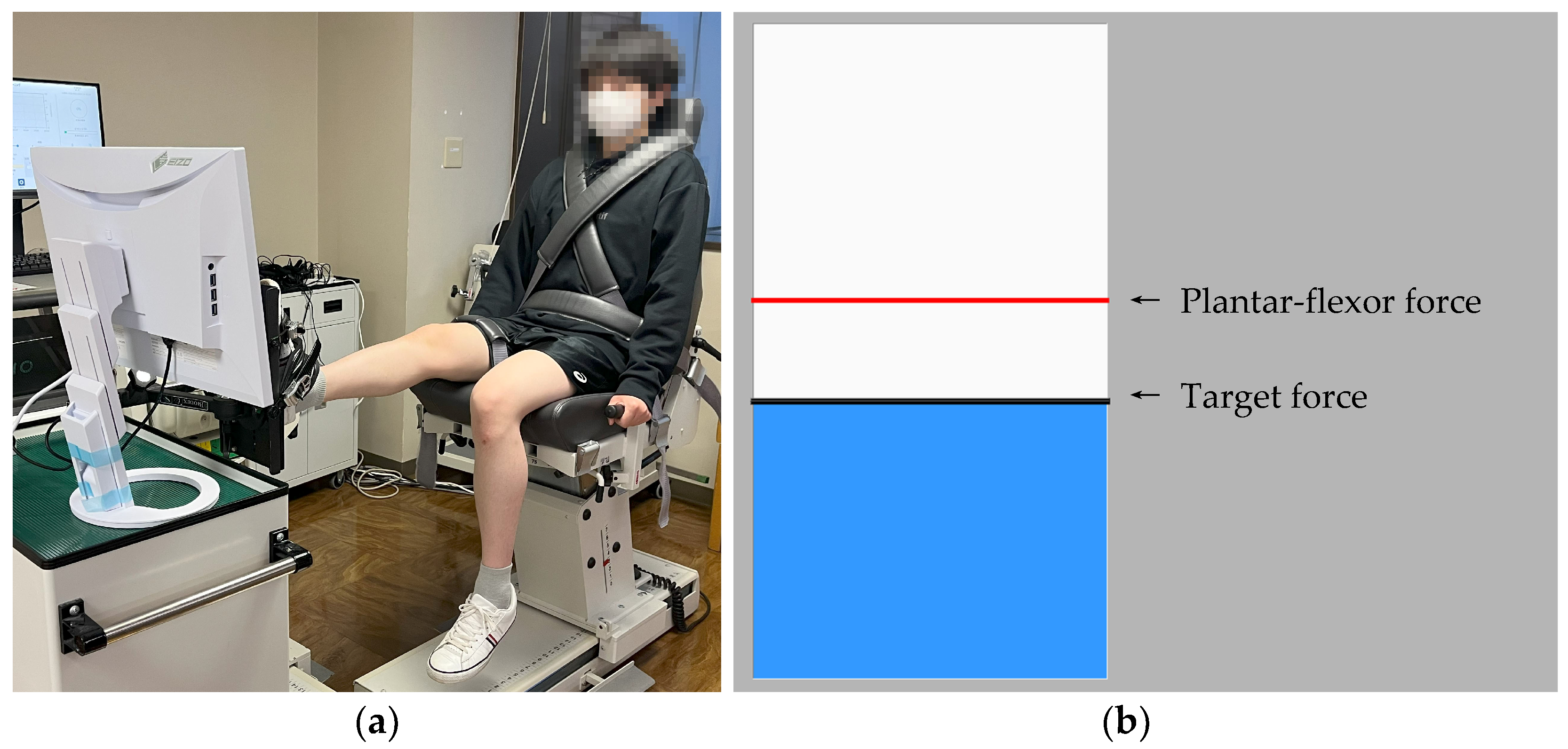
Figure 1. (a) Experimental setup for the force tracking task using the Biodex System 4 dynamometer. The participant was seated with the hip flexed at 70°, knee fully extended, and ankle in a neutral position. The trunk and pelvis were stabilized using the chest and waist straps. (b) Example of visual feedback displayed on the monitor during the task. The black line indicates the target force and the red line represents the participant’s actual force output. A greater plantar flexion force resulted in an upward movement of the feedback.
Applsci 15 07294 g001
Figure 2. Target and actual force trajectories used in force-tracking tasks. (a) Low range task: 10–30% MVIC. (b) High range task: 40–60% MVIC. Both tasks used sinusoidal waveforms with a 20% MVIC range and 10% MVIC amplitude (±10% from center), at 0.2 Hz. The black line represents the target trajectory, and the gray line indicates a representative actual force output from one trial.
Applsci 15 07294 g002
Figure 3. Schematic illustration of the definition and calculation of musculotendinous stiffness. A second-order polynomial was fitted to the passive torque–angle curve, and stiffness was calculated as the average slope (first derivative) at four equally spaced points within the final 13 degrees of each participant’s pre-stretching range of motion. Vertical dashed lines indicate the four stiffness evaluation points.
Applsci 15 07294 g003
Table 1. Changes in ankle dorsiflexion range of motion and musculotendinous stiffness before and after static stretching and control interventions.
OutcomesConditionPrePostp-ValueCohen’s d
Maximal angle of
ankle dorsiflexion (°)
SS26.4 ± 9.728.3 ± 10.4<0.0011.57
Control27.0 ± 9.827.4 ± 9.8>0.9990.50
Musculotendinous stiffness (Nm/°)SS1.30 ± 0.511.20 ± 0.470.0340.78
Control1.44 ± 0.561.42 ± 0.58>0.9990.14
Values are presented as mean ± standard deviation. p-values represent within-condition comparisons (Pre vs. Post) using Bonferroni correction tests. d = Cohen’s d, indicating effect size.
Table 2. RMSE during low- and high-range force tracking tasks, separated by force phase (generation and release) before and after static stretching and control conditions.
TaskForce PhaseConditionPrePostChange (%)95%CI of Change (%)p-ValueCohen’s d
Low rangeGenerationSS1.40 ± 0.441.30 ± 0.33−3.12−16.04 to 9.81>0.9990.32
Control1.39 ± 0.361.19 ± 0.20−11.00−21.55 to −0.450.1010.65
ReleaseSS1.53 ± 0.481.44 ± 0.34−1.55−13.43 to 10.33>0.9990.26
Control1.51 ± 0.591.40 ± 0.39−3.37−13.23 to 6.49>0.9990.28
High rangeGenerationSS1.82 ± 0.571.92 ± 0.71+5.65−6.85 to 18.16>0.9990.21
Control1.77 ± 0.391.69 ± 0.36−2.07−12.09 to 7.95>0.9990.24
ReleaseSS1.82 ± 0.721.47 ± 0.42−14.52−26.33 to −2.790.0300.79
Control1.54 ± 0.381.37 ± 0.45−10.38−21.80 to 1.050.4460.46
Values are presented as mean ± standard deviation. p-values represent within-condition comparisons (Pre vs. Post) using Bonferroni correction tests. d = Cohen’s d, indicating effect size. A positive value of d reflects the standardized magnitude of change, regardless of direction. RMSE values are expressed as a percentage of maximal voluntary isometric contraction (%MVIC). Change (%) indicates the percent change from Pre to Post values, calculated as ((Post − Pre)/Pre) × 100. CI stands for confidence interval.
Disclaimer/Publisher’s Note: The statements, opinions and data contained in all publications are solely those of the individual author(s) and contributor(s) and not of MDPI and/or the editor(s). MDPI and/or the editor(s) disclaim responsibility for any injury to people or property resulting from any ideas, methods, instructions or products referred to in the content.