Review

Efficacy of Maxillary Expansion with Clear Aligner in the Mixed Dentition: A Systematic Review

1
Department of Human Science and Innovation for the Territory, University of Insubria, 21100 Varese, Italy
2
Department of Medicine and Innovative Technologies, University of Insubria, 21100 Varese, Italy
3
School of Medicine, University of Insubria, 21100 Varese, Italy
4
Department of Biotechnology and Life Sciences, University of Insubria, 21100 Varese, Italy
*
Author to whom correspondence should be addressed.
Appl. Sci. 2025, 15(13), 7233; https://doi.org/10.3390/app15137233
Submission received: 23 March 2025 / Revised: 12 June 2025 / Accepted: 20 June 2025 / Published: 27 June 2025

Abstract

The purpose of this systematic review is to analyze the dentoalveolar changes resulting from the use of clear aligners in the treatment of transverse maxillary deficiency among growing children in the mixed dentition stage. An electronic literature search was carried out using the following databases: PubMed, Scopus, Cochrane Library, Embase, and Web of Science. The review protocol was prospectively registered in the PROSPERO database. Eligible studies included children aged 6 to 12 years in the mixed dentition period, presenting with erupted maxillary first molars and a transverse deficiency of the maxilla, and undergoing treatment with Invisalign® First aligners. The review encompassed various study types including retrospective and prospective designs, randomized controlled trials, preliminary studies, and case series. Two independent reviewers conducted the data extraction process. The quality of evidence was assessed using the GRADE approach. Except for studies by Bruni et al., the risk of bias in selected articles was visually summarized in a traffic light plot using the robvis tool, following the ROBINS-I methodology. For the studies by Bruni et al., a separate visual representation was created using robvis with the RoB2 evaluation framework, prepared by the authors S.E.I. and C.A. In total, 14 studies were included in the final synthesis, selected from 265 records retrieved through electronic searches and an additional 36 identified via manual screening. Several parameters were considered in order to assess dentoalveolar expansion: intercanine width, intercanine transpalatal width, intercanine dentoalveolar width, first and second interdeciduous molar width, first and second interpremolar width, first and second interdeciduous molar transpalatal width, first intermolar width, first intermolar mesial, distal, and transpalatal width, molar inclination, arch depth, and arch perimeter, and intermolar dentoalveolar width. An improvement was recorded in all parameters. The studies comparing treatment with Invisalign® First clear aligners and rapid maxillary expander highlighted that these both determined statistically significant differences compared to the natural growth group. Treatment with Invisalign® First in mixed dentition proved to be very effective for dentoalveolar expansion of the maxillary arch, with good control of the crown angulation of the upper first molar and an increase in the palatal area similar to RME, compared to pre-treatment or to the natural growth group. It could represent an effective and comfortable alternative to the traditional rapid maxillary expander treatment. However, further high-quality studies are required to support our current observations and verifying the stability of treatment outcomes on a long-term basis.

1. Introduction

Interceptive orthodontics addresses the diagnosis and treatment of malocclusions and functional alterations in children before growth ends [1,2]. Its goals include promoting craniofacial development, preventing complex future treatments or surgeries, and improving psychological well-being [3]. Early orthodontic therapy aims to enhance skeletal, dentoalveolar, and muscular development, facilitating the correct eruption of permanent teeth, and is often followed by a second phase to achieve optimal results [3,4]. Early diagnosis of common malocclusions, such as early or late loss of primary teeth, supernumerary teeth, crowding, ectopic eruption, midline diastema, crossbite, harmful oral habits, arch constriction, molar rotation, and Class II or III malocclusions, is crucial for effective treatment using various orthodontic appliances [3,5]. Maxillary transverse deficiency is a common craniofacial problem associated with crowding, tooth protrusion, arch deformations, posterior crossbite, and narrow nasal cavities [6,7,8,9]. Maxillary expansion is indicated for correcting transverse deficiencies and treating related issues, with slow maxillary expansion (SME) using lighter forces for gradual expansion and rapid maxillary expansion (RME) using heavier forces for immediate effects [9,10,11,12,13,14]. Common devices for SME include the Leaf Expander and Quad Helix, while Haas and Hyrax expanders are used for RME [15,16,17]. Invisalign® First, developed in 2018, offers a comfortable and aesthetic option for early mixed dentition, facilitating dentoalveolar expansion similar to SME [8,10,14,15,18,19]. Clear Align Therapy (CAT), introduced by Kesling in 1946 and advanced by others, uses polyurethane aligners to achieve dental alignment with minimal force, requiring more sequential aligners for the desired result and offering benefits such as fewer emergencies, better aesthetics, and improved oral hygiene [16,17,20,21,22,23,24]. Invisalign® First involves two phases: an 18-month interceptive phase, followed by a potential second phase within ten years if needed, using thermoplastic aligners [5,14,22,25].
This systematic review’s objective is to thoroughly examine the body of research in order to assess the dentoalveolar alterations seen in developing patients who have received orthodontic treatment with Invisalign® First clear aligners and presented maxillary transverse deficiency during the mixed dentition phase. By analyzing changes in important dentoalveolar parameters, such as intercanine and intermolar widths, transpalatal and alveolar arch dimensions, molar inclination, and dental arch perimeter and depth, this review aims to evaluate how well Invisalign® First promotes maxillary transverse development. The review also seeks to understand the clinical significance of these results in terms of functional occlusion, arch coordination, and long-term stability, as well as whether these changes are comparable to those made using conventional expansion techniques like rapid maxillary expanders (RME). This review aims to give researchers and clinicians evidence-based insights into the potential role of clear aligners as an alternative approach for early interceptive treatment of transverse maxillary deficiency in pediatric patients by critically evaluating the quality of the included studies and synthesizing their findings.

2. Materials and Methods

This systematic review was conducted following the PRISMA guidelines (Preferred Reporting Items for Systematic Reviews and Meta-Analyses) [26]. The review protocol was previously registered in the PROSPERO database (CRD42024406688).

2.1. Eligibility Criteria

The selection of studies was guided by predefined inclusion and exclusion criteria based on the PICOS framework [27], as detailed in Table 1. The selected research focused on patients in the mixed dentition phase presenting with transverse maxillary deficiency, treated with Invisalign® First clear aligners to promote dentoalveolar expansion of the maxilla.

2.2. Information Sources

An electronic search was performed in PubMed, Scopus, Cochrane, Embase, and Web of Science databases. The last access to the databases was performed on 29 August 2024. A manual search was also completed in order to find further relevant articles.

2.3. Search Strategy

One query string was applied in order to automatically select as many articles as possible on the same topic. A controlled vocabulary was applied using the MeSH terms “Orthodontic Appliances, Removable”[Mesh] AND “Dentition, Mixed”[Mesh]. Additionally, reference lists were carefully reviewed to identify any relevant studies not captured by the electronic database search. The reference lists of included articles were manually screened to identify any additional studies potentially missed during the electronic database search.

2.4. Selections Process

The articles were screened and reviewed by two researchers (S.E.I. and C.A.) according to title and abstract and using the inclusion and exclusion criteria. All articles that did not satisfy the eligibility criteria were ruled out after a preliminary screening. Then, the two authors obtained the full-text versions of the selected articles and of those whose content was not clear based on the data of the title and/or abstract, and they separately read the full-text of the remaining studies utilizing the eligibility criteria. Any doubt and disagreement were overcome through dialogue between the two authors, consulting a third reviewer if necessary.

2.5. Data Collection Process

Two researchers (S.E.I. and C.A.) determined the relevant criteria for the data collection which they subsequently extracted in double and independently from the selected studies. Any doubt and disagreement were overcome through dialogue between the two authors. The data collected from the selected articles were the following:
  • Study features: authors, year and source of publication, study design;
  • Sample characteristics: size, gender, and age of the population;
  • Clinical evaluation characteristics: type of measured outcomes;
  • The type of intervention;
  • Characteristics of the results: Maxillary arch changes.

2.6. Data Items

The information extracted from the selected studies included the study design, sample size, average age and gender distribution of participants, inclusion and exclusion criteria, the type of aligner used for maxillary expansion, and outcomes measured, as well as the duration of treatment and follow-up periods.

2.7. Study Risk of Bias Assessment

The assessment of risk of bias was conducted using the Cochrane Risk of Bias Tool for randomized controlled trials (RCTs), whereas non-randomized studies were evaluated with the ROBINS-I tool.

2.8. Effect Measures and Synthesis Methods

Bruni et al. [1,2] compared palatal morphology change calculating its volume in both treatment group and control group to determine the sample size calculation, while the studies by Lombardo et al. [15], Lombardo et al. [26], Lione et al. [27], and Loberto et al. [18] used previous pilot studies.
In the articles by Lombardo et al. [15], Lombardo et al. [26], Lione et al. [27], Lu et al. [28], Lu et al. [29], and Loberto et al. [18], all measurements were repeated twice over 2 weeks. Lombardo et al. [26] randomly selected 20 maxillary dental casts and re-digitalized them 10 days after the first session.
Data distribution was determined using the Shapiro–Wilk test in the studies by Bruni et al. [1], Levrini et al. [7], Lione et al. [27], Lu et al. [28], Lu et al. [29], Torbaty et al. [19], Pamukçu et al. [25], and Loberto et al. [18]; the Skewness and kurtosis test in the studies by Bruni et al. [1]; and the Kolmogorov–Smirnov test in the study by Gonçalves et al. [30]. Torbaty et al. [19] used an analysis of variance with the Bonferroni post hoc test to determine disequalities between the groups.
Parametric tests were selected to compare the T2–T1 changes when the data had a normal data distribution.
The unpaired t-test was utilized for inferential analysis of independent continuous variables in several studies: by Bruni et al. [1,2], it was applied to analyze intergroup differences; Lu et al. [28,29] used it to compare outcomes between the First group and the Haas group; Lombardo et al. [15,26] employed it for analyzing starting forms and for statistical comparison of T2–T1 changes across all measurements between First and RME groups; Gonçalves et al. [30] used the test to assess differences in maxillary and mandibular arch values as well as predicted versus achieved movements within each arch; and Pamukçu et al. [25] applied it to compare two groups with normally distributed data. Additionally, Fisher’s exact test was implemented in the same study to evaluate gender distribution comparability between the groups.
For paired comparisons, the paired t-test was selected by Levrini et al. [7], Lombardo et al. [15,26], Lione et al. [27], Lu et al. [28,29], and Loberto et al. [18] to detect significant changes between T0 and T1 measurements. Bruni et al. [1,2] and Pamukçu et al. [25] also used this test to evaluate intragroup differences for all parameters between T0 and T1 in both groups.
Descriptive analyses of all variables were reported by Gonçalves et al. [30], while Pinho et al. [9] provided a descriptive overview of movement and cephalometric metrics in all children. The one-sided Cochran–Armitage Trend test, adjusted with a Bonferroni correction for multiple comparisons, was used to examine trends. Additionally, the Wilcoxon signed-rank test assessed changes in cephalometric metrics, derotation, expansion, and space recovery pre- and post-treatment. The Dahlberg method was applied to estimate the standard deviation of intra-investigator error for validating cephalometric measurements.
The Mann–Whitney–Wilcoxon test was performed by Bruni et al. [1,2] to assess the existence of selection bias in the cohort at the baseline (T0), and by Pamukçu et al. [25] for nonnormally distributed data and demographic statistical analysis.
Pamukçu et al. [25] also used the analysis of variance and Kruskal–Wallis test to assess differences.
In the study by Lombardo et al. [26], the magnitude of the random error was evaluated thanks to the method of moments estimator.
The method of moment’s estimator (MME) was applied by Loberto et al. [18] to evaluate the random error. The comparison between before and after treatment and the predictability of Clincheck were investigated with a Friedman ANOVA for repeated measures and the Tukey post hoc test.
The Intraclass Correlation Coefficient (ICC) was employed to evaluate the reliability of initial and follow-up measurements in the studies conducted by Levrini et al. [7], Lombardo et al. [11], Lione et al. [27], Lu et al. [28,29], Bruni et al. [1,2], and Pamukçu et al. [25]. Procrustes analysis was applied by Loberto et al. [18] to assess statistical differences between the Invisalign® First and rapid maxillary expansion (RME) groups. Additionally, principal component analysis (PCA) was used to identify the primary patterns of dental shape variation within intra-group comparisons (T2–T1 in both RME and First groups).
Across all studies, statistical significance was set at p < 0.05. Data analysis was performed using various software packages, including IBM SPSS Statistics version 26.0 ([28,29]), SPSS version 18.0 ([27]), SAS version 9.4 [19], Stata version 17 [1], SPSS Statistics version 22.0 [25], Prism 10 [18], IBM SPSS Statistics 27 ([30]), and IBM SPSS Statistics 28 alongside R version 4.1.2 ([9]). In Pinho et al. [9], data organization and graphical presentations were created using Microsoft Excel® (Microsoft Excel for Microsoft 365 Version 2206). Lombardo et al. [11] and Loberto et al. [18] conducted PCA using Viewbox 4.0 (dHAL Software, Kifissia, Greece).

2.9. Certainty Assessment
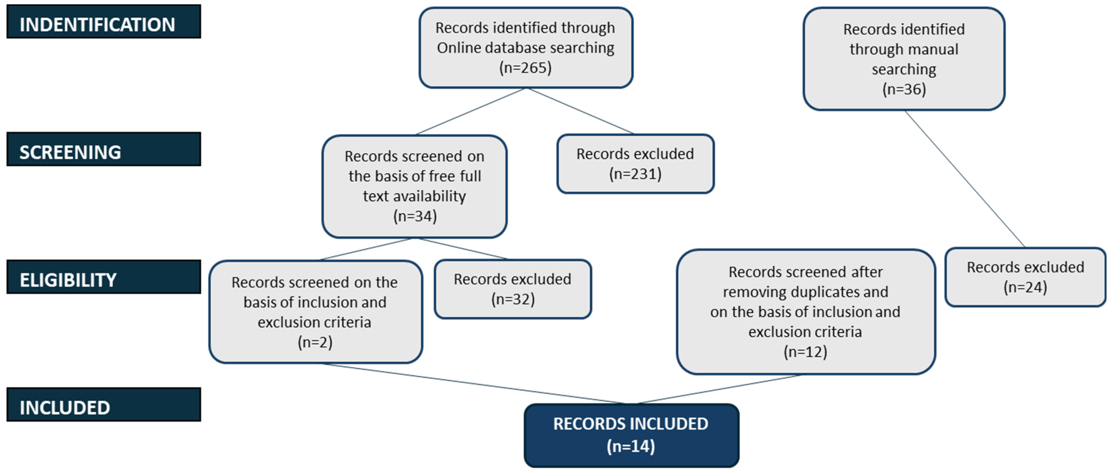
The Grading of Recommendations Assessment, Development, and Evaluation (GRADE) [31,32] allowed evaluation of the certainty of evidence.
Two researchers (S.E.I. and C.A.) assessed the different aspects (ROBINS-I [33], RoB2 [33], inconsistency [34], indirectness [35], imprecision [36], and publication bias [37]) and they classified the quality of evidence into four ratings: high, moderate, low, and very low.

3. Results

Two authors (S.E.I and C.A.) found 265 articles on the electronic databases. After removing duplicates and the articles that did not meet inclusion and exclusion criteria there were twelve items left. In the end, this systematic review included fourteen articles. The article by Gonçalves et al. [30] was a retrospective study and systematic review: only retrospective study was considered for this systematic review. Several articles were excluded due to the extension of the population of the studies to children not in mixed dentition or treated without Invisalign® First system but with other maxillary expanders.
The selection of studies is represented in Figure 1.

3.1. Study Characteristics

Type of Study and Location

Table 2 and Table 3 presented the characteristics of the eighteen included studies. Three of these [27,28,29] were prospective studies, seven were retrospective studies [11,15,18,19,25,26,30], one was a case series [9], one [7] was a preliminary study, and two [1,2] were randomized controlled trials. The studies were conducted at multiple institutions, including the Oral Pathology and Rehabilitation Research Unit (UNIPRO) at the University Institute of Health Sciences (IUCS), Cooperativa de Ensino Superior Politécnico e Universitário (CESPU) [9,30]; the Department of Systems Medicine at the University of Rome ‘Tor Vergata’, Italy [11,15,26]; the Department of Surgical Sciences, CIR Dental School, University of Turin [1,2]; the Department of Orthodontics at Xiangya Stomatological Hospital, Central South University, China [28,29]; the Department of Dentistry at UNSBC, Tirana, Albania [27]; the Department of Human Sciences, Innovation and Territory, School of Dentistry, Postgraduate Orthodontics Program at the University of Insubria, Italy [7]; the Department of Orthodontics, University of the Pacific, Arthur A. Dugoni School of Dentistry, USA [19]; the Department of Oral Health Science, Faculty of Dentistry, University of British Columbia, Canada [19]; the Surgical, Medical and Dental Department, University of Modena and Reggio Emilia, Italy [1]; the Dentistry Unit, Department of Health Sciences, University of Catanzaro “Magna Graecia”, Italy [1]; the Department of Orthodontics, School of Dentistry, Başkent University, Türkiye [25]; the Department of Orthodontics, School of Dentistry, İzmir Tınaztepe University, Türkiye [25]; and the Department of Health Science, Unicamillus-Saint Camillus International Medical University, Italy [18]. These investigations took place in Portugal [9,30], Italy [1,2,7,11,15,26], Albania [27], China [28,29], the USA [19], and Türkiye [25]. The studies were published between 2021 [7,27], 2022 [2,9,11,15], 2023 [26,28,29,30], and 2024 [1,18,19,25]. All of them involved young patients in the mixed dentition stage presenting with maxillary transverse deficiency, who were treated with Invisalign® First clear aligners aimed at achieving dentoalveolar maxillary expansion.

3.2. Characteristics of the Participants

The selected articles presented similar inclusion and exclusion criteria.
The studies by Bruni et al. [2], Levrini et al. [7], Lombardo et al. [11], Lombardo et al. [15], Lombardo et al. [26], Lione et al. [27], Lu et al. [28], Lu et al. [29] and Torbaty et al. [19], Bruni et al. [1], Pamukçu et al. [25], and Loberto et al. [18] included patients in mixed dentition with first molars erupted. Also, the study by Gonçalves et al. [30] considered children between 6 and 10 years old with first permanent molars fully erupted but also with palatal malocclusion requiring orthodontic treatment. Pinho et al. [9] analyzed cases involving individuals in the early mixed dentition phase who completed both initial and final intraoral digital scans, and who required at least one orthodontic movement of intermediate to high complexity according to a modified Align® protocol. Bruni et al. [2], Lu et al. [28], and Lu et al. [29] included only cases presenting posterior transverse discrepancies between the maxillary and mandibular arches of 5 mm or less. In contrast, Lombardo et al. [11,15,26], Lione et al. [27], Bruni et al. [1], and Loberto et al. [18] evaluated patients with discrepancies up to 6 mm.
Moreover, the studies by Lione et al. [27] and Lombardo et al. [11,15,26] focused on individuals of European descent who demonstrated a high level of treatment compliance. These latter studies also reported cases exhibiting either a mesial step or flush terminal plane molar relationship.
In the study by Levrini et al. [7] the inclusion criteria were children between 6 and 12 years who needed an arch expansion performed with Invisalign® First and with adequate diagnostic records.
Torbaty et al. [19] considered patients without anterior crossbite and bilateral posterior crossbite.
The study samples of Lu et al. [28] and Lu et al. [29] presented mild or moderate crowding and a prepuberal stage of development (CS1-CS3 in cervical vertebral maturation), like those of Bruni et al. [1,2], which had a cervical vertebral maturation stage (CVMS) not exceeding 4. Those patients, with good standards of oral hygiene, also had indications for maxillary expansion treatment and presented the upper second premolar cusps position apical to half pulp chamber (HPC) line of the ipsilateral upper first permanent molars on pre-treatment panoramic radiographs.
In the work by Pamukçu et al. [25] patients with a high level of compliance, with crowding that did not require extractions and sagittal corrections, treated using exclusively Invisalign attachments, were included.
In the included articles no patients dropped out or were excluded during treatment, except for the studies by Bruni et al. [1,2].

3.3. Characteristics of the Intervention and Comparisons

The selected studies evaluated the efficacy of Invisalign® First system on maxillary arch. Invisalign® First is an orthodontic treatment by Align Technology which consists of thermoplastic devices used for the treatment of young patients aged between 6 and 10 years [5,7]. All patients treated with Invisalign® First had to wear the masks for 20–22 h a day. A good level of compliance was required.
Nine of these articles [1,2,11,15,19,25,26,28,29] compared the treatment with this appliance with RME, in particular with Hyrax-type maxillary expander [1,2,19], and Haas [28], butterfly palatal expander [11,15,26], acrylic splint expander [29], and removable acrylic expander [25]. Furthermore, Lu et al. [28,29] and Torbaty et al. [19] also considered a natural growth group.
All studies obtained digital impressions before and after treatment using the iTero® intraoral scanner (Align Technology Inc., Tempe, AZ, USA), with the exception of Torbaty et al. [19], who imported digital casts into Ortho Analyzer software (3Shape Inc., Copenhagen, Denmark). Except for the work by Bruni et al. [2], all studies utilized ClinCheck® planning for each patient. Measurements were conducted on pre-treatment and post-treatment digital dental models to assess changes in the maxillary arch. In this study [9] Pinho et al. performed cephalometric measurements and they quantified tooth movement using commonly used measurement tools.

3.4. Characteristics of the Outcomes

The outcomes analyzed in the selected studies were the morphological changes of the maxillary arch using Invisalign® First System. The aim of the studies by Lu et al. [28,29] was to examine the changes of intercanine, first interpremolar, second interdeciduous molar and first intermolar width, measured in millimeters, arch depth and perimeter, measured in millimeters, intercanine and intermolar dentoalveolar width, measured in millimeters, and inclination of the molars, measured in degrees. The article of Lione et al. [27] and Loberto et al. [18] analyzed intercanine, first interdeciduous molar, second interdeciduous molar and first intermolar mesial, distal and transpalatal width, measured in millimeters.
Gonçalves et al. [30] considered maxillary dental arch width by the linear measurements of interdental widths at three different positions, and the expansion efficiency calculated as a percentage of the achieved movement compared to the predicted using a specific equation. Lombardo et al. [11] evaluated 14 landmarks for maxillary dentition, while in the other studies [15,26], nine variables were considered: intercanine width, first and second interdeciduous molar width, first intermolar mesial, distal, and transpalatal width, intercanine transpalatal width, and first and second interdeciduous molar transpalatal width, measured in millimeters.
Levrini et al. [7] considered eleven variables in their study, including ten linear measurements in millimeters and one angular measurement in degrees. These variables comprised canine gingival width, gingival width of the first and second deciduous molars, gingival width of the first permanent molar, dental widths of the canine, first and second deciduous molars, and first permanent molar, as well as arch perimeter, arch depth, and intermolar angle. Similarly, Bruni et al. [1,2] evaluated linear measurements of the upper arch, such as intercanine and intermolar widths at both cusp and gingival levels, along with palatal volume and surface area.
Pinho et al. [9] analyzed outcomes including molar derotation (in degrees), dentoalveolar expansion and space recovery (in millimeters), molar sagittal malocclusion, posterior crossbite, open bite, midline discrepancy (in millimeters), and crowding. Based on these parameters, cases were then classified into mild, moderate, or severe categories.
Torbaty et al. [19] considered intercanine width, first and second interdeciduous molar width, and first intermolar width, measured in millimeters.
In the article by Pamukçu et al. [25] the intercanine and intermolar width, arch depth, buccolingual inclination of the first molars, expansion, palatal volume, and surface area were assessed.

3.5. Reporting Bias Assessment

Risk of bias of selected studies, except for the articles by Bruni et al. [1,2], was reproduced graphically (Figure 2) using robvis, the risk of bias traffic light plot of ROBINS-I assessments [38].

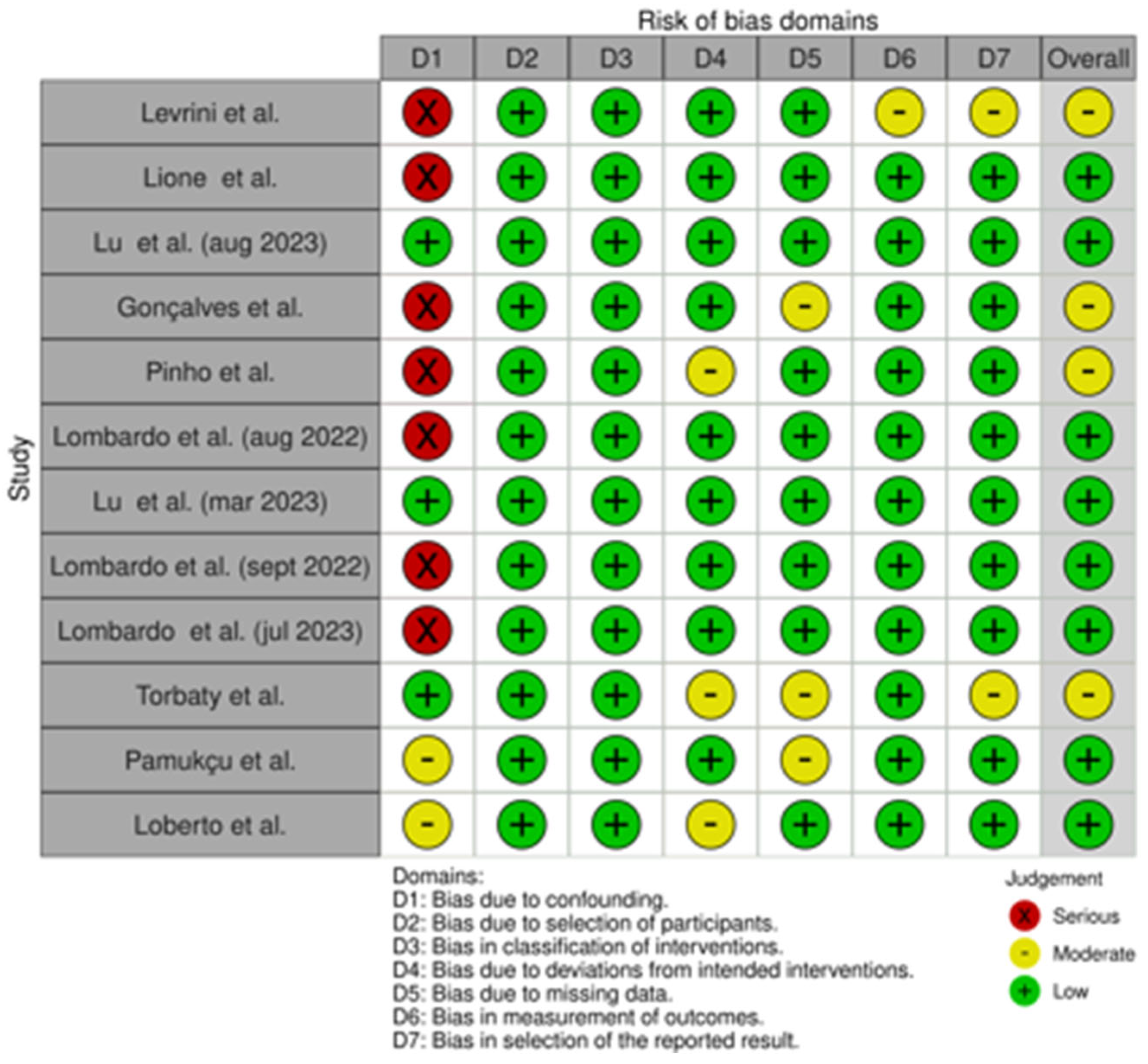
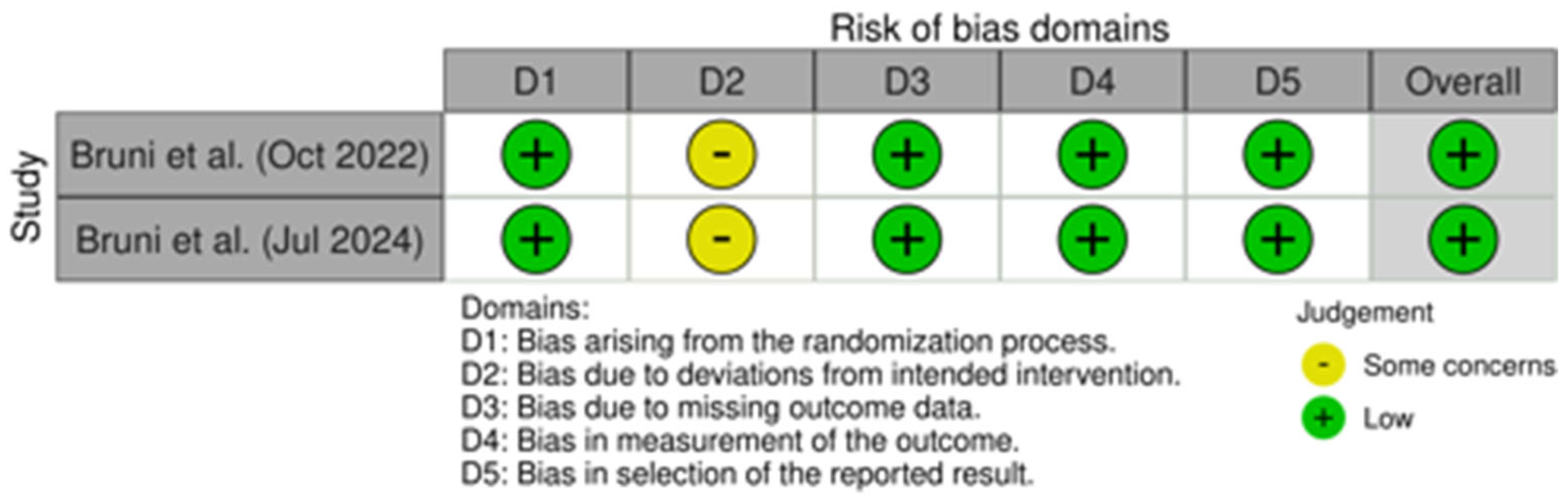
3.6. Assessment of Risk of Bias Within Studies and Quality of Evidence

The risk of bias of the three prospective studies [27,28,29], the seven retrospective studies [11,15,18,19,25,26,30], the preliminary study [7], and the one case series [9] was calculated using ROBINS-I, a tool for assessing risk of bias in non-randomized studies of interventions [39].
The risk of bias of the two randomized controlled trials [1,2] was evaluated using RoB 2 (Figure 3), a revised Cochrane risk-of-bias tool for randomized trials [40].

3.7. Overall Risk

From the risk of bias traffic light plot of ROBINS-I assessments the following can be observed:
  • Levrini et al. [7] was considered at moderate risk of bias because it showed a high risk of bias in D1 and a moderate risk in D6 and D7.
  • Lione et al. [27] was considered at low risk of bias because it only showed a high risk of bias in D1.
  • Lu et al. [29] was considered at low risk of bias because it showed a low risk in all domains.
  • Gonçalves et al. [30] was considered at moderate risk of bias because it showed a high risk of bias in D1 and a moderate risk in D5.
  • Pinho et al. [9] was considered at moderate risk of bias because it showed a high risk of bias in D1 and a moderate risk in D4.
  • Lombardo et al. [11] was considered at low risk of bias because it only showed a high risk of bias in D1.
  • Lu et al. [28] was considered at low risk of bias because it showed a low risk in all domains.
  • Lombardo et al. [15] was considered at low risk of bias because it only showed a high risk of bias in D1.
  • Lombardo et al. [26] was considered at low risk of bias because it only showed a high risk of bias in D1.
  • Torbaty et al. [19] was considered at moderate risk of bias because it showed a moderate risk of bias in D4, D5 and D7.
  • Pamukçu et al. [25] was considered at low risk of bias because it only showed a moderate risk of bias in D1 and D5.
  • Loberto et al. [18] was considered at low risk of bias because it only showed a moderate risk of bias in D1 and D4.
From the risk of bias traffic light plot of RoB 2 assessments the following can be observed:
  • Bruni et al. [2] was considered at low risk of bias because it only showed a moderate risk of bias in D2.
  • Bruni et al. [1] was considered at low risk of bias because it only showed a moderate risk of bias in D2.
A moderate risk of bias was identified in four studies: Levrini et al. [7], Gonçalves et al. [30], Pinho et al. [9], and Torbaty et al. [19]. The moderate risk in Levrini et al. [7] was related to confounding factors, outcome measurements, and selective reporting. Gonçalves et al. [30] showed moderate bias mainly due to confounding and incomplete data. In Pinho et al. [9], confounding and deviations from intended interventions contributed to the bias, while Torbaty et al. [19] faced issues related to deviations from planned interventions, missing data, and selective result reporting.

3.8. Certainty of Evidence and Risk of Reporting Biases in Syntheses

The quality of evidence for the included studies was assessed using the GRADE framework (Grading of Recommendations Assessment, Development and Evaluation). The overall certainty was rated as moderate across all variables, primarily due to moderate risk of bias detected in three of the fourteen studies, alongside concerns related to imprecision.

4. Discussion

The purpose of this systematic review was to evaluate dentoalveolar changes using Invisalign® First clear aligners in patients in the mixed dentition with maxillary transverse deficiency. Invisalign® First was created by Align Technology in 2018 in order to perform phase 1 of orthodontic treatment, which was the interceptive orthodontic treatment for growing patients in the mixed dentition.
This systematic review included fourteen articles (retrospective studies, prospective studies, preliminary studies, randomized controlled trials, and case series), which were selected from a total of 265 records identified through online database searching and 36 records identified through manual searching. Several parameters were considered in order to assess dentoalveolar expansion, such as intercanine width, intercanine transpalatal width, intercanine dentoalveolar width, first and second interdeciduous molar width, first and second interdeciduous molar transpalatal width, first and second interpremolar width, first intermolar mesial, distal, and transpalatal width, intermolar dentoalveolar width, first intermolar width, space recovery, overbite, overjet, molar inclination, arch depth and perimeter, palatal volume, and surface area.

4.1. Intercanine Width

Intercanine width (linear distance between cusp tips of the deciduous canines) was assessed in twelve [1,2,7,15,18,19,25,26,27,28,29,30] of the fourteen articles considered. In the studies by Lu et al. [28,29], Lione et al. [27], Levrini et al. [7], Bruni et al. [1,2], Torbaty et al. [19], Pamukçu et al. [25], and Loberto et al. [18], a statistically significant increase in the intercanine width was recorded between T0 and T1 (p < 0.05), in patients treated with Invisalign® First. Lu et al. [28,29], Bruni et al. [1,2], Torbaty et al. [19], and Pamukçu et al. [25] showed that there was an increase of intercanine width between T0 and T1 in RME group with statistical significance (p < 0.05), while in NG group analyzed in the articles by Lu et al. [28,29] and Torbaty et al. [19] there was a small increase in the same values without statistical significance. The studies by Lombardo et al. [15,26] reported an increase in intercanine width both in IF group and RME group, but do not specify whether this difference is statistically significant. Also, the article by Gonçalves et al. [30] noted that there was an improvement of this parameter, but statistical analysis is missing.

4.2. Intercanine Transpalatal Width

Five articles [1,2,7,15,26], analyzed intercanine transpalatal width (linear distance between the groove of the deciduous canines at the mucosa). Levrini et al. [7] and Bruni et al. [1,2], reported a significant increase (p < 0.05) in these data between T0 and T1 in patients treated with Invisalign® First, such as the RME group evaluated by Bruni et al. [1,2]. The studies by Lombardo et al. [15,26] reported an increase in intercanine transpalatal width both in IF group and RME group, but they do not specify whether this difference is statistically significant.

4.3. First and Second Interdeciduous Molar Width

First interdeciduous molar width (linear distance between the vestibular cusp tips of the first deciduous molars) and second interdeciduous molar width (linear distance between the sulcus of the second deciduous molars) were assessed in five [7,15,18,26,27] of the fourteen articles considered. The studies by Lione et al. [27], Levrini et al. [7], and Loberto et al. [18] demonstrated that Invisalign® First system determined a significant increase (p < 0.05) of first and second interdeciduous molar width between T0 and T1. The articles by Lombardo et al. [15,26] reported an increase in these parameters both in IF group and RME group, but they do not specify whether this difference is statistically significant.

4.4. Second Interdeciduous Molar Width, First Interpremolar Width, and Intercanine Dentoalveolar Width

Two articles [28,29] evaluated the following three parameters in the three different groups:
-
Intercanine dentoalveolar width (from the most prominent buccal bulge on the alveolus superior to the maxillary first canine)
-
Second interdeciduous molar width (linear distance between the sulcus of the second deciduous molars, deciduous teeth were substituted when present)
-
First interpremolar width (linear distance between the buccal cusp tips of the first premolars)
The difference between T0 and T1 was statistically significant (p < 0.05) both in IF group and RME group, while no significance was found in NG group.

4.5. First and Second Interdeciduous Molar Transpalatal Width

The first and second interdeciduous molar transpalatal widths—defined as the linear distances measured between the grooves of the first and second deciduous molars at the mucosal level—were evaluated in several studies [7,15,19,26]. Levrini et al. [7] reported a significant increase (p < 0.05) in both measurements from T0 to T1. Similarly, Torbaty et al. [19] found a significant increase in the width of both the first and second interdeciduous molars in the Invisalign® First (IF) and rapid maxillary expansion (RME) groups. The natural growth (NG) group also showed statistically significant changes in the second interdeciduous molar width, which were attributed to normal growth and arch development.

4.6. First Intermolar Mesial Width

The following ten studies [1,2,7,15,18,25,26,27,28,29] evaluated the first intermolar mesial width (linear distance between the mesiobuccal cusp tips of the first molars). Lu et al. [28,29] and Bruni et al. [1,2] demonstrated that there was a significant (p < 0.05) increase in the first intermolar mesial width between T0 and T1 in patients treated with Invisalign® First or with rapid maxillary expander, while it was not significant in NG group. The articles by Lione et al. [27], Levrini et al. [7], Pamukçu et al. [25], and Loberto et al. [18] also revealed a significant improvement (p < 0.05) in these data. The articles by Lombardo et al. [15,26] noted that there was an increase in first intermolar mesial width in patients treated with Invisalign® First and rapid maxillary expander, but statistical analysis is missing.

4.7. First Intermolar Distal Width

In the studies by Lione et al. [27], Lombardo et al. [15,26], Pamukçu et al. [25], and Loberto et al. [18], a statistically significant increase in the first intermolar distal width (linear distance between the distobuccal cusp tips of the first molars) in IF group was registered between T0 and T1 (p < 0.05), and Pamukçu et al. [25] also noted that there was a statistically significant increase in RME group. The studies by Lombardo et al. [15,26] observed an improvement of the considered data both in IF group and RME group, but they do not specify the statistical significance.

4.8. First Intermolar Transpalatal Width

First intermolar transpalatal width (linear distance between the groove of the first molars at the mucosa) was measured in patients treated with Invisalign® First and rapid maxillary expander at T0 and T1. The difference between T0 and T1 was statistically significant (p < 0.05) in both the IF group [1,2,7,18,19,27] and the RME group [1,2,19]. Also, the articles by Lombardo et al. [15,26] noted that there was an improvement of these data, but it did not report the statistical significance. Torbaty et al. [19] reported a significant increase in first intermolar width in NG group due to normal growth and arch development.

4.9. Molar Inclination, Arch Depth, and Arch Perimeter

Three studies [7,28,29] examined the molar inclination, defined as the angle formed by lines connecting the mesial buccal and mesial lingual cusp tips of both the right and left first molars, as well as the arch perimeter, measured as the path starting from the mesial contact point of one molar, passing through the mesial and distal contact points of the six anterior teeth, and ending at the mesial contact point of the opposite molar.
Additionally, four studies [7,25,28,29] assessed arch depth, which is the length of the perpendicular line drawn from the contact point between the mesial contact points of the central incisors to the line connecting the contact points between the second deciduous molars and the first permanent molars. A statistically significant (p < 0.05) difference between T0 and T1 emerged in patients treated with Invisalign® First [7,25,28,29] and with rapid maxillary expander [25,28,29], while no significance was found in NG group [28,29]. The only exception is represented by the study by Lu et al. [28,29], because arch depth in RME group is not statistically significant.

4.10. Intermolar Dentoalveolar Width

The difference of intermolar dentoalveolar width at T0 and T1, which was calculated from the most prominent buccal bulge on the alveolus superior to the maxillary first molar, was statistically significant (p < 0.05) in the IF group [9,28,29] and the RME group [28,29], while they did not find a significant difference in the NG group [28,29].

4.11. First and Second Interdeciduous Molar Width, First and Second Interpremolar Width, and First Intermolar Width

Gonçalves et al. [30] measured upper inter first premolar (palatal cusp tip), upper inter second premolar (palatal cusp tip), upper inter first temporary molar (mesiopalatal cusp tip), upper inter second temporary molar (mesiopalatal cusp tip) and upper inter first permanent molar (mesiopalatal cusp tip) in patients treated with Invisalign® First between T0 and T1. They observed an increase in all these measurements, but they did not report the statistical significance.
The studies included in the review demonstrated a significant increase in intercanine width [1,2,7,15,18,19,25,26,27,28,29,30] in patients treated with Invisalign® First clear aligners, like patients treated with rapid maxillary expander [1,2,19,25,28,29], while no significance was found in NG group [19,28,29]. The studies by Lombardo et al. [15,26] and Gonçalves et al. [30] reported an increase in this parameter, but they do not specify whether this difference is statistically significant.
It was registered also an improvement of intercanine transpalatal width [1,2,7,15,26], first and second interdeciduous molar width [7,15,18,26,27,28,29,30], first interpremolar width [28,29,30], intercanine dentoalveolar width [28,29], first and second interdeciduous molar transpalatal width [7,15,19,26], first intermolar mesial width [1,2,7,15,18,25,26,27,28,29], first intermolar distal width [15,18,25,26,27], first intermolar transpalatal width [1,2,7,15,18,26,27,30], first intermolar width [30], inclination of the molars and arch perimeter [7,28,29], arch depth [7,25,28,29], intermolar dentoalveolar width [9,28,29], and second interpremolar width [30]. From the analysis of the results, it emerged that the greatest increases were achieved at deciduous molars. Compared to the other teeth, the difference between the increases was minimal, therefore a near-parallel pattern of maxillary dental arch expansion was observed.
The studies comparing treatment with Invisalign® First clear aligners and rapid maxillary [7,15,26,27,28,29] expander highlighted that both determined statistically significant differences compared to NG group.
Lombardo et al. [11] represented graphically the changes of the maxillary arch after the treatment with Invisalign® First clear aligners and RME: the IF group presented an improvement in the maxillary arch shape, while the RME group maintained the initial triangular shape.
Except for arch depth, Bruni et al. [1,2], Lu et al. [28,29], Gonçalves et al. [30], Torbaty et al. [19], and Pamukçu et al. [25] reported that RME allowed a greater expansion with larger increases in all width indicators compared to Invisalign® First, indicating that it may be a preferable option for severe malocclusions. Lombardo et al. [15,26] also recorded a larger increase in first intermolar mesial, distal, and transpalatal width in patients with RME compared to Invisalign®, but not for intercanine transpalatal width, first and second interdeciduous molar width, or first and second interdeciduous molar transpalatal width.
Invisalign® First, a clear orthodontic system created especially for kids with mixed dentition, has been shown to be a creative and clinically effective way to treat transverse problems with the maxillary arch. By influencing the position of the teeth as well as the adaptation of the alveolar bone that supports them, it has demonstrated exceptional effectiveness in causing dentoalveolar extension of the upper arch. Good control of the upper first molar’s crown angulation, a crucial factor in guaranteeing the long-term stability of orthodontic treatment and averting future recurrences or malpositions, is among the most pertinent outcomes seen. The notable increase in palate area, which indicates effective expansion not just at the dental level but also at the skeletal level, is another important factor. This outcome is similar to that of the conventional treatment for maxillary transverse narrowing, the rapid maxillary expander (RME). However, Invisalign® First offers a less invasive method that is more accepted by juvenile patients than RME, which necessitates the use of a fixed appliance and may result in discomfort, trouble maintaining good dental hygiene, and potential problems (such as pain or palatine microlesions). Invisalign® First treatment has also been demonstrated to be effective when compared to the NG group of untreated participants. Notable differences have been observed in terms of transverse expansion, arch symmetry, and the amount of space available for the eruption of permanent teeth. Since craniofacial development is still occurring at this age, early correction is crucial to avoiding more complicated malocclusions in adolescence or adulthood. Given these factors, an increasing number of orthodontists are incorporating this technique into their routines in order to better forecast and monitor craniofacial growth and treat transverse abnormalities early.
In conclusion, maxillary dentoalveolar expansion accomplished with Invisalign® First is not only a clinically feasible substitute for traditional expansion techniques, but it is also a more sanitary, psychologically acceptable, and comfortable treatment option for the child and their family.
The articles analyzed in this systematic review had some limitations. The main limitations were the short-term design (less than one year of treatment, except for the studies by Gonçalves et al. [30] and Pinho et al. [9], without any follow-up in the years following), the small sample size of the treated groups and the absence of a randomized design, with the exception of the works by Bruni et al. [1,2]. Therefore, further studies with a randomized design are necessary to increase the sample size and to analyze the stability of the results obtained in the long term.
Other limitations were the absence of a control group, apart from the articles by Lu et al. [28,29] and Torbaty et al. [19] which also considered a natural growth group, the presence of missing records, and the use of digital model instead of Cone-Beam Computed Tomography to avoid radiation exposure. This systematic review is further limited by the dearth of meta-analyses currently available in the literature. Although this gap makes it more difficult to compare our results in a more comprehensive quantitative framework, it also highlights how important our study is in offering a structured synthesis in situations when quantitative aggregation is still not possible.
The treatment with Invisalign® First removable aligners must consider a potential noncompliance and lack of motivation from the patients to follow the orthodontist’s instructions. The questionnaire has not been tested for validity, and its reliability could be subject to various forms of bias such as mood bias and false reporting bias, because children may report greater use of the aligners than what occurs in reality [9].
Furthermore, Bruni et al. [1,2] and Gonçalves et al. [30] stated the lack of some measurements due to the absence of some teeth during the mixed dentition.

5. Conclusions

The therapeutic potential of Invisalign® First clear aligners in patients with mixed dentition and transverse maxillary deficiency is highlighted by the results of this systematic review. According to the data gathered, the use of clear aligners significantly improved maxillary dentoalveolar expansion when compared to the pre-treatment state or subjects in the untreated control group (NG). The outcomes were similar to those achieved with the conventional palate expander. More precisely, the review’s included research demonstrated several quantifiable and clinically significant dimensions and functional alterations. The first and second molar interdeciduous amplitudes increased in tandem with the transpalatal and dentoalveolar intercanine amplitudes. Along with the amplitude of the first and second intermolars, the molars’ transpalatal expansion also demonstrated noticeable improvements. The first intermolar’s mesial, distal, and transpalatal amplitudes showed positive findings, indicating efficient and carefully managed three-dimensional expansion. Moreover, there was a notable molar derotation, an expansion of the arch depth and perimeter, an improvement in molar inclination without a loss of control, and an increase in intermolar dentoalveolar breadth. These enhancements imply that, despite being based on a low-impact, removable orthodontic technique, Invisalign® First treatment can result in multifactorial, successful transverse modifications by precisely and carefully influencing different areas of the dental arch and the alveolar plane. Since Invisalign® First clear aligners provide a contemporary, efficient, and well-tolerated alternative for the repair of transverse maxillary deficiency in mixed dentition, they should be regarded as a feasible treatment choice for patients who are growing. This method offers similar clinical outcomes to conventional devices, but it also offers benefits in terms of comfort, appearance, oral cleanliness, and patient compliance. As a result, it is an orthodontic solution that meets the demands of modern clinical practice and families. The comprehensive analysis of the studies reviewed supports the conclusion that Invisalign® First represents an effective and promising solution for addressing transverse maxillary deficiency in growing patients. While variability exists among the included studies, the overall evidence indicates that this treatment modality offers favorable outcomes in terms of space management and maxillary expansion. Further high-quality research is encouraged to strengthen these findings and optimize clinical protocols.

Author Contributions

Conceptualization, A.C. and L.L.; methodology, P.A.Z.; software, A.D.; validation, E.I.S., M.C. and S.S.; formal analysis, M.B.; investigation, M.C.; resources, A.C.; data curation, A.C.; writing—original draft preparation, E.I.S.; writing—review and editing, A.D.; visualization, P.A.Z.; supervision, L.L.; project administration, A.C.; funding acquisition, M.B. All authors have read and agreed to the published version of the manuscript.

Funding

This research received no external funding.

Acknowledgments

A.C. is a Ph.D. student of the Life Sciences and Biotechnology course at the University of Insubria.

Conflicts of Interest

The authors declare no conflicts of interest.

References

  1. Bruni, A.; Ferrillo, M.; Gallo, V.; Parrini, S.; Garino, F.; Castroflorio, T.; Deregibus, A. Efficacy of clear aligners vs. rapid palatal expanders on palatal volume and surface area in mixed dentition patients: A randomized controlled trial. Am. J. Orthod. Dentofac. Orthop. 2024, 166, 203–214. [Google Scholar] [CrossRef]
  2. Bruni, A.; Gallo, V.; Parrini, S.; Ferrillo, M.; Garino, F.; Deregibus, A.; Castroflorio, T. Effectiveness of Aligners Versus Rapid Palatal Expander on Palatal Volume in Mixed Dentition Patients: A Randomized Controlled Trial. Res. Sq. 2022. [Google Scholar] [CrossRef]
  3. Artese, F. A broader look at Interceptive Orthodontics: What can we offer? Dent. Press. J. Orthod. 2019, 24, 7–8. [Google Scholar] [CrossRef]
  4. Oancea, R.; Funieru, C.; Sfeatcu, R.; Jumanca, D. Interceptive Orthodontics in Primary and Mixed Dentition: The Importance of Early Diagnosis. Jurnalul Pediatrului 2019, 18. [Google Scholar]
  5. Lione, R.; Gazzani, F.; Moretti, S.; Danesi, C.; Cretella Lombardo, E.; Pavoni, C. Gingival Margins’ Modifications during Orthodontic Treatment with Invisalign First((R)): A Preliminary Study. Children 2022, 9, 1423. [Google Scholar] [CrossRef]
  6. Howe, R.P.; McNamara, J.A., Jr.; O’Connor, K.A. An examination of dental crowding and its relationship to tooth size and arch dimension. Am. J. Orthod. 1983, 83, 363–373. [Google Scholar] [CrossRef] [PubMed]
  7. Levrini, L.; Carganico, A.; Abbate, L. Maxillary expansion with clear aligners in the mixed dentition: A preliminary study with Invisalign(R) First system. Eur. J. Paediatr. Dent. 2021, 22, 125–128. [Google Scholar] [CrossRef]
  8. McNamara, J.A. Maxillary transverse deficiency. Am. J. Orthod. Dentofac. Orthop. 2000, 117, 567–570. [Google Scholar] [CrossRef]
  9. Pinho, T.; Rocha, D.; Ribeiro, S.; Monteiro, F.; Pascoal, S.; Azevedo, R. Interceptive Treatment with Invisalign((R)) First in Moderate and Severe Cases: A Case Series. Children 2022, 9, 1176. [Google Scholar] [CrossRef]
  10. Bouserhal, J.; Bassil-Nassif, N.; Tauk, A.; Will, L.; Limme, M. Three-dimensional changes of the naso-maxillary complex following rapid maxillary expansion. Angle Orthod. 2014, 84, 88–95. [Google Scholar] [CrossRef]
  11. Cretella Lombardo, E.; Paoloni, V.; Fanelli, S.; Pavoni, C.; Gazzani, F.; Cozza, P. Evaluation of the Upper Arch Morphological Changes after Two Different Protocols of Expansion in Early Mixed Dentition: Rapid Maxillary Expansion and Invisalign((R)) First System. Life 2022, 12, 1323. [Google Scholar] [CrossRef]
  12. Kilic, N.; Oktay, H. Effects of rapid maxillary expansion on nasal breathing and some naso-respiratory and breathing problems in growing children: A literature review. Int. J. Pediatr. Otorhinolaryngol. 2008, 72, 1595–1601. [Google Scholar] [CrossRef] [PubMed]
  13. Machado-Junior, A.J.; Zancanella, E.; Crespo, A.N. Rapid maxillary expansion and obstructive sleep apnea: A review and meta-analysis. Med. Oral Patol. Oral Cir. Bucal 2016, 21, e465–e469. [Google Scholar] [CrossRef] [PubMed]
  14. Rutili, V.; Nieri, M.; Franceschi, D.; Pierleoni, F.; Giuntini, V.; Franchi, L. Comparison of rapid versus slow maxillary expansion on patient-reported outcome measures in growing patients: A systematic review and meta-analysis. Prog. Orthod. 2022, 23, 47. [Google Scholar] [CrossRef]
  15. Lombardo, E.C.; Fanelli, S.; Paoloni, V.; Cozza, P.; Lione, R. Comparison between two different expansion protocols in early mixed dentition:Clear Aligners vs. Rapid Maxillary Expansion. Semin. Orthod. 2022. [Google Scholar] [CrossRef]
  16. Rossini, G.; Parrini, S.; Castroflorio, T.; Deregibus, A.; Debernardi, C.L. Efficacy of clear aligners in controlling orthodontic tooth movement: A systematic review. Angle Orthod. 2015, 85, 881–889. [Google Scholar] [CrossRef] [PubMed]
  17. Weir, T. Clear aligners in orthodontic treatment. Aust. Dent. J. 2017, 62 (Suppl. S1), 58–62. [Google Scholar] [CrossRef]
  18. Loberto, S.; Pavoni, C.; Fanelli, S.; Lugli, L.; Cozza, P.; Lione, R. Predictability of expansion movements performed by clear aligners in mixed dentition in both arches: A retrospective study on digital casts. BMC Oral Health 2024, 24, 694. [Google Scholar] [CrossRef]
  19. Moravedje Torbaty, P.; Suh, H.; Tai, S.K.; Baird, M.; Boyd, R.L.; Oh, H. Vertical and transverse treatment effects of Invisalign First system compared to Hyrax maxillary expanders with fixed appliances in mixed dentition patients. Angle Orthod. 2024, 94, 496–503. [Google Scholar] [CrossRef]
  20. Galluccio, G.; De Stefano, A.A.; Horodynski, M.; Impellizzeri, A.; Guarnieri, R.; Barbato, E.; Di Carlo, S.; De Angelis, F. Efficacy and Accuracy of Maxillary Arch Expansion with Clear Aligner Treatment. Int. J. Environ. Res. Public Health 2023, 20, 4634. [Google Scholar] [CrossRef]
  21. Lin, E.; Julien, K.; Kesterke, M.; Buschang, P.H. Differences in finished case quality between Invisalign and traditional fixed appliances. Angle Orthod. 2022, 92, 173–179. [Google Scholar] [CrossRef] [PubMed]
  22. Lynch, N.M.; Shroff, B.; Carrico, C.K.; Sawicki, V.; Sabol, M.; Lindauer, S.J. Clear aligner therapy in the mixed dentition: Indications and practitioner perspectives. Am. J. Orthod. Dentofac. Orthop. 2023, 164, 172–182. [Google Scholar] [CrossRef] [PubMed]
  23. Papadimitriou, A.; Mousoulea, S.; Gkantidis, N.; Kloukos, D. Clinical effectiveness of Invisalign(R) orthodontic treatment: A systematic review. Prog. Orthod. 2018, 19, 37. [Google Scholar] [CrossRef]
  24. Upadhyay, M.; Arqub, S.A. Biomechanics of clear aligners: Hidden truths & first principles. J. World Fed. Orthod. 2022, 11, 12–21. [Google Scholar] [CrossRef]
  25. Pamukcu, H.; Ozsoy, S.; Aksoy, P.C.; Polat Ozsoy, O. Evaluation of maxillary dimensional changes in the mixed dentition: Clear aligners vs. acrylic expanders. Angle Orthod. 2024, 94, 392–399. [Google Scholar] [CrossRef] [PubMed]
  26. Lombardo, E.C.; Fanelli, S.; Pavoni, C.; Cozza, P.; Lione, R. Maxillary Response Induced by Rapid Palatal Expansion vs. Clear Aligners: A Short-Term Retrospective Evaluation of the Dento-Alveolar Effects in Mixed Dentition. Appl. Sci. 2023, 13, 8675. [Google Scholar] [CrossRef]
  27. Lione, R.; Cretella Lombardo, E.; Paoloni, V.; Meuli, S.; Pavoni, C.; Cozza, P. Upper arch dimensional changes with clear aligners in the early mixed dentition: A prospective study. J. Orofac. Orthop. 2023, 84, 33–40. [Google Scholar] [CrossRef]
  28. Lu, L.; Zhang, L.; Li, C.; Yi, F.; Lei, L.; Lu, Y. Effects of maxillary expansion in mixed dentition with Invisalign First System and Hass: A prospective cohort study. Rev. Prepr. 2023. [Google Scholar] [CrossRef]
  29. Lu, L.; Zhang, L.; Li, C.; Yi, F.; Lei, L.; Lu, Y. Treatment effects after maxillary expansion using invisalign first system vs. acrylic splint expander in mixed dentition: A prospective cohort study. BMC Oral Health 2023, 23, 598. [Google Scholar] [CrossRef]
  30. Goncalves, A.; Ayache, S.; Monteiro, F.; Silva, F.S.; Pinho, T. Efficiency of Invisalign First(R) to promote expansion movement in mixed dentition: A retrospective study and systematic review. Eur. J. Paediatr. Dent. 2023, 24, 112–123. [Google Scholar] [CrossRef]
  31. Kirmayr, M.; Quilodran, C.; Valente, B.; Loezar, C.; Garegnani, L.; Franco, J.V.A. The GRADE approach, Part 1: How to assess the certainty of the evidence. Medwave 2021, 21, e8109. [Google Scholar] [CrossRef]
  32. Quilodran, C.; Kirmayr, M.; Valente, B.; Perez-Bracchiglione, J.; Garegnani, L.; Franco, J.V.A. The GRADE approach, Part 2: Evidence to decision frameworksoutlining decision-making in health. Medwave 2021, 21, e8182. [Google Scholar] [CrossRef]
  33. Guyatt, G.H.; Oxman, A.D.; Vist, G.; Kunz, R.; Brozek, J.; Alonso-Coello, P.; Montori, V.; Akl, E.A.; Djulbegovic, B.; Falck-Ytter, Y.; et al. GRADE guidelines: 4. Rating the quality of evidence--study limitations (risk of bias). J. Clin. Epidemiol. 2011, 64, 407–415. [Google Scholar] [CrossRef] [PubMed]
  34. Guyatt, G.H.; Oxman, A.D.; Kunz, R.; Woodcock, J.; Brozek, J.; Helfand, M.; Alonso-Coello, P.; Glasziou, P.; Jaeschke, R.; Akl, E.A.; et al. GRADE guidelines: 7. Rating the quality of evidence--inconsistency. J. Clin. Epidemiol. 2011, 64, 1294–1302. [Google Scholar] [CrossRef] [PubMed]
  35. Guyatt, G.H.; Oxman, A.D.; Kunz, R.; Woodcock, J.; Brozek, J.; Helfand, M.; Alonso-Coello, P.; Falck-Ytter, Y.; Jaeschke, R.; Vist, G.; et al. GRADE guidelines: 8. Rating the quality of evidence--indirectness. J. Clin. Epidemiol. 2011, 64, 1303–1310. [Google Scholar] [CrossRef]
  36. Guyatt, G.H.; Oxman, A.D.; Kunz, R.; Brozek, J.; Alonso-Coello, P.; Rind, D.; Devereaux, P.J.; Montori, V.M.; Freyschuss, B.; Vist, G.; et al. GRADE guidelines 6. Rating the quality of evidence--imprecision. J. Clin. Epidemiol. 2011, 64, 1283–1293. [Google Scholar] [CrossRef]
  37. Guyatt, G.H.; Oxman, A.D.; Montori, V.; Vist, G.; Kunz, R.; Brozek, J.; Alonso-Coello, P.; Djulbegovic, B.; Atkins, D.; Falck-Ytter, Y.; et al. GRADE guidelines: 5. Rating the quality of evidence--publication bias. J. Clin. Epidemiol. 2011, 64, 1277–1282. [Google Scholar] [CrossRef]
  38. McGuinness, L.A.; Higgins, J.P.T. Risk-of-bias VISualization (robvis): An R package and Shiny web app for visualizing risk-of-bias assessments. Res. Synth. Methods 2021, 12, 55–61. [Google Scholar] [CrossRef]
  39. Sterne, J.A.; Hernan, M.A.; Reeves, B.C.; Savovic, J.; Berkman, N.D.; Viswanathan, M.; Henry, D.; Altman, D.G.; Ansari, M.T.; Boutron, I.; et al. ROBINS-I: A tool for assessing risk of bias in non-randomised studies of interventions. BMJ 2016, 355, i4919. [Google Scholar] [CrossRef]
  40. Sterne, J.A.C.; Savovic, J.; Page, M.J.; Elbers, R.G.; Blencowe, N.S.; Boutron, I.; Cates, C.J.; Cheng, H.Y.; Corbett, M.S.; Eldridge, S.M.; et al. RoB 2: A revised tool for assessing risk of bias in randomised trials. BMJ 2019, 366, l4898. [Google Scholar] [CrossRef]
Figure 1. Information flow of search and selection of studies.
Applsci 15 07233 g001
Figure 2. Risk of bias traffic light plot of ROBINS-I assessments [7,9,11,15,18,19,25,26,27,28,29,30].
Applsci 15 07233 g002
Figure 3. Risk of bias traffic light plot of RoB2 assessments [1,2].
Applsci 15 07233 g003
Table 1. PICOS criteria.
Participants
  • Growing patients from 6 to 12 years old in the mixed dentition with maxillary transverse deficiency.
  • Erupted maxillary first molars
  • Planned arch expansion to be performed with Invisalign® First clear aligners.
The exclusion criteria were: adult, syndromic patients and patients treated with orthognathic surgery
InterventionMaxillary expansion treatment using Invisalign® First clear aligners
ComparisonPatients treated with other orthodontic expanders and patients not treated orthodontically
OutcomeMaxillary arch changes achievable with Invisalign® First treatment
Study designRetrospective studies, prospective studies, preliminary studies, randomized controlled trials, and case series
The exclusion criteria were: meta-analyses, systematic reviews, literature reviews, case reports, and expert opinions
Table 2. Characteristics of studies. N.R.: not reported; IF: Invisalign® First; RME: rapid maxillary expander, NG: natural growth.
References (Authors, Year of Publication)Study DesignSample Size and (Number of Female)Mean Age ± DS and/or Interval (Years)Type of Appliance UsedMeasurement TechniqueAverage Duration of Treatment (Months)
Lu et al., 2023 [29]Prospective cohort study51 (N.R.)
IF group: 17
RME group: 17
NG group: 17
6–10IF, RMEDigital Impression6
Lione et al., 2023 [27]Prospective study23 (9)9.4 ± 1.2IFDigital Impression8.1
Gonçalves et al., 2023 [30]Retrospective study24 (13)6–12IFDigital Impression18
Levrini et al., 2021 [7]Preliminary study20 (12)8.9 (ranged from 6.9 to 11.2)IFDigital Impression8
Pinho et al., 2022 [9]Case series23 (13)Children in the mixed dentitionIFDigital Impression18
Lombardo et al., 2022 [11]Retrospective study32 (17)
IF group: 15 (8)
RME Group: 17 (9)
RME group: 8.1 ± 0.8
IF group: 8.4 ± 1.1
IF, RMEDigital Impression8
Bruni et al., 2022 [2]Randomized Controlled Trial39 (22)
IF group: 19 (14)
RME group: 20 (8)
IF group: 8.48 ± 1.42
RME group: 7.83 ± 1.19
IF, RMEDigital ImpressionIF group: 8 ± 3
RME group: 9 ± 2
Lu et al., 2023 [28]Prospective cohort study51 (N.R.)
IF group: 17
RME group: 17
NG group: 17
6–10IF, RMEDigital Impression8
Lombardo et al., 2022 [15]Retrospective study32 (17)
IF group: 15 (8)
RME Group: 17 (9)
RME group: 8.1 ± 0.8
IF group: 8.4 ± 1.1
IF, RMEDigital Impression8
Lombardo et al., 2023 [26]Retrospective study32 (17)
IF group: 15 (8)
RME Group: 17 (9)
RME group: 8.1 ± 0.8
IF group: 8.4 ± 1.1
IF, RMEDigital Impression8
Torbaty et al., 2024
[19]
Retrospective study120 (72)
IF group: 40 (21)
RME group: 40 (26)
NG group: 40 (25)
IF group: 8.91 ± 1.2
RME group: 9.07 ± 1.08
NG group: 8.88 ± 1.25
IF, RMEDigital ImpressionIF group: 1.19 ± 0.45
RME group: 1.22 ± 0.46
NG group: 1.32 ± 0.51
Bruni et al., 2024
[1]
Randomized Controlled Trial41 (22)
IF group: 19 (14)
RME group: 20 (8)
IF group: 8.48 ± 1.42
RME group: 7.83 ± 1.19
IF, RMEDigital ImpressionIF group: 8 ± 3
RME group: 9 ± 1
Pamukçu et al., 2024
[25]
Retrospective study34 (20)
IF group: 17 (11)
RME group: 17 (9)
IF group: 8.79 ± 0.82
RME group: 8.83 ± 1.02
IF, RMEDigital ImpressionIF group: 7.66 ± 3.09
RME group: 8.86 ± 5.67
Loberto et al., 2024
[18]
Retrospective study36 (20)8.3 ± 1.5IFDigital Impression15 ± 2.2
Table 3. Characteristics of studies. N.R.: not reported; IF: Invisalign® First; RME: rapid maxillary expander, NG: natural growth.
References (Authors, Year of Publication)Inclusion CriteriaExclusion CriteriaType of Outcomes Evaluated
Lu et al., 2023 [29]
-
mixed dentition with first molars fully erupted;
-
posterior transverse discrepancy ≤ 5 mm;
-
mild or moderate crowding;
-
prepubertal stage of development (CS1–CS3 in cervical vertebral maturation)
-
Class III malocclusion;
-
previous orthodontic treatment;
-
congenitally missing teeth;
-
disturbance syndrome of the temporomandibular joint;
-
cleft lip and palate;
-
use of additional orthodontic devices during the observation period.
-
Intercanine width
-
First interpremolar width
-
Second interdeciduous molar width
-
First intermolar width
-
Arch depth
-
Arch perimeter
-
Intercanine/intermolar dentoalveolar width
-
Inclination of the molars
Lione et al., 2023 [27]
-
European ancestry
-
Posterior transverse discrepancy between maxillary and mandibular arches up to 6 mm
-
Mixed dentition stage
-
Presence of first molars
-
Good compliance
-
Multiple and/or advanced caries
-
Tooth agenesis
-
Supernumerary teeth
-
Cleft lip and/or palate
-
Periodontal diseases
-
Intercanine width (III-III)
-
First (IV-IV) and second (V–V) interdeciduous molar width
-
First intermolar mesial (6–6 mesial cusps), distal (6–6 distal cusps), and transpalatal (6–6 transpalatal) width
Gonçalves et al., 2023 [30]
-
6–10 years old with palatal malocclusion requiring orthodontic treatment;
-
First permanent molars fully erupted
-
Periodontal, dental, or systemic disease that can affect tooth movement
-
Orofacial malformations or syndromes
-
Patients requiring an auxiliary treatment during the arch expansion stage.
-
Maxillary dental arch width (The landmarks used to perform the measurements: mesiopalatal cusp tip of the temporary and permanent molars, palatal cusp tip of the premolars, and cusp tip of temporary and permanent canine)
-
Expansion efficiency
Levrini et al., 2021 [7]
-
6–12 years old
-
Mixed dentition
-
Erupted maxillary first molars
-
Planned arch expansion to be performed with IF treatment
-
Adequate diagnostic records
-
Previous orthodontic treatment
-
Presence of complex malocclusion
-
Presence of craniofacial abnormalities or syndromes.
-
Extraction cases
-
Canine gingival and dental width
-
First and second deciduous molar gingival width
-
First permanent molar gingival and dental width
-
First and second deciduous molar dental width
-
Arch perimeter
-
Arch depth
-
Intermolar angle
Pinho et al., 2022 [9]
-
Early mixed dentition treated with IF in both arches.
-
Children who completed an initial and final intraoral digital scan
-
Cases requiring at least one movement with intermediate to high complexity based on an adaptation of the Align® protocol.
-
Previous/concomitant orthodontic treatments and craniofacial malformations (including cleft lip or palate), history of dental trauma, oral neoformations, or other oral cavity pathologies
-
Molar derotation
-
Dentoalveolar expansion
-
Space recovery
-
Molar sagittal malocclusion
-
Posterior crossbite
-
Open bite
-
Midline discrepancy
-
Crowding
Lombardo et al., 2022 [11]
-
European ancestry
-
Posterior transverse discrepancy < 6 mm
-
Mixed dentition
-
Presence of first permanent molars
-
High level of compliance
-
Multiple and/or advanced caries
-
Tooth agenesis
-
Supernumerary teeth
-
Cleft lip and/or palate
-
Periodontal diseases
-
Morphological changes of the upper arch by evaluating 14 landmarks for maxillary dentition
Bruni et al., 2022 [2]
-
Indications for maxillary expansion treatment
-
Mixed dentition
-
Cervical vertebral maturation stage (CVMS) < 4
-
Erupted first molars
-
Transverse discrepancy ≤ 5 mm
-
Upper second premolar cusps position apical to HPC line in x-ray
-
Good standards of oral hygiene.
-
Subjects with craniofacial malformations (including cleft lip or palate)
-
History of dental trauma, oral neoformations and other oral cavity pathologies
-
Previous or concomitant orthodontic treatment
-
Palatal volume and surface area
-
Linear measures of upper arch (intercanine width at cusp and gingival level; intermolar width at cusp and gingival level)
Lu et al., 2023 [28]
-
Mixed dentition with first molars fully erupted
-
Posterior transverse discrepancy ≤ 5 mm
-
Mild or moderate crowding
-
Prepubertal stage of development (CS1–CS3 in cervical vertebral maturation)
-
Class III malocclusion
-
Previous orthodontic treatment
-
Congenitally missing teeth
-
Disturbance syndrome of temporomandibular joint
-
Cleft lip and palate
-
Use of additional orthodontic devices during the observation period.
-
Intercanine width
-
First interpremolar width
-
Second interdeciduous molar width
-
First intermolar width
-
Arch depth
-
Arch perimeter
-
Intercanine/intermolar dentoalveolar width
-
Inclination of the molars
Lombardo et al., 2022 [15]
-
European ancestry
-
Posterior transverse discrepancy < 6 mm
-
Mixed dentition
-
Mesial step or flush terminal plane molar relationship
-
Fully erupted first molars
-
High level of compliance
-
Multiple and/or advanced caries
-
Tooth agenesis
-
Supernumerary teeth
-
Cleft lip and/or palate
-
Periodontal diseases.
-
Intercanine width
-
First and second interdeciduous molar width
-
First intermolar mesial, distal and transpalatal width
-
Intercanine transpalatal width
-
First and second interdeciduous molar transpalatal width
Lombardo et al., 2023 [26]
-
European ancestry
-
Posterior transverse discrepancy < 6 mm
-
Mixed dentition
-
Mesial step or flush terminal plane molar relationship
-
Fully erupted first molars
-
High level of compliance
-
Multiple and/or advanced caries
-
Tooth agenesis
-
Supernumerary teeth
-
Cleft lip and/or palate
-
Periodontal diseases.
-
Intercanine width
-
First and second interdeciduous molar width
-
First intermolar mesial, distal and transpalatal width
-
Intercanine transpalatal width
-
First and second interdeciduous molar transpalatal width
Torbaty et al., 2024
[19]
-
Mixed dentition,
-
Eruption of the permanent first maxillary molars
-
Absence of anterior crossbite and bilateral posterior crossbite
RME group:
-
Transferred out
-
Switched to Invisalign
-
No upper fixed appliances
-
Missing records
-
Facemask therapy
-
Bilateral posterior crossbite
IF group:
-
Submission cancellations
-
Still in active treatment
-
<4 mm expansion in maxillary intermolar region
-
Mandibular advancement
-
Fixed appliance, RME
-
Missing records
-
Intercanine width
-
First and second interdeciduous molar width
-
First intermolar width
Bruni et al., 2024
[1]
-
Posterior transverse interarch discrepancy < 6 mm;
-
Mixed dentition phase with CVMS < 3;
-
Erupted first molars
-
Upper second premolar cusps position apical to HPC line in x-ray
-
Subjects with craniofacial malformations (including cleft lip or palate)
-
History of dental trauma, oral neoformations and other oral cavity pathologies
-
Previous or concomitant orthodontic treatment
-
Palatal volume and surface area
-
Linear measures of upper arch (intercanine width at cusp and gingival level; intermolar width at cusp and gingival level)
Pamukçu et al., 2024
[25]
-
Mixed dentition malocclusion treated for crowding
-
Nonextraction
-
Fully erupted upper first molars
-
No additional mechanics other than Invisalign attachments,
-
No sagittal correction
-
High level of compliance
-
Patients with sucking habits
-
Previous orthodontic treatment
-
Missing primary canines
-
Supernumerary teeth
-
Tooth agenesis
-
Use of auxiliary appliances
-
Systemic disease
-
Dental anomalies
-
Syndromes and craniofacial deformities
-
Intercanine width
-
Intermolar width
-
Arch depth,
-
Buccolingual inclination of the first molars
-
Palatal volume and surface area
-
Expansion
Loberto et al., 2024
[18]
-
European ancestry
-
Posterior transverse discrepancy between maxillary and mandibular arches up to 6 mm
-
Mixed dentition stage
-
Presence of first molars
-
Good compliance
-
Missing primary canines or molars
-
Multiple and/or advanced caries
-
Previous orthodontic treatment
-
Use of auxiliary appliances
-
Tooth agenesis
-
Supernumerary teeth
-
Periodontal diseases
-
Intercanine width (III-III)
-
First (IV-IV) and Second (V–V) interdeciduous molar width
-
First intermolar mesial (6–6 mesial cusps), distal (6–6 distal cusps), and transpalatal (6–6 transpalatal) width
Disclaimer/Publisher’s Note: The statements, opinions and data contained in all publications are solely those of the individual author(s) and contributor(s) and not of MDPI and/or the editor(s). MDPI and/or the editor(s) disclaim responsibility for any injury to people or property resulting from any ideas, methods, instructions or products referred to in the content.

