Review

Kiwifruit Allergy—Molecular Basis, Diagnostics and Treatment

1
Department of Biochemistry and Molecular Biology, Michigan State University, East Lansing, MI 48824, USA
2
Department of Chemistry, Davidson College, Davidson, NC 28035, USA
3
Department of Obstetrics and Gynecology, Albany Medical Center, Albany, NY 12208, USA
4
Department of Family Medicine, Rush University Medical Center, Chicago, IL 60612, USA
5
Institute of Biosciences and BioResources (I.B.B.R.), National Research Council of Italy (C.N.R.), 80131 Naples, Italy
6
Department of Allergology and Internal Medicine, Medical University of Bialystok, 15-276 Bialystok, Poland
7
Department of Experimental Allergology and Immunology, Medical University of Bialystok, 15-276 Bialystok, Poland
*
Author to whom correspondence should be addressed.
Appl. Sci. 2025, 15(13), 7182; https://doi.org/10.3390/app15137182
Submission received: 6 June 2025 / Revised: 20 June 2025 / Accepted: 24 June 2025 / Published: 26 June 2025
(This article belongs to the Special Issue New Diagnostic and Therapeutic Approaches in Food Allergy)

Abstract

Featured Application

The knowledge of kiwifruit allergic proteins at the molecular level can find a practical application in the fields of diagnosis, immunotherapy, and overall kiwifruit allergic patient management. In fact, precision medicine in the field of allergy is based on the concept of diagnosis at the molecular level leading to personalized treatments.

Abstract

Kiwifruit allergy was first described over 40 years ago and is becoming increasingly common worldwide. This is most likely related to the fact that kiwifruit production and consumption increased by almost two orders of magnitude during the last 50 years. Currently, there are thirteen officially registered allergens belonging to the species Actinidia deliciosa (green kiwifruit), and three officially registered allergens belonging to the species Actinidia chinensis (golden kiwifruit). The molecular properties of the kiwifruit allergens are summarized, and their features are discussed, considering the protein families to which they belong. At present, kiwifruit allergens are found to belong to 13 protein families. Allergic reactions caused by these molecules can be local, for example, related to the oral cavity, but in some cases systemic responses, such as anaphylaxis, are also observed. Generally, kiwifruit allergy should not be considered as a homogenous disorder, as it was noted that there are distinct groups of patients with different sensitization profiles. Therefore, the diagnostic process may be challenging, as in many cases other food allergies must be considered. Frequently cross-reactivity between kiwifruit allergens and their homologs originating from other organisms has a significant impact on the wellbeing of the affected individuals.

1. Introduction

Kiwifruit from the Actinidiaceae family, also known as Chinese gooseberry, is a popular exotic fruit. The fruit is native to China, but commercial cultivation and global export started in the 1950–1960s from New Zealand, from where the fruit obtained its popular nickname, “kiwi” [1]. Today, the commercial value of kiwifruit as an economic crop has increased significantly due to its rich nutritional and medicinal benefits. Kiwifruit is rich in antioxidants, vitamins, and dietary fiber, and has been found to be effective for digestive and metabolic health [2]. Additionally, kiwifruit is also popular in the baking and food-processing industries. With the growing use of kiwifruit and its derived products, cases of allergic reaction to this fruit have grown significantly, making it an increasingly common food allergy source [2].
Overall, there are approximately 70 kiwifruit species that belong to the Actinidia genus [3]. However, only two species of this fruit are known to be sources of allergens registered by the World Health Organization and International Union of Immunological Societies (WHO/IUIS), because their capability to cause allergic reactions in humans has been demonstrated. In fact, the Allergen Nomenclature Sub-Committee of the WHO/IUIS (accessed June 2025) recognizes 13 kiwifruit allergens belonging to the species Actinidia deliciosa (green kiwifruit), and 3 allergens belonging to the species Actinidia chinensis (golden kiwifruit) [4,5]. Initially classified as one species, A. deliciosa and A. chinensis differ slightly in physical characteristics; A. chinensis generally has less hair, more yellowish flesh, and tastes sweeter [6]. Moreover, the two species differ in allergenicity and immunogenicity. While both can elicit an allergic response, allergenicity to A. chinensis appears to be milder than that to deliciosa [7]. The kiwifruits differ in their protein compositions and have discrete immunoglobulin E (IgE) recognition patterns [7]. Kiwifruit allergens belong to 13 protein families, detailed in Table 1. While this review focuses on allergens and allergies associated with A. deliciosa and A. chinensis, Allergome [8] also reports potential allergens, originating from Actinidia arguta (hardy kiwi or kiwiberry) and Actinidia eriantha, which are not yet registered by the WHO/IUIS Allergen Nomenclature Sub-Committee.
Medically, hypersensitivity to kiwifruit was first recorded in 1981 in a woman in the USA, who developed allergic symptoms, including hives, wheezing, itchy eyes, and difficulty breathing, after peeling and slicing the fruit [9]. Subsequently, a growing number of clinical cases of kiwifruit allergies have been reported globally. A study by James et al. (2023) [3] reports findings from the literature that showed that the prevalence of kiwifruit allergy among the 0–18 age group ranges from 0.1 to 0.2% in an Isle of Wight birth cohort study in the UK [10], and up to 60% among children with an allergy to fruit and vegetables in Portugal [11]. Similarly, for adult populations (18–97 years), allergy to kiwifruit was reported among 0.35% of birch pollen-sensitized patients in Korea [12] and among 38.4% of university students in Finland who may or may not have clinical symptoms of an allergy [13]. It is also worth mentioning that from the time the first allergic reaction to kiwifruit was reported, the world’s production of this fruit has increased by approximately two orders of magnitude, with countries like China, New Zealand, Italy, Greece, Iran, Chile, Turkey, Portugal, France, and the United States being the major providers [14].
Symptoms associated with kiwifruit allergy include urticaria, abdominal pain, dyspnea, rhinitis, cyanosis, and systemic responses such as anaphylaxis [15,16]. Similarly to hypersensitivity to other sources, the allergy to this fruit cannot be treated as a homogenous disorder, as different clinical subgroups can be established [17]. Allergy to kiwifruit is developed either by direct sensitization to the allergens of this fruit or by sensitization to homologous molecules of other organisms, which can cause cross-reactivity. An example of indirect sensitization to a food is pollen food allergy syndrome (PFAS), which occurs when people with pollen allergies develop an allergic reaction upon the ingestion of various foods, including kiwifruit [18,19]. Therefore, clinical associations of kiwifruit allergy with allergies to pollens, latex, and other organisms should be taken into consideration [20]. PFAS develops due to IgE-mediated cross-reactivity between allergenic proteins from pollens and food from botanically related plant species, due to their structural and sequence similarity [21]. For example, individuals who are allergic to birch pollen often develop PFAS to kiwifruit, apples, hazelnuts, and peaches [19,22]. It is reported that 65–72% of cases of kiwifruit allergy are associated with PFAS.
In summary, kiwifruit allergy is not a homogenous disorder, and, due to the increased popularity of kiwifruits as a nutritious food, this allergy will most likely become more frequent. At the same time, there are different challenges related to the diagnosis and treatment of kiwifruit allergy. Therefore, this review focuses on the molecules responsible for kiwifruit allergy and provides the necessary molecular basis needed to understand the pathogenesis, diagnostics, and potential treatment of this disease.

2. Materials and Methods

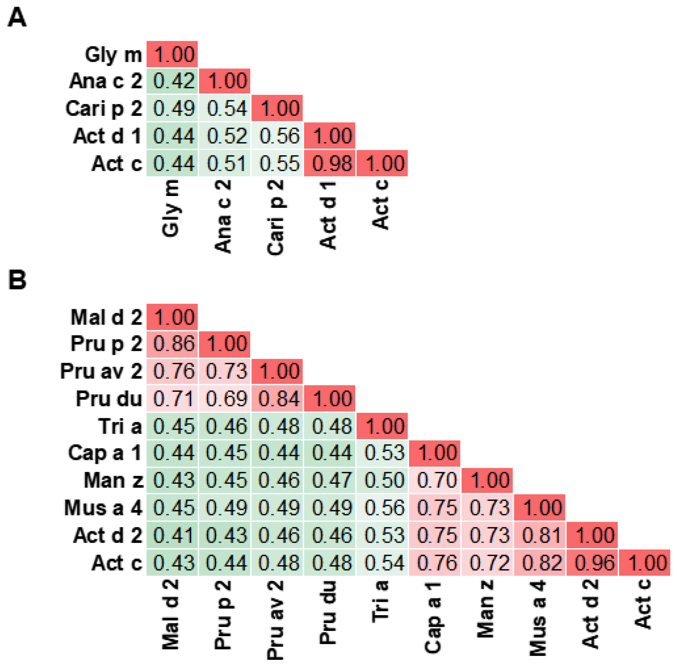
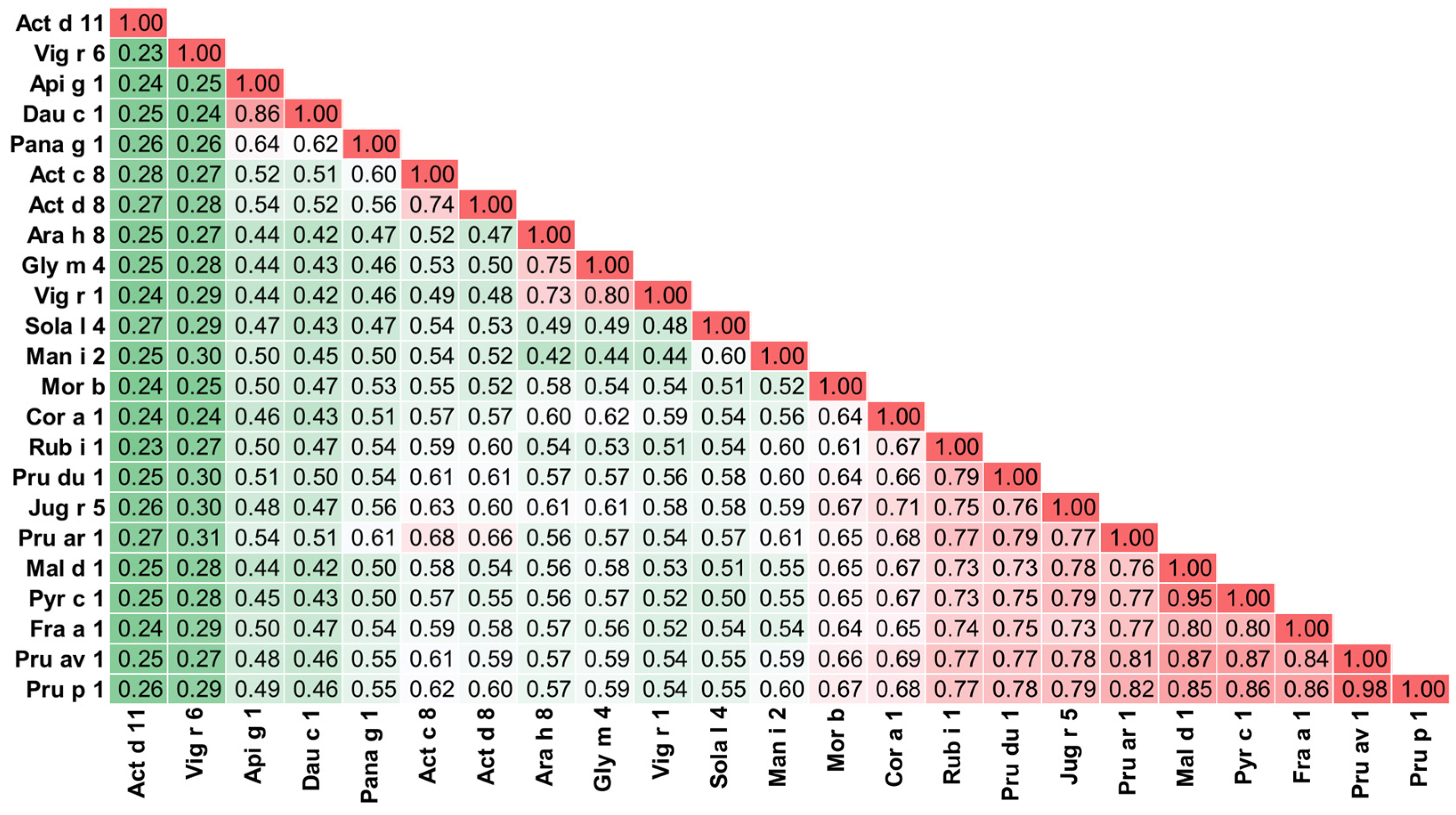
The Allergens’-Relative Identity, Similarity and Cross-reactivity (A-RISC) index provides a method for the prediction of cross-reactivity between allergens of the same family [23]. The A-RISC index suggests the likelihood of cross-reactivity between allergens from the same protein family, with the assumption that allergic cross-reactivity occurs between homologous proteins and will be more likely the more similar that proteins are. A-RISC plots that include the kiwifruit allergens were generated as previously described [23]. Briefly, the A-RISC index is calculated by averaging the sequence identity and the sequence similarity of two protein sequences, using the SIAS (Sequence Identity And Similarity) server (http://imed.med.ucm.es/Tools/sias.html; accessed on 8 May 2025) and the default parameters. Sequences for A-RISC diagrams of registered food allergens were obtained from AllFam [24] and the database of officially registered allergens approved by the WHO/IUIS Allergen Nomenclature Sub-Committee [23]. Sequences corresponding to mature proteins were used during the analysis. A-RISC plots are colored such that red indicates a high likelihood of IgE-mediated cross-reactivity, green indicates a low likelihood, and intermediate colors suggest intermediate cross-reactivity. Structures for allergens without structures available in the Protein Data Bank (PDB) were generated with AlphaFold3 [25] or SwissModel [26]. Images of structures were generated with PyMOL [27].
The results of Allergy Explorer 2 (ALEX2) testing were obtained in four patients who participated in a study evaluating the immune response to plant-derived pollen/food allergens. This study was approved by the local bioethics committee (R-I-002/590/2019). All the subjects signed an informed consent.

3. Protein Families

There are 16 officially registered allergens that originate from kiwifruit and contribute to its allergenic potential [23]. They belong to specific protein families, apart from a poorly characterized glycoprotein, Act d 3, which is not currently classified within a particular family. The protein families to which the kiwifruit allergenic proteins belong are cysteine protease, thaumatin-like protein (TLP), phytocystatin, kiwellin, pectin methylesterase inhibitor (PMEI), pectin methylesterase (PME), pathogenesis-related class 10 proteins (PR-10), profilin, non-specific lipid transfer protein (nsLTP), major latex protein/ripening-related protein (MLP/RRP), 11S globulin, and 2S albumin (Figure 1).

3.1. Cysteine Protease

Actinidin was reported as a major allergen from kiwifruit in different populations [28,29]. It is a 30 kDa cysteine protease enzyme belonging to the peptidase C1 family of papain-like proteases [29]. Actinidin is found in both the green (A. deliciosa) and golden (A. chinensis) varieties (Figure 2A); however, actinidin levels in green kiwifruit are higher than those in the golden variety [30,31]. As is common for most thiol group enzymes, actinidin is secreted in its inactive form as a zymogen (molecular weight of 39 kDa) and then converted into the active form (actinidin) after the cleavage of the precursor peptide [29]. As a proteolytic enzyme, actinidin breaks down complex proteins into peptides to aid in digestion and fruit ripening in the kiwifruit plant. This proteolytically active form is responsible for the allergenicity of this protein [29,32]. Studies have shown that inactive actinidin has lower allergenicity than the proteolytically active form [33]. Proteolytically active actinidin has higher thermal stability (Tm = 73.9 °C) than the inactive form and retains activity in a wide pH range [33,34]. Studies have also shown that immunogenic and proteolytic properties, including the conformational fold of actinidin, are well preserved in a simulated gastric and intestinal digestion experiment, suggesting actinidin’s resistance to pepsin and trypsin degradation [33]. Since actinidin can reach the intestinal mucosa in a proteolytically active form, it can increase the intestinal epithelial permeability by cleaving tight junctions’ proteins in a similar fashion to clinically relevant inhalant allergens (e.g., Der p/f 1), which disrupt the nasal epithelial barrier integrity, and allow facilitated allergen delivery to the intraepithelial dendritic cells [35,36,37]. This has been shown by studies in which actinidin was able to proteolytically truncate intestinal epithelial tight junctions’ proteins like ZO-1 and occludin, in time- and concentration-dependent manners, and increase intestinal permeability in in vitro and in vivo models [34,38]. It was also shown that actinidin upregulates the pro-TH2 cytokines IL-25, IL-33, and thymic stromal lymphopoietin in mice intestines, suggesting its role in triggering innate immunity [39].

3.2. Thaumatin-like Protein

Thaumatin-like protein (TLP) is a major kiwifruit allergen, which has been registered as Act d 2 [40,41,42]. It is a 21–24 kDa single-chain protein. Two isoforms of Act d 2, which differ in their isoelectric point, have been reported. Both isoforms exhibit similar IgE binding ability in skin prick test responses, but their content varies depending on kiwifruit varieties. Belonging to the pathogenesis-related group 5 (PR-5) family, these proteins engage in plant defense mechanisms by inhibiting fungal growth [43]. The antifungal activity of Act d 2 has been demonstrated for Saccharomyces carlsbergenis and Candida albicans [40].
Act d 2 exhibits high thermal stability and resistance to digestive degradation [44,45]. Studies have shown that at least 25% of Act d 2 remained intact after in vitro gastric digestion, and it retained comparable IgE reactivity to untreated Act d 2 fractions [44]. However, the application of high-intensity ultrasound disrupted its secondary structure, decreased its solubility, and increased digestive degradation [46].
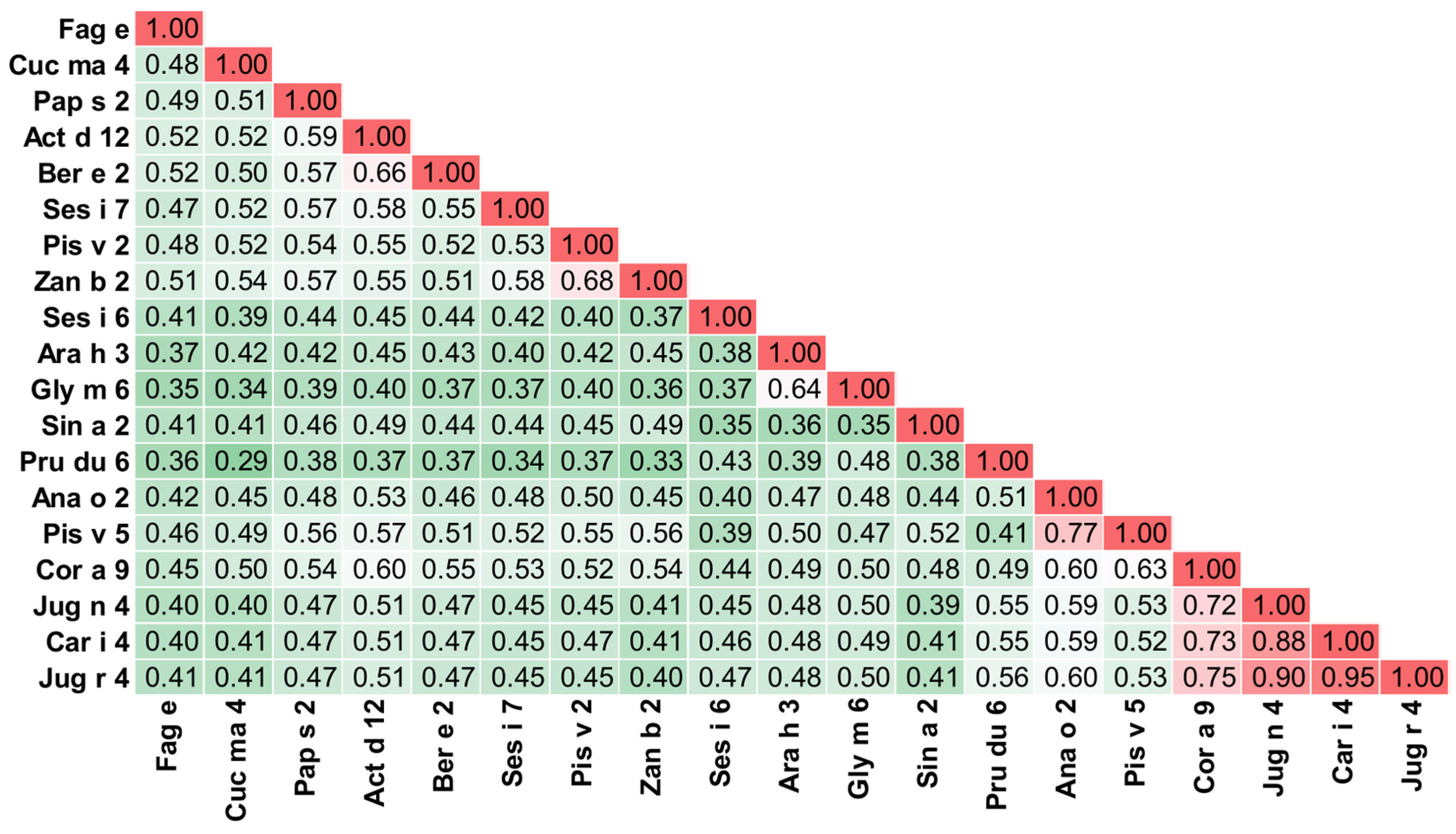
A study reported above 60% specific IgE levels against Act d 2 in kiwifruit-sensitized subjects’ sera and 50% positive skin prick test responses in tested patients [47]. Unlike Act d 1, Act d 2 is frequently a cause of cross-reactions due to the structural similarity of this protein with the homologous molecules contained in other fruits and vegetables [48]. The A-RISC index (Figure 2B) suggests likely cross-reactivity between Act d 2 and the TLPs from banana, mango, pepper, and golden kiwifruit. One study shows that the TLP from apple (Mal d 2) shares allergenic epitopes with peach (Pru p 2) and Act d 2 [49]. The cross-reactivity between Act d 2 and other fruit TLPs is also associated with PFAS [50]. Furthermore, association between Act d 2 and Alt a 1 (allergen from mold Alternaria) reactivity has also been frequently reported, such that 89% of Act d 2-sensitized subjects were also sensitized to Alt a 1 [1]. In fact, it is suggested that sensitization is an outcome of an electrostatic interaction between Alt a 1 and Act d 2, due to the mold penetration inside the fruit pulp [1].

3.3. Phytocystatin

The phytocystatin from kiwifruit, Act d 4, is an 11 kDa cysteine protease inhibitor protein belonging to the cystatin protein family. As a protease inhibitor, Act d 4 plays a significant role in protease activity regulation in plant defense mechanisms [51]. Less is known about the allergenic properties of Act d 4, but studies have suggested its contribution to the clinical symptoms of kiwifruit allergy [52]. Act d 4 was first identified from IgE binding to kiwifruit extract [53]. Dot blot and Western blot analysis on kiwifruit-allergic patient sera suggested conformational IgE epitopes on Act d 4 [52], although this allergen has not yet been structurally characterized.

3.4. Kiwellin

Allergens Act c 5 and Act d 5 are classified as kiwellin. These are approximately 28 kDa, cysteine-rich proteins that account for nearly one-third of the total protein content in kiwifruit [54]. Structurally, kiwellin comprises two domains: a small, N-terminal domain known as kissper (residues 1–39), containing six cysteine residues, and a large, C-terminal domain known as KiTH (residues 40–189), with eight cysteine residues [55,56]. The kissper domain has been shown to have pH- and voltage-dependent pore-forming activity, anion selectivity, and channeling, and anti-inflammatory/antioxidant effects [57,58]. The kissper domain is highly flexible and NMR experiments demonstrated multiple conformations in solution, even with the presence of the three disulfide bonds [55,56,58]. In ripe green kiwifruit, actinidin can proteolytically degrade kiwellin, resulting in a 4 kDa N-terminal kissper and 16 kDa C-terminal KiTH. Such proteolytic degradation of kiwellin also occurs in golden kiwifruit, but to a lesser extent due to the lower abundance of actinidin in golden kiwifruit varieties [59].
Skin prick tests (SPTs) among 29 kiwifruit-allergic subjects showed that 38% were positive to kiwellin [56]. IgE recognition to kiwellin can also occur after proteolytic digestion with actinidin, with some sera reacting solely to KiTH in immunoblot assays [56].

3.5. Pectin Methylesterase Inhibitor

The pectin methylesterase inhibitor (PMEI) Act d 6 is an 18 kDa minor allergen from kiwifruit. PMEI is a natural inhibitor of pectin methylesterase and has wide application in fruit and vegetable processing and preservation [60]. Act d 6 has a low isoelectric point, and an increase in pH can cause structural changes, resulting in unfolding via disulfide bond breakage and the consecutive loss of inhibition activity [60]. PMEIs share structural similarities with invertase inhibitors, and both are grouped in the same protein family as major pollen allergen Pla a 1 from plane trees [42,61]. A study among 49 kiwifruit-allergic subjects showed 73% Act d 6-positive sera in an immunoblot assay; however, only one serum of those tested showed monospecificity to this allergen [42]. Furthermore, skin prick testing and immune solid-phase allergen chip (ISAC89 version) testing showed only 1% (of 36) and 0.05% (of 9107) of kiwifruit-allergic subjects, respectively, to be positive to Act d 6, indicating the weaker allergenic prevalence of this protein [42].

3.6. Pectin Methylesterase

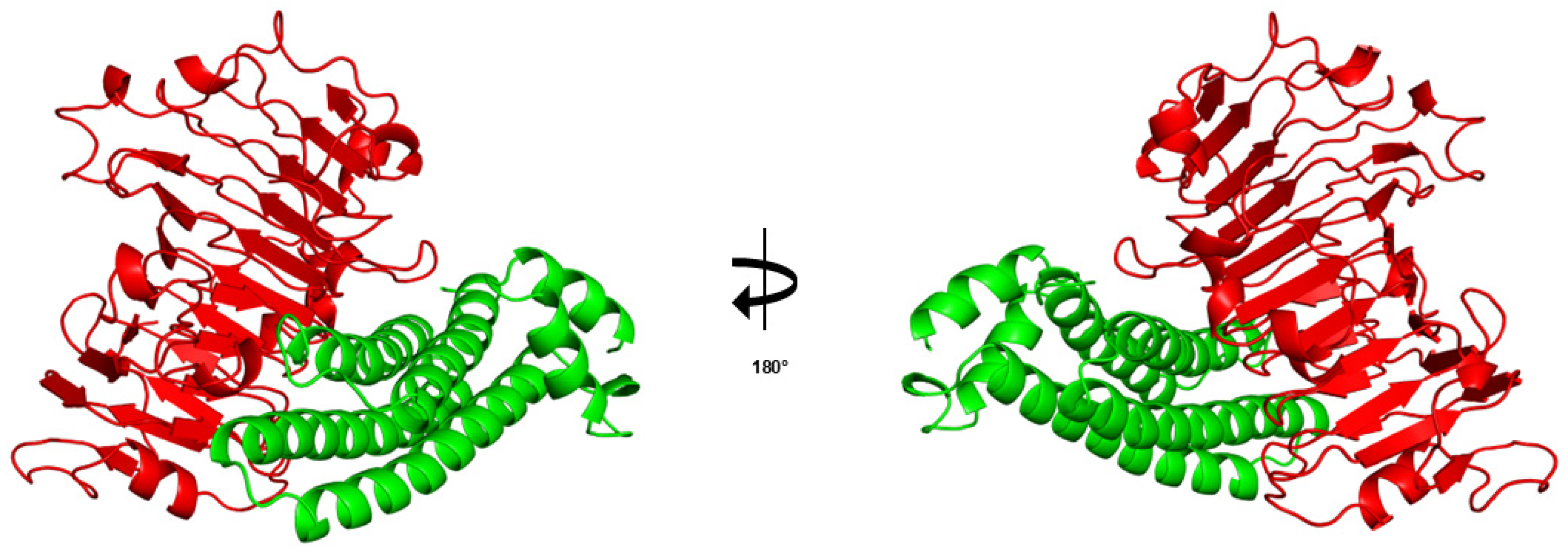
Pectin methylesterase (PME) allergen Act d 7 is a 50 kDa glycoprotein and is considered a minor kiwifruit allergen. PMEs are ubiquitously expressed in plants and are enzymes that affect the mechanical properties of the cell wall [60]. It was demonstrated that the complex between Act d 6 and Act d 7 (Figure 3) is stable in a wide range of pHs, and it dissociates in conditions with a pH above 10 [62]. A study among 49 kiwifruit-allergic patients showed that 32% had positive sera to Act d 7 via immunoblotting, and 18% of patients were reported positive for Act d 7 via ISAC testing (ISAC89 version). In the second group of 36 patients from the same study, 11% of patients had a positive SPT to Act d 7; however, of these patients, none showed specific IgE for Act d 7 [42,63].

3.7. Pathogenesis-Related Class 10 Proteins

Act c 8 and Act d 8 are members of the pathogenesis-related group 10 (PR-10) or Bet v 1-like protein family, each with a molecular weight of 17 kDa. Act d 8 is considered a major allergen in birch-endemic regions but globally is classified as a minor allergen [3]. The classification status of Act c 8 is less defined due to a lack of specific IgE prevalence data independent of Act d 8. However, both Act d 8 and Act c 8 are considered clinically significant for PFAS due to significant IgE cross-reactivity with Bet v 1 [3,64,65]. The A-RISC index (Figure 4) also predicts cross-reactivity between Act c 8 as well as Act d 8 and ingested PR-10 allergens, such as those from apricot and walnut. The concentrations of both allergens remain unclear, but are more abundant in the fruit’s skin than in the pulp [65]. Act c 8 and Act d 8 have more recently been structurally characterized by NMR and maintain the conserved overall fold of PR-10 proteins [65,66]. Variations in the structures of Act c 8 and Act d 8 involve the positioning of the two short α1 and α2 helices, along with the longer C-terminal α3 helix [65]. Furthermore, Act d 8 possesses a loop between β5 and β6 that is shorter by one residue compared to Act c 8, leading to a slightly more rigid local structure [65]. Despite these structural differences, the two allergens exhibit comparable immunological behavior [64].

3.8. Profilin

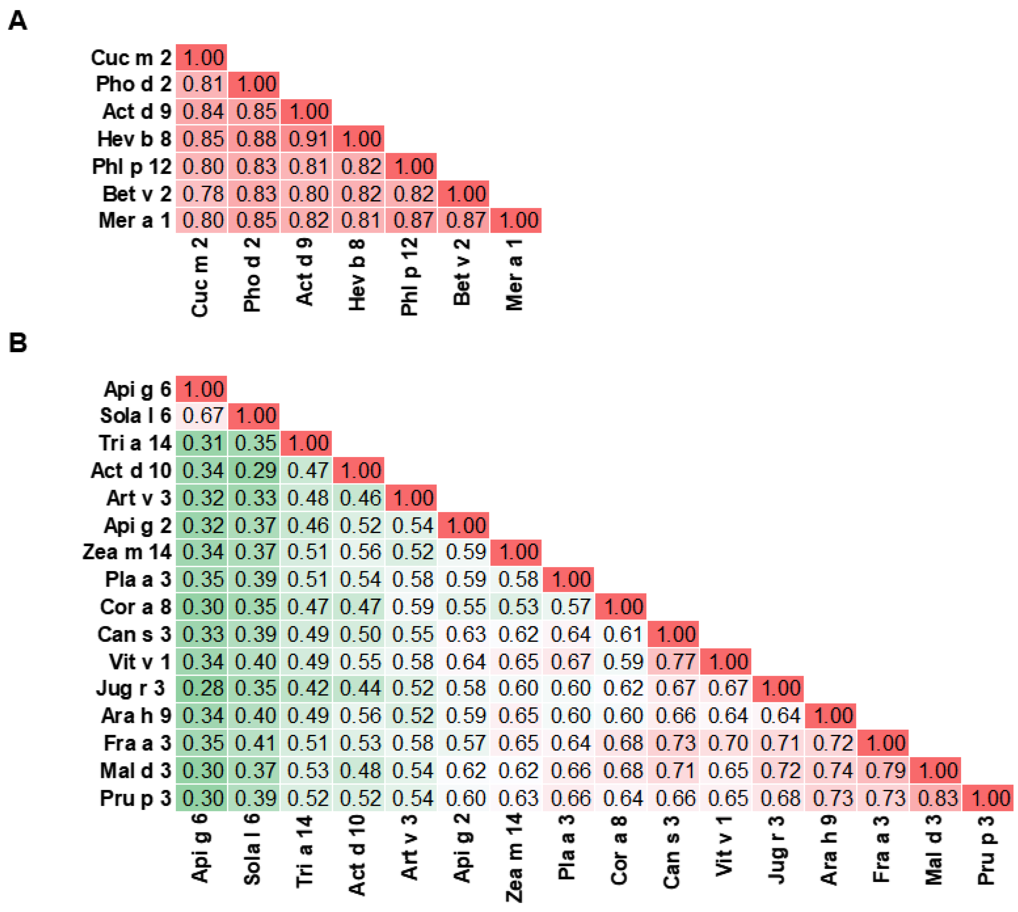
The profilin from kiwifruit, Act d 9, is a 14 kDa protein and is classified as a minor kiwifruit allergen. Profilins are panallergens found in all eukaryotic cells. They are cytosolic proteins that regulate actin polymerization [67]. Profilins play a key role in reorganizing plant microfilaments during critical processes like cytokinesis, cytoplasmic streaming, cell elongation, and pollen tube or root hair growth [68,69,70]. Profilins exhibit a highly conserved sequence and similar structural folding. These observations are in line with the A-RISC index (Figure 5A) predicting cross-reactivity between Act d 9 and virtually all ingested profilin allergens [71,72]. Monosensitization to Act d 9 is uncommon; sensitization is rarely linked to severe systemic reactions and usually associated with mild symptoms, PFAS, and latex–food syndrome. Act d 9 may also be responsible for cross-reactivity between kiwifruit and timothy grass, as well as birch pollen [73]. In a study of 30 patients with confirmed kiwifruit allergy, IgE reactivity to Act d 9 was observed predominantly in polysensitized patients with pollen and/or latex allergy, rather than in those monosensitized to kiwifruit [28]. Latex–food syndrome with kiwi–latex cross-reactivity is often caused by similarity between latex profilin Hev b 8 and Act d 9 [74].

3.9. Non-Specific Lipid Transfer Proteins

Act c 10 and Act d 10 are characterized as non-specific lipid transfer proteins (nsLTPs). These proteins are known for their ability to bind and transport lipids as well as other hydrophobic ligands [75,76,77]. nsLTPs constitute a large protein family and are found in all land plants with a broad range of studied functions [78]. While the biological role of nsLTPs is still not fully understood, they are often involved in plant defense and reproductive mechanisms, can hinder fungal pathogen growth in vitro, and may have roles in antimicrobial activity and structural adaptation [75,77,79,80].
The plant nsLTP family consists of two subfamilies: the larger 9 kDa nsLTP1 and the smaller 7 kDa nsLTP2 [79,81], with Act c 10 and Act d 10 being categorized as nsLTP1s. Both subfamilies have a similar structure, consisting of four α-helices stabilized by four disulfide bridges. These bridges form an internal hydrophobic cavity that can hold a variety of lipids and other hydrophobic ligands [82]. While they are similar in terms of folding structure, neither the cysteine pairings nor the overall amino acid sequences are highly consistent between subfamilies [75,83].
nsLTPs are classified as panallergens, and many members of this family are classified as potent food allergens, with peach allergen Pru p 3 considered the prototypic nsLTP due to its high sensitization capacity [75,84,85]. nsLTPs are highly stable and resistant to heat treatment and digestion, leading to severe allergic symptoms [75,86]. Both Act c 10 and Act d 10 are considered minor kiwifruit allergens and were found in the seed extract but not the pulp extract of kiwifruit [79]. Thus far, Act c 10 has been structurally characterized [75]. In the initial study characterizing the allergenicity of Act d 10, 804 randomly chosen allergic patients were tested for IgE against Act d 10; of these, 21.1% (170 patients) had positive SPTs [87]. Additionally, 89.4% of these 170 patients were also positive for IgE to Pru p 3 [87]. The same study showed high IgE correlations between Act c 10 and Act d 10. Many individuals sensitized to Act c 10 were shown to also be IgE-positive to Act d 10. However, in vivo studies occasionally demonstrated a non-completely overlapping behavior of Act c 10 and Act d 10 [87]. Similarly, it was observed that a few patients (4.3% of 346 patients) were IgE-positive to Act c 10 but negative to Act d 10 isoforms, suggesting structural differences in one or more IgE-binding epitopes of these allergens [75,87]. Interestingly, the A-RISC index (Figure 5B) predicts low cross-reactivity between Act d 10, Act c 10, and other nsLTP allergens.

3.10. Major Latex Protein/Ripening-Related Protein

Act d 11, also known as kirola, is a 17 kDa protein belonging to the major latex protein/ripening-related protein (MLP/RRP) family, which is also part of the Bet v 1-like family [22,88]. Act d 11 is found in high quantities in ripened golden and green kiwifruit. This protein is ripening-related, and the concentration present is influenced by natural ripening and post-harvesting treatments [22,58,88]. Act d 11 has been structurally characterized into multiple crystal forms [22]. Although the sequence identity is low, the 3D structure of Act d 11 is highly similar to other Bet v 1-like allergens [22]. Due to this high structural similarity, specifically within the glycine-rich loop region, the immunological correlation and cross-reactivity of Act d 11 with other PR-10 family allergens, such as Bet v 1 from birch pollen, are observed [22,70].
Act d 11 is classified as a minor kiwifruit allergen. A study of 91 kiwifruit-allergic patients reported that 22/91 (24.2%) patients had IgE to the natural purified Act d 11 when sera were tested by an ISAC microarray system [88]. In the same study, 18/91 (22%) of patients reacted positively to SPTs when tested with natural purified Act d 11 [88]. Sensitization to Act d 11 is reported to primarily occur through Bet v 1-sensitized individuals, suggesting that Act d 11 is involved in cross-reactivity rather than primary sensitization [1,22].
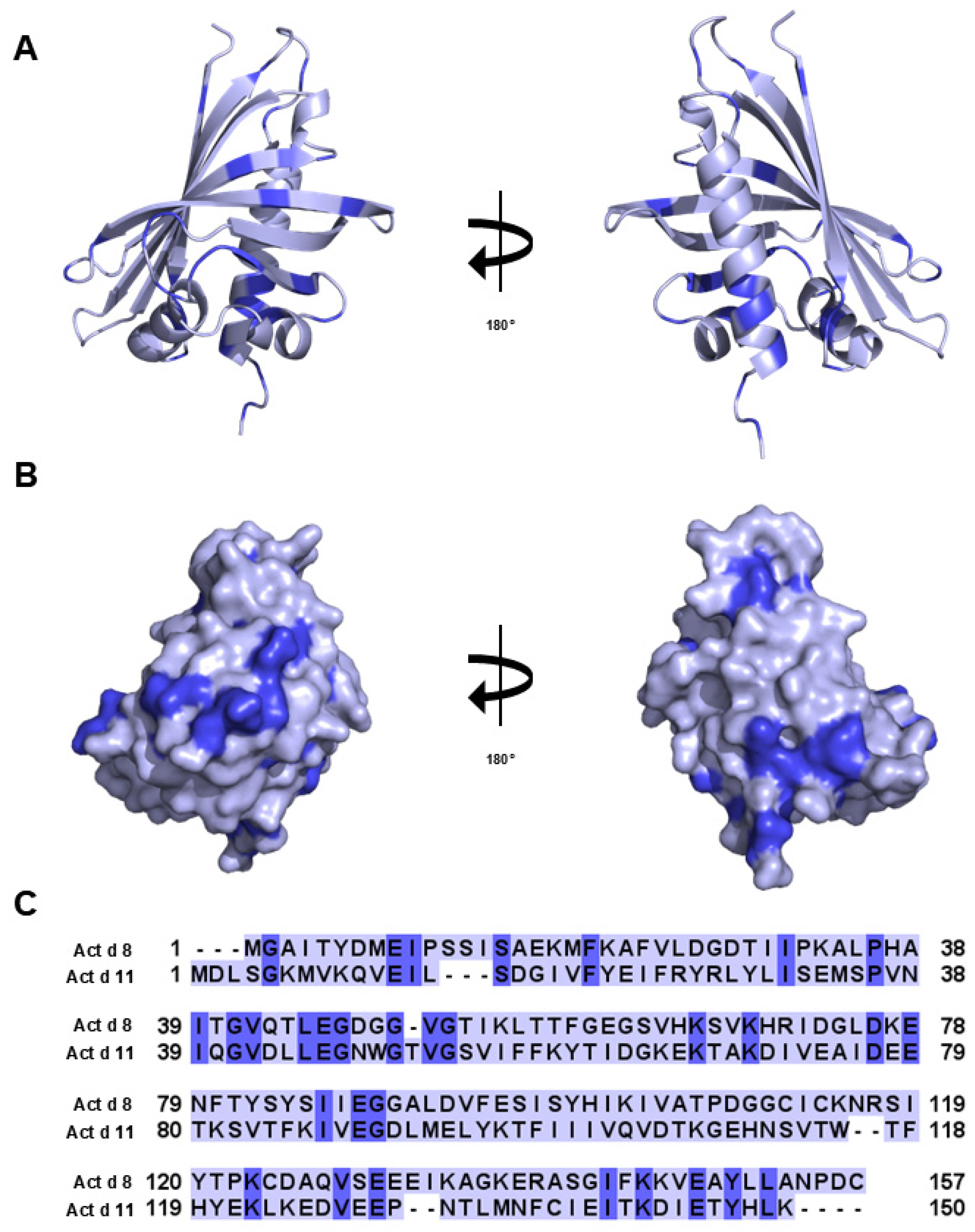
Additionally, it is interesting to compare the structural similarity of Act d 8 with Act d 11 (Figure 6). Despite sharing less than 25% sequence identity with Act d 8, the structure of Act d 11 shows hallmarks of the Bet v 1 superfamily. This structural similarity suggests that Act d 8 and Act d 11 may present similar IgE-binding epitopes, which explains why IgE antibodies generated against one of these allergens may be able to recognize the other as well.

3.11. S Globulin and 2S Albumin

Act d 12, 11S globulin (cupin), and Act d 13, 2S albumin, are kiwifruit-seed-specific storage proteins from two different protein families. These proteins are non-glycosylated and highly stable, composed of two polypeptide chains linked by disulfide bridges [89]. Previous studies have characterized other proteins from these families as potent food allergens that are able to induce primary allergic sensitization upon ingestion [90,91,92]. Due to the structural similarities to cupin proteins, individuals allergic to Act d 12 may also react to other foods containing 11S globulins, such as peanuts, soybeans, and certain tree nuts [89]. The A-RISC index (Figure 7), for example, predicts cross-reactivity between Act d 12 and Ber e 2, the cupin from Brazil nut.
A study of 55 kiwifruit-allergic patients found that Act d 12 represents a major allergen recognized by more than 50% (39/55) of patients tested, whereas Act d 13 represents a minor allergen (10/55) [93]. The same study also found a significant correlation between the reactivity observed in enzyme-linked immunosorbent assay (ELISA) and immunoblotting for both allergens. A mixture of Act d 12 and Act d 13 inhibited approximately 80% of the IgE reactivity to kiwifruit seed extract [93]. This demonstrated that within the study’s population, most of the IgE epitopes of kiwifruit seeds are represented by Act d 12 and Act d 13. Importantly, the same study showed that Act d 12 and Act d 13 were able to induce in vitro IgE cross-linking from kiwifruit-allergic patients, demonstrating that both purified allergens retain allergenic capacity [93].

4. Diagnostics in Kiwifruit Allergy

There are four main diagnostic tests that can be performed or ordered by a physician to diagnose an individual with a kiwifruit allergy, in addition to their clinical history. These are skin prick testing, prick-to-prick testing, the oral food challenge, and an allergen-specific IgE in serum test [94]. The National Institute of Allergy and Infectious Diseases (NIAID) established guidelines to diagnose and manage food allergies in the United States, and similar guidelines are developed in other countries. A related publication by the European Academy of Allergy and Clinical Immunology summarizes information on the molecular basis of allergies, their diagnostics, and treatment; however, it is not considered to be a guideline [94]. The NIAID states that the gold standard for diagnosing a food allergy is the oral food challenge test due to its high specificity and bias reduction compared to other diagnostic options [95]. A food elimination diet is also a viable option.
Generally, medical testing should be both sensitive and specific. In the case of 100% sensitivity, it is expected that all individuals with a particular disease will be identified. Similarly, if a test has 100% specificity, it indicates that all individuals in the tested group that do not have a particular disease are correctly identified.

4.1. Skin Prick Test

In skin prick tests (SPTs), a small amount of kiwifruit protein extract is placed on the surface of the skin and scratched lightly, breaking the skin to allow the allergen to enter the body. After a 15 min wait, the SPT is measured. SPTs were reported to have high sensitivity (93%), but poor specificity (45%), compared to the prick-to-prick test (PPT) (n = 45) [15].
There are several different factors that must be taken into consideration when preparing kiwifruit extract or using commercially available extract. For example, one must remember that green and golden kiwifruits have a different ratio of allergens present (e.g., difference in concentrations of actinidin, kiwellin, and thaumatin-like protein) [50]. Moreover, the allergen content depends on the ripening stage of fruit, the way in which the fruits are stored, and the protocol used to prepare the extract [58]. It was also noted that the allergens may concentrate in different parts of the fruit (e.g., peel, pulp, and seeds) [79].
Many studies have indicated problems with extracts obtained from kiwifruits [28,58], and, in some cases, the quality of extracts was mentioned as a potential reason for poor test performance. In the case of kiwifruit extract, particularly of green kiwifruit, the major issue is related to the high concentration of Act d 1, which is a protease, and therefore can degrade other proteins present in the solution. In fact, kiwellin Act d 5 was found to be a natural substrate of Act d 1, which cleaves kiwellin into 4 kDa N-terminal kissper and 16 kDa C-terminal KiTH [96,97].

4.2. Prick-to-Prick Test

Many physicians use prick-to-prick testing (PPT) to diagnose a kiwifruit allergy. PPTs are comparable to SPTs, except for the source of the allergen. In PPTs, a fresh fruit (kiwifruit) is pricked with a lancet and then the patient’s skin is pricked with the same lancet. After a 15 min wait, the growth of the wheal is measured. PPTs show high sensitivity (81.8%) and high specificity (94.1%) (n = 30) [18]. One study showed that 93% of subjects tested positive for PPTs with fresh kiwifruit who had confirmed kiwifruit allergy via a double-blind placebo-controlled food challenge (DBPCFC) [15]. It was also shown that fresh kiwifruit may be substituted with frozen juice [98]. However, even though PPT is one of the most common techniques for clinical investigation, it should be emphasized that testing with fresh fruits is not standardized [20].

4.3. Oral Food Challenge Test

In the oral food challenge test, the patient ingests increasing doses of various foods while being monitored for an allergic reaction. There are three types of oral food challenge tests: a DBPCFC, a single-blind food challenge, and an open-food challenge test [95]. In the DBPCFC, neither the physician nor the patient knows whether it is an allergen or placebo. In the single-blind food challenge, the physician knows it is an allergen, but the patient does not. In the open-food challenge, both the patient and physician know it is an allergen. The DBPCFC is the preferred method as it minimizes bias.
The first study using DBPCFCs, published in 2004, involved 33 patients reporting symptoms after eating kiwifruits [17]. It was shown that 70% of patients that participated in a DBPCFC had a positive oral food challenge test. This study also revealed the existence of several clinical subgroups of patients. The subgroups were defined based on the allergic reactions (mild or moderate/severe), presence or absence of associated pollinosis, and the presence or absence of a latex allergy. Soon after, another study found that 53% of patients with a history of kiwifruit allergy that undertook the oral food challenge test confirmed that they had a kiwifruit allergy (n = 45) [15]. In another study involving a larger group of patients (n = 92), DBPCFCs were performed on 52 individuals [47].
There are several drawbacks to the oral food challenge test. This test poses some risk to the patients as they are ingesting a potential allergen; therefore, testing must be performed by a trained healthcare professional in a healthcare facility with the means to treat a severe allergic reaction, should one occur. The DBPCFC test can also be expensive; therefore, physicians can perform the single-blind or open-food challenge instead [95].

4.4. Allergen-Specific IgE in Serum

Another method to diagnose a kiwi allergy is for physicians to order the quantification of allergen-specific IgE in serum. Here, the discovery and characterization of allergenic proteins originating from kiwifruit were critical for the establishment of component-resolved diagnostics. One of the most used is ImmunoCAP, which can be used to test IgE antibodies against kiwifruit proteins. ImmunoCAP sIgE consistently has high specificity (90%) when kiwifruit allergens Act d 1, Act d 2, Act d 4, and Act d 5 are combined, but lower values of sensitivity that tend to vary by study (45–54%) [15,28]. It was also noted that the sum of the single allergens performed better than commercial extracts when sensitivity was taken into consideration (77% vs. 17%); however, specificities were 30% and 100%, respectively.
The availability of highly purified allergens as well as allergen extracts enabled the development of high-throughput multiplex assays in which IgE binding to many allergens may be measured simultaneously. Currently, there are several multiplex platforms that are available for allergy diagnostics, like ImmunoCAP ISAC E112i (the newest version of ISAC), Allergy Explorer 2 (ALEX2), and FABER 244 [99,100]. The ISAC test includes only purified molecules of kiwifruit, namely Act d 1, Act d 2, Act d 5, and Act d 8. The FABER system allows for the testing of both extracts and purified molecules. The kiwifruit extracts spotted on the FABER biochip are Act d and Act c, derived from green and golden kiwifruit, respectively; the six available purified molecules are Act d 1, Act d 2, Act d 5, Act d 10, Act c kirola, and Act c chitinase IV [101]. Like FABER, the ALEX2 test also includes both extracts and purified molecules of several organisms, and contains the kiwifruit allergens Act d 1, Act d 2, Act d 5, and Act d 10. Therefore, the last two multiplex platforms, due to the presence of kiwifruit extracts, can provide more detailed information on kiwifruit-allergic patient sensitization.

5. Cross-Reactivity of Kiwifruit Allergens

Allergens originating from different species within the Actinidia genus are obvious culprits in different cross-reactive reactions; for example, the clinical risk of reactions to allergens from golden and green kiwifruits is well established [6,50]. However, a significant fraction of kiwifruit-allergic individuals also react to allergens originating from pollen (e.g., timothy grass or birch) or latex [73,102]. In most of these cases, the reactions are caused by cross-reactivity, but co-sensitization cannot be excluded. Kiwifruit-allergic individuals who have pollinosis most often react to different panallergens that originate from the PR-10, nsLTP, and profilin families. Cross-reactivity between Bet v 1 and both Act d 8 and Act c 8 is relatively common, as it was reported that almost 48% of Bet v 1-sensitized individuals react after eating kiwifruit [103]. The molecular basis of cross-reactivity between the PR-10 allergens was recently explained, and it was shown that PR-10 proteins from kiwifruit are very similar to their homolog from birch [65]. Similarly, cross-reactivity (or IgE co-recognition) between both Act d 10 and Act c 10, as well as different nsLTPs, was also reported [79]. Kiwifruit profilin, similarly to PR-10 and nsLTP allergens, often plays a role in PFAS [104]. Some kiwifruit allergens are also among molecules that are responsible for “latex–fruit syndrome,” which is an example of PFAS [1].
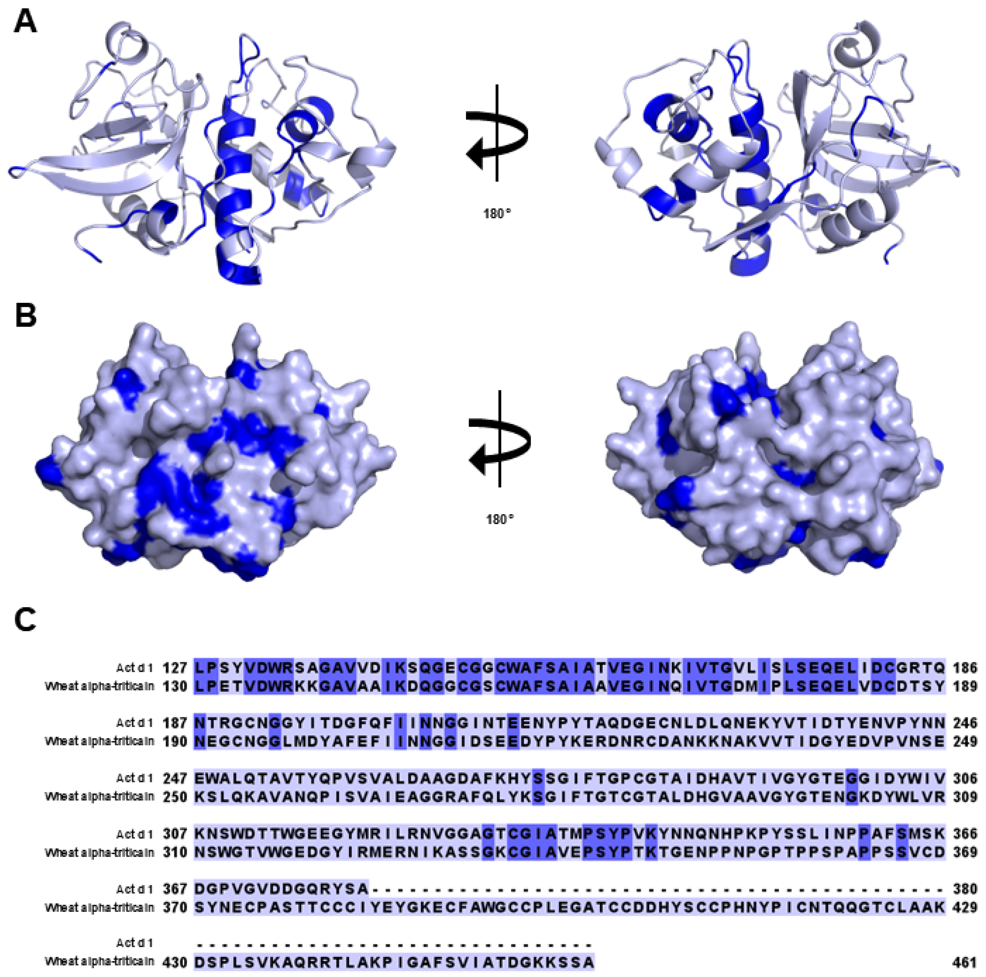
Not only panallergens originating from kiwifruit are implicated in cross-reactive reactions. Cross-reactivity has been demonstrated between actinidin (Act c 1 and Act d 1) and homologous proteins from fig, papaya, or wheat [86,105,106]. The cross-reactivity between Act d 1 and wheat was indicated in patients with baker’s asthma who were also allergic to kiwifruit. It was suggested that the IgE co-recognition of actinidin and alpha-triticain was responsible for this condition (Figure 8) [86]. In the case of thaumatin-like protein (Act d 2 and Act c 2), cross-reactivity was reported with similar proteins originating from avocado, banana, and grape [50]. It was also reported that IgE levels to Act d 12 (11S globulin), but not Act d 13 (2S albumin), correlate positively with IgE levels to Ara h 3, suggesting cross-reactivity. This study was performed on a cohort of patients who were peanut-allergic, and 39% of these individuals were reported to have kiwifruit allergy as well [107]. Interestingly, the same study revealed the presence of kiwifruit 7S globulin that cross-reacted with Ara h 1, which implies the existence of a new unregistered kiwifruit allergen.
The A-RISC index provides a method for the prediction of molecular and clinical cross-reactivity between allergens of the same family [23]. Use of the A-RISC index with kiwifruit and other fruit allergens allows for the prediction of other allergic responses that may arise as a result of sensitization to kiwifruit. For example, the comparison of allergens in the profilin family suggests a high likelihood of cross-reactivity between Act d 9 and all other profilin allergens, primarily to the profilins from pepper (Cap a 2) and potato (Figure 5A). For allergies based on the thaumatin-like protein Act d 2, cross-reactivity may be expected if eating pepper, mango, and banana. The remaining families suggest a lower likelihood of cross-reactivity, although clinical cross-reactivity may differ from the predictions shown here. Use of the A-RISC index for kiwifruit allergy can allow clinicians to predict additional reactions to other foods, including PFAS symptoms and anaphylaxis.

6. Patterns of Sensitization in Kiwifruit Allergy

The development of component-resolved kiwifruit allergy diagnostics improved our ability to investigate sensitization patterns. In studies that involved 92 kiwi-sensitized patients from Spain, it was shown that Act d 1, Act d 2, and Act d 3 were major allergens in the studied population [47]. It was also noticed that high levels of IgE to Act d 1 and Act d 3 were linked with severe symptoms after kiwifruit ingestion. Another study that included 30 Swiss individuals with suspected kiwifruit allergy divided patients into three groups: monosensitized to kiwifruit (n = 8), individuals with concomitant pollen allergy (n = 17), and a group that had kiwifruit, pollen, and latex allergies (n = 5) [28]. It was shown that monosensitized patients reacted more often to Act d 1 in comparison with polysensitized individuals, who reacted more often to Act d 8. This study also did not find any significant correlation between IgE reactivity and reaction severity in kiwifruit allergy. Later, a large European study reported that only Act d 1 was associated with disease severity, and individuals with severe symptoms (when compared with patients with mild symptoms) were often sensitized to actinidin [108]. It was also noticed that sensitization to Act d 1 was less frequent in patients that had kiwifruit allergy for more than 5 years. The same study highlighted differences in sensitization profiles in patients living in different parts of Europe. For example, individuals from Iceland were mainly affected by Act d 1, living in southern Europe was associated with sensitization to Act d 9 and Act d 10, and individuals sensitized to Act d 8 were most frequently living in western, central, and eastern Europe. Sensitization to Act d 8 in Europe is quite frequent, and it is related to cross-reactivity with Bet v 1 as well as other allergens from the PR-10 family. Interestingly, a group from Japan showed that, among the local population, a negative specific response to Act d 8 was linked with more severe kiwifruit allergy [109].
Some exemplary cases of kiwifruit allergy with ALEX2 results, detailed in Table 2, are provided below to illustrate different patterns of allergic reactions. The reactions are not exclusive to kiwifruit, and they can be explained by analyzing the multiplex assay results. The selected patients provided a history of an allergic response to kiwifruit, but they were not necessarily allergic solely to kiwifruit.
Patient 1: A 25-year-old female experienced multiple allergic reactions upon eating foods containing vegetables, corn, cereals, and nuts. Symptoms seemed to be more severe with physical activity after meals but occurred even without exercise. The patient consumed kiwifruit assuming she was not allergic to it; however, consumption resulted in anaphylaxis. The patient profile showed selective sensitization to nsLTP and no sensitization to specific kiwifruit allergens Act d 1, Act d 2, Act d 5, or cross-reactive 11S globulins or 2S albumins, which are homologous to Act d 12 or Act d 13, respectively.
Patient 2: A 19-year-old male suffered from seasonal allergic rhinitis and conjunctivitis for 10 years. The rhinitis and conjunctivitis symptoms were exasperated during tree pollen season (March–May). The patient experienced PFAS to hazelnuts and apples, and underwent unexpected anaphylaxis after eating kiwifruit. No known cofactors were identified. The patient profile showed sensitization to Act d 1. PR-10 co-sensitization did not seem to be responsible for anaphylaxis. Not sensitized to nsLTP, 11S globulins, or 2S albumins.
Patient 3: A 35-year-old male suffered from seasonal allergic rhinitis and conjunctivitis for more than 20 years. The rhinitis and conjunctivitis symptoms increased during tree, grass, and weed pollen season (March–October). Symptoms of PFAS were noted to many fruits (stone fruits) and hazelnuts. The patient was on a strict diet, excluding stone fruits, nuts, carrot, celery, tomato, and corn. He tried kiwifruit based on no clinical history of an allergy. No known cofactors were identified. Anaphylaxis was experienced after eating fresh, raw kiwifruit, this being an example of the real-life problem of the selection of an individual protein as an exclusive culprit of anaphylaxis. The patient showed sensitization to PR-10, but it is an unlikely cause of anaphylaxis due to protein instability. One candidate is nsLTP, a second is TLP, and a third is 11S globulins or 2S albumins. Sensitization to TLP is also associated with sensitization to Alt a 1.
Patient 4: An 18-year-old male suffered from allergic rhinitis and conjunctivitis for more than 12 years. The rhinitis and conjunctivitis symptoms increased during tree pollen season (March–May). Symptoms of PFAS were noted to hazelnuts, apples, cherries, soy milk, and fresh, raw kiwifruit. Unfortunately, both PR-10 and profilin can be a culprit in PFAS. The patient profile showed no sensitization to Act d 1, Act d 2, cross-reactive nsLTP, 11S globulins, or 2S albumins.

7. Management of Kiwifruit Allergies

The general principles of management for all allergic diseases also apply to this group of patients. There are three areas of action: allergen avoidance, pharmacotherapy, and allergen immunotherapy. In kiwifruit allergy, management strongly depends on the clinical presentation, which may range from benign PFAS to severe, life-threatening anaphylaxis [70].
In patients with a history of PFAS, ideally supported by the demonstration of sensitization to unstable proteins such as PR-10s or profilins, but not to stable proteins such as nsLTP, 2S albumins, or actinidin, complete abstinence from kiwifruit consumption is not necessary [110]. Moreover, meals containing processed kiwifruit usually do not trigger an allergic response in such patients [5].
Should symptoms triggered by accidental exposure to raw kiwifruit appear, the use of oral H1 antihistamines such as cetirizine or loratadine should be effective in reducing the severity of symptoms [110]. However, it is usually not advised to use pretreatment with H1 antihistamines to consume foods known to trigger PFAS. If PR-10-dependent PFAS is demonstrated, particularly in patients who experience birch pollen allergy, allergen immunotherapy to birch may improve the tolerance of kiwifruit in some patients, but this is not a guarantee [65,110].
It is worth emphasizing that component-resolved diagnosis may be helpful in determining the risk of more severe clinical reactions upon kiwifruit ingestion in patients who have reported only local responses [111].
Patients that experienced anaphylaxis upon kiwifruit ingestion should undergo a careful evaluation of potential allergen sources, ideally at the molecular level, to provide sufficient information concerning food avoidance methods, including the assessment of the potential impact of food processing on the risk of anaphylaxis upon the ingestion of different kiwifruit-containing meals [112]. Moreover, searching for co-factors that affect the appearance of anaphylaxis upon kiwifruit ingestion and/or affect its intensity is highly warranted [113]. Cases of food-dependent exercise-induced anaphylaxis to kiwifruit have been described [16].
All patients who have experienced anaphylaxis should be equipped with epinephrine autoinjectors and be instructed how to use them [112]. Allergen immunotherapy with kiwifruit extract is not commercially available. However, kiwifruit-induced allergy and anaphylaxis have been successfully treated with sublingual immunotherapy containing extracts from kiwifruit [114].
Thermal treatment of fruit was reported to be effective in the reduction in allergic reactions to kiwifruit [115]. In a trial that involved 20 children, it was found that thermally processed kiwifruit elicited responses in only five of the children when SPTs was used, while all 20 participants reacted to raw fruit [115]. Later, it was also confirmed that structurally intact and non-denatured kiwifruit allergens that were resistant to digestion could be responsible for systemic reactions [115]. It was also suggested that patients with hypoacidic medical conditions may have impaired the pepsin digestion of kiwifruits and therefore may have an increased risk of allergic reactions. Interestingly, it was also shown that, while the heat-induced denaturation of Act d 1 was not reversible when the allergen was transferred to an acidic solution, this was not true in the case of Act d 2 [44]. However, recent studies suggest that the microwave processing of kiwifruit may reduce its allergenicity related to the presence of Act d 2 [116].

8. Conclusions

Kiwifruit allergy has become increasingly common, driven by the fruit’s global availability, rich nutritional value, and rising popularity. This allergy is characterized by a diverse array of major and minor allergens from different protein families, which complicates diagnosis and management. The clinical relevance of kiwifruit allergy is further complicated by cross-reactivity with allergens from fruits, vegetables, and plants, including birch and latex. This phenomenon is largely attributed to shared allergenic epitopes as well as sequence and structural similarities between kiwifruit allergens and those from other sources. The variability in allergenicity among kiwifruit proteins, influenced by differing allergen abundances, directly affects the severity of allergic reactions and allows for the categorization of different patient groups.
Future research holds significant potential for improving the diagnosis, management, and prevention of kiwifruit allergy. Such advances need to include the characterization of additional kiwifruit allergens, as well as improvement in the accuracy, accessibility, and reliability of both allergy diagnostic and treatment methods. For example, we expect that there are still unidentified kiwifruit allergens, such as a kiwifruit allergen homologous to Ara h 1 [107]. Additionally, more recent research has shown that other cofactors, such as physical exercise, can trigger severe allergic responses, such as exercise-induced anaphylaxis, in individuals that are sensitized to kiwifruit [16]. Increasing interest is also being given to small molecular ligands, which might affect the onset and intensity of allergic reactions by impacting immune mechanisms or altering allergen absorption [117,118].
IgE epitopes on kiwifruit allergens are currently uncharacterized [119,120], and this has a significant impact on the design of potential hypoallergens or passive immunotherapy for affected individuals. Similarly, reports on successful immunotherapy are very scarce, and there are only limited descriptions of successful sublingual/oral allergen immunotherapy [121]. Therefore, further studies and improved understanding of the molecular mechanisms of kiwifruit allergenicity are critical for developing effective strategies for managing and preventing allergic reactions to this fruit.

Author Contributions

E.M.W.: Conceptualization; investigation; methodology; formal analysis; visualization; validation; writing—original draft; writing—review and editing. A.O.: Conceptualization; investigation; methodology; formal analysis; visualization; validation; writing—original draft; writing—review and editing. K.K. (Kriti Khatri): Conceptualization; investigation; methodology; formal analysis; validation; writing—original draft; writing—review and editing. R.P.: Conceptualization; investigation; methodology; formal analysis; validation; writing—original draft; writing—review and editing. L.R.O.: Conceptualization; investigation; methodology; formal analysis; validation; writing—original draft; writing—review and editing. E.C.: Conceptualization; investigation; methodology; formal analysis; validation; writing—original draft; writing—review and editing. T.R.: Conceptualization; investigation; methodology; formal analysis; validation; writing—original draft; writing—review and editing. M.A.C.: Conceptualization; investigation; methodology; formal analysis; writing—original draft; writing—review and editing; validation; visualization. K.K. (Krzysztof Kowal): Conceptualization; investigation; methodology; formal analysis; writing—original draft; writing—review and editing; validation; visualization. M.C.: Conceptualization; formal analysis; visualization; project administration; supervision; resources; writing—original draft; writing—review and editing; funding acquisition; validation. All authors have read and agreed to the published version of the manuscript.

Funding

The research reported in this publication was partially supported by the National Institute of Allergy and Infectious Diseases of the National Institutes of Health under award number R01AI077653-13 (to M.C.). The content is solely the responsibility of the authors and does not necessarily represent the official views of the National Institutes of Health.

Conflicts of Interest

The authors declare no conflict of interest.

Abbreviations

The following abbreviations are used in this manuscript:
WHOWorld Health Organization
IUISInternational Union of Immunological Societies
IgEImmunoglobulin E
PFASPollen food allergy syndrome
A-RISCAllergens’-Relative Identity, Similarity and Cross-Reactivity
PDBProtein Data Bank
ALEX2Allergy Explorer 2
TLPThaumatin-like protein
PMEIPectin methylesterase inhibitor
PMEPectin methylesterase
PR-10Pathogenesis-related proteins group 10
nsLTPNon-specific lipid transfer protein
MLP/RRPMajor latex protein/ripening-related protein
PR-5Pathogenesis-related proteins group 5
SPTSkin prick test
ISACImmune solid-phase allergen chip
ELISAEnzyme-linked immunosorbent assay
NIAIDNational Institute of Allergy and Infectious Diseases
PPTPrick-to-prick testing
DBPCFCDouble-blind, placebo-controlled food challenge

References

  1. Bringheli, I.; Brindisi, G.; Morelli, R.; Marchetti, L.; Cela, L.; Gravina, A.; Pastore, F.; Semeraro, A.; Cinicola, B.; Capponi, M.; et al. Kiwifruit’s Allergy in Children: What Do We Know? Nutrients 2023, 15, 3030. [Google Scholar] [CrossRef]
  2. Richardson, D.P.; Ansell, J.; Drummond, L.N. The nutritional and health attributes of kiwifruit: A review. Eur. J. Nutr. 2018, 57, 2659–2676. [Google Scholar] [CrossRef] [PubMed]
  3. James, C.A.; Welham, S.; Rose, P. Update on the global prevalence and severity of kiwifruit allergy: A scoping review. Int. J. Food Sci. Tech. 2023, 58, 6158–6181. [Google Scholar] [CrossRef]
  4. Pomes, A.; Davies, J.M.; Gadermaier, G.; Hilger, C.; Holzhauser, T.; Lidholm, J.; Lopata, A.L.; Mueller, G.A.; Nandy, A.; Radauer, C.; et al. WHO/IUIS Allergen Nomenclature: Providing a common language. Mol. Immunol. 2018, 100, 3–13. [Google Scholar] [CrossRef]
  5. Uberti, F.; Penas, E.; Manzoni, Y.; di Lorenzo, C.; Ballabio, C.; Fiocchi, A.; Terracciano, L.; Restani, P. Molecular characterization of allergens in raw and processed kiwifruit. Pediatr. Allergy Immunol. 2015, 26, 139–144. [Google Scholar] [CrossRef] [PubMed]
  6. Lucas, J.S.; Lewis, S.A.; Trewin, J.B.; Grimshaw, K.E.; Warner, J.O.; Hourihane, J.O. Comparison of the allergenicity of Actinidia deliciosa (kiwi fruit) and Actinidia chinensis (gold kiwi). Pediatr. Allergy Immunol. 2005, 16, 647–654. [Google Scholar] [CrossRef] [PubMed]
  7. Bublin, M.; Mari, A.; Ebner, C.; Knulst, A.; Scheiner, O.; Hoffmann-Sommergruber, K.; Breiteneder, H.; Radauer, C. IgE sensitization profiles toward green and gold kiwifruits differ among patients allergic to kiwifruit from 3 European countries. J. Allergy Clin. Immunol. 2004, 114, 1169–1175. [Google Scholar] [CrossRef]
  8. Mari, A.; Rasi, C.; Palazzo, P.; Scala, E. Allergen databases: Current status and perspectives. Curr. Allergy Asthma Rep. 2009, 9, 376–383. [Google Scholar] [CrossRef]
  9. Fine, A.J. Hypersensitivity reaction to kiwi fruit (Chinese gooseberry, Actinidia chinensis). J. Allergy Clin. Immunol. 1981, 68, 235–237. [Google Scholar] [CrossRef]
  10. Venkataraman, D.; Erlewyn-Lajeunesse, M.; Kurukulaaratchy, R.J.; Potter, S.; Roberts, G.; Matthews, S.; Arshad, S.H. Prevalence and longitudinal trends of food allergy during childhood and adolescence: Results of the Isle of Wight Birth Cohort study. Clin. Exp. Allergy 2018, 48, 394–402. [Google Scholar] [CrossRef]
  11. Vieira, T.; Cunha, L.; Neves, E.; Falcao, H. Diagnostic usefulness of component-resolved diagnosis by skin prick tests and specific IgE to single allergen components in children with allergy to fruits and vegetables. Allergol. Immunopathol. 2014, 42, 127–135. [Google Scholar] [CrossRef]
  12. Kim, J.H.; Kim, S.H.; Park, H.W.; Cho, S.H.; Chang, Y.S. Oral Allergy Syndrome in Birch Pollen-Sensitized Patients from a Korean University Hospital. J. Korean Med. Sci. 2018, 33, e218. [Google Scholar] [CrossRef] [PubMed]
  13. Mattila, L.; Kilpelainen, M.; Terho, E.O.; Koskenvuo, M.; Helenius, H.; Kalimo, K. Food hypersensitivity among Finnish university students: Association with atopic diseases. Clin. Exp. Allergy 2003, 33, 600–606. [Google Scholar] [CrossRef] [PubMed]
  14. Karakaya, E.; Uzundumlu, A.S. Kiwi Production Forecasts for the Leading Countries in the Period 1983–2027. Appl. Fruit Sci. 2025, 67, 66. [Google Scholar] [CrossRef]
  15. Lucas, J.S.; Grimshaw, K.E.; Collins, K.; Warner, J.O.; Hourihane, J.O. Kiwi fruit is a significant allergen and is associated with differing patterns of reactivity in children and adults. Clin. Exp. Allergy 2004, 34, 1115–1121. [Google Scholar] [CrossRef]
  16. Ukleja-Sokolowska, N.; Zacniewski, R.; Lis, K.; Zbikowska-Gotz, M.; Kuzminski, A.; Bartuzi, Z. Exercise induced anaphylaxis in kiwi allergic patient: Case report. Allergy Asthma Clin. Immunol. 2021, 17, 91. [Google Scholar] [CrossRef]
  17. Aleman, A.; Sastre, J.; Quirce, S.; de las Heras, M.; Carnes, J.; Fernandez-Caldas, E.; Pastor, C.; Blazquez, A.B.; Vivanco, F.; Cuesta-Herranz, J. Allergy to kiwi: A double-blind, placebo-controlled food challenge study in patients from a birch-free area. J. Allergy Clin. Immunol. 2004, 113, 543–550. [Google Scholar] [CrossRef]
  18. D’Amelio, C.M.; Bernad, A.; Garcia-Figueroa, B.E.; Garrido-Fernandez, S.; Azofra, J.; Beristain, A.; Bueno-Diaz, C.; Garrido-Arandia, M.; Gastaminza, G.; Ferrer, M.; et al. Unraveling the Diagnosis of Kiwifruit Allergy: Usefulness of Current Diagnostic Tests. J. Investig. Allergol. Clin. Immunol. 2022, 32, 206–212. [Google Scholar] [CrossRef]
  19. Gall, H.; Kalveram, K.J.; Forck, G.; Sterry, W. Kiwi fruit allergy: A new birch pollen-associated food allergy. J. Allergy Clin. Immunol. 1994, 94, 70–76. [Google Scholar] [CrossRef]
  20. Lucas, J.S.; Lewis, S.A.; Hourihane, J.O. Kiwi fruit allergy: A review. Pediatr. Allergy Immunol. 2003, 14, 420–428. [Google Scholar] [CrossRef]
  21. Kashyap, R.R.; Kashyap, R.S. Oral Allergy Syndrome: An Update for Stomatologists. J. Allergy 2015, 2015, 543928. [Google Scholar] [CrossRef] [PubMed]
  22. Chruszcz, M.; Ciardiello, M.A.; Osinski, T.; Majorek, K.A.; Giangrieco, I.; Font, J.; Breiteneder, H.; Thalassinos, K.; Minor, W. Structural and bioinformatic analysis of the kiwifruit allergen Act d 11, a member of the family of ripening-related proteins. Mol. Immunol. 2013, 56, 794–803. [Google Scholar] [CrossRef]
  23. Chruszcz, M.; Kapingidza, A.B.; Dolamore, C.; Kowal, K. A robust method for the estimation and visualization of IgE cross-reactivity likelihood between allergens belonging to the same protein family. PLoS ONE 2018, 13, e0208276. [Google Scholar] [CrossRef]
  24. Radauer, C.; Bublin, M.; Wagner, S.; Mari, A.; Breiteneder, H. Allergens are distributed into few protein families and possess a restricted number of biochemical functions. J. Allergy Clin. Immunol. 2008, 121, 847–852.e847. [Google Scholar] [CrossRef] [PubMed]
  25. Abramson, J.; Adler, J.; Dunger, J.; Evans, R.; Green, T.; Pritzel, A.; Ronneberger, O.; Willmore, L.; Ballard, A.J.; Bambrick, J.; et al. Accurate structure prediction of biomolecular interactions with AlphaFold 3. Nature 2024, 630, 493–500. [Google Scholar] [CrossRef]
  26. Waterhouse, A.; Bertoni, M.; Bienert, S.; Studer, G.; Tauriello, G.; Gumienny, R.; Heer, F.T.; de Beer, T.A.P.; Rempfer, C.; Bordoli, L.; et al. SWISS-MODEL: Homology modelling of protein structures and complexes. Nucleic Acids Res. 2018, 46, W296–W303. [Google Scholar] [CrossRef]
  27. The PyMOL Molecular Graphics System, 3.0; Schrödinger, LLC: New York, NY, USA, 2025; Available online: https://pymol.org/support.html (accessed on 23 June 2025).
  28. Bublin, M.; Pfister, M.; Radauer, C.; Oberhuber, C.; Bulley, S.; Dewitt, A.M.; Lidholm, J.; Reese, G.; Vieths, S.; Breiteneder, H.; et al. Component-resolved diagnosis of kiwifruit allergy with purified natural and recombinant kiwifruit allergens. J. Allergy Clin. Immunol. 2010, 125, 687–694.e1. [Google Scholar] [CrossRef]
  29. Pastorello, E.A.; Conti, A.; Pravettoni, V.; Farioli, L.; Rivolta, F.; Ansaloni, R.; Ispano, M.; Incorvaia, C.; Giuffrida, M.G.; Ortolani, C. Identification of actinidin as the major allergen of kiwi fruit. J. Allergy Clin. Immunol. 1998, 101, 531–537. [Google Scholar] [CrossRef] [PubMed]
  30. Kaur, L.; Mao, B.; Bailly, J.; Oladeji, O.; Blatchford, P.; McNabb, W.C. Actinidin in Green and SunGold Kiwifruit Improves Digestion of Alternative Proteins-An In Vitro Investigation. Foods 2022, 11, 2739. [Google Scholar] [CrossRef]
  31. Nieuwenhuizen, N.J.; Beuning, L.L.; Sutherland, P.W.; Sharma, N.N.; Cooney, J.M.; Bieleski, L.R.F.; Schroder, R.; MacRae, E.A.; Atkinson, R.G. Identification and characterisation of acidic and novel basic forms of actinidin, the highly abundant cysteine protease from kiwifruit. Funct. Plant Biol. 2007, 34, 946–961. [Google Scholar] [CrossRef]
  32. Giangrieco, I.; Ciardiello, M.A.; Tamburrini, M.; Tuppo, L.; Mari, A.; Alessandri, C. Plant and Arthropod IgE-Binding Papain-like Cysteine Proteases: Multiple Contributions to Allergenicity. Foods 2024, 13, 790. [Google Scholar] [CrossRef] [PubMed]
  33. Grozdanovic, M.M.; Ostojic, S.; Aleksic, I.; Andjelkovic, U.; Petersen, A.; Gavrovic-Jankulovic, M. Active actinidin retains function upon gastro-intestinal digestion and is more thermostable than the E-64-inhibited counterpart. J. Sci. Food Agric. 2014, 94, 3046–3052. [Google Scholar] [CrossRef]
  34. Grozdanovic, M.M.; Cavic, M.; Nesic, A.; Andjelkovic, U.; Akbari, P.; Smit, J.J.; Gavrovic-Jankulovic, M. Kiwifruit cysteine protease actinidin compromises the intestinal barrier by disrupting tight junctions. Biochim. Biophys. Acta 2016, 1860, 516–526. [Google Scholar] [CrossRef]
  35. Fukuoka, A.; Yoshimoto, T. Barrier dysfunction in the nasal allergy. Allergol. Int. 2018, 67, 18–23. [Google Scholar] [CrossRef] [PubMed]
  36. Soh, W.T.; Zhang, J.; Hollenberg, M.D.; Vliagoftis, H.; Rothenberg, M.E.; Sokol, C.L.; Robinson, C.; Jacquet, A. Protease allergens as initiators-regulators of allergic inflammation. Allergy 2023, 78, 1148–1168. [Google Scholar] [CrossRef] [PubMed]
  37. Yazici, D.; Ogulur, I.; Pat, Y.; Babayev, H.; Barletta, E.; Ardicli, S.; Bel Imam, M.; Huang, M.; Koch, J.; Li, M.; et al. The epithelial barrier: The gateway to allergic, autoimmune, and metabolic diseases and chronic neuropsychiatric conditions. Semin. Immunol. 2023, 70, 101846. [Google Scholar] [CrossRef]
  38. Cavic, M.; Grozdanovic, M.M.; Bajic, A.; Jankovic, R.; Andjus, P.R.; Gavrovic-Jankulovic, M. The effect of kiwifruit (Actinidia deliciosa) cysteine protease actinidin on the occludin tight junction network in T84 intestinal epithelial cells. Food Chem. Toxicol. 2014, 72, 61–68. [Google Scholar] [CrossRef]
  39. Nesic, A.; Cavic, M.; Popovic, M.; Zlatanova, M.; Pieters, R.; Smit, J.; Gavrovic-Jankulovic, M. The Kiwifruit Allergen Act d 1 Activates NF-kappaB Signaling and Affects mRNA Expression of TJ Proteins and Innate Pro-Allergenic Cytokines. Biomolecules 2019, 9, 816. [Google Scholar] [CrossRef]
  40. Gavrovic-Jankulovic, M.; cIrkovic, T.; Vuckovic, O.; Atanaskovic-Markovic, M.; Petersen, A.; Gojgic, G.; Burazer, L.; Jankov, R.M. Isolation and biochemical characterization of a thaumatin-like kiwi allergen. J. Allergy Clin. Immunol. 2002, 110, 805–810. [Google Scholar] [CrossRef]
  41. Gavrovic-Jankulovic, M.; Spasic, M.; Cirkovic Velickovic, T.; Stojanovic, M.; Inic-Kanada, A.; Dimitrijevic, L.; Lindner, B.; Petersen, A.; Becker, W.M.; Jankov, R.M. Quantification of the thaumatin-like kiwi allergen by a monoclonal antibody-based ELISA. Mol. Nutr. Food Res. 2008, 52, 701–707. [Google Scholar] [CrossRef]
  42. Palazzo, P.; Tuppo, L.; Giangrieco, I.; Bernardi, M.L.; Rafaiani, C.; Crescenzo, R.; Tamburrini, M.; Zuzzi, S.; Alessandri, C.; Mari, A.; et al. Prevalence and peculiarities of IgE reactivity to kiwifruit pectin methylesterase and its inhibitor, Act d 7 and Act d 6, in subjects allergic to kiwifruit. Food Res. Int. 2013, 53, 24–30. [Google Scholar] [CrossRef]
  43. Wang, H.; Ng, T.B. Isolation of an antifungal thaumatin-like protein from kiwi fruits. Phytochemistry 2002, 61, 1–6. [Google Scholar] [CrossRef]
  44. Bublin, M.; Radauer, C.; Knulst, A.; Wagner, S.; Scheiner, O.; Mackie, A.R.; Mills, E.N.C.; Breiteneder, H. Effects of gastrointestinal digestion and heating on the allergenicity of the kiwi allergens Act d 1, actinidin, and Act d 2, a thaumatin-like protein. Mol. Nutr. Food Res. 2008, 52, 1130–1139. [Google Scholar] [CrossRef] [PubMed]
  45. Wang, J.; Vanga, S.K.; Raghavan, V. Structural responses of kiwifruit allergen Act d 2 to thermal and electric field stresses based on molecular dynamics simulations and experiments. Food Funct. 2020, 11, 1373–1384. [Google Scholar] [CrossRef]
  46. Wang, J.; Wang, J.; Kranthi Vanga, S.; Raghavan, V. Influence of high-intensity ultrasound on the IgE binding capacity of Act d 2 allergen, secondary structure, and In-vitro digestibility of kiwifruit proteins. Ultrason. Sonochem. 2021, 71, 105409. [Google Scholar] [CrossRef] [PubMed]
  47. Palacin, A.; Rodriguez, J.; Blanco, C.; Lopez-Torrejon, G.; Sanchez-Monge, R.; Varela, J.; Jimenez, M.A.; Cumplido, J.; Carrillo, T.; Crespo, J.F.; et al. Immunoglobulin E recognition patterns to purified Kiwifruit (Actinidinia deliciosa) allergens in patients sensitized to Kiwi with different clinical symptoms. Clin. Exp. Allergy 2008, 38, 1220–1228. [Google Scholar] [CrossRef]
  48. Palacin, A.; Rivas, L.A.; Gomez-Casado, C.; Aguirre, J.; Tordesillas, L.; Bartra, J.; Blanco, C.; Carrillo, T.; Cuesta-Herranz, J.; Bonny, J.A.; et al. The involvement of thaumatin-like proteins in plant food cross-reactivity: A multicenter study using a specific protein microarray. PLoS ONE 2012, 7, e44088. [Google Scholar] [CrossRef]
  49. Smole, U.; Bublin, M.; Radauer, C.; Ebner, C.; Scheiner, O.; Breiteneder, H. Allergenic fruit TLPs possess different degrees of IgE cross-reactivity. J. Allergy Clin. Immun. 2006, 117, S49. [Google Scholar] [CrossRef]
  50. Maddumage, R.; Nieuwenhuizen, N.J.; Bulley, S.M.; Cooney, J.M.; Green, S.A.; Atkinson, R.G. Diversity and relative levels of actinidin, kiwellin, and thaumatin-like allergens in 15 varieties of kiwifruit (Actinidia). J. Agric. Food Chem. 2013, 61, 728–739. [Google Scholar] [CrossRef]
  51. Martinez, M.; Santamaria, M.E.; Diaz-Mendoza, M.; Arnaiz, A.; Carrillo, L.; Ortego, F.; Diaz, I. Phytocystatins: Defense Proteins against Phytophagous Insects and Acari. Int. J. Mol. Sci. 2016, 17, 1747. [Google Scholar] [CrossRef]
  52. Popovic, M.M.; Milovanovic, M.; Burazer, L.; Vuckovic, O.; Hoffmann-Sommergruber, K.; Knulst, A.C.; Lindner, B.; Petersen, A.; Jankov, R.; Gavrovic-Jankulovic, M. Cysteine proteinase inhibitor Act d 4 is a functional allergen contributing to the clinical symptoms of kiwifruit allergy. Mol. Nutr. Food Res. 2010, 54, 373–380. [Google Scholar] [CrossRef] [PubMed]
  53. Popovic, M.; Grozdanovic, M.; Gavrovic-Jankulovic, M. Kiwifruit as a food allergen source. J. Serb. Chem. Soc. 2013, 78, 333–352. [Google Scholar] [CrossRef]
  54. Tamburrini, M.; Cerasuolo, I.; Carratore, V.; Stanziola, A.A.; Zofra, S.; Romano, L.; Camardella, L.; Ciardiello, M.A. Kiwellin, a novel protein from kiwi fruit. Purification, biochemical characterization and identification as an allergen*. Protein J. 2005, 24, 423–429. [Google Scholar] [CrossRef] [PubMed]
  55. Hamiaux, C.; Maddumage, R.; Middleditch, M.J.; Prakash, R.; Brummell, D.A.; Baker, E.N.; Atkinson, R.G. Crystal structure of kiwellin, a major cell-wall protein from kiwifruit. J. Struct. Biol. 2014, 187, 276–281. [Google Scholar] [CrossRef]
  56. Offermann, L.R.; Giangrieco, I.; Perdue, M.L.; Zuzzi, S.; Santoro, M.; Tamburrini, M.; Cosgrove, D.J.; Mari, A.; Ciardiello, M.A.; Chruszcz, M. Elusive Structural, Functional, and Immunological Features of Act d 5, the Green Kiwifruit Kiwellin. J. Agric. Food Chem. 2015, 63, 6567–6576. [Google Scholar] [CrossRef]
  57. Ciacci, C.; Russo, I.; Bucci, C.; Iovino, P.; Pellegrini, L.; Giangrieco, I.; Tamburrini, M.; Ciardiello, M.A. The kiwi fruit peptide kissper displays anti-inflammatory and anti-oxidant effects in in-vitro and ex-vivo human intestinal models. Clin. Exp. Immunol. 2014, 175, 476–484. [Google Scholar] [CrossRef]
  58. Ciardiello, M.A.; Giangrieco, I.; Tuppo, L.; Tamburrini, M.; Buccheri, M.; Palazzo, P.; Bernardi, M.L.; Ferrara, R.; Mari, A. Influence of the natural ripening stage, cold storage, and ethylene treatment on the protein and IgE-binding profiles of green and gold kiwi fruit extracts. J. Agric. Food Chem. 2009, 57, 1565–1571. [Google Scholar] [CrossRef]
  59. Tuppo, L.; Giangrieco, I.; Palazzo, P.; Bernardi, M.L.; Scala, E.; Carratore, V.; Tamburrini, M.; Mari, A.; Ciardiello, M.A. Kiwellin, a Modular Protein from Green and Gold Kiwi Fruits: Evidence of in Vivo and in Vitro Processing and IgE Binding. J. Agric. Food Chem. 2008, 56, 3812–3817. [Google Scholar] [CrossRef]
  60. Bonavita, A.; Carratore, V.; Ciardiello, M.A.; Giovane, A.; Servillo, L.; D’Avino, R. Influence of pH on the Structure and Function of Kiwi Pectin Methylesterase Inhibitor. J. Agric. Food Chem. 2016, 64, 5866–5876. [Google Scholar] [CrossRef]
  61. Asturias, J.A.; Ibarrola, I.; Eraso, E.; Arilla, M.C.; Martinez, A. The major Platanus acerifolia pollen allergen Pla a 1 has sequence homology to invertase inhibitors. Clin. Exp. Allergy 2003, 33, 978–985. [Google Scholar] [CrossRef]
  62. Ciardiello, M.A.; D’Avino, R.; Amoresano, A.; Tuppo, L.; Carpentieri, A.; Carratore, V.; Tamburrini, M.; Giovane, A.; Pucci, P.; Camardella, L. The peculiar structural features of kiwi fruit pectin methylesterase: Amino acid sequence, oligosaccharides structure, and modeling of the interaction with its natural proteinaceous inhibitor. Proteins 2008, 71, 195–206. [Google Scholar] [CrossRef] [PubMed]
  63. Bublin, M.; Dennstedt, S.; Buchegger, M.; Antonietta Ciardiello, M.; Bernardi, M.L.; Tuppo, L.; Harwanegg, C.; Hafner, C.; Ebner, C.; Ballmer-Weber, B.K.; et al. The performance of a component-based allergen microarray for the diagnosis of kiwifruit allergy. Clin. Exp. Allergy 2011, 41, 129–136. [Google Scholar] [CrossRef] [PubMed]
  64. Oberhuber, C.; Bulley, S.M.; Ballmer-Weber, B.K.; Bublin, M.; Gaier, S.; DeWitt, A.M.; Briza, P.; Hofstetter, G.; Lidholm, J.; Vieths, S.; et al. Characterization of Bet v 1-related allergens from kiwifruit relevant for patients with combined kiwifruit and birch pollen allergy. Mol. Nutr. Food Res. 2008, 52 (Suppl. S2), S230–S240. [Google Scholar] [CrossRef]
  65. Zeindl, R.; Franzmann, A.L.; Fernandez-Quintero, M.L.; Seidler, C.A.; Hoerschinger, V.J.; Liedl, K.R.; Tollinger, M. Structural Basis of the Immunological Cross-Reactivity between Kiwi and Birch Pollen. Foods 2023, 12, 3939. [Google Scholar] [CrossRef] [PubMed]
  66. Fernandes, H.; Michalska, K.; Sikorski, M.; Jaskolski, M. Structural and functional aspects of PR-10 proteins. FEBS J. 2013, 280, 1169–1199. [Google Scholar] [CrossRef]
  67. Carlsson, L.; Nystrom, L.E.; Sundkvist, I.; Markey, F.; Lindberg, U. Actin polymerizability is influenced by profilin, a low molecular weight protein in non-muscle cells. J. Mol. Biol. 1977, 115, 465–483. [Google Scholar] [CrossRef]
  68. Breiteneder, H.; Radauer, C. A classification of plant food allergens. J. Allergy Clin. Immunol. 2004, 113, 821–830, quiz 831. [Google Scholar] [CrossRef]
  69. van Ree, R.; Voitenko, V.; van Leeuwen, W.A.; Aalberse, R.C. Profilin is a cross-reactive allergen in pollen and vegetable foods. Int. Arch. Allergy Immunol. 1992, 98, 97–104. [Google Scholar] [CrossRef]
  70. Wang, J.; Vanga, S.K.; McCusker, C.; Raghavan, V. A Comprehensive Review on Kiwifruit Allergy: Pathogenesis, Diagnosis, Management, and Potential Modification of Allergens Through Processing. Compr. Rev. Food Sci. Food Saf. 2019, 18, 500–513. [Google Scholar] [CrossRef]
  71. Teran, M.G.; Garcia-Ramirez, B.; Mares-Mejia, I.; Ortega, E.; O’Malley, A.; Chruszcz, M.; Rodriguez-Romero, A. Molecular Basis of Plant Profilins’ Cross-Reactivity. Biomolecules 2023, 13, 608. [Google Scholar] [CrossRef]
  72. Valenta, R.; Duchene, M.; Ebner, C.; Valent, P.; Sillaber, C.; Deviller, P.; Ferreira, F.; Tejkl, M.; Edelmann, H.; Kraft, D.; et al. Profilins constitute a novel family of functional plant pan-allergens. J. Exp. Med. 1992, 175, 377–385. [Google Scholar] [CrossRef]
  73. Pastorello, E.A.; Pravettoni, V.; Ispano, M.; Farioli, L.; Ansaloni, R.; Rotondo, F.; Incorvaia, C.; Asman, I.; Bengtsson, A.; Ortolani, C. Identification of the allergenic components of kiwi fruit and evaluation of their cross-reactivity with timothy and birch pollens. J. Allergy Clin. Immunol. 1996, 98, 601–610. [Google Scholar] [CrossRef] [PubMed]
  74. Schuler, S.; Ferrari, G.; Schmid-Grendelmeier, P.; Harr, T. Microarray-based component-resolved diagnosis of latex allergy: Isolated IgE-mediated sensitization to latexprofilin Hev b8 may act as confounder. Clin. Transl. Allergy 2013, 3, 11. [Google Scholar] [CrossRef]
  75. O’Malley, A.; Pote, S.; Giangrieco, I.; Tuppo, L.; Gawlicka-Chruszcz, A.; Kowal, K.; Ciardiello, M.A.; Chruszcz, M. Structural Characterization of Act c 10.0101 and Pun g 1.0101-Allergens from the Non-Specific Lipid Transfer Protein Family. Molecules 2021, 26, 256. [Google Scholar] [CrossRef] [PubMed]
  76. Pasquato, N.; Berni, R.; Folli, C.; Folloni, S.; Cianci, M.; Pantano, S.; Helliwell, J.R.; Zanotti, G. Crystal structure of peach Pru p 3, the prototypic member of the family of plant non-specific lipid transfer protein pan-allergens. J. Mol. Biol. 2006, 356, 684–694. [Google Scholar] [CrossRef]
  77. Skypala, I.J.; Asero, R.; Barber, D.; Cecchi, L.; Diaz Perales, A.; Hoffmann-Sommergruber, K.; Pastorello, E.A.; Swoboda, I.; Bartra, J.; Ebo, D.G.; et al. Non-specific lipid-transfer proteins: Allergen structure and function, cross-reactivity, sensitization, and epidemiology. Clin. Transl. Allergy 2021, 11, e12010. [Google Scholar] [CrossRef] [PubMed]
  78. Salminen, T.A.; Blomqvist, K.; Edqvist, J. Lipid transfer proteins: Classification, nomenclature, structure, and function. Planta 2016, 244, 971–997. [Google Scholar] [CrossRef]
  79. Bernardi, M.L.; Giangrieco, I.; Camardella, L.; Ferrara, R.; Palazzo, P.; Panico, M.R.; Crescenzo, R.; Carratore, V.; Zennaro, D.; Liso, M.; et al. Allergenic lipid transfer proteins from plant-derived foods do not immunologically and clinically behave homogeneously: The kiwifruit LTP as a model. PLoS ONE 2011, 6, e27856. [Google Scholar] [CrossRef]
  80. Marion, D.; Bakan, B.; Elmorjani, K. Plant lipid binding proteins: Properties and applications. Biotechnol. Adv. 2007, 25, 195–197. [Google Scholar] [CrossRef]
  81. Giangrieco, I.; Alessandri, C.; Rafaiani, C.; Santoro, M.; Zuzzi, S.; Tuppo, L.; Tamburrini, M.; D’Avino, R.; Ciardiello, M.A.; Mari, A. Structural features, IgE binding and preliminary clinical findings of the 7kDa Lipid Transfer Protein from tomato seeds. Mol. Immunol. 2015, 66, 154–163. [Google Scholar] [CrossRef]
  82. Scheurer, S.; Schulke, S. Interaction of Non-Specific Lipid-Transfer Proteins with Plant-Derived Lipids and Its Impact on Allergic Sensitization. Front. Immunol. 2018, 9, 1389. [Google Scholar] [CrossRef] [PubMed]
  83. Yeats, T.H.; Rose, J.K. The biochemistry and biology of extracellular plant lipid-transfer proteins (LTPs). Protein Sci. 2008, 17, 191–198. [Google Scholar] [CrossRef]
  84. Asero, R.; Pravettoni, V.; Villalta, D.; Cecchi, L.; Scala, E. IgE-mediated reactivity to non-specific lipid transfer protein (nsLTP): Clinical implications and management—A consensus document of the Association of Italian Territorial and Hospital Allergists and Immunologists (AAIITO). Eur. Ann. Allergy Clin. Immunol. 2024, 56, 176–182. [Google Scholar] [CrossRef]
  85. Mothes-Luksch, N.; Raith, M.; Stingl, G.; Focke-Tejkl, M.; Razzazi-Fazeli, E.; Zieglmayer, R.; Wohrl, S.; Swoboda, I. Pru p 3, a marker allergen for lipid transfer protein sensitization also in Central Europe. Allergy 2017, 72, 1415–1418. [Google Scholar] [CrossRef] [PubMed]
  86. Palacin, A.; Quirce, S.; Sanchez-Monge, R.; Fernandez-Nieto, M.; Varela, J.; Sastre, J.; Salcedo, G. Allergy to kiwi in patients with baker’s asthma: Identification of potential cross-reactive allergens. Ann. Allergy Asthma Immunol. 2008, 101, 200–205. [Google Scholar] [CrossRef] [PubMed]
  87. Bernardi, M.L.; Giangrieco, I.; Camardella, L.; Panico, M.R.; Zennaro, D.; Ferrara, R.; Palazzo, P.; Tuppo, L.; Tamburrini, M.; Carratore, V.; et al. Biochemical and immunological characterisation of Act d 10 a lipid transfer proteins from green kiwifruit. Clin. Transl. Allergy 2011, 1, O23. [Google Scholar] [CrossRef]
  88. D’Avino, R.; Bernardi, M.L.; Wallner, M.; Palazzo, P.; Camardella, L.; Tuppo, L.; Alessandri, C.; Breiteneder, H.; Ferreira, F.; Ciardiello, M.A.; et al. Kiwifruit Act d 11 is the first member of the ripening-related protein family identified as an allergen. Allergy 2011, 66, 870–877. [Google Scholar] [CrossRef]
  89. Mills, E.N.; Jenkins, J.; Marigheto, N.; Belton, P.S.; Gunning, A.P.; Morris, V.J. Allergens of the cupin superfamily. Biochem. Soc. Trans. 2002, 30, 925–929. [Google Scholar] [CrossRef] [PubMed]
  90. Beyer, K.; Bardina, L.; Grishina, G.; Sampson, H.A. Identification of sesame seed allergens by 2-dimensional proteomics and Edman sequencing: Seed storage proteins as common food allergens. J. Allergy Clin. Immunol. 2002, 110, 154–159. [Google Scholar] [CrossRef]
  91. Menéndez-Arias, L.; Moneo, I.; Domínguez, J.; Rodríguez, R. Primary structure of the major allergen of yellow mustard (Sinapis alba L.) seed, Sin. a I. Eur. J. Biochem. 1988, 177, 159–166. [Google Scholar] [CrossRef]
  92. Palomares, O.; Vereda, A.; Cuesta-Herranz, J.; Villalba, M.; Rodriguez, R. Cloning, sequencing, and recombinant production of Sin a 2, an allergenic 11S globulin from yellow mustard seeds. J. Allergy Clin. Immunol. 2007, 119, 1189–1196. [Google Scholar] [CrossRef]
  93. Sirvent, S.; Canto, B.; Gomez, F.; Blanca, N.; Cuesta-Herranz, J.; Canto, G.; Blanca, M.; Rodriguez, R.; Villalba, M.; Palomares, O. Detailed characterization of Act d 12 and Act d 13 from kiwi seeds: Implication in IgE cross-reactivity with peanut and tree nuts. Allergy 2014, 69, 1481–1488. [Google Scholar] [CrossRef]
  94. Dramburg, S.; Hilger, C.; Santos, A.F.; de Las Vecillas, L.; Aalberse, R.C.; Acevedo, N.; Aglas, L.; Altmann, F.; Arruda, K.L.; Asero, R.; et al. EAACI Molecular Allergology User’s Guide 2.0. Pediatr. Allergy Immunol. 2023, 34 (Suppl. S28), e13854. [Google Scholar] [CrossRef]
  95. Panel, N.I.-S.E.; Boyce, J.A.; Assa’ad, A.; Burks, A.W.; Jones, S.M.; Sampson, H.A.; Wood, R.A.; Plaut, M.; Cooper, S.F.; Fenton, M.J.; et al. Guidelines for the diagnosis and management of food allergy in the United States: Report of the NIAID-sponsored expert panel. J. Allergy Clin. Immunol. 2010, 126, S1–S58. [Google Scholar] [CrossRef]
  96. Bernardi, M.L.; Picone, D.; Tuppo, L.; Giangrieco, I.; Petrella, G.; Palazzo, P.; Ferrara, R.; Tamburrini, M.; Mari, A.; Ciardiello, M.A. Physico-chemical features of the environment affect the protein conformation and the immunoglobulin E reactivity of kiwellin (Act d 5). Clin. Exp. Allergy 2010, 40, 1819–1826. [Google Scholar] [CrossRef] [PubMed]
  97. Ciardiello, M.A.; Meleleo, D.; Saviano, G.; Crescenzo, R.; Carratore, V.; Camardella, L.; Gallucci, E.; Micelli, S.; Tancredi, T.; Picone, D.; et al. Kissper, a kiwi fruit peptide with channel-like activity: Structural and functional features. J. Pept. Sci. 2008, 14, 742–754. [Google Scholar] [CrossRef] [PubMed]
  98. Terlouw, S.; van Boven, F.E.; Borsboom-van Zonneveld, M.; de Graaf-In‘t Veld, T.; Gerth van Wijk, R.; van Daele, P.L.A.; van Maaren, M.S.; Kuijpers, J.; Veenbergen, S.; de Jong, N.W. Comparison of skin prick test and prick-to-prick test with fruits and vegetables in the diagnosis of food allergy. Clin. Transl. Allergy 2024, 14, e12375. [Google Scholar] [CrossRef]
  99. Alessandri, C.; Ferrara, R.; Bernardi, M.L.; Zennaro, D.; Tuppo, L.; Giangrieco, I.; Tamburrini, M.; Mari, A.; Ciardiello, M.A. Diagnosing allergic sensitizations in the third millennium: Why clinicians should know allergen molecule structures. Clin. Transl. Allergy 2017, 7, 21. [Google Scholar] [CrossRef]
  100. Nosslinger, H.; Mair, E.; Oostingh, G.J.; Ahlgrimm-Siess, V.; Ringauf, A.; Lang, R. Multiplex Assays in Allergy Diagnosis: Allergy Explorer 2 versus ImmunoCAP ISAC E112i. Diagnostics 2024, 14, 976. [Google Scholar] [CrossRef]
  101. Tuppo, L.; Giangrieco, I.; Tamburrini, M.; Alessandri, C.; Mari, A.; Ciardiello, M.A. Detection of Allergenic Proteins in Foodstuffs: Advantages of the Innovative Multiplex Allergen Microarray-Based Immunoassay Compared to Conventional Methods. Foods 2022, 11, 878. [Google Scholar] [CrossRef]
  102. Wagner, S.; Breiteneder, H. The latex-fruit syndrome. Biochem. Soc. Trans. 2002, 30, 935–940. [Google Scholar] [CrossRef] [PubMed]
  103. Ebo, D.G.; Bridts, C.H.; Mertens, C.H.; Sabato, V. Principles, potential, and limitations of ex vivo basophil activation by flow cytometry in allergology: A narrative review. J. Allergy Clin. Immunol. 2021, 147, 1143–1153. [Google Scholar] [CrossRef]
  104. Carlson, G.; Coop, C. Pollen food allergy syndrome (PFAS): A review of current available literature. Ann. Allergy Asthma Immunol. 2019, 123, 359–365. [Google Scholar] [CrossRef] [PubMed]
  105. Giangrieco, I.; Ciardiello, M.A.; Tamburrini, M.; Tuppo, L.; Rafaiani, C.; Mari, A.; Alessandri, C. Comparative Analysis of the Immune Response and the Clinical Allergic Reaction to Papain-like Cysteine Proteases from Fig, Kiwifruit, Papaya, Pineapple and Mites in an Italian Population. Foods 2023, 12, 2852. [Google Scholar] [CrossRef] [PubMed]
  106. Jiang, N.; Yin, J.; Wen, L. Papain Induced Occupational Asthma with Kiwi and Fig Allergy. Allergy Asthma Immunol. Res. 2016, 8, 170–173. [Google Scholar] [CrossRef]
  107. van Odijk, J.; Sjolander, S.; Brostedt, P.; Borres, M.P.; Englund, H. High frequency of IgE sensitization towards kiwi seed storage proteins among peanut allergic individuals also reporting allergy to kiwi. Clin. Mol. Allergy 2017, 15, 18. [Google Scholar] [CrossRef]
  108. Le, T.-M.; Bublin, M.; Breiteneder, H.; Fernández-Rivas, M.; Asero, R.; Ballmer-Weber, B.; Barreales, L.; Bures, P.; Belohlavkova, S.; De Blay, F.; et al. Kiwifruit allergy across Europe: Clinical manifestation and IgE recognition patterns to kiwifruit allergens. J. Allergy Clin. Immun. 2013, 131, 164–171. [Google Scholar] [CrossRef]
  109. Asaumi, T.; Yanagida, N.; Sato, S.; Takahashi, K.; Ebisawa, M. Negative Act d 8 indicates systemic kiwifruit allergy among kiwifruit-sensitized children. Pediatr. Allergy Immunol. 2017, 28, 291–294. [Google Scholar] [CrossRef]
  110. Al-Shaikhly, T.; Cox, A.; Nowak-Wegrzyn, A.; Cianferoni, A.; Katelaris, C.; Ebo, D.G.; Konstantinou, G.N.; Brucker, H.; Yang, H.J.; Protudjer, J.L.P.; et al. An International Delphi Consensus on the Management of Pollen-Food Allergy Syndrome: A Work Group Report of the AAAAI Adverse Reactions to Foods Committee. J. Allergy Clin. Immunol. Pract. 2024, 12, 3242–3249.e1. [Google Scholar] [CrossRef]
  111. Martelli, A.; Calvani, M.; Foiadelli, T.; Tosca, M.; Pingitore, G.; Licari, A.; Marseglia, A.; Ciprandi, G.; Caffarelli, C. Component resolved diagnosis and risk assessment in food allergy. Acta Biomed. 2021, 92, e2021528. [Google Scholar] [CrossRef]
  112. Anagnostou, A.; Abrams, E.M.; Anderson, W.C.; Carver, M.; Eftekhari, S.; Golden, D.B.K.; Jaffee, H.; Lieberman, J.A.; Mack, D.P.; Mustafa, S.S.; et al. Development of a validated, updated North American pediatric food allergy anaphylaxis management plan. Ann. Allergy Asthma Immunol. 2025; online ahead of print. [Google Scholar] [CrossRef]
  113. Shin, M. Food allergies and food-induced anaphylaxis: Role of cofactors. Clin. Exp. Pediatr. 2021, 64, 393–399. [Google Scholar] [CrossRef]
  114. Mempel, M.; Rakoski, J.; Ring, J.; Ollert, M. Severe anaphylaxis to kiwi fruit: Immunologic changes related to successful sublingual allergen immunotherapy. J. Allergy Clin. Immunol. 2003, 111, 1406–1409. [Google Scholar] [CrossRef]
  115. Fiocchi, A.; Restani, P.; Bernardo, L.; Martelli, A.; Ballabio, C.; D’Auria, E.; Riva, E. Tolerance of heat-treated kiwi by children with kiwifruit allergy. Pediatr. Allergy Immunol. 2004, 15, 454–458. [Google Scholar] [CrossRef]
  116. Wang, J.; Zhang, L.; Shi, J.; Vanga, S.K.; Raghavan, V. Effect of microwave processing on the nutritional properties and allergenic potential of kiwifruit. Food Chem. 2023, 401, 134189. [Google Scholar] [CrossRef] [PubMed]
  117. Cubells-Baeza, N.; Gomez-Casado, C.; Tordesillas, L.; Ramirez-Castillejo, C.; Garrido-Arandia, M.; Gonzalez-Melendi, P.; Herrero, M.; Pacios, L.F.; Diaz-Perales, A. Identification of the ligand of Pru p 3, a peach LTP. Plant Mol. Biol. 2017, 94, 33–44. [Google Scholar] [CrossRef] [PubMed]
  118. Khatri, K.; O’Malley, A.; Linn, C.; Kowal, K.; Chruszcz, M. Role of Small Molecule Ligands in IgE-Mediated Allergy. Curr. Allergy Asthma Rep. 2023, 23, 497–508. [Google Scholar] [CrossRef] [PubMed]
  119. Pomes, A.; Smith, S.A.; Chruszcz, M.; Mueller, G.A.; Brackett, N.F.; Chapman, M.D. Precision engineering for localization, validation, and modification of allergenic epitopes. J. Allergy Clin. Immunol. 2024, 153, 560–571. [Google Scholar] [CrossRef]
  120. Smith, S.A.; Chruszcz, M.; Chapman, M.D.; Pomes, A. Human Monoclonal IgE Antibodies-a Major Milestone in Allergy. Curr. Allergy Asthma Rep. 2023, 23, 53–65. [Google Scholar] [CrossRef]
  121. Kerzl, R.; Simonowa, A.; Ring, J.; Ollert, M.; Mempel, M. Life-threatening anaphylaxis to kiwi fruit: Protective sublingual allergen immunotherapy effect persists even after discontinuation. J. Allergy Clin. Immunol. 2007, 119, 507–508. [Google Scholar] [CrossRef]
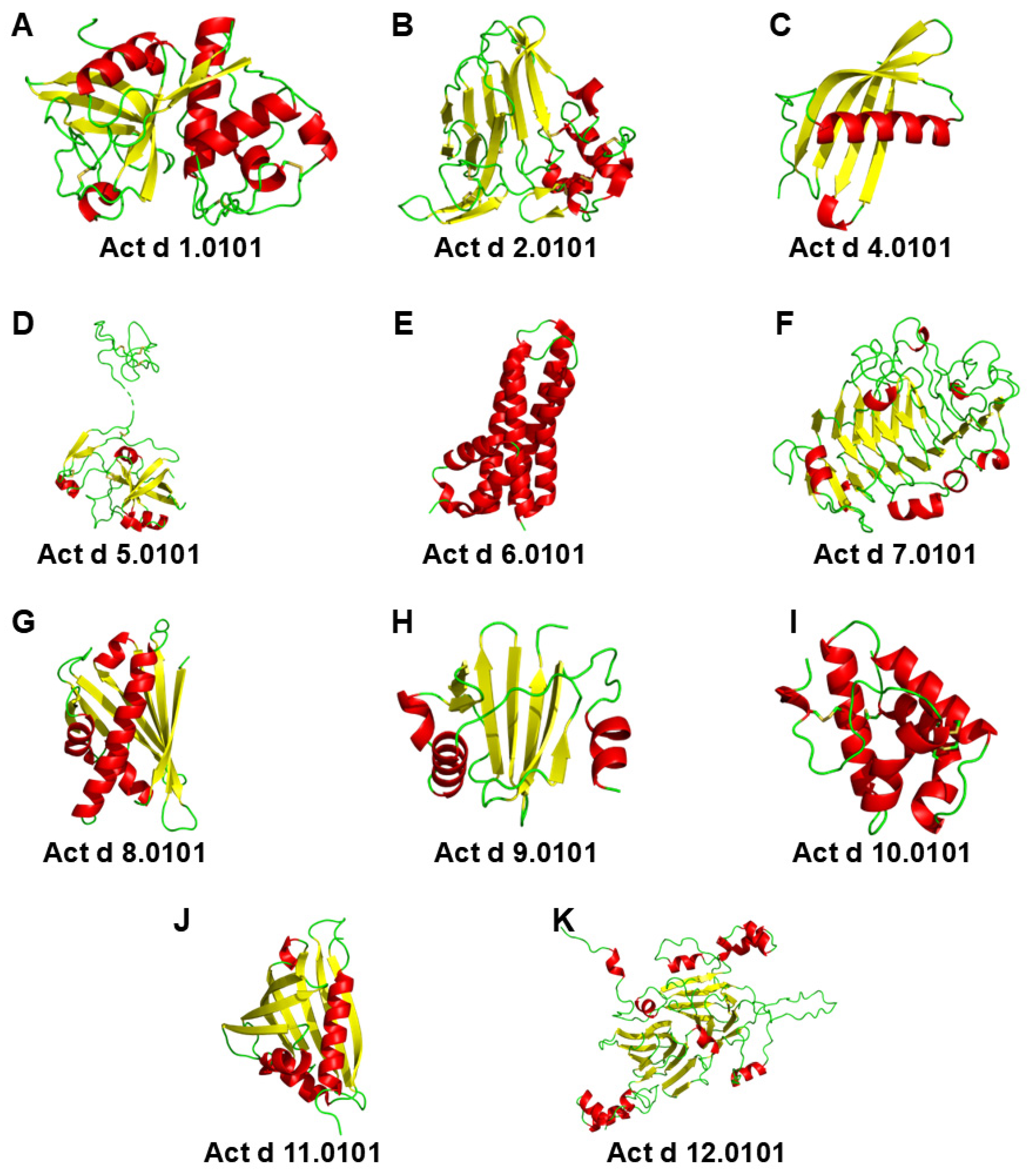
Figure 1. Structures of all Actinidia deliciosa allergens registered with the WHO/IUIS. Full nomenclature allergens’ names are provided below the corresponding structures. Act d 3 and Act d 13 are excluded since their sequences are incomplete. (A,C,F,HK) AlphaFold3 models. (B) PDB: 4BCT. (D) PDB: 4X9U. (E) PDB: 1XG2. (G) PDB: 8QHH. (J) PDB: 4IHR.
Applsci 15 07182 g001
Figure 2. A-RISC indices for (A) papain-like cysteine proteases and (B) thaumatin-like proteins. Sequences were selected from the WHO/IUIS (proteins with full allergen nomenclature) and AllFam (proteins without numbers) databases. All A-RISC diagrams in this work are scaled against each other, such that red indicates a high likelihood of IgE-mediated cross-reactivity, green indicates a low likelihood, and intermediate colors suggest intermediate cross-reactivity.
Applsci 15 07182 g002
Figure 3. AlphaFold3 model of the putative complex between Act d 6 (green) and Act d 7 (red).
Applsci 15 07182 g003
Figure 4. A-RISC index for PR-10 proteins registered as ingestion allergens. Sequences were selected from the WHO/IUIS (proteins with full allergen nomenclature) and AllFam (proteins without numbers) databases. All A-RISC diagrams in this work are scaled against each other, such that red indicates a high likelihood of IgE-mediated cross-reactivity, green indicates a low likelihood, and intermediate colors suggest intermediate cross-reactivity.
Applsci 15 07182 g004
Figure 5. A-RISC index for inhalant and ingestion (A) profilins and (B) nsLTPs found in the ALEX2 macroarray. Sequences were selected from the WHO/IUIS database. All A-RISC diagrams in this work are scaled against each other, such that red indicates a high likelihood of IgE-mediated cross-reactivity, green indicates a low likelihood, and intermediate colors suggest intermediate cross-reactivity.
Applsci 15 07182 g005
Figure 6. Mapping of sequence conservation of Act d 8.0101 on Act d 11 and sequence alignment. (A) X-ray crystal structure of natural Act d 11 (PDB: 4IGV); ribbon representation is shown in two orientations related by 180° rotation. Residues conserved between Act d 8.0101 and Act d 11 are shown in dark blue. (B) Same as shown in (A), but as a surface representation. (C) Sequence alignment of mature Act d 11 and Act d 8.0101, with conserved residues shown in dark blue.
Applsci 15 07182 g006
Figure 7. A-RISC index for 11S globulins registered as ingestion allergens. Sequences were selected from the WHO/IUIS (proteins with full allergen nomenclature) and AllFam (proteins without numbers) databases. All A-RISC diagrams in this work are scaled against each other, such that red indicates a high likelihood of IgE-mediated cross-reactivity, green indicates a low likelihood, and intermediate colors suggest intermediate cross-reactivity.
Applsci 15 07182 g007
Figure 8. Mapping of sequence conservation of wheat alpha-triticain on Act d 1 (actinidin) and sequence alignment. (A) SwissModel of mature Act d 1; ribbon representation is shown in two orientations related by 180° rotation. Conserved residues from wheat alpha-triticain are shown in dark blue. (B) Same as shown in (A), but as a surface representation. (C) Sequence alignment of mature Act d 1 (residues 127–380) and mature wheat alpha-triticain (residues 130–461), with conserved residues shown in dark blue.
Applsci 15 07182 g008
Table 1. List of allergens originating from green and golden kiwifruits. Allergens marked with asterisks (*) are included in Allergome [8] but not registered by the Allergen Nomenclature Sub-Committee of the International Union of Immunological Societies.
Actinidia deliciosa
(Green Kiwifruit)
Actinidia chinensis
(Golden Kiwifruit)
Protein Family Biochemical Name
Act d 1Act c 1 *Cysteine protease (actinidin)
Act d 2Act c 2 *Thaumatin-like protein (TLP)
Act d 3----
Act d 4Act c 4 *Phytocystatin
Act d 5Act c 5Kiwellin (kissper + KiTH)
Act d 6--Pectin methylesterase inhibitor (PMEI)
Act d 7--Pectin methylesterase (PME)
Act d 8Act c 8Pathogenesis-related protein (PR-10)
Act d 9--Profilin
Act d 10Act c 10Non-specific lipid transport protein (nsLTP1)
Act d 11--Major latex protein/ripening-related protein (MLP/RRP)
Act d 12--11S globulin (cupin)
Act d 13--2S albumin
Table 2. Examples of sensitization profiles obtained using ALEX2. Only IgE titers higher than 0.3 kUA/mL are shown.
IgE Titer (kUA/mL)
Allergen Protein Patient 1 Patient 2 Patient 3 Patient 4
Act d 1Cysteine protease 8.16
Act d 2TLP 10.32
Act d 5Kiwellin
Act d 10nsLTP3.47 16.88
Aln g 1PR-10 1.54 35.63
Alt a 1 5.45
Api g 1PR-10 0.776.91
Api g 2nsLTP1.23
Api g 6nsLTP1.57 0.72
Ara h 17/8 globulin 4.65
Ara h 62S albumin 0.97
Ara h 8PR-10 1.598.2626.13
Ara h 9nsLTP5.88 10.44
Art v 3nsLTP10.45
Bet v 1PR-10 23.7939.7938.32
Bet v 2Profilin 7.99
Can s 3nsLTP1.38
Cor a 1.0103PR-10 4.6417.3338.08
Cor a 1.0401PR-10 2.995.5127.13
Cor a 117/8 globulin 3.29
Cor a 8nsLTP4.08 1.31
Cor a 911S globulin 6.26
Cuc m 2Profilin 24.43
Dau c 1PR-10 6.08
Fag s 1PR-10 4.317.2323.49
Fra a 1 + 3PR-10 + nsLTP1.040.899.1210.48
Gly m 4PR-10 3.189.8326.68
Hev b 8Profilin 6.26
Jug r 3nsLTP0.57
Jug r 411S globulin 11.47
Mal d 1PR-10 0.930.6716.36
Mal d 3nsLTP4.03 2.24
Mer a 1Profilin 13.90
Phl p 12Profilin 8.32
Pho d 2Profilin 19.87
Pis v 211S globulin 1.84
Pis v 37/8 globulin 4.48
Pla a 3nsLTP1.01
Pru p 3nsLTP5.16 1.60
Ses i 1 0.53
Sola l 6nsLTP2.16 2.01
Tria a 14nsLTP1.17
Vit v 1nsLTP4.06
Zea m 14nsLTP10.58 15.68
Disclaimer/Publisher’s Note: The statements, opinions and data contained in all publications are solely those of the individual author(s) and contributor(s) and not of MDPI and/or the editor(s). MDPI and/or the editor(s) disclaim responsibility for any injury to people or property resulting from any ideas, methods, instructions or products referred to in the content.