Article

3D Printed Posterior Connector Dimensions’ Effect on Fracture Properties of Provisional Two-Unit Fixed Dental Prostheses

by
Turki S. Alkhallagi
1,*,
Manal A. Alqahtani
2 and
Thamer Y. Marghalani
1
1
Oral and Maxillofacial Prosthodontics Department, Faculty of Dentistry, King Abdulaziz University, Jeddah 21589, Saudi Arabia
2
Faculty of Dentistry, King Abdulaziz University, Jeddah 21589, Saudi Arabia
*
Author to whom correspondence should be addressed.
Appl. Sci. 2025, 15(13), 7171; https://doi.org/10.3390/app15137171
Submission received: 28 March 2025 / Revised: 2 June 2025 / Accepted: 20 June 2025 / Published: 25 June 2025
(This article belongs to the Special Issue 3D Printed Materials Dentistry II)

Abstract

This in vitro study aims to investigate the fracture properties of 3D-printed resin provisional material designed with different connector dimensions for two-unit fixed dental prostheses (FDPs). The master model was digitally designed following Shillingburg’s all-ceramic restoration tooth preparation guidelines and milled from aluminum. Four two-unit FDPs with different connector dimensions were designed: 2 × 3 mm, 3 × 3 mm, 3 × 4 mm, and 4 × 4 mm (width × length) (Groups A, B, C, and D, respectively; n = 10 for each group). These specimens were printed using 3D-printed resin material (Detax FREEPRINT® temp). Forty specimens were subjected to a three-point test using a universal testing machine until fracture. The failure mode was examined under a stereomicroscope. The Kruskal–Wallis test at α = 0.05 revealed non-significant differences in fracture resistance load but significantly different elastic modulus, yield strength, and compressive strength (p = 0.061, p < 0.001, p < 0.001, and p < 0.001, respectively) among the different groups. The 2 × 3 mm connectors had higher means of modulus, yield strength, and compressive strength compared to the other groups. The study found that the maximum load causing fractures in 3D-printed provisional material connectors was consistent, regardless of connector cross-section variations. The 2 × 3 mm group performed best, while the 4 × 4 mm group performed worst.

1. Introduction

In prosthodontics dental rehabilitation, fixed dental prostheses (FDPs) are the most common choice in dental clinics, enhancing patient desire and satisfaction [1]. FDPs are any prosthesis fixed to natural teeth or dental implants the patient cannot remove [2]. The main goals of fabricating FDPs are restoring patients’ function, aesthetics, and comfort [3]. Furthermore, FDPs contain multiple components. The main FDP components are pontic, retainer, connector, and abutment [3]. The FDP components are designed, constructed, and manufactured in the dental laboratory. These laboratory steps should be finished before FDPs are cemented in the oral cavity [3].
According to the material used, these FDPs could be a definitive or provisional prosthesis [3]. Several materials can be used to fabricate the FDPs, such as ceramic, porcelain fused to metal, lithium disilicate-based ceramics, zirconia, and densely sintered alumina [4]. Nowadays, the most common material is ceramic, which has extraordinary properties. It has high aesthetic characteristics and good mechanical properties. This is in addition to more biocompatibility than metal fused to the porcelain prosthesis [4].
Although dental implants have a higher success rate than FDPs, there are still some limitations to placing them for all patients [5]. Additionally, dental implant treatment has some contraindications, such as periodontal disease, poor oral hygiene, heavy smoking, parafunctional habits like bruxism, and certain systemic diseases that affect the periodontium, bleeding control, or healing after surgical procedures [6]. Therefore, FDPs remain a valid and reliable treatment option in prosthodontics.
FDP abutments should be protected between dental visits using provisional FDPs [3]. A dental provisional prosthesis is used to improve the current condition of the oral cavity. It is a dental interim prosthesis used for a limited period before a permanent dental prosthesis is replaced [2]. A dental provisional prosthesis is designed to enhance esthetics, provide stabilization, and facilitate function [2]. It aids in avoiding gingival growth or inflammation of the periodontium and eliminates the need to remake the definitive impression [3].
Provisional FDPs could be fabricated using different provisional materials [7,8]. Polymethyl methacrylate (PMMA) is one of these materials that is widely used in dental applications [8]. These materials could be manufactured conventionally as cold-cured or heat-cured PMMA materials. In addition, these provisional FDPs could be manufactured digitally using subtractive or additive manufacturing technology [7,8].
Subtractive technology is defined as the machining process to remove material from a material block [2]. 3D-printing technology, which is known as additive manufacturing technology, is a general term for a manufacturing approach that transfers an object from liquid to solid structure by curing layer over layer until it reaches the final 3D structure [9,10]. These objects were designed primarily with specific designing software. Then, this 3D design is manufactured from varying resin materials using a 3D printer machine [9,11].
In dentistry, 3D-printing technology has a multi-advantage covering rapid manipulation, high precision, lower cost, simplicity and flexibility of the complex workflow, reduced amount of wasted raw material, good marginal adaptation, and reduced internal gap of dental prosthesis compared to other manufacturingNb methods [11,12,13,14,15]. A 3D-printed resin is one of these materials that is widely used in prosthodontics [7,11,12]. Nowadays, 3D-printed resin material is used for provisional crowns or FDPs [7].
The connector is a part that unites the retainer(s) and pontic(s) of FDPs, which could be a rigid or nonrigid connector [2]. In addition, the connectors should be large enough to improve the strength without impeding plaque control [3]. However, it is considered the weaker portion of FDPs. So, the connector dimension and type of connector are important variables that play a critical role in the longevity of the FDPs [16,17,18,19]. Flexural strength, fracture toughness, color stability, yield strength, and hardness are the most measurable properties in the literature regarding dental application [20]. In terms of mechanical parameters of any material, fracture strength is defined as the ability of a material to resist failure and is designated according to the mode of load application, such as compressive or tensile [21].
Although all previous studies used an all-ceramic material to measure the most suitable connector dimension, the literature lacks information on adequate 3D Printed Posterior Connector Dimensions. Therefore, this in vitro study aims to investigate the fracture properties of 3D-printed resin provisional material designed with different connector dimensions for two-unit FDPs.
The null hypothesis is that the 3D-printed posterior connector dimensions do not influence the fracture properties of provisional two-unit FDPs.

2. Materials and Methods

2.1. Master Model Design

Two adjacent teeth were fabricated digitally to simulate a clinical situation of maxillary posterior teeth to be restored with two-unit FDPs. Teeth were prepared digitally using (Autodesk Fusion 360 Professional, Autodesk, San Francisco, CA, USA) software following Shillingburg tooth preparation guidelines for all-ceramic restoration, with rounded line angles, deep chamfer finish line, teeth dimensions simulate the average measurement of maxillary first molar for both abutments and occluso-gingival preparation height 5.5 mm [22,23].

2.2. Master Model Manufacturing

The prepared model was fabricated using Aluminum material with CAM software MasterCam 2023 (Mastel Aluminium-Halbzeuge GmbH, Talheim, Germany) to generate the tool path for a 3-axis CNC milling machine. Then, the metal model is polished with a metal polish compound. Finally, the metal model was scanned using a Ceramill Map 400+ scanner (AmannGirrbach, GmbH for Ceramill Map 400 Scanner, Koblach, Austria), and the master digital model was exported as a Standard Tessellation Language (STL) file.

2.3. FPDs Design

Two-unit FDPs were designed on the master model STL file using Exocad software (DentalCAD 3.1 Rijeka, Exocad, Darmstadt, Germany). The two-unit FDPs were connected in the design with a triangular connector shape. The connector had four different dimensions selected based on the manufacturer’s recommendations (4 × 4 mm) and previous literature for the other groups [16,18,24,25,26]. Table 1 shows the sizes of the tested connectors. Figure 1 shows the design of the group D connector.

2.4. FPDs Manufacturing

Ten specimens from each design (a total of 40 specimens) were printed using a 3D mixture of acrylic/methacrylic resin material (Detax FREEPRINT® temp, Detax GmbH, Ettlingen, Germany) and a Straumann P30+ 3D printer (Straumann, Andover, MA, USA). After printing, the printed specimens were washed, cleaned, and post-cured according to the manufacturer’s recommendations. Finally, supporting structures were removed, and specimens were finished and polished. However, all specimens were designed and printed by a single examiner.

2.5. Specimens Testing

This study evaluated and characterized connector failure in two-unit FDPs. Mechanical performance was assessed by measuring fracture load, compressive strength, yield strength, and failure modes under in vitro static loading. All forty specimens were examined under the stereomicroscope for cracks or fractures. Any specimen with cracks or fractures was excluded, and another specimen was printed using the same protocol. All groups were cemented to the metal model using temporary cement, Temp-Bond™ Clear (Kerr Dental, Orange, CA, USA). Each specimen of these groups was subjected to a three-point test using a universal testing machine, Instron 5969 (Instron, MA, USA). The metal model is positioned in the center of the universal testing machine at a 0° angulation. All specimens were subjected to vertical load until visually fractured in a universal test machine. The load was transferred through a 2.5 mm diameter steel ball at a 0.5 mm/minute crosshead speed at one point in the connector area of two units of FDPs.

2.6. Specimens Evaluation

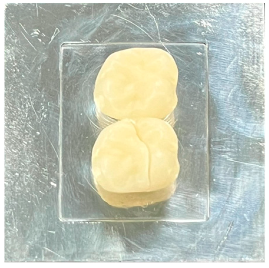
The mode of failure was examined by one experienced examiner and classified according to the mode of failure mentioned in Table 2. Figure 2 shows an example of a type 1 fracture.

2.7. Statistical Analysis

Descriptive statistics were performed according to the assumptions for statistical tests: normality, independence, homogeneity of variance, and outliers; the Kruskal–Wallis test at α = 0.05 was used as a statistical test.

3. Results

Descriptive statistics were performed. Means, medians, and standard deviations are shown in Table 3. Factors were Independent. According to the Shapiro-Wilk test used for normality assessment, there were few significant values for the dependent variables and groups p < 0.05. Therefore, the data is not normally distributed and considered using a nonparametric variant of the ANOVA test, the Kruskal–Wallis’s test. The Kruskal–Wallis test was conducted to determine whether there were significant differences in fracture resistance load by connectors’ dimension.
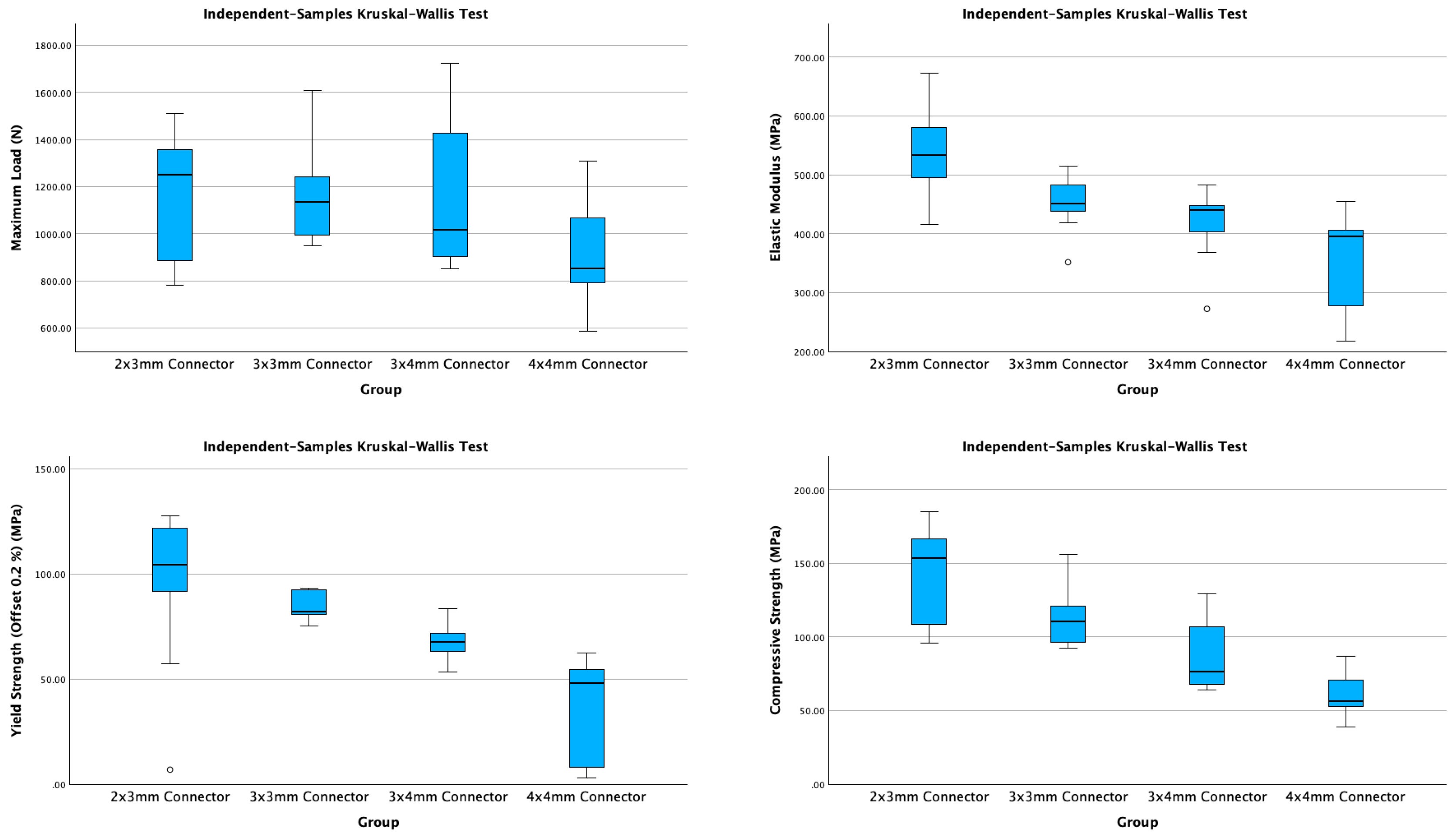
The homogeneity of variance is a key factor in the study analysis. The data were homogenous for the maximum load and modulus (p > 0.05), which is favorable for the study analysis. However, for yield and compressive strength, the data were heterogeneous (p < 0.05), potentially affecting the conclusions. Preliminary screening for outliers showed few outside the favorable range recommended by Tukey (1977), Hoaglin (1983), and Hoaglin (1985) [27,28,29]. The outliers were managed by imputation, replacing the outlier in question with the median according to the method described by Tukey (1977), Hampel (1986), and Rousseeuw (1987) [29,30,31]. After managing outliers, there were no outliers in maximum load and compressive strengths, while few outliers were in the modulus and yield strength dependent variables within the acceptable range, as shown in Figure 3.
The independent-sample Kruskal–Wallis test was as follows: The maximum load (N) was 7.374, df = 3, p = 0.061; The modulus (MPa) was 22.365 df = 3, p < 0.001; for yield strength (Offset 0.2%) (MPa) was 24.221, df = 3, p < 0.001; for compressive strength (MPa) was 26.770, df = 3, p < 0.001.
Figure 4 and Table 4 show the pairwise comparisons of group results.
Table 5 shows the distribution of fractures according to location in the specimens. Fracture type 12 (multiple-direction fracture) was the most common type for all groups. Fracture types 2, 3, 5, and 7 had not occurred in any specimens.

4. Discussion

The null hypothesis was retained for the maximum load across the different tested groups. At the same time, it was rejected for elastic modulus, Yield Strength (Offset 0.2%), and Compressive Strength when the significance level was p < 0.001.
Temporary or interim fixed prosthodontics are commonly used as a step towards fabricating final fixed prostheses [3,22]. 3D-printed temporaries or milled PMMA can serve as an interim treatment option [32]. One observation from an author during the clinical experience was that most fractures in 3D-printed temporary prostheses occurred in the connector area. This prompted the authors’ interest in evaluating the factors that may lead to connector failures. Connectors are between two adjacent retainers and between a retainer and a pontic [2]. To characterize the effects of chewing and mastication on connectors, we begin with the simplest form—connectors between two crowns, namely, splinted crowns. Then, we will progress to more complex connector designs in future studies. Long-span bridges or fixed procedures rely on splinting the retainers at the end of the prosthesis to ensure a strong structure. The dimensions of the connectors are crucial factors that can affect the outcome of fixed prosthodontics treatment [22].
This study differs from previous studies in isolating the connector area and testing the effects of direct static loading on the connector area of two splinted crowns [16,18,19,24,25,26,33,34].
The methodology utilized two adjacent abutments on metal dies that were not previously used in any study. We employed a metal die made of milled aluminum, with dimensions standardized according to Shillingburg’s recommendations and average tooth dimensions [22,23]. The production of these 3D milled dies uses 3D software Fusion 360 (Autodesk Fusion 360 Professional, Autodesk, San Francisco, CA, USA), making it more accurate than other methods of fabricating metal dies to specified dimensions. The two connected retainers were also designed with the 3D software Exocad (DentalCAD 3.1 Rijeka, Exocad, Darmstadt, Germany), which was used to select the measurements of the connector area during the design process.
Testing using the Instron machine (Instron 5969, Norwood, MA, USA) utilized a 2.5-mm three-dimensional generated rod that simulates the loading of a cusp area directly on the connector. The loading was configured at a 0-degree angle from the connector’s vertical axis.
In the literature, the connector area is commonly tested on three-unit, four-unit, or long-span FDPs, regardless of the type of materials [18,19,25,26]. Although the pontic design of FDPs is more critical with connector area testing in the literature, the fabrication of two-unit FDPs in this study aids in minimizing the factors that could influence the results. If there is a pontic design, the force distribution in the three-point universal testing may affect the fracture resistance load and the mode of fracture readings. Additionally, the retainers (crowns) were designed with exact tooth dimensions that follow the abutment preparation to ensure the connector area is in the middle of the metal model. To resemble a clinical scenario with triangular connector designs, avoiding an unequal force distribution on the connector area that could influence the fracture properties of 3D-printed resin material is our priority in this study.
A study by Harshitha et al. in 2013 [34] tested the stress distribution using finite element analysis. The study found that the maximum Von Misses stresses are concentrated at the connector area, especially in the cervical region [34]. The cylindrical connector design was less stressed than the triangular design [34]. All-ceramic FDPs framework strength depends on the connector dimension [3]. Hence, it is necessary to enhance the strength of 3D-printed resin FDPs by manufacturing an adequate connector dimension.
In addition, Rezaei et al. in 2014 found the failure probability decreased with the high width of the connector [25]. For all ceramic prostheses, the connector dimension is more important than the abutment core thickness, as found by Ambre et al. in 2013 [16].
In another study, Modi et al. found that nonrigid connectors produced less stress than rigid ones [33]. In this study, evaluating the connector dimension on fracture properties for a ridge connector is essential. According to Shillingburg et al., the nonrigid connector transfers the stress to the supporting structure rather than concentrating it in the connector area [22]. In addition, Junker et al. (2019) recommended that minimum connector dimensions prove crucial for the load-bearing capability of monolithic e.max CAD FPDs [24]. Significantly, the reduction of connector dimension increased the strength of 3D-printed resin FDPs. According to the results, the mean of Modulus, Yield Strength, and Compressive Strength of 2 × 3 mm connectors were significantly larger among the other groups. In triangular connector shape design, the cross-sectional connector area decreases as the connector dimensions decrease, Table 1, which is the denominator that the fracture load will be divided on to produce the stress values in megapascals. We should remember that usually, in three-point bends of specimens that have relatively comparable cross-sections, stress can be compared easily since the denominator is almost equal. However, in this current study, we are comparing loads, moduli, and strengths that are different in cross-section, which makes it look odd that the fracture strengths were reduced as the cross-section decreased. Another way to look at it is to normalize those loads and strengths by unifying the denominators across the tested groups. Then, the results may look more relevant and comparable.
Larsson et al. tested all ceramic zirconia-based FDP connector diameters [26]. The authors evaluated the fracture strength properties of varied materials. This in vitro study recommended a minimum connector diameter of 4.0 mm for long spans or replacing molar teeth [26]. Compared to 3D-printed resin material, all ceramic material is harder than 3D-printed resin [35]. Hence, the thinning of the external wall around the large connector of 3D-printed resin FDPs could decrease the strength resistance load.
Observation of the failure mode on those dies shows almost similar fractures on both sides of the connectors and the crowns, yet no other fractures passed through the connectors themselves. This seems logical because fracture occurs in the weakest area of the designed restoration [19,36]. The highest number of fractures, counts, and percentages were in the 3 × 4 mm connectors compared to the other connectors dimensions. Regarding the location of the fractures and the fracture lines, it was observed that they are mostly on the occlusal and distal parts of the two adjacent crowns.
This in vitro study provides valuable insight, as 3D-printed resin is a relatively new material for provisional prostheses. Knowing its influence on fracture properties may help improve the design and printing process of these prostheses.
The manufacturing of each clinical or laboratory step must be precise or have a guideline for any new technology, which is why further investigation is needed. However, each material has an indication depending on its mechanical and physical properties and availability of either provisional or permanent prosthesis.
This study is in vitro, which may have a different outcome than the patient’s mouth and has some limitations. One limitation is that the specimens were tested statically using a universal testing machine. In addition, the tested provisional material and 3D printer are from one company. Adding another 3D-printed material and different connector-shaped designs will be helpful in the future. In addition, the specimens should be tested in a dynamic testing machine to simulate patient function.

5. Conclusions

The study found that the maximum load causing fractures in 3D-printed provisional material connectors was consistent, regardless of connector cross-section variations. Elastic modulus, yield strength, and compressive strength differed between 4 × 4 mm and 2 × 3 mm connectors, with the smallest showing the highest values. More connector fractures correlated with lower mechanical performance. The 2 × 3 mm group performed best, while the 4 × 4 mm group performed worst. Lower fracture counts, associated with single-direction fractures, indicated higher mechanical strengths, whereas multiple-direction fractures were linked to lower strengths.

Author Contributions

Conceptualization, T.S.A.; methodology, T.S.A., M.A.A. and T.Y.M.; software, M.A.A.; validation, T.S.A. and T.Y.M.; formal analysis, T.Y.M.; resources, M.A.A.; writing—original draft preparation, T.S.A., M.A.A. and T.Y.M.; writing—review and editing, T.S.A. and T.Y.M.; visualization, T.S.A. and T.Y.M.; supervision, T.Y.M.; project administration, T.S.A. All authors have read and agreed to the published version of the manuscript.

Funding

This research received no external funding.

Institutional Review Board Statement

Ethical review and approval were waived for this study because it is an in vitro study, and there is no human and animal participation.

Informed Consent Statement

Not applicable.

Data Availability Statement

The raw data supporting the conclusions of this article will be made available by the authors upon request.

Acknowledgments

The authors acknowledge with thanks the Advanced Technology Dental Research Laboratory (ATDRL), Faculty of Dentistry, King Abdulaziz University, Jeddah, Saudi Arabia, for their technical support.

Conflicts of Interest

The authors declare no conflicts of interest.

References

  1. ELsyad, M.A.; Elgamal, M.; Mohammed Askar, O.; Youssef Al-Tonbary, G. Patient satisfaction and oral health-related quality of life (OHRQoL) of conventional denture, fixed prosthesis and milled bar overdenture for All-on-4 implant rehabilitation. A crossover study. Clin. Oral Implant. Res. 2019, 30, 1107–1117. [Google Scholar] [CrossRef]
  2. The Glossary of Prosthodontic Terms 2023: Tenth Edition. J. Prosthet. Dent. 2023, 130 (Suppl. S1), e1–e3. [CrossRef] [PubMed]
  3. Rosenstiel, S.F.; Land, M.F.; Fujimoto, J. Contemporary Fixed Prosthodontics; Mosby/Elsevier: Amsterdam, The Netherlands, 2006. [Google Scholar]
  4. Saeed, F.; Muhammad, N.; Khan, A.S.; Sharif, F.; Rahim, A.; Ahmad, P.; Irfan, M. Prosthodontics dental materials: From conventional to unconventional. Mater. Sci. Eng. C Mater. Biol. Appl. 2020, 106, 110167. [Google Scholar] [CrossRef] [PubMed]
  5. Goodacre, C.J.; Naylor, W.P. Single implant and crown versus fixed partial denture: A cost-benefit, patient-centred analysis. Eur. J. Oral Implantol. 2016, 9 (Suppl. S1), S59–S68. [Google Scholar] [PubMed]
  6. Kullar, A.S.; Miller, C.S. Are There Contraindications for Placing Dental Implants? Dent. Clin. N. Am. 2019, 63, 345–362. [Google Scholar] [CrossRef]
  7. Park, S.M.; Park, J.M.; Kim, S.K.; Heo, S.J.; Koak, J.Y. Flexural Strength of 3D-Printing Resin Materials for Provisional Fixed Dental Prostheses. Materials 2020, 13, 3970. [Google Scholar] [CrossRef]
  8. Zafar, M.S. Prosthodontic Applications of Polymethyl Methacrylate (PMMA): An Update. Polymers 2020, 12, 2299. [Google Scholar] [CrossRef]
  9. Dawood, A.; Marti Marti, B.; Sauret-Jackson, V.; Darwood, A. 3D printing in dentistry. Br. Dent. J. 2015, 219, 521–529. [Google Scholar] [CrossRef]
  10. Liu, Q.; Leu, M.C.; Schmitt, S.M. Rapid prototyping in dentistry: Technology and application. Int. J. Adv. Manuf. Technol. 2005, 29, 317–335. [Google Scholar] [CrossRef]
  11. Barazanchi, A.; Li, K.C.; Al-Amleh, B.; Lyons, K.; Waddell, J.N. Additive Technology: Update on Current Materials and Applications in Dentistry. J. Prosthodont. 2017, 26, 156–163. [Google Scholar] [CrossRef]
  12. Tian, Y.; Chen, C.; Xu, X.; Wang, J.; Hou, X.; Li, K.; Lu, X.; Shi, H.; Lee, E.S.; Jiang, H.B. A Review of 3D Printing in Dentistry: Technologies, Affecting Factors, and Applications. Scanning 2021, 2021, 9950131. [Google Scholar] [CrossRef] [PubMed]
  13. Lin, L.; Fang, Y.; Liao, Y.; Chen, G.; Gao, C.; Zhu, P. 3D Printing and Digital Processing Techniques in Dentistry: A Review of Literature. Adv. Eng. Mater. 2019, 21, 1801013. [Google Scholar] [CrossRef]
  14. Zanetti, E.M.; Aldieri, A.; Terzini, M.; Calì, M.; Franceschini, G.; Bignardi, C. Additively Manufactured Custom Load-Bearing Implantable Devices. Australas. Med. J. 2017, 10, 694. [Google Scholar] [CrossRef]
  15. Alharbi, N.; Alharbi, S.; Cuijpers, V.; Osman, R.B.; Wismeijer, D. Three-dimensional evaluation of marginal and internal fit of 3D-printed interim restorations fabricated on different finish line designs. J. Prosthodont. Res. 2018, 62, 218–226. [Google Scholar] [CrossRef] [PubMed]
  16. Ambre, M.J.; Aschan, F.; Vult von Steyern, P. Fracture strength of yttria-stabilized zirconium-dioxide (Y-TZP) fixed dental prostheses (FDPs) with different abutment core thicknesses and connector dimensions. J. Prosthodont. 2013, 22, 377–382. [Google Scholar] [CrossRef]
  17. Peñate, L.; Basilio, J.; Roig, M.; Mercadé, M. Comparative study of interim materials for direct fixed dental prostheses and their fabrication with CAD/CAM technique. J. Prosthet. Dent. 2015, 114, 248–253. [Google Scholar] [CrossRef]
  18. Motta, A.B.; Pereira, L.C.; da Cunha, A.R.; Duda, F.P. The influence of the loading mode on the stress distribution on the connector region of metal-ceramic and all-ceramic fixed partial denture. Artif. Organs 2008, 32, 283–291. [Google Scholar] [CrossRef]
  19. Oh, W.S.; Anusavice, K.J. Effect of connector design on the fracture resistance of all-ceramic fixed partial dentures. J. Prosthet. Dent. 2002, 87, 536–542. [Google Scholar] [CrossRef] [PubMed]
  20. Astudillo-Rubio, D.; Delgado-Gaete, A.; Bellot-Arcis, C.; Montiel-Company, J.M.; Pascual-Moscardo, A.; Almerich-Silla, J.M. Mechanical properties of provisional dental materials: A systematic review and meta-analysis. PLoS ONE 2018, 13, e0193162. [Google Scholar] [CrossRef]
  21. Orthodontic Applications of Biomaterials; Elsevier: Amsterdam, The Netherlands, 2017. [CrossRef]
  22. Shillingburg, H.T. Fundamentals of Fixed Prosthodontics; Quintessence Pub. Co.: New Malden, UK, 1997. [Google Scholar]
  23. Scheid, R.C.; Woelfel, J.B. Woelfel’s Dental Anatomy; Wolters Kluwer/Lippincott Williams & Wilkins Health: Philadelphia, PA, USA, 2012. [Google Scholar]
  24. Junker, R.; Holler, M.; Yoshida-Anastasova, Y.; Frank, W.; Nothdurft, F.P. Influence of Connector Diameter on Fracture Load of CAD/CAM-Processed Monolithic Lithium Disilicate Fixed Partial Dentures. Int. J. Prosthodont. 2019, 32, 68–70. [Google Scholar] [CrossRef]
  25. Rezaei, S.M.; Heidarifar, H.; Arezodar, F.F.; Azary, A.; Mokhtarykhoee, S. Influence of Connector Width on the Stress Distribution of Posterior Bridges under Loading. J. Dent. 2011, 8, 67–74. [Google Scholar]
  26. Larsson, C.; Holm, L.; Lovgren, N.; Kokubo, Y.; Vult von Steyern, P. Fracture strength of four-unit Y-TZP FPD cores designed with varying connector diameter. An in-vitro study. J. Oral Rehabil. 2007, 34, 702–709. [Google Scholar] [CrossRef] [PubMed]
  27. Hoaglin, D.C.; Mosteller, F.; Tukey, J.W. Understanding Robust and Exploratory Data Analysis; Wiley: Hoboken, NJ, USA, 1983. [Google Scholar]
  28. Hoaglin, D.C.; Mosteller, F.; Tukey, J.W. Exploring Data Tables, Trends, and Shapes; Wiley: Hoboken, NJ, USA, 1985. [Google Scholar]
  29. Tukey, J.W. Exploratory Data Analysis; Addison-Wesley Pub. Co.: Princeton, NJ, USA, 1977. [Google Scholar]
  30. Hampel, F.R. Robust Statistics: The Approach Based on Influence Functions; Wiley: Hoboken, NJ, USA, 1986. [Google Scholar]
  31. Rousseeuw, P.J.; Leroy, A.M. Robust Regression and Outlier Detection; Wiley: Hoboken, NJ, USA, 1987. [Google Scholar]
  32. Masri, R.; Driscoll, C.F. Clinical Applications of Digital Dental Technology; Wiley-Blackwell: Hoboken, NJ, USA, 2023. [Google Scholar]
  33. Modi, R.; Kohli, S.; Rajeshwari, K.; Bhatia, S. A three-dimension finite element analysis to evaluate the stress distribution in tooth supported 5-unit intermediate abutment prosthesis with rigid and nonrigid connector. Eur. J. Dent. 2015, 9, 255–261. [Google Scholar] [CrossRef] [PubMed]
  34. Harshitha Gowda, B.H.; Satish Babu, C.L. Connector design in a long-span-fixed dental prosthesis: A three-dimensional finite element analysis. Indian J. Dent. Res. 2013, 24, 178–182. [Google Scholar] [CrossRef]
  35. Shen, C. Phillips’ Science of Dental Materials; Elsevier: Amsterdam, The Netherlands, 2022. [Google Scholar]
  36. Luthy, H.; Filser, F.; Loeffel, O.; Schumacher, M.; Gauckler, L.; Hammerle, C. Strength and reliability of four-unit all-ceramic posterior bridges. Dent. Mater. 2005, 21, 930–937. [Google Scholar] [CrossRef]
Figure 1. The design of group D connector.
Applsci 15 07171 g001
Figure 2. Example of a type 1 fracture (single mesial crown fracture, mesial to distal direction, without connector fracture).
Applsci 15 07171 g002
Figure 3. Box plot of means of maximum load, elastic modulus, yield strength, and compressive strength by connectors’ dimension.
Applsci 15 07171 g003
Figure 4. Pairwise comparisons of groups.
Applsci 15 07171 g004
Table 1. The tested groups and their connector’s dimensions.
GroupWidth (mm)Height (mm)Cross-Sectional Area (mm2)
A238.16
B3310.30
C3413.33
D4415.08
Table 2. The mode of failure that occurred in the specimens.
Crown Fracture
TypeFirst (Mesial)Second (Distal)Fracture DirectionConnector Fracture
1YesNoOne direction (mesial to distal)No
2YesNoMultiple directionsNo
3YesNoOne direction (mesial to distal)Yes
4YesNoMultiple directionsYes
5NoYesOne direction (mesial to distal)No
6NoYesMultiple directionsNo
7NoYesOne direction (mesial to distal)Yes
8NoYesMultiple directionsYes
9YesYesOne direction (mesial to distal)No
10YesYesMultiple directionsNo
11YesYesOne direction (mesial to distal)Yes
12YesYesMultiple directionsYes
Table 3. Median, mean, 95% confidence interval, standard deviation (SD), and sample size for the maximum load (N), elastic modulus (MPa), yield strength (MPa), and compressive strengths (MPa) by connectors’ dimensions.
GroupABCD
n10101010
Maximum Load (N)Median1251.321136.061016.78853.59
Mean1176.91159.61161904.7
95% Confidence Interval
for Mean
989.22–1364.641021.50–1297.71920.50–1401.48742.61–1066.85
SD262.4193.06336.19226.6
Elastic Modulus (MPa)Median533.92451.09440.57394.83
Mean537.62451.9421.79359.5
95% Confidence Interval
for Mean
488.91–586.34418.79–485.01377.27–466.32302.09–416.91
SD68.146.2862.2480.25
Yield Strength
(Offset 0.2%) (MPa)
Median104.4482.2167.4848.00
Mean94.1484.7867.7737.98
95% Confidence Interval
for Mean
67.98–120.2979.72–89.8561.89–73.6520.81–55.15
SD36.567.088.2224
Compressive Strength (MPa)Median153.35110.3076.2856.61
Mean144.23112.5887.0960
95% Confidence Interval
for Mean
121.23–167.2499.17–125.9969.05–105.1449.25–70.75
SD32.1618.7425.2215.03
Table 4. Pairwise comparisons of groups.
Test StatisticSig.Adj. Sig. a
Maximum Load (N) across Group
Group D-Group C10.300.050.29
Group D-Group B11.800.02 *0.14
Group D-Group A12.300.02 *0.11
Group C-Group B1.500.771.00
Group C-Group A2.000.701.00
Group B-Group A0.500.921.00
Elastic Modulus (MPa) across Group
Group D-Group C8.000.130.76
Group D-Group B13.100.01 *0.07
Group D-Group A24.10<0.001 *0.00
Group C-Group B5.100.331.00
Group C-Group A16.100.00 *0.01
Group B-Group A11.000.04 *0.21
Yield Strength (Offset 0.2%) (MPa) across Group
Group D-Group C11.000.04 *0.21
Group D-Group B20.25<0.001 *0.00
Group D-Group A23.35<0.001 *0.00
Group C-Group B9.250.080.46
Group C-Group A12.350.02 *0.11
Group B-Group A3.100.551.00
Compressive Strength (MPa) across Group
Group D-Group C10.100.050.32
Group D-Group B18.80<0.001 *0.00
Group D-Group A25.50<0.001 *0.00
Group C-Group B8.700.100.58
Group C-Group A15.400.00 *0.02
Group B-Group A6.700.201.00
Each row tests the null hypothesis that the group 1 and group 2 distributions are the same. Asymptotic significances (2-sided tests) are displayed. * The significance level is 0.050. a Significance values have been adjusted using the Bonferroni correction for multiple tests.
Table 5. The distribution of fractures is in counts according to the location of the specimens.
Maximum Load
(N)
Elastic Modulus
(MPa)
Yield Strength
(Offset 0.2%) (MPa)
Compressive Strength
(MPa)
GroupFracture TypesCountMeanSDMeanSDMeanSDMeanSD
A621378.6556.36543.74181.6849.3159.98168.956.91
811509.82.536.94.122.54.185.03.
113918.70206.29548.4735.8286.5925.45112.5925.28
1241186.52215.08526.6041.82115.1111.31145.4126.36
B411241.98.448.84.93.25.120.58.
1011606.83.462.14.82.46.156.00.
1281093.41115.64451.0052.3284.027.26106.1611.23
C91961.81.368.82.71.59.72.15.
101849.49.447.29.62.87.63.73.
1121016.7876.13360.07123.9960.7210.3576.285.71
1261294.17381.23446.9530.4070.308.0597.0928.60
D15779.57113.26368.1266.8240.1921.7851.707.51
112783.20147.54308.17128.3423.7628.9351.949.79
1231194.35121.81379.3491.6943.7730.9979.208.08
Disclaimer/Publisher’s Note: The statements, opinions and data contained in all publications are solely those of the individual author(s) and contributor(s) and not of MDPI and/or the editor(s). MDPI and/or the editor(s) disclaim responsibility for any injury to people or property resulting from any ideas, methods, instructions or products referred to in the content.