Article

Unravelling the Potential of Seven Microalgae Species: Nutritional, Antioxidant, and Antimicrobial Properties and Application

by
Valter F. R. Martins
1,
Ana I. Lopes
1,
Diana Gomes
2,
Celina Parreira
2,
Sara M. Badenes
2,
Luís Costa
2,
Manuela Pintado
1,
Alcina M. M. B. Morais
1,* and
Rui M. S. C. Morais
1,*
1
CBQF—Centro de Biotecnologia e Química Fina—Laboratório Associado, Escola Superior de Biotecnologia, Universidade Católica Portuguesa, Rua Diogo Botelho 1327, 4169-005 Porto, Portugal
2
A4F—Algae for Future, Campus do Lumiar, Estrada do Paço do Lumiar, Edif. E, R/C, 1649-038 Lisboa, Portugal
*
Authors to whom correspondence should be addressed.
Appl. Sci. 2025, 15(12), 6691; https://doi.org/10.3390/app15126691 (registering DOI)
Submission received: 1 May 2025 / Revised: 30 May 2025 / Accepted: 7 June 2025 / Published: 14 June 2025

Abstract

:

Featured Application

The present study highlights the rich biochemical profiles and bioactivities of seven microalgal species and their respective extracts, emphasizing their broad and versatile range of potential applications. These microalgae serve as promising sources of valuable biomolecules, notably proteins, especially in Limnospira sp., and lipids, with Nannochloropsis sp. being particularly rich in the latter. Limnospira sp. generally exhibited the strongest antioxidant activity and demonstrated good antimicrobial properties, while Nannochloropsis sp. showed the most pronounced antimicrobial effects against the majority of tested bacterial strains. These species hold significant potential across various sectors, from sustainable nutrition (e.g., functional foods, aquafeeds) and therapeutics (e.g., pharmaceuticals, wound care), as well as in next-generation cosmetics, bio-based industrials, and environmental bioremediation—placing them at the forefront of green biotechnological innovation. A specific application explored in this work involves the development of an edible film incorporating lipid- and bioactive-rich extracts from Nannochloropsis sp. and a bioactive-rich extract from Limnospira sp., which could be used in active food packaging. The implementation of integrated biorefinery approaches is expected to enhance the commercial viability and sustainability of these microalgae. In conclusion, the seven microalgae and their extracts investigated in this study present sustainable and multifunctional solutions for the health, nutrition, cosmetic, and environmental sectors. Their inherent bioactivity and compositional versatility continue to stimulate growing interest in biotechnological development and commercial exploitation.

Abstract

Microalgae are a rich renewable source of a wide variety of bioactive compounds. This study focuses on seven microalgae—Limnospira sp., Dunaliella sp., Lobosphaera sp., Nannochloropsis sp., Odontella sp., Porphyridium sp., and Tetraselmis sp.— analyzing their nutritional compositions and the potential bioactivity of their hydroethanolic extracts obtained via ultrasound-assisted extraction. The total phenolic content (TPC) and antioxidant activity (ABTS, DPPH, and ORAC), as well as the antimicrobial activity of the extracts were determined. The protein content of the microalgae ranged from 22.9 ± 0.1 to 59.8 ± 1.6%, the fat content ranged from 5.3 ± 0.0 to 36.6 ± 0.0%, and the carbohydrates ranged from 24.0 ± 3.0 to 46.1 ± 1.0%. The highest contents of protein, fat, and carbohydrates were found in Limnospira sp., Nannochloropsis sp., and Lobosphaera sp., respectively. The TPC of the extracts varied between 0.53 ± 0.09 and 3.18 ± 0.53 mg GAE/100 mg DW. Antioxidant activity values ranged from 1.09 ± 0.15 and 2.85 ± 0.45 μmol TE/100 mg DW for ABTS, 0.25 ± 0.06 and 2.28 ± 0.06 μmol TE/100 mg DW for DPPH, and 2.37 ± 0.58 and 18.32 ± 1.00 μmol TE/100 mg DW for ORAC. The extract from Limnospira sp. exhibited the highest antioxidant activity and also showed notable antimicrobial effects. Meanwhile, the Nannochloropsis sp. extract demonstrated the strongest antimicrobial activity against most tested bacterial strains. Lipids were successfully extracted from Nannochloropsis sp. Furthermore, alginate and zein films incorporated with bioactive-rich extracts from Limnospira sp. and Nannochloropsis sp., along with the lipid-rich extract from Nannochloropsis sp., were developed. These films showed significant antioxidant activity and effective antimicrobial activity against Listeria monocytogenes.

1. Introduction

Microalgae can produce large amounts of lipids, proteins, and carbohydrates—key components in the formulation of bio-based products such as bioplastics [1,2] and edible films and coatings [3]—offering a sustainable alternative to conventional plastic packaging.
Microalgae are rich in a wide range of bioactive compounds, including proteins (essential amino acids and bioactive peptides), polyunsaturated fatty acids (EPA and DHA), pigments (chlorophylls, carotenoids, and phycobiliproteins), polysaccharides, vitamins, polyphenols, and phytosterols [4,5]. However, data on the chemical composition and nutritional value of algal biomass are available for only a limited number of commercially utilized species [6]. These data vary considerably due to the immense diversity and heterogeneity of algal species, as well as the wide variations in environmental conditions during cultivation, harvesting, and biomass processing [7,8]. Microalgae contain pigments that impart different colors. These pigments are classified into chlorophylls (a and b), carotenoids (carotenes and xanthophylls), and phycobiliproteins. Due to structural differences, chlorophyll a appears blue-green in color, whereas chlorophyll b exhibits a green-yellow hue. Carotenoids are responsible for orange and red coloration. Phycobiliproteins—complexes of phycobilins bound to proteins—exhibit distinct spectral properties. The four main classes of phycobiliproteins are allophycocyanin (bluish green), phycocyanin (blue), phycoerythrin (red), and phycoerythrocyanin (orange) [9].
Seven microalgae species were studied as part of the Extratoteca Project, with biomass produced by the company A4F, due to their high potential for sustainable biotechnological applications and market relevance. Each species offers unique metabolic profiles and bioactive compounds that are of growing interest in fields such as functional foods, nutraceuticals, cosmetics, aquaculture, and pharmaceuticals.
Limnospira sp. (former Arthrospira sp.) is a genus of cyanobacteria capable of adapting to diverse aquatic environments, including freshwater and alkaline brackish lakes. It represents a promising component of the aquaculture bioeconomy due to its high biomass productivity and expected market growth. Limnospira sp. is rich in proteins, photosynthetic pigments, and various bioactive compounds. Traditionally used in nutrition and therapeutic applications, it is also employed in wastewater treatment and biofuel production. More recently, it has demonstrated potential in the development of pharmaceuticals, wound-healing dressings, photosensitizers for photodynamic therapy, tissue engineering, and anticancer therapies. Additionally, Limnospira sp. is being explored in industrial applications for biopolymer production, fuel cells, and photovoltaic technologies, thereby contributing to climate change mitigation [10].
Dunaliella sp. is a genus of predominant marine chlorophyte algae, with freshwater species being relatively rare. It is widely used for the recovery of high-value compounds (β-carotene, glycerol, proteins, lipids), which can be extracted from this microalga, or as whole-cell biomass, which can be used in e.g., animal feed, functional foods, and dietary supplements. Due to its high carotenoid content, Dunaliella sp. exhibits therapeutic properties, including antioxidant, anticancer, and anti-inflammatory effects. It is particularly rich in β-carotene, making it valuable to the food and cosmetics industries as a natural colorant and antioxidant. Lipids from Dunaliella sp., particularly fatty acids, are of interest for biofuel production, and the whole organism is also used in bioremediation applications such as wastewater treatment [11].
Lobosphaera sp. is primarily a freshwater chlorophyte, although some species are found in marine environments. It produces high-value metabolites with potential pharmaceutical and nutritional applications. Its lipid profile—particularly the presence of arachidonic acid, α-linolenic acid, and eicosapentaenoic acid—underscores its potential for biotechnological applications. Lobosphaera sp. has also shown promise in biofuel production and may serve as a biosorbent in water treatment processes [12,13].
Nannochloropsis sp. is found in both marine and freshwater environments. It has been studied for a range of applications, including cosmetics, food, feed, and carbon dioxide capture. This microalga is notable for its skin-protective properties and contains several bioactive compounds such as polyunsaturated fatty acids (PUFAs including EPA), carotenoids (canthaxanthin, β-carotene, zeaxanthin, violaxanthin), and phenolic compounds. Nannochloropsis sp. is beneficial for aquaculture and human nutrition and is used in forms such as pastes, dry powders, or extracts [14,15,16].
Odontella sp. is a genus of marine diatoms rich in bioactive metabolites, including fucoxanthin and omega-3 fatty acids. Fucoxanthin has been studied for its antiproliferative effects on bronchopulmonary and epithelial cancer cell lines, and for its anti-inflammatory properties. This microalga also contains high levels of PUFAs, β-carotene, and other antioxidant pigments, as well as in sulphated polysaccharides and sterols with potential antimicrobial activity [17,18].
Porphyridium sp. produces sulphated polysaccharides with applications as gelling agents, thickeners, stabilizers, and emulsifiers. These compounds exhibit antioxidant activity and have potential uses as hypolipidaemic, hypoglycaemic, biolubricant agents, and drag reducers. Porphyridium sp. is also employed in the pharmaceutical and cosmetics industries. It contains a range of metabolites, including chlorophyll, carotenoids, and phycoerythrin [19,20,21].
Tetraselmis sp. is a marine microalga known for its broad tolerance to variations in salinity and temperature. It has potential uses in aquaculture wastewater reclamation and biofuel production. This species exhibits several biological activities, including antioxidant, tyrosinase-inhibitory, and antifungal properties [22,23,24].
The Food and Agriculture Organization (FAO) and the World Health Organization (WHO) have advocated for dietary shifts to reduce calorie intake from animal sources and to promote the consumption of sustainable, nutrient-dense, and calorie-efficient food products. Among the emerging candidates for functional foods, microalgae have garnered considerable global interest due to their abundance of high-value bioactive compounds with potential health-promoting properties [5]. The use of green extraction techniques employing environmentally friendly solvents is essential for producing extracts suitable for food applications and for enhancing the valorization of these microalgae. Innovative applications, such as the development of edible films and coatings based on or enriched with microalgae extracts, may significantly expand their industrial potential [25].
This study aims to evaluate the nutritional value and bioactive properties of seven microalgae species and to investigate the application of their extracts in edible films for food use. Specifically, it focuses on the determination of the nutritional compositions of these microalgae and the bioactivity of their hydroethanolic extracts (obtained via ultrasound- assisted extraction), particularly their antioxidant and antimicrobial activities. Additionally, the application of selected extracts— especially a lipid-rich extract from Nannochloropsis sp. and bioactive-rich extracts with high antioxidant and antimicrobial properties—is explored for the production of edible films.
Therefore, this study presents a comprehensive comparative analysis of the biochemical composition and bioactive properties of Limnospira sp., Dunaliella sp., Lobosphaera sp., Nannochloropsis sp., Odontella sp., Porphyridium sp., and Tetraselmis sp. A key innovation lies in the inclusion of Lobosphaera sp. and Odontella sp., which are underrepresented in current bioactive and antimicrobial research, respectively. The study pioneers the development of an edible, biodegradable film for active food packaging, leveraging synergistic extracts from Nannochloropsis sp. and Limnospira sp. This novel application combines nutritional and preservative functions, showcasing a forward-looking approach to microalgae-based product innovation.

2. Materials and Methods

All reagents were p.a. grade.

2.1. Microalgae Biomass

Seven species of microalgae—Limnospira sp., Dunaliella sp., Lobosphaera sp., Nannochloropsis sp., Odontella sp., Porphyridium sp., and Tetraselmis sp.—were selected for this study. All microalgae used in the present work were cultivated by A4F (Lisbon, Portugal) under non-limiting nutrient conditions. Each species was cultured under species-specific conditions using proprietary media developed and optimized by A4F. Although the detailed composition of these media is confidential due to industrial protection, all cultivation procedures followed standard microalgal culture protocols as described by Richmond [26], ensuring comparability with widely accepted methods. Only solar irradiance was employed, except for Dunaliella sp., which was cultivated with supplemental artificial light to enhance biomass productivity. Limnospira sp., Dunaliella sp., Lobosphaera sp., Nannochloropsis sp., and Porphyridium sp. were cultivated under a semi-continuous regime, whereas Odontella sp. and Tetraselmis sp. were cultivated in a batch regime. The cultivation conditions of pH, temperature, and light are listed in Table 1.
The biomass of each strain was harvested by centrifugation, frozen, and subsequently freeze-dried. All biomass samples were stored at −20 °C before freeze-drying.

2.2. Biomass Characterization

2.2.1. Color Analysis of the Microalgae Biomass

The color of each microalga was analyzed using a Chroma Meter CR 400 colorimeter (Konica Minolta Sensing, Osaka, Japan), calibrated with a white standard color plate. Five measurements were taken for each of the three replicates. Color measurements were performed according to the recommendations of the C.I.E. [27]. The parameters obtained included a* (red/green coordinate), b* (yellow/blue coordinate), and L* (lightness). Hue and chroma (C*) were determined using the following equations:
Hue = arctan(b*/a*)
C * = a * 2 + b * 2 .

2.2.2. Determination of the Nutritional Composition of Microalgae Biomass

The dry matter, moisture, ash, fat, protein, fiber, and carbohydrate contents and the energetic value of all microalgae biomass were determined.
  • Moisture content
A sample of about 1 g was placed in a Petri dish inside an oven at 105 °C for 24 h and subsequently weighed [28]. Three replicates were performed. The moisture content was calculated through the following equation:
Moisture content (%) = (weight at time 0 − weight of dried sample)/weight at time 0 × 100
  • Ash
A sample of about 2.5 g was incinerated in a muffle furnace at 550 ± 15 °C until constant weight [29]. The assay was performed in triplicate.
  • Fat
A sample of approximately 10 g was boiled in a beaker under reflux conditions with dilute hydrochloric acid (4 M) for 30 min, then filtered. The fat was extracted for 4 hours using petroleum ether in a Soxhlet extractor into a pre-weighed round-bottom flask. The solvent was evaporated using a rotary evaporator (Buchi R-210, Buchi Labortechnik AG, Flawil, Switzerland), and the residue was dried to a constant weight at 102 ± 2 °C [29].
  • Protein
The protein content was determined according to ISO 1871:2009 [30]. Briefly, a sample of approximately 0.5 g was digested in a mineralization block (Kjeltec Foss) with concentrated sulfuric acid (96% w/w) in the presence of a catalyst. The nitrogen content (N) was calculated based on the quantity of ammonia produced. The protein content was then determined using the following formula: %protein = %N × 6.25. A blank test was also conducted simultaneously. The assay was performed in triplicate.
  • Fiber
The methods used were based on AOAC 991.43 [31] and AOAC 985.29 [32], with the assay performed in triplicate. Approximately 1 g of sample was subjected to enzymatic digestion using three enzymes: α-amylase (30 min at 90 °C), followed by protease (30 min at 60 °C), and amyloglucosidase (30 min at 60 °C). Insoluble fibers were separated from soluble fibers in the first filtration (using Celite). The residue was washed with 10 mL of water at 70 °C, followed by 10 mL of 95% ethanol (EtOH) and 10 mL of acetone. Four volumes of pre-heated EtOH (60 °C) were added to isolate the soluble fiber, and the residue was then washed with 15 mL each of 75% EtOH, 95% EtOH, and acetone. The residue was dried, and protein and ash contents were determined, with the remainder considered insoluble fiber, subtracting the weight of the Celite. Total fiber content was calculated by summing the insoluble and soluble fiber contents.
  • Carbohydrates
The carbohydrates were determined by difference according to the European Commission [33] and using the following formula:
Carbohydrates = 100 − (moisture + ash + fat + protein)
  • Energetic value
For the determination of the energetic value, the following formula (Regulation UE n° 1169) [33] was used:
Energetic value (kcal/100 g DW) = 4 × (massprotein + masscarbohydrates) + 9 × massfat

2.3. Evaluation of the Bioactive Potential

2.3.1. Extraction of Bioactive Compounds from the Microalgae Biomass

Solvent extraction was carried out using a hydroalcoholic solution (water/ethanol, 1:9 v/v). Freeze-dried microalgal powder (1 g) was added to 30 mL of the solution and incubated at 50 °C with shaking at 120 rpm (Orbital Shaker, MaxQ 6000, Thermo Scientific, Waltham, MA, USA) for 120 min; this process was repeated twice. The mixture was then homogenized using an ultrasound probe (Sonics, Vibra Cell, Newtown, CT, USA) with 20 kHz pulses of 30 s for a total of 10 min. The resulting solution was filtered, and the ethanol was evaporated using a rotary evaporator (Buchi R-210, Buchi Labortechnik AG, Flawil, Switzerland). The final extract was lyophilized to obtain a dried product [34].
A residue (slurry) resulted from each extraction. The residue from Nannochloropsis sp. was used to extract the non-polar lipids (Section 2.4.1).

2.3.2. Determination of the Bioactivity of Microalgae Extracts

Solutions were prepared with the extracts from Section 2.3.1 at 20 mg/mL.
  • Total Phenolic Content (TPC)
The total phenolic content (TPC) was determined using the Folin–Ciocalteu method, following the procedure described by Martins et al. [34]. Absorbance was measured at 765 nm using a microplate reader (Synergy H1, Biotek, Winooski, VT, USA) in a 96-well microplate (Sarstedt, Nümbrecht, Germany). Gallic acid was used as the standard for the calibration curve, and results were expressed as milligrams of gallic acid equivalents per 100 mg of extract dry weight (mg GAE/100 mg DW). Three independent analyses were performed for each of the triplicates.
  • Antioxidant Activity (AA)
The AA of the extract solutions mentioned above (20 mg/mL) was determined using three different assays.
  • ABTS
The ABTS (2,2′-azinobis(3-ethylbenzothiazoline-6-sulphonic acid)) assay was performed as described by Martins et al. [34]. Trolox was used as the standard for the calibration curve, and the results were expressed as micromoles of Trolox equivalents per 100 mg of extract dry weight (µmol TE/100 mg DW). Three independent analyses were performed for each of the triplicates.
2.
DPPH
The DPPH (2,2-diphenyl-1-picrylhydrazyl) assay was carried out following the procedure described by Martins et al. [34]. Trolox was used as the standard for the calibration curve, and the results were expressed as micromoles of Trolox equivalents per 100 mg of extract dry weight (µmol TE/100 mg DW). Three independent analyses were performed for each of the triplicates.
3.
ORAC
The ORAC assay was performed as described by Martins et al. [34]. The results were expressed as µmol of Trolox equivalent per one hundred milligrams of extract dry weight (µmol TE/100 mg DW). Three independent analyses were performed in each of the triplicates.
  • Antimicrobial activity
With some modifications, the protocol of Wiegand et al. [35] was used to assess the antibacterial activity of the microalgal extracts. In summary, the procedure comprises five principal steps.
The first step involves preparing a bacterial suspension and seeding isolated bacteria onto Mueller–Hinton agar nutrient medium (Biokar Diagnostics, Allonne, France). The Petri dishes are then incubated in an oven at 37 °C for 18–24 h. Three to five morphologically similar colonies are selected and suspended in the same liquid medium (Mueller–Hinton broth; Biokar Diagnostics, Allonne, France), with vortex agitation. The bacterial suspension is adjusted to 108 CFU by adding bacterial colonies or liquid medium as necessary, aiming for a solution absorbance between 0.08 and 0.13 (excitation wavelength 625 nm, Shimadzu UV mini 1240, Tokyo, Japan).
In the second step, the microalgal extract solutions and serial dilutions are prepared in a laminar flow cabinet (Telstar Bio-II-A, Terrassa, Spain) by dissolving the extracts in liquid medium. A 96-well microplate is used for the microdilution assay. Each well receives 50 µL of microalgal extract solution and 50 µL of bacterial suspension. Two controls are included: a positive control containing 50 µL of bacterial suspension and 50 µL of liquid medium, and a negative control with 100 µL of liquid medium alone. Before addition, the bacterial suspension (108 CFU) is diluted 1:100 in the liquid medium. The microplate is incubated at 37 °C for 16–20 h.
The third step is the confirmation of CFU present in the bacterial suspension. Before incubation of the microplate, 10 µL from the positive control is transferred to an Eppendorf tube, mixed with 990 µL of Mueller–Hinton broth, and serially diluted 1:10. Then, 100 µL of the final dilution is seeded onto a Petri dish and incubated at 37 °C for 16–20 h. The number of colonies is counted, and the assay is deemed accurate if approximately 50 colonies are present.
The fourth step is the reading of results. Bacterial growth alters the color of the liquid medium in the microplate. To facilitate visualization, 40 µL of iodonitrotetrazolium chloride (INT) (Sigma-Aldrich Chemie GmbH, St. Louis, MO, USA) at 0.2 mg/mL is added to each well and incubated at 37 °C for 30 min. The presence of live cells causes a color change from yellow to pink. Pink wells indicate bacterial growth, while yellow wells indicate inhibition of growth. The lowest concentration at which a yellow well appears corresponds to the minimum inhibitory concentration (MIC).
The fifth step is the determination of the minimum bactericidal concentration (MBC). For this, 50 µL from each yellow well (where no bacterial growth occurred) is seeded onto Mueller–Hinton agar plates. The MBC is the lowest concentration at which no bacterial colonies grow on the agar.
The bacterial strains used, obtained from the CBQF collection, included both rabbit- and human-derived clinical isolates. The species tested were either Gram-negative—Escherichia coli ATCC 25922, Yersinia enterocolitica NCTC 10406, and Salmonella enterica serovar Enteritidis ATCC 13076—or Gram-positive—Staphylococcus aureus ATCC 6538, Bacillus cereus NCTC 2599, and Listeria monocytogenes NCTC 10357.

2.4. Evaluation of Application Potential

2.4.1. Extraction of Non-Polar Lipids from Nannochloropsis sp.

The apolar lipid extraction was performed according to the methodology described by Yap et al. [36]. The slurry (residue) resulting from the ethanolic extraction (approximately 4 g) was transferred into a glass vial equipped with a lid, to which 20 mL of 95% n-hexane (Sigma-Aldrich Chemie GmbH, St. Louis, MO, USA) was added. The vial was rotated at 8 rpm for 72 h at room temperature to facilitate the mixing of the biomass with the solvent.
Subsequently, the mixture was subjected to centrifugation (1367× g for 10 min) to separate the n-hexane–lipid phase from the biomass residue. Post-centrifugation, three distinct phases were observed: a clear upper layer containing the n-hexane–lipid mixture, a middle emulsified biomass phase, and a lower hydroalcoholic layer. The upper hexane–lipid phase was carefully transferred into a pre-weighed glass vial.
The collected n-hexane–lipid mixture was then evaporated under a stream of nitrogen to remove the solvent. Gravimetric analysis was subsequently employed to quantify the total amount of non-polar lipids extracted.

2.4.2. Film Production

Based on the antioxidant activity and minimum inhibitory concentration (MIC) of the bioactive-rich extracts, as well as the nutritional profile of Nannochloropsis sp. (notably its high lipid content), an alginate-based film was developed. This film incorporated bioactive-rich extracts from Limnospira sp. and Nannochloropsis sp., as well as a lipid-rich extract from Nannochloropsis sp.
The films were prepared using the casting method, adapted from the procedure described by Lade et al. [37], with slight modifications. The composition of the film (designated as film B) included 3% (w/v) sodium alginate, 1% (w/v) zein, 0.25% (w/v) lipid-rich extract from Nannochloropsis sp., 0.125% (w/v) bioactive-rich extract from Nannochloropsis sp., and 0.125% (w/v) bioactive-rich extract from Limnospira sp.
Zein (Sigma-Aldrich Chemie GmbH, St. Louis, MO, USA) (1 g) was first dissolved in 20 mL of 85% ethanol and heated at 65 ± 5 °C for 5 min. A separate solution was prepared by dissolving the lipid-rich extract (0.25 g), the Nannochloropsis-derived bioactive extract (0.125 g), and the Limnospira-derived bioactive extract (0.125 g) in 20 mL of 20% ethanol. This solution was added to the zein solution and stirred on a magnetic stirrer at 200 rpm for 5 min.
Separately, 3 g of sodium alginate (Sigma-Aldrich Chemie GmbH, St. Louis, MO, USA) was dissolved in 60 mL of water and added to the combined solution. The resulting mixture was stirred manually with a glass rod until a gel-like consistency was achieved. The final film-forming suspension, containing sodium alginate, zein, and the three extracts, was heated to 50 ± 5 °C for 5 min and mixed thoroughly.
To form the films, 10 mL of the final solution was poured into plastic Petri dishes (10 cm diameter) to ensure uniform thickness. The films were then dried in an oven (Memmert GmbH, Schwabach, Germany) at 40 °C for 24 to 36 h. A control film (film A) was also prepared, consisting solely of 3% sodium alginate and 1% zein.

2.4.3. Color Analysis of the Films

The color of each film was analyzed using a Chroma Meter CR 400 colorimeter, which was calibrated with a white standard color plate. Five measurements were taken for each of the three replicates. Color measurements were recorded in the L*, a*, and b* system [35]. Hue and chroma values were calculated using Equations 1 and 2.

2.4.4. Film Thickness Determination

A digital thickness gauge (Adamel Lhomargy, Ivry-sur-Seine, France) was employed to measure the thickness of the films. For each sample, three replicates were prepared, and five measurements were taken at random positions on each replicate [34].

2.4.5. Determination of the Film Bioactivity

  • Antioxidant activity
The antioxidant activity of each film was assessed using the ABTS and DPPH radical scavenging assays, following the methodology described by Lopes et al. [38]. For the ABTS assay, the solution was diluted with water to achieve an initial absorbance of 0.700 ± 0.020 at 734 nm. The DPPH working solution (90 µM) was prepared in methanol to obtain an absorbance of 0.600 ± 0.100 at 515 nm (Synergy H1, Biotek, Winooski, VT, USA).
Each film sample was cut into 1 mg pieces and placed into test tubes, into which 2 to 8 mL of either the ABTS or DPPH solution was added. The tubes were kept at room temperature (25 °C), shielded from light. The reaction times were 6 min for ABTS and 30 min for DPPH. Absorbance readings were taken at 734 nm for ABTS and 515 nm for DPPH.
Results were expressed as micromoles of Trolox equivalents per milligram of film (µM TE/mg film). All measurements were conducted in quadruplicate.
  • Antimicrobial activity
The antibacterial activity of the films was evaluated using the viable cell count method, as outlined by Lopes et al. [38]. An overnight liquid culture of each selected bacterial strain was prepared in Mueller–Hinton broth, and the optical density was adjusted to 0.2 at 610 nm, corresponding to approximately 108 CFU/mL. The culture was subsequently diluted in Mueller–Hinton broth to obtain an inoculum concentration of 10⁵–10⁶ CFU/mL.
Film samples were cut into 1 cm discs (approximately 10 mg) and sterilized by exposure to UV light for 10 min on each side. The sterilized discs were then placed into sterile tubes, and 200 µL of the bacterial inoculum was added to each one. The films were incubated with the inoculum at 37 °C for designated time intervals (0, 4, 8, 16, and 24 h). Following incubation, 1.8 mL of peptone water was added to each tube, and the mixture was homogenized until the film was fully dissolved.
A series of four ten-fold serial dilutions was carried out in sterile peptone water. From each dilution, 20 µL aliquots were plated on Mueller–Hinton agar and incubated at 37 °C for 24 h. The number of colonies was then recorded.
Results were expressed as log CFU/g of film, with the detection limit set at 1.6 log CFU/g of film. Each film was tested in duplicate per time point, and every dilution was plated in duplicate, resulting in an average of four colony counts per film per time point.
The bacterial strains used, sourced from the CBQF collection, included clinical isolates from rabbits and humans. The Gram-negative strain tested was Salmonella enterica serovar Enteritidis ATCC 13076, while the Gram-positive strain was Listeria monocytogenes NCTC 10357.

2.5. Statistical Analysis

The results were expressed as the mean ± standard deviation of three independent replicates (n = 3). Shapiro–Wilk and Levene tests were conducted on the residuals of the fitted model to assess normality and homogeneity of variance, respectively. All data exhibited a normal distribution and were statistically analyzed using one-way ANOVA, followed by Tukey’s test (p ≤ 0.05) in the case of homogeneous variance, or Dunnett’s C test (p ≤ 0.05) for heterogeneous variance. Student’s t-test was employed to identify significant differences between the two groups. Statistical analysis was performed using SPSS Base 23.0 for Windows (SPSS Inc., Armonk, NY, USA). To summarize and interpret the data, a principal component analysis (PCA) was conducted.

3. Results and Discussion

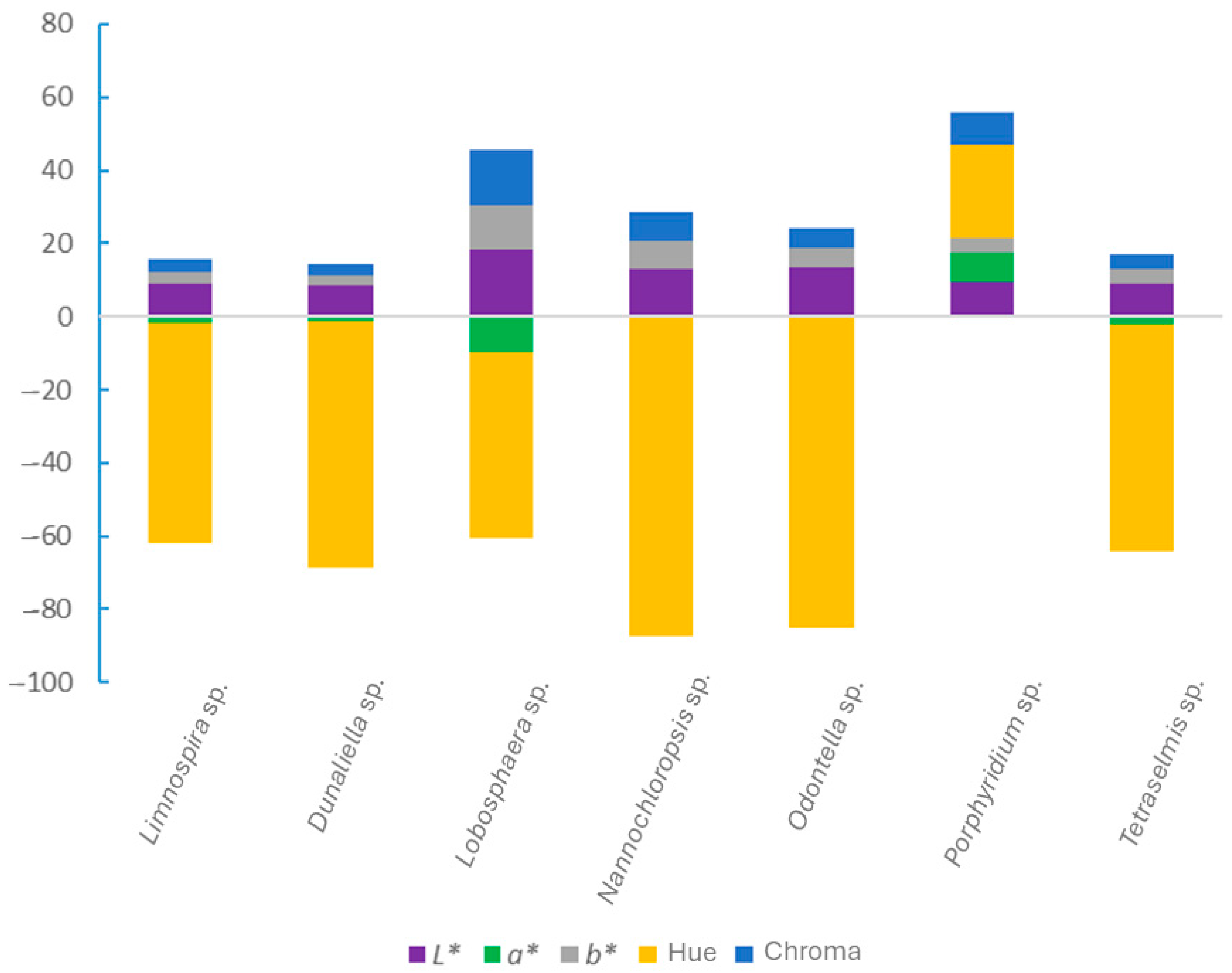
3.1. Color of the Microalgae Biomass

In the present study, the colorimetric parameters L* (lightness), a* (red–green axis), b* (yellow–blue axis), hue, and chroma were analyzed. The values of these parameters were generally low (Table 2), indicating dark colors, as the biomass was in powder form (freeze-dried), which typically becomes lighter in aqueous solution.
Among the studied microalgae, Lobosphaera sp. exhibited the highest lightness (L*) value. This species also showed the lowest a* value, indicating a greener hue, whereas Porphyridium sp. displayed the highest a* value, corresponding to a red color. Dunaliella sp. recorded the lowest b* value, suggesting a shift towards the yellow end of the spectrum. In line with the a* and b* values, Lobosphaera sp. also showed the highest chroma (Table 2 and Figure 1). The highest hue value was observed in Porphyridium sp., owing to its specific a* and b* coordinates, while all other species exhibited negative hue values (Table 2 and Figure 1).

3.2. Nutritional Composition of the Microalgae Biomass

The cultivation conditions of the seven microalgae species were largely comparable. Except for Limnospira sp., the pH remained within the range of 7–8 for all species. Temperature fluctuations ranged from below 20 °C to 30.3 ± 1.6 °C. The photosynthetically active radiation (PAR) varied from 10.8 ± 3.15 to 46.0 ± 6.89 mol/m2/day, with all values of similar magnitude and yielding a mean of 33.7 mol/m2/day (Table 1).
Despite these variation in cultivation conditions, all biomass samples were characterized with respect to their moisture, ash, lipid, protein, fiber, and carbohydrate content.
Protein was the predominant macronutrient in the biomass, ranging from 22.9 ± 0.1% to 59.8 ± 1.6% dry weight (DW) across the different microalgae species (Table 3 and Figure 2). Under the cultivation conditions outlined in Section 2.1, Limnospira sp. exhibited the highest protein content, while Nannochloropsis sp. recorded the highest lipid content. Lobosphaera sp., Nannochloropsis sp., and Tetraselmis sp. were particularly rich in fiber (Table 3).
Gabr et al. [39] reported that Limnospira platensis biomass contained 53.75 g/100 g DW protein, 7.8 g/100 g DW total lipids, and 9.21 g/100 g DW crude fiber, which are values comparable to those found in the present study (Table 3).
Ambrico et al. [40] analyzed wet biomass of Dunaliella sp. and obtained values of 10.03 ± 0.57% (w/w, dry basis) for protein, 25.31 ± 1.55% carbohydrates, 3.49 ± 0.10% lipids, and 8.97 ± 0.50% total dietary fiber. These results are lower than those in the present study (Table 3).
Bahi et al. [41], in their evaluation of various microalgal species as potential feed ingredients for aquaculture, found that Nannochloropsis gaditana had the highest protein content, up to 52%, which exceeds the 22.9 ± 0.1% observed in this study. Additionally, Porphyridium cruentum was reported to have high carbohydrate (40–57%) and lipids (18%) contents, both higher than the corresponding values obtained here (Table 3). For Tetraselmis sp., their reported values of 31% protein, 17% lipids, and 12.1% carbohydrates are comparable for protein, but indicate higher lipid and lower carbohydrate contents relative to the findings in the present work (Table 3).

3.3. Yield of Extraction of Bioactive Compounds from the Microalgae Biomass

The extraction yields of bioactive compounds from the various microalgae species ranged from 11.64 ± 0.28% to 52.96 ± 3.25% (Table 4). Dunaliella sp. exhibited the highest extraction yield among the microalgae studied, using hydroalcoholic ultrasound-assisted extraction, followed by Odontella sp. (Table 4). The lowest yields were recorded for Tetraselmis sp. and Limnospira sp.
Ferreira-Santos et al. [42] reported a water extraction yield of 35 ± 1% (37 °C, 60 min) from dried spirulina (Limnospira sp.), which is higher than that observed in the present study (Table 4). However, their subsequent ethanol (96%) extraction yielded 9.6 ± 0.2%, which is closer to the values obtained here. Gabr et al. [39] performed both aqueous and 80% ethanol extractions on dried spirulina powder, with the resulting extracts containing varying concentrations of carotenoids, chlorophyll a, total phenols, flavonoids, phycocyanin, free amino acids, total proteins, and carbohydrates.
Ambrico et al. [40] carried out an ethanol extraction from dried Dunaliella sp., obtaining yields of 8.8% (ethanol), 6.1% (hexane), and 17.0% (chloroform/methanol, 1:1). These values are lower than those reported in the present study, underscoring the influence of solvent polarity on extraction efficiency. Among all analyzed microalgae, Dunaliella sp. consistently exhibited the highest extraction yield.
Gnanakani et al. [43] reported an extraction yield of 23.33% using an ethanolic solution for Nannochloropsis sp., which is comparable to the 27.72% obtained in the present work.
Cuong et al. [44] achieved yields of 20.4% using a 70% hydroethanolic solution and 23.4% with hot water (125 °C, 15 min) for Odontella sp., both of which are lower than the 31.01% yield found in the present study.
Bueno et al. [45] conducted water extraction (25 °C, 60 min) on freeze-dried Porphyridium cruentum powder, resulting in a yield of 36.14 ± 0.18%, which exceeds the 26.12% yield reported here.
Jo et al. [46] obtained a yield of 21.7% from freeze-dried Tetraselmis suecica using methanol and a protease enzyme, which is significantly higher than the 11.64% yield observed in the current study.
Khawli et al. [47] generally reported improved extraction efficiencies with ultrasound-assisted extraction compared to conventional methods. The use of enzymes and thermal treatment also contributed to enhance yields. Similarly, Martins et al. [34] observed increased extraction efficiency when using viscoenzyme and cellulase.

3.4. Bioactivity of the Microalgae Extracts

3.4.1. Total Phenolic Content (TPC) and Antioxidant Activity (AA) of the Microalgae Extracts

In the present study, the TPC varied significantly among the microalgae species (Table 5). Odontella sp. exhibited the highest total phenolic content (TPC) among the microalgae species investigated. Limnospira sp. presented the highest antioxidant activity (AA) in the ABTS, DPPH, and ORAC assays. Conversely, Porphyridium sp., Nannochloropsis sp., and Tetraselmis sp. showed the lowest TPC, with Tetraselmis sp. and Nannochloropsis sp. generally demonstrating the weakest antioxidant activities (Table 5). Recent studies consistently demonstrate that TPC varies significantly among microalgae species. This variation is primarily due to species-specific metabolic pathways, genetic differences, and environmental responses [48]. Such inter-species differences have important implications for selecting microalgae strains for specific bioactive applications (e.g., nutraceuticals, antioxidants).
Kumar et al. [49] determined the TPC of an ethanolic extract from Limnospira sp., reporting a value of 9.919 ± 0.446 mg GAE/g of extract dry weight (DW), which is similar to the value observed in the present study (9.7 ± 0.6 mg GAE/g DW). Guler et al. [50] assessed the antioxidant activity of an aqueous extract from Limnospira platensis, reporting DPPH and ABTS scavenging activities of 40.65 ± 0.21% and 81.93 ± 0.61%, respectively. Gabr et al. [39] noted that the TPC in spirulina (51.20 µg/mL) was higher than that in its ethanolic extract (49.48 µg/mL), while the aqueous extract had the lowest value (15.26 µg/mL).
Mondal et al. [51] obtained an ethanolic extract from Dunaliella tertiolecta using ultrasound-assisted extraction, with a TPC of 515.72 ± 4.67 µg GAE/g and a DPPH radical scavenging activity of 40 ± 1.41%.
Corrêa et al. [52] investigated the TPC and AA of ethanolic extracts of Lobosphaera sp., evaluating the effects of high-pressure processing (HPP) on frozen versus freeze-dried biomass. The highest TPC (11.50 ± 0.21 mg GAE/g DW) was achieved when HPP was applied to frozen biomass, which is comparable to the value found in the present study (10.7 mg GAE/g DW). The lowest TPC (4.61 ± 0.06 mg GAE/g DW) was observed following HPP on freeze-dried biomass. Additionally, freeze-drying was found to significantly reduce DPPH radical scavenging activity (by approximately 3%) compared to freezing (around 2%).
Wali et al. [53] evaluated the ABTS and DPPH assays of a methanolic extract of Nannochloropsis oculata obtained via ultrasound-assisted extraction. IC50 values were 96.95 ± 1.68 µg/mL for ABTS and 52.10 ± 0.85 µg/mL for DPPH. Gnanakani et al. [43] reported a TPC of 26.42 ± 1.5 mg GAE/g for an ethanolic extract, while an ethyl acetate extract followed by hexane separation (EAH) yielded a higher TPC of 40.61 ± 1.8 mg GAE/g. In the DPPH assay, IC50 values were 13.9 µg/mL (EAH), 31.84 µg/mL (ethanol), and 48.01 µg/mL (acetone). Banskota et al. [54] reported a TPC of 43.6 ± 1.2 µmol GAE/g DW and an ORAC of 6948 ± 1532 µmol TE/100 g DW for a methanolic extract of Nannochloropsis granulate.
Cuong et al. [44] reported a TPC of 0.894 ± 0.20 mg GAE/g DW for a 70% hydroethanolic extract of Odontella sp., which is significantly lower than the value obtained in the present study (31.8 ± 5.3 mg GAE/g DW using a 90% hydroethanolic solution. For a chloroform/methanol extract (8 mg/mL), EC50 values were 1.821 ± 0.25 and 5.463 ± 0.47 mg/mL for the ABTS and DPPH assays, respectively—both higher than those observed for the hydroethanolic extract.
For Porphyridium sp., Tounsi et al. [55] analyzed an extract prepared by cellular lysis (0.1 mg/mL), reporting IC50 values of 45 µg/mL (DPPH) and 658.81 µg/mL (ABTS). Assunção et al. [56] investigated Porphyridium aerugineum and Porphyridium sordidum in the ABTS assay, obtaining values of 67.95 ± 0.36 and 32.48 ± 0.04 mg TE/100 g extract, respectively. The IC50 values in the DPPH assay were 157.53 ± 0.35 and 286.24 ± 0.25 mg/mL, respectively.
For Tetraselmis sp., Trentin et al. [57] obtained a TPC of 20.45 ± 1.87 and 25.19 ± 1.26 mg GAE/g DW in water and methanolic extracts, respectively. These are notably higher than the value recorded in the present study (0.57 ± 0.01 mg GAE/100 mg DW using a 90% hydroethanolic extract). They also evaluated AA via ABTS and DPPH assays, reporting no detectable activity for the water extract at concentrations of 1, 5, and 10 mg/mL. The methanolic extract showed no ABTS activity, but it exhibited DPPH activity of 17.29 ± 0.35% and 24.94 ± 0.71% at 5 mg/mL and 10 mg/mL, respectively.
It is important to highlight the considerable variability in units and methodologies used in literature, which complicates direct comparisons. Nevertheless, it is evident that extraction conditions—including the choice of solvent, the extraction method (e.g., infusion, maceration), the solvent polarity, and the use of techniques like heating, enzymatic treatment, microwave irradiation, and ultrasound— greatly influence the composition and resultant bioactivity of extracts. These factors can substantially affect both the antioxidant activity and total phenolic content of the microalgal extracts.

3.4.2. Antimicrobial Activity of Microalgae Extracts Against Bacteria

Among the microbial extracts tested, Nannochloropsis sp. exhibited the highest inhibitory effect, as evidenced by the lowest minimal inhibitory concentrations (MIC) against all bacteria strains assessed, with the exception of Bacillus cereus (Table 6). Extracts of Limnospira sp., Odontella sp., and Lobosphaera sp. also showed notable antimicrobial activity, inhibiting the growth of all tested bacteria strains. In contrast, extracts from Porphyridium sp. and Dunaliella sp. did not show significant inhibitory effects at the concentrations tested. The extract of Tetraselmis sp., although less potent than that of Nannochloropsis sp., displayed inhibitory activity against all bacteria except B. cereus (Table 6).
Maadane et al. [58] prepared ethanolic extracts from several microalgae and reported the following MIC values (mg/mL): for Dunaliella sp., 4.3 ± 0.1 against Pseudomonas aeruginosa and Escherichia coli, and >5 against Staphylococcus aureus; for Nannochloropsis gaditana, 3.4 ± 0.2 against E. coli and 4.5 ± 0.6 against S. aureus; and for Tetraselmis sp., 2.6 ± 0.2 against E. coli and 3 ± 0.1 against S. aureus.
Najdenski et al. [59] determined the MICs of aqueous extracts from Porphyridium cruentum and Porphyridium aerugineum. No inhibitory effect was observed against B. cereus, E. coli, or Salmonella typhimurium for P. aerugineum, while the extract of P. cruentum exhibited an MIC of 7.0 mg/mL against the same bacteria. Interestingly, P. aerugineum showed enhanced efficacy against Staphylococcus aureus, with an MIC of 0.29 mg/mL.
Pradhan et al. [60] suggested that the antimicrobial activity of Limnospira sp. is associated with its content of pigments, polyphenols, fatty acids, and carbohydrates, and is particularly effective against E. coli. Similarly, they attributed the antimicrobial properties of P. cruentum to pigments, polyphenols, and fatty acids (against S. aureus), and carbohydrates (against E. coli).
Ilieva et al. [61] reported that some strains of Limnospira platensis displayed potent antimicrobial activity, with MIC values ranging from 2–15 μg/mL against various fish pathogens, including species of Bacillus and Vibrio. Additionally, Dunaliella tertiolecta displayed MIC values of 25 μg/mL and 50 μg/mL against certain Staphylococcus spp. isolated from bovine and goat mastitis, respectively. These values were considerably lower than those found in the present study.
As with antioxidant activity, the extraction method—including the choice of solvent, technique (e.g., maceration, sonication), and conditions (e.g., temperature, duration)—significantly influences the bioactivity of the resulting extracts. Moreover, the use of different bacterial strains introduces further variability. These factors may explain the discrepancies between the present findings and those previously reported in the literature.
It is worth noting that for all tested bacteria strains, the minimum bactericidal concentration (MBC) was above 5 mg/100 μL, indicating that none of the microalgae extracts exhibited bactericidal effects at the tested concentrations.

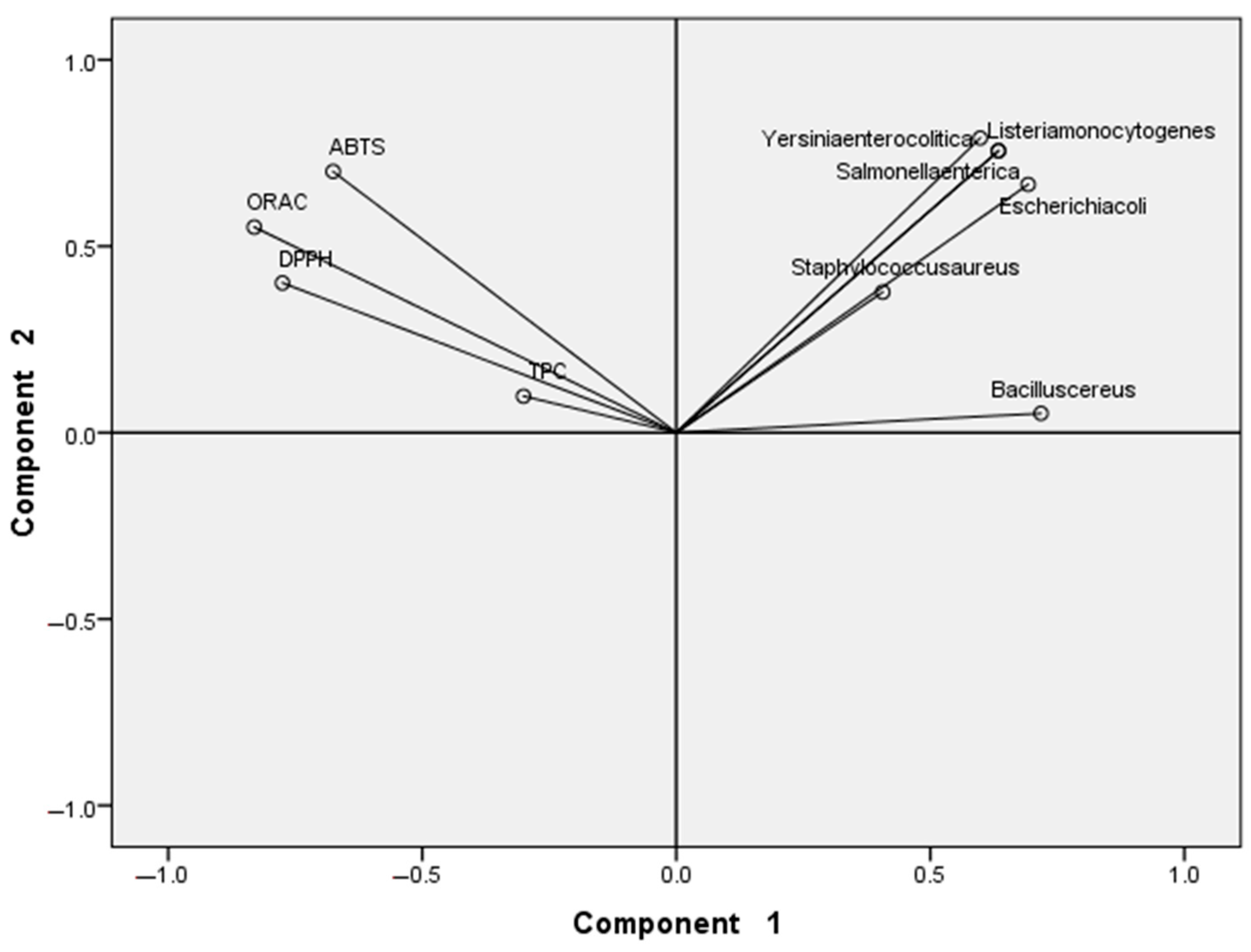
3.5. Principal Components Analysis (PCA)
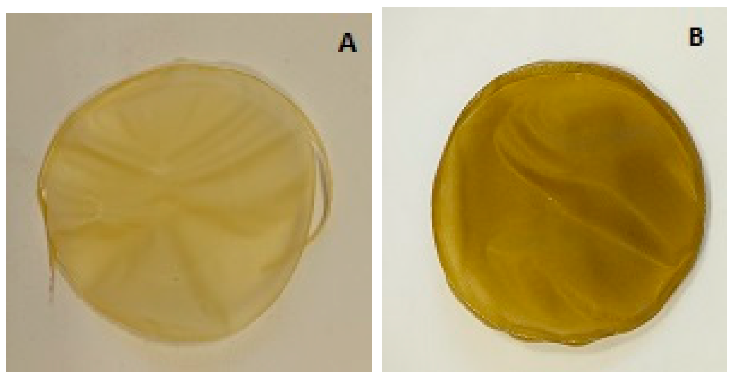
Principal component analysis (PCA) was employed to summarize, clarify, and simplify the dataset generated from the measured variables. In this analysis, the seven microalgal species were treated as independent variables, while the total phenolic content (TPC), antioxidant activity (ABTS, DPPH, and ORAC), and minimal inhibitory concentrations (MICs) against the bacteria served as the dependent variables. By preserving the intrinsic variability among the microalgae species, PCA enabled a graphical representation of the relative distances between them, thereby facilitating the identification of clusters of species with similar properties. Additionally, PCA allowed for the recognition of the most suitable microalgae in relation to specific variables or bioactivities (Figure 3 and Figure 4).
Figure 3 displays the projection of the variables. The variation parameter ratio, 76.07%, is the aggregate of the components resulting from the aggregate of the individual components (component 1, 46,71%, and component 2, 29.36%), indicating how much of the variance each component contains. Component 1 is more strongly correlated with all the variables than component 2. The minimal inhibitory concentrations (MICs) for the bacteria are positively correlated with component 1, whereas the total phenolic content (TPC) and antioxidant activity exhibit negative correlations. In contrast, all variables—including MICs, TPC, and antioxidant activity—show positive correlations with component 2.
The analysis of Figure 3 and Figure 4 reveals three distinct groups. Limnospira sp., Lobosphaera sp., and Odontella sp. form a group (pink circle, Figure 4) with high TPC, strong antioxidant activity, and relatively low MIC values against all tested bacteria (noting that a lower MIC indicates a more effective extract, as a smaller concentration is required to inhibit bacterial growth). A second group (green circle, Figure 4), comprising Dunaliella sp. and Porphyridium sp., shows good TPC and antioxidant activity but poor antimicrobial efficacy, as reflected by high MIC values. The third group (yellow circle, Figure 4) includes Nannochloropsis sp. and Tetraselmis sp., which display strong antimicrobial activity (low MICs) but low antioxidant potential.
The PCA results thus offer a clear visualization and validation of the observed differences among the microalgal species, which were discussed in Section 3.4.1 and Section 3.4.2, corroborating the findings presented in Figure 3 and Figure 4. PCA allows for better identification of the most suitable microalgal extracts to produce a synergistic effect once incorporated into an edible film.

3.6. Films Produced

The film produced using the casting method exhibited good film-forming capacity, even with the incorporation of extracts rich in non-polar lipids and bioactives. These additions resulted in the formation of an emulsion comprising alginate, zein, and microalgal extracts, which successfully yielded an edible film.
The most noticeable difference between the films was their visual appearance, particularly in terms of color. Film A (Figure 5) exhibited a lighter, more uniform color, whereas film B presented a darker appearance with a subtle brownish tint. This variation is likely attributed to the inclusion of the non-polar lipids and bioactive extracts into the formulation of film B. These additions affected the color parameters, leading to a decrease in L* and an increase in a*, b*, hue, and chroma (Table 7). Additionally, film B was thicker (0.079 ± 0.006 mm) than film A (0.065 ± 0.014 mm), a difference also attributable to the incorporation of these extracts.
Linares-Castañeda et al. [62] also reported that modifications in the concentrations of alginate and zein in film formulations led to variations in both the thickness and color characteristics of the resulting films. The increase of zein concentration produced an increase in both b* parameter and thickness, whereas the increase in alginate produced a decrease in the thickness.

3.7. Antioxidant Activity of the Films

Incorporation of all extracts rich in lipids and bioactives into the films resulted in an increased antioxidant activity, with statistically significant differences observed in both ABTS and DPPH assays (Table 8). Film B exhibited higher ABTS and DPPH values than the control film (film A). These findings align with previous reports on alginate films enriched with protein-rich (P-rich) and bioactive-rich extracts from Limnospira sp. [63], indicating that the enhanced antioxidant activity is largely due to the presence of bioactive-rich extracts.
The DPPH value for film B was similar to that reported by Martins et al. [34] for a 2% alginate film containing 0.5% P-rich and 0.25% bioactive-rich extracts from Lobosphaera sp., which exhibited 212.81 ± 39.12 µM TE/mg film. Kia et al. [64] also demonstrated that the incorporation of Arthrospira platensis into a biocomposite film made with Zedo gum and sodium caseinate significantly improved antioxidant activity.
Golmakani et al. [65] reported that zein fibers containing phycocyanin and a 70% hydroethanolic spirulina extract showed DPPH antioxidant activities of 42.86 ± 0.13% and 47.98 ± 0.64%, respectively. The strong antioxidant properties of spirulina extracts are attributed to their high total phenolic and flavonoid contents, alongside contributions from β-carotene and chlorophyll, known antioxidant agents.

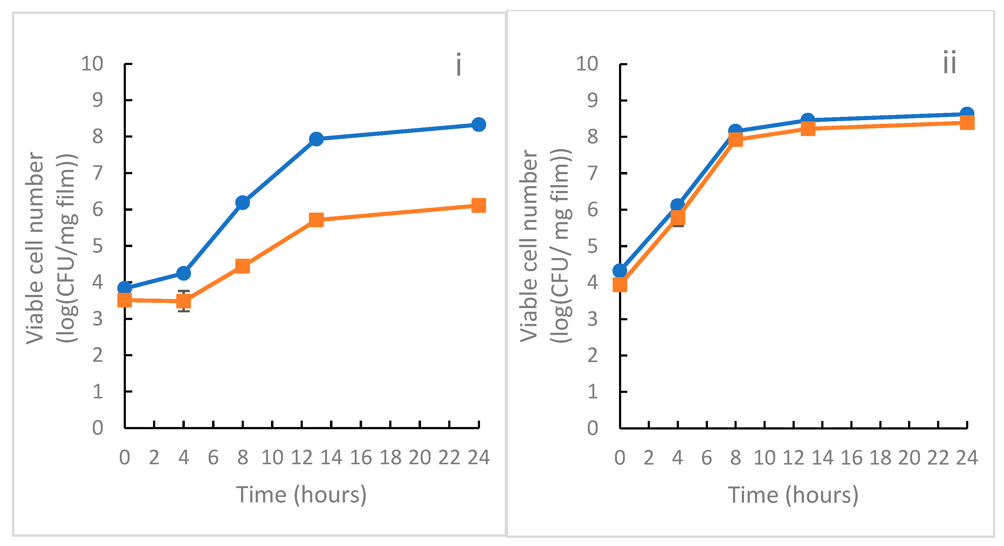
3.8. Antimicrobial Activity of the Films

The antimicrobial efficacy tests demonstrated that incorporating a lipid-rich extract from Nannochloropsis sp. and bioactive-rich extracts from Nannochloropsis sp. and Limnospira sp. significantly reduced the growth of the Gram-positive bacterium Listeria monocytogenes compared to the control film (film A). In contrast, the inclusion of microalgal extracts did not affect the growth of the Gram-negative bacterium Salmonella enterica serovar Enteritidis, with bacterial proliferation remaining comparable to that of the control film composed of 3% alginate and 1% zein (Figure 6). These results are consistent with those reported by Martins et al. [34] for alginate films combined with either protein-rich, polysaccharide-rich, or bioactive-rich extracts from Lobosphaera sp.
Lopes et al. [38] suggested that the antibacterial efficacy of extracts embedded in alginate films might be reduced due to potential chemical interactions between the hydroxyl groups of the alginate matrix and the phenolic compounds in the extracts. Such interactions may block the active sites of the bioactives, thereby inhibiting their antimicrobial effect, particularly against Gram-negative bacteria. Similarly, Golmakani et al. [65] observed that Gram-negative bacteria are generally less susceptible to inhibition than Gram-positive bacteria, attributing this to the hydrophilic nature of the lipopolysaccharide-rich outer membrane, which hinders the diffusion of hydrophobic compounds into the cell. These authors further noted that a 70% hydroethanolic extract of spirulina exhibited greater antimicrobial activity than isolated phycocyanin, likely due to the phenolic compounds in spirulina disrupting bacterial cell membranes and inhibiting growth.
Amaro et al. [66] emphasized that antibiotics and antimicrobial compounds are generally less effective against Gram-negative bacteria because their complex, multilayered cell walls act as a barrier to penetration. These collective findings help to explain the lack of inhibitory effect observed in the present study against S. enterica serovar Enteritidis (Gram-negative) in films containing microalgal extracts (Figure 6). Conversely, the observed inhibition of L. monocytogenes may be attributable to the presence of lipids in the extract, such as eicosapentaenoic acid, known for its antimicrobial properties [45].

4. Conclusions

The findings of the present study demonstrate that the investigated microalgae represent promising sources of valuable macronutrients: Limnospira sp. exhibited the highest protein content; Nannochloropsis sp. was particularly rich in lipids; and Lobosphaera sp. had a high carbohydrate content. Nannochloropsis sp. also showed the highest energy value among the species studied.
Ultrasound-assisted hydroethanolic extraction yielded microalgal extracts with varying degrees of bioactivity. Overall, Limnospira sp. demonstrated the most favorable combination of antioxidant and antimicrobial activities, while Nannochloropsis sp. exhibited the strongest antimicrobial effects against most of the tested bacterial strains.
A promising and innovative direction involves combining extracts from two or more microalgae species to leverage synergistic effects. Edible films composed of alginate and zein, enriched with bioactive-rich extracts from Limnospira sp. and Nannochloropsis sp., as well as a lipid-rich extract from Nannochloropsis sp., were successfully formulated. These films exhibited notable antioxidant activity and effectively inhibited the growth of Listeria monocytogenes. Thus, edible films incorporating lipid-rich extracts from Nannochloropsis sp. and bioactive-rich extracts from Limnospira sp. show promising potential for active food packaging.
In summary, the microalgal extracts obtained in this study show strong potential for applications as natural preservatives in food products (e.g., incorporated in bread, cookies, and pasta), particularly via incorporation into edible films and coatings. For the validation and practical implementation of the results of this study, the different microalgal extracts could, for example, be used in food product formulations as food additives, nutraceutical ingredients, or functional food components. To enable real-world application, first, formulation studies are recommended to ensure the extract stability, compatibility, and sensory acceptability in target products. Next, pilot-scale trials should be performed to test production scalability and consistency. Finally, regulatory and safety requirements could be met through toxicological assessments and compliance with relevant food or health product regulations.
The studied microalgae represent, therefore, a valuable and sustainable source of multifunctional solutions for health, nutrition, cosmetics, and environmental sectors (e.g., eco-friendly packaging), highlighting their role in green biotechnological innovation and commercial viability. Exploring inter-species combinations may unlock next-generation applications that outperform single-extract solutions in both efficacy and commercial value.

Author Contributions

Conceptualization, A.M.M.B.M. and R.M.S.C.M.; methodology, V.F.R.M., A.I.L., A.M.M.B.M. and M.P.; software, V.F.R.M.; validation, A.M.M.B.M.; formal analysis, V.F.R.M., and A.M.M.B.M.; investigation, V.F.R.M. and A.I.L.; resources, M.P., A.M.M.B.M., R.M.S.C.M., D.G., C.P., S.M.B. and L.C.; data curation, V.F.R.M.; writing—original draft preparation, V.F.R.M., and A.M.M.B.M.; writing—review and editing, A.M.M.B.M. and R.M.S.C.M.; visualization, V.F.R.M., and A.M.M.B.M.; supervision, A.M.M.B.M., R.M.S.C.M. and M.P.; project administration, A.M.M.B.M., R.M.S.C.M. and M.P.; funding acquisition, R.M.S.C.M. and M.P. All authors have read and agreed to the published version of the manuscript.

Funding

This work was supported by National Funds from FCT—Fundação para a Ciência e a Tecnologia through Project UIDB/50016/2020 and by project EXTRATOTECA—COMPETE2020 POCI-01-0247-FEDER-033784. Valter Martins acknowledges FCT for the PhD research grant with reference UI/BD/152825/2022.

Institutional Review Board Statement

Not applicable.

Informed Consent Statement

Not applicable.

Data Availability Statement

Data are contained within the article. Further inquiries can be directed to the corresponding authors.

Conflicts of Interest

The authors declare no conflicts of interest. The funders had no role in the design of the study; in the collection, analyses, or interpretation of data; in the writing of the manuscript; or in the decision to publish the results.

References

  1. Morais, A.M.M.B.; Morais, R.M.S.C.; Lackner, M. Biodegradable Bio-Based Plastics Toward Climate Change Mitigation. In Handbook of Climate Change Mitigation and Adaptation; Lackner, M., Sajjadi, B., Chen, W.Y., Eds.; Springer: New York, NY, USA, 2024; pp. 1–48. [Google Scholar] [CrossRef]
  2. Ilhami, S.; Rahmen, S.N.S.A.; Iqhrammullah, M.; Hamid, Z.; Chai, Y.O.; Lam, M.K. Polyhydroxyalkanoates production from microalgae for sustainable bioplastics: A review. Biotechnol. Adv. 2024, 79, 108529. [Google Scholar] [CrossRef] [PubMed]
  3. Weng, S.; Marcet, I.; Rendueles, M.; Diaz, M. Edible Films from the Laboratory to Industry: A Review of the Different Production Methods. Food Bioprocess Technol. 2025, 18, 3245–3271. [Google Scholar] [CrossRef]
  4. Levasseur, W.; Perre, P.; Pozzobon, V. A review of high value-added molecules production by microalgae in light of the classification. Biotechnol. Adv. 2020, 41, 107545. [Google Scholar] [CrossRef]
  5. Matos, J.; Cardoso, C.; Bandarra, N.M.; Afonso, C. Microalgae as healthy ingredients for functional food: A review. Food Funct. 2017, 8, 2672–2685. [Google Scholar] [CrossRef] [PubMed]
  6. Guihéneuf, F.; Stengel, D.B. Impact of Temperature on Fatty Acid Composition and Nutritional Value in Eight Species of Microalgae. Appl. Microbiol. Biotechnol. 2018, 102, 5279–5297. [Google Scholar]
  7. Tibbetts, S.M.; Milley, J.E.; Lall, S.P. Chemical Composition and Nutritional Properties of Freshwater and Marine Microalgal Biomass Cultured in Photobioreactors. J. Appl. Phycol. 2015, 27, 1109–1119. [Google Scholar] [CrossRef]
  8. Paterson, S.; Gómez-Cortés, P.; de la Fuente, M.A.; Hernández-Ledesma, B. Bioactivity and Digestibility of Microalgae Tetraselmis Sp. and Nannochloropsis Sp. as Basis of Their Potential as Novel Functional Foods. Nutrients 2023, 15, 477. [Google Scholar] [CrossRef] [PubMed]
  9. Begum, H.; Yusoff, F.M.D.; Banerjee, S.; Khatoon, H.; Shariff, M. Availability and Utilization of Pigments from Microalgae. Crit. Rev. Food Sci. Nutr. 2016, 56, 2209–2222. [Google Scholar] [CrossRef]
  10. Abreu, A.P.; Martins, R.; Nunes, J. Emerging Applications of Chlorella Sp. and Spirulina (Arthrospira) Sp. Bioengineering 2023, 10, 955. [Google Scholar] [CrossRef]
  11. Silva, M.R.O.B.; Moura, Y.A.S.; Converti, A.; Porto, A.L.F.; Viana Marques, D.A.; Bezerra, R.P. Assessment of the Potential of Dunaliella Microalgae for Different Biotechnological Applications: A Systematic Review. Algal. Res. 2021, 58, 102396. [Google Scholar] [CrossRef]
  12. Lee, S.; Lim, S.R.; Jeong, D.G.; Kim, J.H. Characterization of an Oleaginous Unicellular Green Microalga, Lobosphaera Incisa (Reisigl, 1964) Strain K-1, Isolated from a Tidal Flat in the Yellow Sea, Republic of Korea. Front. Microbiol. 2018, 9, 2159. [Google Scholar] [CrossRef]
  13. Vasilieva, S.; Lukyanov, A.; Antipova, C.; Grigoriev, T.; Lobakova, E.; Chivkunova, O.; Scherbakov, P.; Zaytsev, P.; Gorelova, O.; Fedorenko, T.; et al. Interactive Effects of Ceftriaxone and Chitosan Immobilization on the Production of Arachidonic Acid by and the Microbiome of the Chlorophyte Lobosphaera Sp. IPPAS C-2047. Int. J. Mol. Sci. 2023, 24, 10988. [Google Scholar] [CrossRef] [PubMed]
  14. Raja, R.; Coelho, A.; Hemaiswarya, S.; Kumar, P.; Carvalho, I.S.; Alagarsamy, A. Applications of Microalgal Paste and Powder as Food and Feed: An Update Using Text Mining Tool. Beni-Suef. Univ. J. Basic Appl. Sci. 2018, 7, 740–747. [Google Scholar] [CrossRef]
  15. Alami, A.H.; Tawalbeh, M.; Alasad, S.; Ali, M.; Alshamsi, M.; Aljaghoub, H. Cultivation of Nannochloropsis Algae for Simultaneous Biomass Applications and Carbon Dioxide Capture. Energy Sources Part A Recovery Util. Environ. Eff. 2021, 47, 8471–8482. [Google Scholar] [CrossRef]
  16. Kim, S.Y.; Kwon, Y.M.; Kim, K.W.; Kim, J.Y.H. Exploring the Potential of Nannochloropsis Sp. Extract for Cosmeceutical Applications. Mar. Drugs 2021, 19, 690. [Google Scholar] [CrossRef]
  17. Moreau, D.; Tomasoni, C.; Jacquot, C.; Kaas, R.; Le Guedes, R.; Cadoret, J.P.; Muller-Feuga, A.; Kontiza, I.; Vagias, C.; Roussis, V.; et al. Cultivated Microalgae and the Carotenoid Fucoxanthin from Odontella Aurita as Potent Anti-Proliferative Agents in Bronchopulmonary and Epithelial Cell Lines. Environ. Toxicol. Pharmacol. 2006, 22, 97–103. [Google Scholar] [CrossRef]
  18. Heo, J.; Cho, D.-H.; Kim, U.; Kim, H.-S. Isolation and Characterization of Indigenous Diatom, Odontella Sp. BS-003 as Potential Fucoxanthin and Omega-3 Fatty Acid Producer. J. Mar. Biosci. Biotechnol. 2018, 10, 26–33. [Google Scholar]
  19. Tannin-Spitz, T.; Bergman, M.; Van-Moppes, D.; Grossman, S.; Arad, S. Antioxidant Activity of the Polysaccharide of the Red Microalga Porphyridium Sp. J. Appl. Phycol. 2005, 17, 215–222. [Google Scholar] [CrossRef]
  20. Tounsi, L.; Ben Hlima, H.; Elhadef, K.; Hentati, O.; Blavignac, C.; Fendri, I.; Smaoui, S.; Michaud, P.; Abdelkafi, S. B-Phycoerythrin of Porphyridium Cruentum UTEX 161: A Multifunctional Active Molecule for the Development of Biodegradable Films. Eur. Polym. J. 2024, 208, 112851. [Google Scholar] [CrossRef]
  21. De Jesus Raposo, M.F.; De Morais, R.M.S.C.; De Morais, A.M.M.B. Bioactivity and Applications of Sulphated Polysaccharides from Marine Microalgae. Mar Drugs 2013, 11, 233–252. [Google Scholar] [CrossRef]
  22. Mehariya, S.; Annamalai, S.N.; Thaher, M.I.; Quadir, M.A.; Khan, S.; Rahmanpoor, A.; Abdurahman, k.; Faisal, M.; Sayadi, S.; Al Hawari, A.; et al. A Comprehensive Review on Versatile Microalga Tetraselmis: Potentials Applications in Wastewater Remediation and Bulk Chemical Production. J. Environ. Manage. 2024, 365, 121520. [Google Scholar] [CrossRef] [PubMed]
  23. Schwenzfeier, A.; Wierenga, P.A.; Gruppen, H. Isolation and Characterization of Soluble Protein from the Green Microalgae Tetraselmis Sp. Bioresour. Technol. 2011, 102, 9121–9127. [Google Scholar]
  24. Amna Kashif, S.; Hwang, Y.J.; Park, J.K. Potent Biomedical Applications of Isolated Polysaccharides from Marine Microalgae Tetraselmis Species. Bioprocess. Biosyst. Eng. 2018, 41, 1611–1620. [Google Scholar] [CrossRef]
  25. Benbettaïeb, N.; Debeaufort, F.; Karbowiak, T. Bioactive edible films for food applications: Mechanisms of antimicrobial and antioxidant activity. Crit. Rev. Food Sci. Nutr. 2019, 59, 3431–3455. [Google Scholar] [CrossRef] [PubMed]
  26. Richmond, A. (Ed.) Handbook of Microalgal Culture: Biotechnology and Applied Phycology; Blackwell Science: Oxford, UK; Ames, IA, USA, 2004. [Google Scholar]
  27. Commission Internationale de L’Eclairage (C.I.E.). Recommendations on Uniform Color Spaces, Color-Difference Equations, Psychometric Color Terms; Supplement No. 2 to C.I.E. Publication No. 15, (E-1.3.1) 1971 (TC-1-3); C.I.E.: Paris, France, 1971. [Google Scholar]
  28. A.O.A.C. Official Methods of Analysis, 18th ed.; Method A.O.A.C. 930.15; Association of the Official Analytical Chemists: Arlington, VA, USA, 2005.
  29. Slack, P.T. Analytical Methods Manual, 3rd ed.; Leatherhead Food R.A.: London, UK, 1997; Issue 2. [Google Scholar]
  30. ISO 1871:2009; Food and Feed Products—General Guidelines for the Determination of Nitrogen by the Kjeldahl Method. International Organization for Standardization: Geneva, Switzerland, 2009.
  31. A.O.A.C. Official Methods of Analysis; Method A.O.A.C. 991.43; Association of the Official Analytical Chemists: Arlington, VA, USA, 2003.
  32. A.O.A.C. Official Methods of Analysis; Method A.O.A.C. 985.29; Association of the Official Analytical Chemists: Arlington, VA, USA, 2000.
  33. Regulation (EC) No 1169/2011 of the European Parliament and of the council of 25 October 2011 on the provision of food information to consumers. Off. J. Eur. Union 2011, 304.
  34. Martins, V.F.R.; Lopes, A.I.; Machado, M.; Costa, E.M.; Ribeiro, T.B.; Poças, F.; Pintado, M.; Morais, R.M.S.C.; Morais, A.M.M.B. Biodegradable Films with Polysaccharides, Proteins, and Bioactive Compounds from Lobosphaera sp.: Antioxidant and Antimicrobial Activities. Foods 2025, 14, 1327. [Google Scholar] [CrossRef]
  35. Wiegand, I.; Hilpert, K.; Hancock, R.E.W. Agar and Broth Dilution Methods to Determine the Minimal Inhibitory Concentration (MIC) of Antimicrobial Substances. Nat. Protoc. 2008, 3, 163–175. [Google Scholar] [CrossRef]
  36. Yap, B.H.J.; Crawford, S.A.; Dumsday, G.J.; Scales, P.J.; Martin, G.J.O. A mechanistic study of algal cell disruption and its effect on lipid recovery by solvent extraction. Algal Res. 2014, 5, 112–120. [Google Scholar] [CrossRef]
  37. Lade, B.D.; Dhowlaghar, N.; Pillai, S.S.; Patil, B.S. Physicochemical, mechanical, and antimicrobial properties of sodium alginate films as carriers of zein emulsion with pelargonic acid and eugenol for active food packing. Food Packag. Shelf Life. 2023, 40, 101202. [Google Scholar] [CrossRef]
  38. Lopes, A.I.; Melo, A.; Caleja, C.; Pereira, E.; Finimundy, T.C.; Afonso, T.B.; Silva, S.; Ivanov, M.; Soković, M.; Tavaria, F.K.; et al. Evaluation of Antimicrobial and Antioxidant Activities of Alginate Edible Coatings Incorporated with Plant Extracts. Coatings 2023, 13, 1487. [Google Scholar] [CrossRef]
  39. Gabr, G.A.; El-Sayed, S.M.; Hikal, M.S. Antioxidant Activities of Phycocyanin: A Bioactive Compound from Spirulina Platensis. J. Pharm. Res. Int. 2020, 32, 73–85. [Google Scholar] [CrossRef]
  40. Ambrico, A.; Trupo, M.; Magarelli, R.; Balducchi, R.; Ferraro, A.; Hristoforou, E.; Marino, T.; Musmarra, D.; Casella, P.; Molino, A. Effectiveness of Dunaliella Salina Extracts against Bacillus Subtilis and Bacterial Plant Pathogens. Pathogens 2020, 9, 613. [Google Scholar] [CrossRef] [PubMed]
  41. Bahi, A.; Ramos-Vega, A.; Angulo, C.; Monreal-Escalante, E.; Guardiola, F.A. Microalgae with Immunomodulatory Effects on Fish. Rev. Aquac. 2023, 15, 1522–1539. [Google Scholar] [CrossRef]
  42. Ferreira-Santos, P.; Miranda, S.M.; Belo, I.; Spigno, G.; Teixeira, J.A.; Rocha, C.M.R. Sequential Multi-Stage Extraction of Biocompounds from Spirulina Platensis: Combined Effect of Ohmic Heating and Enzymatic Treatment. Innov. Food Sci. Emerg. Technol. 2021, 71, 102707. [Google Scholar] [CrossRef]
  43. Gnanakani, P.E.; Santhanam, P.; Kumar, K.E.; Dhanaraju, M.D. Chemical Composition, Antioxidant, and Cytotoxic Potential of Nannochloropsis Species Extracts. J. Nat. Sci. Biol. Med. 2019, 10, 167–177. [Google Scholar]
  44. Cuong, D.M.; Yang, S.H.; Kim, J.S.; Moon, J.Y.; Choi, J.; Go, G.M.; Cho, S.K. Evaluation of Antioxidant and Anti-Inflammatory Activity and Identification of Bioactive Compound from the Marine Diatom, Odontella Aurita Extract. Appl. Biol. Chem. 2024, 67, 46. [Google Scholar] [CrossRef]
  45. Bueno, M.; Gallego, R.; Chourio, A.M.; Ibáñez, E.; Herrero, M.; Saldaña, M.D.A. Green Ultra-High Pressure Extraction of Bioactive Compounds from Haematococcus Pluvialis and Porphyridium Cruentum Microalgae. Innov. Food Sci. Emerg. Technol. 2020, 66, 102532. [Google Scholar] [CrossRef]
  46. Jo, W.S.; Choi, Y.J.; Kim, H.J.; Nam, B.H.; Hong, S.H.; Lee, G.A.; Lee, S.W.; Seo, S.Y.; Jeong, M.H. Anti-Inflammatory Effect of Microalgal Extracts from Tetraselmis Suecica. Food. Sci. Biotechnol. 2010, 19, 1519–1528. [Google Scholar] [CrossRef]
  47. Khawli, F.A.; Marti-Quijal, F.J.; Pallares, N.; Barba, F.J.; Ferrer, E. Ultrasound Extraction Mediated Recovery of Nutrients and Antioxidant Bioactive Compounds from Phaeodactylum tricornutum Microalgae. Appl. Sci. 2021, 11, 1701. [Google Scholar] [CrossRef]
  48. Machu, L.; Misurcova, L.; Vavra Ambrozova, J.; Orsavova, J.; Mlcek, J.; Sochor, J.; Jurikova, T. Phenolic content and antioxidant capacity in algal food products. Molecules 2015, 20, 1118–1133. [Google Scholar] [CrossRef]
  49. Kumar, A.; Ramamoorthy, D.; Verma, D.K.; Kumar, A.; Kumar, N.; Kanak, K.R.; Marwein, B.M.; Mohan, K. Antioxidant and Phytonutrient Activities of Spirulina Platensis. Energy Nexus 2022, 6, 100070. [Google Scholar] [CrossRef]
  50. Guler, B.A.; Demirel, Z.; Imamoglu, E. Induction of Antioxidant Activities of Arthrospira Platensis and Chlorella Vulgaris by Modified Culture Conditions. Bioprocess. Biosyst. Eng. 2024, 47, 275–287. [Google Scholar] [CrossRef]
  51. Mondal, K.; Bhattacharjee, S.K.; Mudenur, C.; Ghosh, T.; Goud, V.V.; Katiyar, V. Development of Antioxidant-Rich Edible Active Films and Coatings Incorporated with de-Oiled Ethanolic Green Algae Extract: A Candidate for Prolonging the Shelf Life of Fresh Produce. RSC. Adv. 2022, 12, 13295–13313. [Google Scholar] [CrossRef] [PubMed]
  52. Corrêa, P.S.; Coimbra, R.S.T.; Caetano, N.S. Exploring the Antioxidant Potential of Lobosphaera Sp. and Odontella Sp. Biomasses under Different Extraction and Preservation Conditions. In Proceedings of the Seagriculture 2023-12th International Seaweed Conference EU, Trondheim, Norway, 21–22 June 2023. [Google Scholar]
  53. Wali, A.F.; Al Dhaheri, Y.; Pillai, J.R.; Mushtaq, A.; Rao, P.G.M.; Rabbani, S.A.; Firdous, A.; Elshikh, M.S.; Al Farraj, D.A. Lc-Ms Phytochemical Screening, in Vitro Antioxidant, Antimicrobial and Anticancer Activity of Microalgae Nannochloropsis Oculata Extract. Separations 2020, 7, 54. [Google Scholar] [CrossRef]
  54. Banskota, A.H.; Sperker, S.; Stefanova, R.; McGinn, P.J.; O’Leary, S.J. Antioxidant properties and lipid composition of selected microalgae. J. Appl. Phycol. 2019, 31, 309–318. [Google Scholar] [CrossRef]
  55. Tounsi, L.; Hlima, H.B.; Duchez, D.; Gardarin, C.; Dubessay, P.; Drira, M.; Fendri, I.; Michaud, P.; Abdelkafi, S. Enhanced growth and metabolite production from a novel strain of Porphyridium sp. Bioengineered 2023, 15, 2294160. Bioengineered 2023, 15, 2294160. [Google Scholar]
  56. Assunção, M.F.G.; Amaral, R.; Martins, C.B.; Ferreira, J.D.; Ressurreição, S.; Santos, S.D.; Varejão, J.M.T.B.; Santos, M.L.A. Screening microalgae as potential sources of antioxidants. J. Appl. Phycol. 2017, 29, 865–877. [Google Scholar] [CrossRef]
  57. Trentin, R.; Custódio, L.; Rodrigues, M.J.; Moschin, E.; Sciuto, K.; da Silva, J.P.; Moro, I. Total Phenolic Levels, In Vitro Antioxidant Properties, and Fatty Acid Profile of Two Microalgae, Tetraselmis Marina Strain IMA043 and Naviculoid Diatom Strain IMA053, Isolated from the North Adriatic Sea. Mar. Drugs 2022, 20, 207. [Google Scholar] [CrossRef]
  58. Maadane, A.; Merghoub, N.; Mernissi, N.E.; Ainane, T.; Amzazi, S.; Wahby, I.; Bakri, Y. Antimicrobial activity of marine microalgae isolated from Moroccan Coastlines. J. Microbiol. Biotechnol. Food Sci. 2017, 6, 1257–1260. [Google Scholar] [CrossRef]
  59. Najdenski, H.M.; Gigova, L.G.; Iliev, I.I.; Pilarski, P.S.; Lukavský, J.; Tsvetkova, I.V.; Ninova, M.S.; Kussovski, V.K. Antibacterial and Antifungal Activities of Selected Microalgae and Cyanobacteria. Int. J. Food Sci. Technol. 2013, 48, 1533–1540. [Google Scholar] [CrossRef]
  60. Pradhan, J.; Das, S.; Das, B.K. Antibacterial activity of freshwater microalgae. Afr. J. Pharm. Pharmacol. 2014, 8, 809–818. [Google Scholar]
  61. Ilieva, Y.; Zaharieva, M.M.; Najdensky, H.; Kroumov, A.D. Antimicrobial Activity of Arthrospira (Former Spirulina) and Dunaliella Related to Recognized Antimicrobial Bioactive Compounds. Int. J. Mol. Sci. 2024, 25, 5548. [Google Scholar] [CrossRef] [PubMed]
  62. Linares-Castañeda, A.; Franco-Hernández, M.O.; Gomez, Y.M.G.; Corzo-Rios, L.J. Physical properties of zein-alginate-glycerol edible films and their application in the preservation of chili peppers (Capsicum annuum L.). Food Sci. Biotech. 2024, 33, 889–902. [Google Scholar] [CrossRef] [PubMed]
  63. Martins, V.F.R.; Poças, F.; Pintado, M.; Morais, R.M.S.C.; Morais, A.M.M.B. Edible Films with Protein and Bioactive Compounds from Arthrospira Sp. Biol. Life Sci. Forum 2025, 40, 6. [Google Scholar]
  64. Kia, E.M.; Ghasempour, Z.; Alizadeh, M. Fabrication of an Eco-Friendly Antioxidant Biocomposite: Zedo Gum/Sodium Caseinate Film by Incorporating Microalga (Spirulina Platensis). J. Appl. Polym. Sci. 2017, 135, 46024. [Google Scholar]
  65. Golmakani, M.T.; Kiani, F.; Hajjari, M.M.; Sharif, N.; Fazaeli, M.; Hosseini, S.M.H. Electrospun zein incorporating phycocyanin and spirulina extract: Fabrication, characterization and potential application. LWT–Food Sci. Tech. 2023, 188, 115408. [Google Scholar] [CrossRef]
  66. Amaro, H.M.; Guedes, A.C.; Malcata, F.X. Antimicrobial activities of microalgae: An invited review. In Book Science Against Microbial Pathogens: Communicating Current Research and Technological Advances; Méndez-Vilas, A., Ed.; Formatex Research Center: Badajoz, Spain, 2011; Volume 2, pp. 1272–1280. [Google Scholar]
Figure 1. Color of the microalgae biomass.
Applsci 15 06691 g001
Figure 2. Composition of the microalgae biomass.
Applsci 15 06691 g002
Figure 3. Principal component analysis (PCA)—projection of variables in the two first components. Variation parameter ratio: 76.07% (component 1, 46.71%, and component 2, 29.36%).
Applsci 15 06691 g003
Figure 4. Principal component analysis (PCA)—scores of each microalga in the sorting space built for the linear regression of the two first components. The different colored circles represent different groups of microalgae.
Applsci 15 06691 g004
Figure 5. Film (A): alginate (3%) + zein (1%); film (B): alginate (3%) + zein (1%) + lipid-rich extract from Nannochloropsis sp. (0.25%) + bioactive-rich extracts from Nannochloropsis sp. (0.125%) and Limnospira sp. (0.125%).
Applsci 15 06691 g005
Figure 6. Growth curves of Listeria monocytogenes (i) and Salmonella enterica serovar Enteritidis (ii) in contact with films A (control, blue line) and B (orange line).
Applsci 15 06691 g006
Table 1. Cultivation conditions of the microalgae biomass.
Microalga SpeciespHTemperature (°C)Photosynthetically Active Radiation
(PAR, mol m−2 d−1)
Industrial Medium
(A4F)
Cultivation Technology
Limnospira sp.9–11<32 24.6 ± 8.9Alkaline Open cascade raceway
Dunaliella sp.7–8<26 35.8 ± 13.1Hypersaline Flat-panel photobioreactor (FP-PBR)
Lobosphaera sp.7–8<3346.0 ± 6.89Freshwater Multilayer horizontal tubular photobioreactor (MHT-PBR)
Nannochloropsis sp.7–8<3044.0 ± 4.64Artificial saltwaterUnilateral horizontal tubular photobioreactor (UHT-PBR)
Odontella sp.7–8<2010.8 ± 3.15Artificial saltwaterFP-PBR
Porphyridium sp.7–8<2537.8 ± 7.91Artificial saltwaterFP-PBR
Tetraselmis sp.7–8<3036.7 ± 10.9Artificial saltwaterFP-PBR
Table 2. Color parameters of the microalgae biomass.
Microalgae
Species
L* a* b* Hue (°)C*
Limnospira sp.8.78 ± 0.47 c−1.88 ± 0.26 d3.20 ± 0.21 e−59.8 ± 2.5 c3.71 ± 0.31 f
Dunaliella sp.8.55 ± 1.34 c−1.15 ± 0.18 c2.78 ± 0.48 f−67.4 ± 1.7 e3.01 ± 0.50 g
Lobosphaera sp.18.27 ± 0.63 a−9.63 ± 0.19 e11.96 ± 0.25 a−51.17 ± 0.10 b15.36 ± 0.31 a
Nannochloropsis sp.13.07 ± 2.37 b−0.43 ± 0.11 b7.73 ± 0.72 b−86.8 ± 0.9 f7.74 ± 0.72 c
Odontella sp.13.51 ± 1.17 b−0.47 ± 0.06 b5.23 ± 0.18 c−84.9 ± 0.8 f5.25 ± 0.18 d
Porphyridium sp.9.35 ± 0.87 c8.09 ± 0.40 a3.85 ± 0.24 d25.48 ± 2.36 a8.97 ± 0.29 b
Tetraselmis sp.9.14 ± 1.66 c−2.01 ± 0.11 d3.78 ± 0.18 d−62.0 ± 1.7 d4.28 ± 0.16 e
Different letters in each column mean significant differences (p < 0.05). Three replicates.
Table 3. Nutritional composition of the microalgae biomass.
Microalgae
Species
AshFatProteinFiberCarbohydratesEnergetic Value
(g/100 g DW)(kcal/100 g DW)
Limnospira sp.8.8 ± 0.1 c7.4 ± 1.5 cd59.8 ± 1.6 a8.2 ± 0.0 c24.0 ± 3.0 cd401.6 ± 7.9 b
Dunaliella sp.24.7 ± 0.4 a11.7 ± 0.5 b31.7 ± 1.2 d3.6 ± 1.1 c32.1 ± 1.5 bc359.7 ± 6.3 c
Lobosphaera sp.6.9 ± 0.0 d7.5 ± 1.7 cd39.2 ± 0.7 bc24.6 ± 1.0 a46.1 ± 1.0 a409.0 ± 8.5 b
Nannochloropsis sp.7.2 ± 0.0 c36.6 ± 0.0 a22.9 ± 0.1 e25.9 ± 0.0 a31.4 ± 0.1 bcd546.3 ± 0.0 a
Odontella sp.24.6 ± 0.5 a6.8 ± 0.1 cd38.9 ± 3.2 bc9.7 ± 1.8 c29.7 ± 3.6 cd335.5 ± 2.4 d
Porphyridium sp.25.8 ± 0.3 a5.3 ± 0.0 d42.5 ± 1.3 b22.0 ± 1.3 ab26.3 ± 1.7 cd322.9 ± 1.7 d
Tetraselmis sp.18.4 ± 1.0 b9.4 ± 0.0 bc34.2 ± 0.3 cd24.0 ± 2.1 a38.0 ± 0.7 b373.4 ± 4.1 c
Different letters in each column mean significant differences (p < 0.05). Three replicates.
Table 4. Yield of extraction of bioactive compounds from the microalgae biomass.
Microalgae SpeciesYield of Extraction (%)
Limnospira sp.12.67 ± 0.88 d
Dunaliella sp.52.96 ± 3.25 a
Lobosphaera sp15.48 ± 1.35 d
Nannochloropsis sp.27.72 ± 0.76 bc
Odontella sp.31.01 ± 0.57 b
Porphyridium sp.26.12 ± 0.86 c
Tetraselmis sp.11.64 ± 0.28 d
Different letters mean significant differences (p < 0.05). Three replicates.
Table 5. Total phenolic compounds and antioxidant activity of microalgae extracts.
Microalgae
Species
TPCABTSDPPHORAC
(mg GAE/100 mg DW)(µmol TE/100 mg DW)
Limnospira sp.0.97 ± 0.06 bc2.85 ± 0.45 a2.28 ± 0.06 a18.38 ± 1.00 a
Dunaliella sp.1.52 ± 0.09 b2.75 ± 0.36 ab1.14 ± 0.06 c13.77 ± 1.38 b
Lobosphaera sp1.07 ± 0.05 bc2.44 ± 0.27 ab1.67 ± 0.15 b11.90 ± 1.22 bc
Nannochloropsis sp.0.57 ± 0.10 c1.35 ± 0.27 c0.25 ± 0.06 d7.53 ± 0.68 cd
Odontella sp.3.18 ± 0.53 a1.91 ± 0.37 abc0.40 ± 0.01 d9.62 ± 0.20 de
Porphyridium sp.0.53 ± 0.09 c1.61 ± 0.36 bc0.34 ± 0.06 d6.25 ± 0.16 e
Tetraselmis sp.0.57 ± 0.01 c1.09 ± 0.15 c0.29 ± 0.05 d2.37 ± 0.58 f
Different letters mean significant differences (p < 0.05). Three replicates.
Table 6. Minimum inhibitory concentration (MIC, mg/100 μL) of the microalgae extracts against selected Gram− and Gram+ bacteria.
Microalgae
Species
Gram−Gram+
Escherichia coliYersinia
enterocolitica
Salmonella enterica
Serovar
Enteritidis
Staphylococcus
aureus
Bacillus
cereus
Listeria
monocytogenes
Limnospira sp.2.51.251.252.52.51.25
Dunaliella sp.>5>5>52.5>5>5
Lobosphaera sp2.52.51.25551.25
Nannochloropsis sp.1.250.631.251.25>51.25
Odontella sp.2.52.52.52.51.252.5
Porphyridium sp.>5>5>5>5>5>5
Tetraselmis sp.52.52.52.5>52.5
Three replicates.
Table 7. Color parameters of the films.
FilmL* a* b* Hue (°)Chroma
A49.98 ± 5.55 a−0.22 ± 0.11 b15.60 ± 0.96 b−89.18 ± 0.36 b15.61 ± 0.96 b
B30.49 ± 2.26 b4.70 ± 0.32 a23.94 ± 1.12 a78.89 ± 0.63 a24.40 ± 1.14 a
Different letters mean significant differences (p < 0.05). Three replicates.
Table 8. Antioxidant activity of the films.
FilmABTSDPPH
(µM TE/mg Film)
A128.81 ± 10.40 b104.98 ± 14.31 b
B316.80 ± 32.29 a240.53 ± 14.75 a
Different letters mean significant differences (p < 0.05). Four replicates.
Disclaimer/Publisher’s Note: The statements, opinions and data contained in all publications are solely those of the individual author(s) and contributor(s) and not of MDPI and/or the editor(s). MDPI and/or the editor(s) disclaim responsibility for any injury to people or property resulting from any ideas, methods, instructions or products referred to in the content.

