Article

Apoptotic Potential of Polyphenol Extract of Mexican Oregano Lippia graveolens Kunth on Breast Cancer Cells MDA-MB-231

by
Marilyn S. Criollo-Mendoza
1,2,
José Basilio Heredia
2,*,
Laura A. Contreras-Angulo
2,
Israel García-Aguiar
3,* and
Erick Paul Gutiérrez-Grijalva
4
1
Posdoc SECIHTI, Centro de Investigación en Alimentación y Desarrollo, A.C., Carretera a Eldorado Km. 5.5, Col. Campo El Diez, Culiacán CP 80110, Mexico
2
Laboratorio de Alimentos Funcionales y Nutracéuticos, Centro de Investigación en Alimentación y Desarrollo, A.C., Carretera a Eldorado Km. 5.5, Col. Campo el Diez, Culiacán CP 80110, Mexico
3
Departamento de Ciencias de la Salud, Universidad Autónoma de Occidente, Blvd. Lola Beltrn and Blvd. Rolando Arjona, Culiacán CP 80020, Mexico
4
IIXM-SECIHTI, Centro de Investigación en Alimentación y Desarrollo, A.C., Carretera a Eldorado Km. 5.5, Col. Campo El Diez, Culiacán CP 80110, Mexico
*
Authors to whom correspondence should be addressed.
Appl. Sci. 2025, 15(12), 6393; https://doi.org/10.3390/app15126393
Submission received: 28 April 2025 / Revised: 30 May 2025 / Accepted: 5 June 2025 / Published: 6 June 2025
(This article belongs to the Special Issue Advanced Phytochemistry and Its Applications)

Abstract

:

Featured Application

Confirmation of the antiproliferative effect of Mexican oregano (Lippia graveolens Kunth) polyphenols in breast cancer and the elucidation of their molecular mechanisms of action could lead to the development of a standardized phytopharmaceutical from its extracts or isolated compounds. Additionally, these compounds could be employed as adjuvants in conventional therapies (chemotherapy, radiotherapy) to mitigate their side effects in healthy cells, thereby improving patients’ quality of life. Generating this information at the in vitro level is important to lay the groundwork for in vivo research into this anticancer effect.

Abstract

Some oregano species have been related to antiproliferative activity against various types of cancer cells, such as colon, liver, and breast; this has been mainly associated with their rich content of flavonoid-type compounds due to their ability to induce the activation of intracellular signaling pathways, such as apoptosis induction. This study aimed to determine the antiproliferative activity mechanism of the polyphenol extract of Mexican oregano (Lippia graveolens Kunth) on MDA-MB-231 breast cancer cells. The flavonoid content with the antiproliferative potential was quantified by ESI-QTOF-MS/MS chromatography, finding naringenin in a higher concentration (7758.71 µg/g extract) compared to the other identified compounds (quercetin, luteolin, and apigenin). Subsequently, the cytotoxicity of the extract was evaluated in the normal human fibroblasts CCD-18Co cell line, where the extract did not present cytotoxic activity at the evaluated concentration (150 µg/mL). In MDA-MB-231 cells treated with the same extract concentration, the activation of proteins associated with apoptosis was observed by western blot. Therefore, the previous antiproliferative activity shown by this extract on breast cancer cells may be due to the activation of this cell death pathway. Thus, the polyphenol extract of Mexican oregano L. graveolens has the potential for future research as an adjuvant in treating breast cancer.

1. Introduction

In Mexico, approximately 40 species of herbaceous plants with characteristic smell and flavor are recognized as oregano; one of the most well-known and used is Lippia graveolens Kunth [1]. Traditionally, it has been used to treat some inflammatory diseases, such as cough and headache, because it contains some compounds with antioxidant and anti-inflammatory activity. Those properties have also been related to reducing the risk of the development or appearance of chronic diseases such as diabetes, cardiovascular diseases, and cancer [2,3,4,5]. In this sense, some oregano species have been related to their antiproliferative activity against various types of cancer cells, such as colon, liver, and breast [6,7,8,9,10]; this has been mainly associated with their rich content of flavonoid-type compounds [11,12].
Breast cancer is a common condition among women that has a mortality rate of 20% and a morbidity rate of 30%, with a daily increased incidence due to the development of human physiological instability and current nutritional eating habits; in addition, the main risk factors are age, premature menstruation, and delayed menopause [13,14,15]. This disease has a very heterogeneous behavior, which can be divided into different molecular and clinical subtypes that permit establishing an adequate prognosis and treatment strategy [16]. Within hormone receptor-positive breast cancers, there are the luminal A and luminal B subtypes, for which treatment consists of endocrine therapy that can be combined with estrogen receptor-targeted agents [17]. In the case of breast cancers expressing human epidermal growth factor receptor 2 (HER2), they are initially treated with targeted agents that inhibit HER2 signaling. In the case of triple-negative breast cancer, the targeted therapeutic options are limited due to its aggressive nature and the lack of estrogen receptor (ER), progesterone receptor (PR), and HER2 expression. This subtype of breast cancer represents one of the leading causes of cancer death in women [18,19]. For this reason, the MDA-MB-231 cell line, derived from human carcinoma, has been used as a model for antiproliferative studies [20].
In this sense, an important aspect to mention is that it was previously believed that increased cancer cell proliferation was only related to a larger number of accumulated cells in the organism; however, it is now known that decreased cell death is also responsible for the continuous cell proliferation characteristic of this cells type [21]. Cell death is an indispensable process in the growth, development, senescence, and death of an organism and can occur through various regulatory mechanisms, including apoptosis, necrosis, necroptosis, pyroptosis, and ferroptosis [22,23]. In normal breast cells, there is a balance between cell proliferation and apoptosis, anti-apoptosis, and proapoptosis-related factors, which maintain cell homeostasis [24]. When this balance is lost, either a deficiency of the anti-apoptotic signaling pathway or the proapoptosis pathway, uncontrolled cell proliferation, therapeutic resistance, and cancer cell recurrence are caused [25]. Due to this, apoptosis induction is considered an important strategy to control excessive breast cancer cell proliferation; it is essential to detect apoptosis inducers from natural products, either as crude plant extracts or as separate components [26,27]. In this sense, apoptotic pathway signaling molecules play an essential role, so these proteins could be considered potential apoptotic biomarkers to which the treatment and development of new anticancer drugs can be directed [28].
Flavonoids have been investigated for their possible applications in cancer treatments due to their ability to induce the activation of intracellular signaling pathways that inhibit the proliferation of cancer cells [9,29,30,31]. For example, in the initiation stage, they act as antioxidants by reversing oxidative damage to cells and antimutagenic by repairing damage caused to DNA; in the promotion stage, by exerting an anti-inflammatory effect, delay of cell cycle progression as well as induction of apoptosis [32]; and, finally, in the progression stage, by acting as inhibitors of angiogenesis [33,34]. As mentioned above, apoptosis is one of the key molecular mechanisms currently studied for the development of new drugs for the treatment of cancer; due to the cell suffering apoptosis, it is rapidly recognized and phagocytosed, thereby reducing inflammation and tissue damage [35]. This process can occur from two central pathways, the extrinsic death receptor pathway and the intrinsic pathway, which are initiated by extracellular death receptors and by intracellular stimuli such as hypoxia, irreparable genetic damage, and severe oxidative stress [36]. The intrinsic pathway, also called mitochondrial, involves alteration of the mitochondrial membrane potential and promotes the release of cytochrome c, which activates caspase-9 and cleaves caspase-3, primarily responsible for cell apoptosis [27,37]. On the other hand, the extrinsic pathway is mediated by external surface receptors such as Fas and TNFR1, which will subsequently activate the effectors caspase-3 and caspase-7 [38]. Extracts from natural sources, such as plants, have been widely used in preclinical breast cancer studies due to their rich compound profile, low toxicity, and good efficacy [39,40], as these can target apoptosis-related signaling pathways to induce death in breast cancer cells; making them a valuable resource in the search for drug candidates for breast cancer treatment [41,42,43]. For this reason, it is important to conduct more research on the antiproliferative mechanisms of action of these extracts and know the cytotoxicity that they can present when exposed to normal human cells. Therefore, in this study, we determined the apoptotic potential of the polyphenolic extract of L. graveolens in the breast cancer cell line MDA-MB-231 from a non-cytotoxic concentration in human CCD-18Co cells.

2. Materials and Methods

2.1. Plant Material

Mexican oregano L. graveolens Kunth was obtained from Santa Gertrudis, Durango (coordinates N 23°32′43.8″ W 104°22′20.8″). The leaves were dried and pulverized to obtain a fine oregano powder and were stored at −20 °C until use. Species identification was performed at the Herbarium of the School of Agriculture of the Universidad Autonoma de Sinaloa. The identification catalog number was FA-UAS-017005.

2.2. Preparation of the Polyphenol Extract

Polyphenolic extract from Mexican oregano L. graveolens Kunth (LG) was obtained according to Gutiérrez-Grijalva et al. [44]. Firstly, 0.2 g of oregano powder was incubated with 10 mL of 80% methanol for 24 h without light. Afterward, the samples were centrifuged at 12,000× g for 15 min, and the supernatant was collected. Three replicates were prepared and stored at −20°C. For cell treatment, 5 mL aliquots of L. graveolens extract were evaporated in SyncorePlus (BÜCHI, Flawil, Switzerland), following the supplier’s instructions for methanol and water.

2.3. Identification of Phenolic Compounds by LC-ESI-QTOF-MS/MS

Flavonoids are a very diverse group of polyphenolic compounds. The identification of these compounds was performed by liquid chromatography in UPLC Acquity class H, coupled to a mass analyzer G2-XS QTof (Waters Corporation, Santa Clara, CA, USA), and following the methodology previously described [29]: the separation was performed with an Acquity UPLC BEH C18 1.7 μm 2.1 × 100 mm column at 40 °C. The mobile phase consisted of phase A (acidified water with 0.1% formic acid) and phase B (acetonitrile), with a flow rate of 0.2 mL/min and 2 μL injection volume. The gradient elution procedure was as follows: 0 min, 90% (A); 3 min, 70% (A); 9 min, 60% (A); 11 min, 50% (A); 12 min, 0% (A), 13 min, 0% (A); 15 min, 90% (A); and 17 min, 90% (A). The ionization of the compounds was performed by electrospray (ESI). The parameters were set as the capillary voltage of 1.5 kV, sampling cone 30, desolvation gas 800 (L/h), and temperature of 500 °C. Collision energies of 10, 20, and 30 V were used. The North America Massbank database (MoNA) was used to identify compounds. Compounds were quantified using a standard curve of quercetin, naringenin, and luteolin.

2.4. Cell Culture

The cell lines CCD-18Co and MDA-MB-231 were acquired from the American Type Culture Collection (ATCC, Manassas, VA, USA) and cultured according to the recommendations of the supplier; they were kept in incubation at 37 °C with 5% CO2 until the appropriate density for the tests. Culture media and fetal bovine serum were purchased from Gibco Life Technologies (Thermo Fisher, Waltham, MA, USA). Penicillin-Streptomycin was purchased from Merck, St. Louis, MO, USA.

2.5. Cytotoxicity Assay

The cytotoxicity of polyphenol extract of L. graveolens was evaluated by the in vitro Toxicology Assay Kit (Merck, St. Louis, MO, USA), following the supplier’s recommendations; this assay is based on lactate dehydrogenase enzyme (LDH) activity, which is released into the cell culture medium upon membrane permeabilization of dead or damaged cells induced by a toxic agent. In a 96-well sterile plate, CCD-18Co and MDA-MB-231 cells were placed at 5 × 10⁴ cells/well 24 h before the experiment and incubated at 37 °C with 5% CO2. The treatments evaluated were control cells (cells in basal state and without treatment), cell death control (cells with lysis solution), cells with polyphenol extract (150 μg/mL), and cells with a reference drug (cisplatin at 250 μM), which were incubated for 5 h at 37 °C with 5% of CO2. One hour before finishing the incubation time, 20 μL of a lysis solution was added in the respective wells, then, 50 μL of supernatant was taken from each well and transferred to a new plate, where 100 μL of the reaction solution was added and the plate was incubated for 30 min protected from light. Finally, 15 μL of 1 N HCl was added to each well to read at 490 nm in a Synergy HT spectrophotometer (Bio-Tek Instruments, Winooski, VT, USA).

2.6. Western Blot

For this experiment, MDA-MB-231 cells were grown to a density of 4 × 10⁶ cells and treated with polyphenol extract of L. graveolens at 150 μg/mL for 24, 48, and 72 h. A total of 250 μM of cisplatin was used as a positive control for apoptosis, and untreated cells were used as a negative control. To extract protein for western blot analysis, cells were washed with 1X phosphate-buffered saline (PBS) and then lysed using 600 µL of RIPA lysis buffer, supplemented with a protease inhibitor (Complete, Roche, Copenhagen, Denmark). The extracts were clarified by centrifugation for 20 min at 9465× g at 4 °C. Protein concentrations were determined by the Bradford protein assay according to the supplier’s specifications (Merck, St. Louis, MO, USA). Soluble protein extracts (20 μg) were mixed with an appropriate amount of 2X or 4X Laemmli sample buffer, then boiled for 10 min, separated by SDS-PAGE, and subsequently transferred to polyvinylidene difluoride (PVDF) membrane (Bio-Rad, Hercules, CA, USA). Membranes were blocked with 5% nonfat dry milk in 1X Tris-buffered saline supplemented with 0.1% Tween-20 and probed overnight using an appropriate primary antibody (1:1000 dilutions were used for the primary antibodies). Dilutions of 1:3000 of anti-rabbit antibody conjugated to horseradish peroxidase were used for detection. Finally, immune complexes were developed using the highest sensitivity substrate SuperSignal West Pico or West Femto (Thermo Fisher Scientific, Waltham, MA, USA) with a ChemiDoc imaging system (Bio-Rad, Hercules, CA, USA) [45]. Most of the PVDF membranes were stripped and reprobed for β-actin. Antibodies for PARP (#9542), caspase-9 (#9502), caspase-3 (#9662), β-actin (#4967), and secondary anti-rabbit IgG (#7074) were purchased from Cell Signaling Technology (Danvers, MA, USA).

2.7. Statistical Analysis

The statistically significant differences among means were estimated by a one-way analysis of variance (ANOVA) and Dunnett test using the statistical package Minitab 17 (Minitab Inc., State College, PA, USA); statistical differences at the level p < 0.05 were significant. Analyses were performed in triplicate (n = 3). Data were expressed as means ± S.E.

3. Results and Discussion

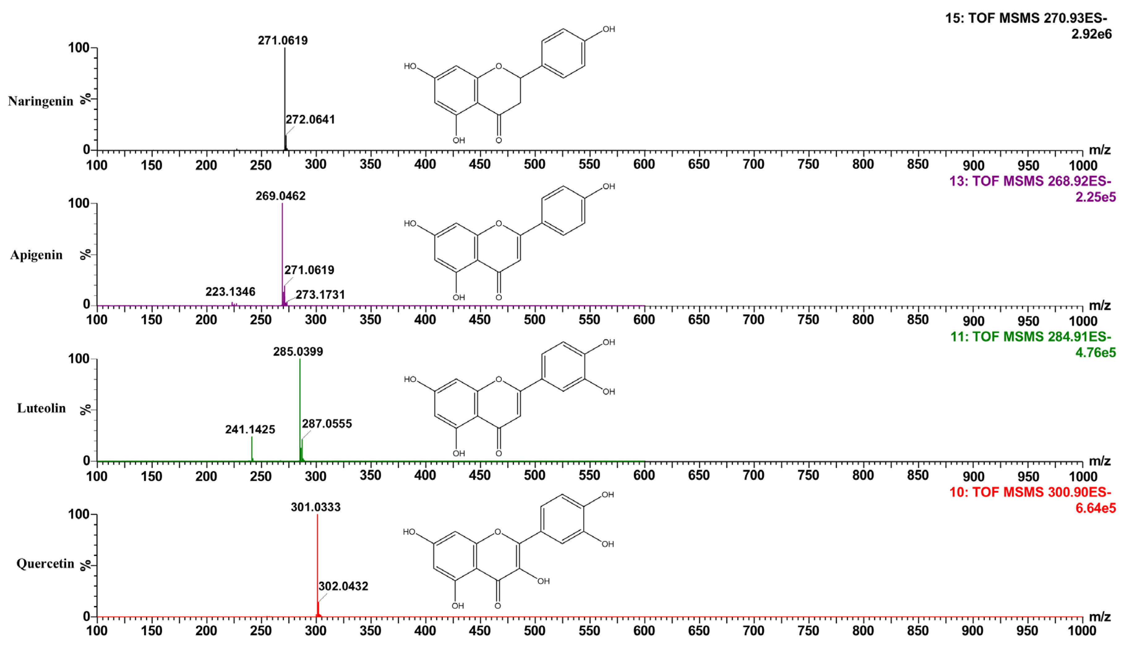
3.1. Identification and Quantification of Flavonoids with Antiproliferative Potential in the Polyphenol Extract of Mexican Oregano L. graveolens

For the analysis and identification of flavonoids, the molecular ion and the fragments obtained in each peak found in the spectra of polyphenol extract (Figure 1) were taken as a reference and compared with the compounds’ data reported for other oregano species as well as in the database Massbank of North America (MoNA). The flavonoid concentration was calculated using the reference standards of each compound. The data obtained can be seen in Table 1.
According to the literature, among the main flavonoids reported with antiproliferative activity in cells of different types of cancer are luteolin, quercetin, naringenin, and apigenin [34,46,47,48,49]. These have been reported to exert this activity by interacting with different cell signaling pathways [50,51]. Some of the chemical characteristics of these phenolic compounds that have been identified with antiproliferative properties are the presence of double bonds between C2 and C3, the presence of an -OH group at the 3′-position of the B-ring of quercetin, unsubstituted allylic hydrogen at C3 and two adjacent -OH groups on the B-ring, the presence of the ortho-dihydroxy group on the B-ring at the 1′ and 2′ positions of trihydroxylfavone derivatives, the presence of amino side chains of the N-heterocyclic ring in the apigenin derivatives, which increases their antiproliferative activity, and the ortho functional group at position 4 and a double bond at C2-C3 [52,53,54,55].
In our oregano extract, we were able to identify the presence of these compounds, finding the highest concentration naringenin at 7758.71 ± 32.15 µg/g extract, followed by quercetin and apigenin at 250.23 ± 18.15 and 122.60 ± 10.65 (EQ) µg/g extract, respectively; the compound with the lowest concentration in our extract was luteolin at 38.24 ± 1.12 µg/g extract. It is important to mention that the total phenol content of this polyphenolic extract is 143.87 ± 1.29 mg of GAE/g, which was already reported in our previous work with this species of oregano. In addition, we were able to identify the presence of other phenolic acids and flavonoids of importance in anticancer research, such as rosmarinic acid, caffeic acid, gallic acid, eriodictyol, and baicalin, among others [29]. The compounds of our polyphenol extract coincide with the data reported for other oregano species, such as Origanum dictamus, Origanum marjorana, and Lippia micromera [56,57,58,59]. In addition, some species of oregano with a similar content of flavonoid compounds have shown antiproliferative activity in breast cancer cells [11,12,60,61]. Identifying and quantifying the presence of these compounds in our extract allowed us to guide our research towards the elucidation of the mechanism of action of the same, in this case, to know if the inhibition of proliferation in breast cancer cells was by activating the apoptosis pathway, one of the most studied and important in the research and development of new drugs for the treatment of this type of disease.

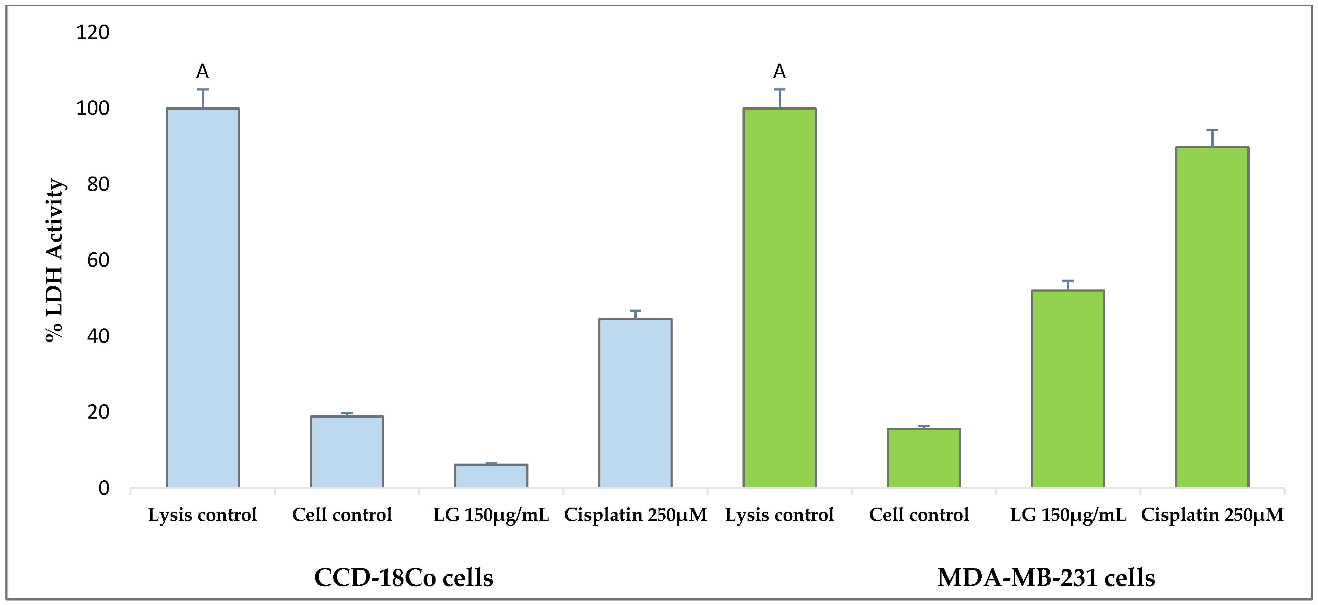
3.2. Non-Cytotoxic Effect of Polyphenol Extract of Mexican Oregano L. graveolens on Normal Human Cells CCD-18Co

The results obtained from the evaluation of the cytotoxicity of the polyphenol extract of L. graveolens are shown in Figure 2. The lactate dehydrogenase enzyme (LDH) activity indicated cell membrane integrity or cell death. In normal conditions, a lysis control can be seen on the left side of Figure 2, with a 100% activity of the LDH enzyme and a cellular control (cells without polyphenol extract). In addition, 250 µM of cisplatin was used as a reference drug to compare the extract’s effect in treating breast cancer. We observed that the polyphenol extract did not show cytotoxicity in this type of normal human cells since the LDH enzyme activity was less than 10% after treatment with the extract for 24 h. Compared to the reference drug, there was a marked difference regarding the behavior of the extract, and cisplatin presented values above 40% of cell death. On the other hand, we also observed a lower percentage of cell death after the treatment with the polyphenol extract of L. graveolens compared to the control cells. This may be because the antioxidant compounds present in the extract can form stable complexes, preventing the catabolic action of the free radicals in the membrane and protecting DNA from oxidative damage, thereby contributing to increased cell viability [31,58].
On the other hand, the behavior observed in the cytotoxicity results for cell line MDA-MB-231 shows a marked selectivity of the extract on these cells, because, at the same conditions and the same concentration of polyphenolic extract of L. graveolens evaluated, a percentage of LDH enzyme activity above 50% was obtained; the effect of the extract on these cancer cells can be seen in Figure 3. In previous work by our research group, we demonstrated that the polyphenol extract of L. graveolens can inhibit the proliferation of breast cancer cells without cytotoxicity in normal mouse cells (NIH3T3) at a concentration of 150 µg/mL of the extract [29], behavior that coincides with the results obtained with these cell lines for the LDH cytotoxicity assay.
According to results, it could be observed that the extract did not cause cytotoxicity in normal human CCD-18Co cells, an important characteristic of this extract to highlight since many of the drugs currently used for the treatment of breast cancer lack cell specificity, causing significant damage in normal cells which leads to the presence of serious side effects for patients, for example, the cisplatin. In addition, another work showed that this oregano species presented cytotoxicity in SK-LU-1 lung cancer cells without causing damage in normal HFF-1 cells [43]. Also, similar behavior was observed in the extract of the Origanum vulgare species, presenting activity against B16-F10 and A375 melanoma cells without cytotoxicity or mutagenicity in normal C2C12 myoblast cells [48].
Following this context, we can find more evidence of the selectivity of flavonoids to inhibit cancer cells than normal cells in the literature. For example, in a study conducted with the flavonoid-rich extract fraction of the plant Rhus verniciflua Stokes on the proliferation and apoptosis of primary embryonic mouse liver cells, normal embryonic liver cells, and a transformed cancer cell line, it was shown to exhibit selective growth inhibition and induction of apoptosis in cancer cells [62]. On the other hand, Sophora flavescens, a plant commonly used in traditional Chinese medicine, was investigated for its potential for the discovery of new active molecules, particularly flavonoid-like structures, which were evaluated in three cancer cell lines (HepG2, A549, and MCF7) and a normal human cell line (LO2 cells), observing remarkable cytotoxicity against the three cancer cell lines and a minimal effect on the normal human cell line [63]. The results of these investigations coincide with what we observed in our experiment. Due to this characteristic, it is important to continue investigating the potential of L. graveolens against cancer cells and to know its antiproliferative mechanism of action, since it could represent a source for developing new treatments against this disease.

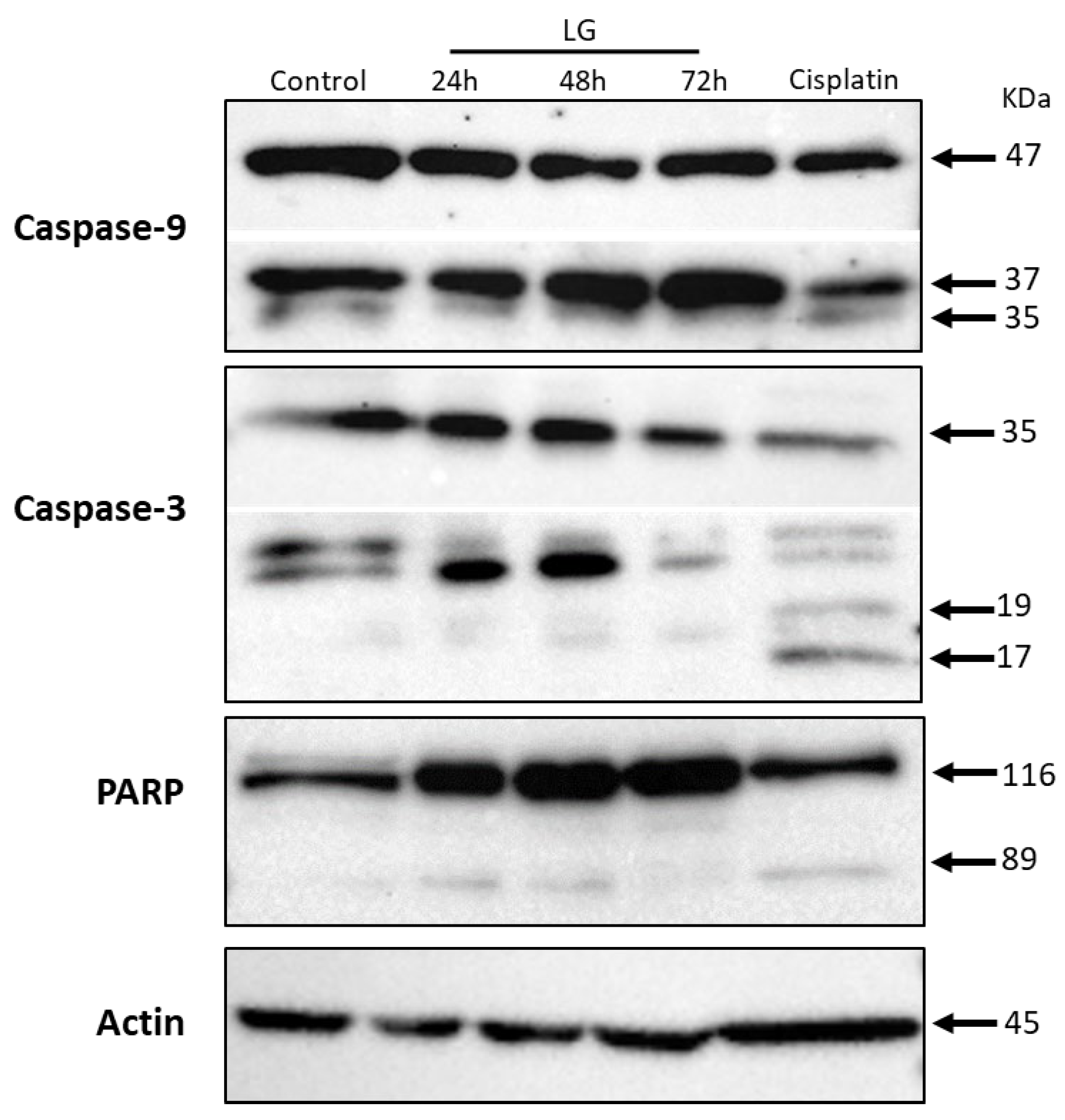
3.3. Polyphenolic Extract of L. graveolens Has Potential to Induce Apoptosis in Breast Cancer Cells

According to the profile of compounds with antiproliferative potential identified in the polyphenol extract of L. graveolens, as well as the information in the literature regarding the induction of apoptosis of these compounds, we decided to evaluate caspase-9 as the initiator molecule of the intrinsic pathway, the processing of caspase-3 (the effector molecule), and PARP cleavage, one of its main substrates. We detected the pro-caspase form of caspase-9 (47 kDa) and the active form of 37 kDa in the treatments with extract at 24, 48, and 72 h. The same processing of caspase-9 was observed in the reference drug cisplatin, which is an intrinsic apoptosis inducer. According to the activation of the initiator caspase-9 in response to the treatment with L. graveolens extract, we decided to continue with the downstream evaluation of the intrinsic pathway, focusing on caspase-3, and we detected fully processed forms (19/17 kDa) in all three treatments, as well as in cisplatin. The pattern of caspase-3 processing suggests activation; so, to further characterize this pathway, we also evaluated PARP processing. As expected, we observed cleaved PARP (89 kDa) in all treatments except the control under basal conditions (Figure 4). Thus, the data obtained suggest that the polyphenol extract of Mexican oregano L. graveolens promotes the intrinsic pathway of apoptosis in MDA-MB-231 breast cancer cells.
The results obtained coincide with those reported by Seo et al. [64] for the treatment with 100 µg of apigenin for 72 h of BT-47 breast cancer cells, which inhibited cell proliferation up to 90%, inducing apoptosis by upregulation of cleaved caspase-3 and PARP cleavage. In combination with an extract (ethyl acetate and water) of Morinda citrifolia, apigenin was also tested on the viability of MDA-MB-231 breast cancer cells. Treatment at 10 µM of the extract and 10 µM of apigenin for 96 h reduced cell viability by around 80% through induction of apoptosis, a similar effect to that shown by our extract alone [48]. On the other hand, luteolin at a concentration of 35 µM combined with lapatinib (a drug used in the treatment of advanced breast cancer) at a concentration of 450 nmol for 72 h inhibited the growth of BT474 cells around 90% by synergistically inducing apoptosis [65]. So, it could be considered a synergistic effect of the polyphenol extract of Mexican oregano L. graveolens with other compounds to promote its antiproliferative effect.
In another study, 10 µM luteolin for 24 h reduced cell proliferation by about 40% in MDA-MB-453 and MCF-7 cells by inducing apoptosis through increased Bax expression, downregulation of Bcl-2, and the promotion of the cleavage of Caspase-3, which coincides with results of our work [47]. In the case of quercetin, it induced apoptosis, through activation of caspase-3 and the mitochondrial-dependent pathway (intrinsic), in MCF-7 and MDA-MB-231 human breast cancer cells, with IC50 values of 80 and 50 μm/mL, respectively [66]. Similarly, Seo et al. [67] proved that 100 μM quercetin for 72 h decreases BT-474 cell proliferation by around 70%. This inhibition was associated with the activation of the extrinsic apoptotic pathway through the upregulation of cleaved caspase-8 and cleaved caspase-3, as well as the induction of PARP cleavage. On the other hand, the ethanolic extract of Thymus vulgaris, rich in naringenin, inhibited the growth of HTB26 breast cancer cells in a dose and time-dependent manner through apoptotic cell death by expression of Bax and caspases 3, 7, 8, and 9 [68].
Therefore, according to these reports, we can attribute the inhibition of antiproliferative activity in MDA-MB-231 breast cancer cells treated with polyphenolic extract of Mexican oregano Lippia graveolens, to the possible induction of apoptosis by the presence of these compounds. However, it is important to mention that the specific effect cannot be attributed to one or two of the compounds present due to possible synergy within the polyphenols of our extract, so it is important to carry out more research in this regard.

4. Conclusions

The polyphenol extract of Mexican oregano L. graveolens was shown to activate the apoptosis pathway in MDA-MB-231 breast cancer cells. It did not exhibit cytotoxicity in normal human fibroblast CCD-18Co cells at the same concentration tested (150 μg/mL). These results demonstrate the antiproliferative mechanism of the extract previously reported in our group of investigations. Also, this is the first study where the possible mechanism of action of polyphenol extracts of this oregano species on breast cancer cells is reported. Therefore, it is important to continue investigating the potential of this species with more precise complementary methods to validate these preliminary findings in order to develop new adjuvant drugs to treat this disease.

Author Contributions

Conceptualization, M.S.C.-M. and J.B.H.; methodology, M.S.C.-M., L.A.C.-A., E.P.G.-G. and I.G.-A.; software, J.B.H., E.P.G.-G. and I.G.-A.; validation, M.S.C.-M., I.G.-A. and J.B.H.; formal analysis, J.B.H.; investigation, M.S.C.-M.; resources, J.B.H., I.G.-A. and L.A.C.-A.; data curation, M.S.C.-M. and L.A.C.-A.; writing—original draft preparation, M.S.C.-M.; writing—review and editing, J.B.H.; visualization, I.G.-A.; supervision, L.A.C.-A. and E.P.G.-G.; project administration, J.B.H.; funding acquisition, J.B.H. and I.G.-A. All authors have read and agreed to the published version of the manuscript.

Funding

This research received no external funding.

Institutional Review Board Statement

Not applicable.

Informed Consent Statement

Not applicable.

Data Availability Statement

The original contributions presented in this study are included in the article. Further inquiries can be directed to the corresponding author.

Acknowledgments

Thanks to Alexis Emus Medina and Rosabel Vélez de la Rocha for their technical support.

Conflicts of Interest

The authors declare no conflicts of interest.

References

  1. Kintzios, S.E. 21–Oregano. In Handbook of Herbs and Spices, 2nd ed.; Peter, K.V., Ed.; Woodhead Publishing: Sawston, UK, 2012; pp. 417–436. [Google Scholar]
  2. Arcila, C.; Loarca, G.; Lecona, S.; González, E. El orégano: Propiedades, composición y actividad biológica de sus componentes. Arch. Latinoam. Nutr. 2004, 54, 100–111. [Google Scholar]
  3. Baser, K. The Turkish Origanum species. J. Med. Aromat. Plants 2002, 109, 109–126. [Google Scholar]
  4. Gutiérrez-Grijalva, E.; Picos-Salas, M.; Leyva-López, N.; Criollo-Mendoza, M.; Vazquez-Olivo, G.; Heredia, J. Flavonoids and Phenolic Acids from Oregano: Occurrence, Biological Activity and Health Benefits. Plants 2018, 7, 2. [Google Scholar] [CrossRef]
  5. Leyva-López, N.; Nair, V.; Bang, W.Y.; Cisneros-Zevallos, L.; Heredia, J.B. Protective role of terpenes and polyphenols from three species of Oregano (Lippia graveolens, Lippia palmeri and Hedeoma patens) on the suppression of lipopolysaccharide-induced inflammation in RAW 264.7 macrophage cells. J. Ethnopharmacol. 2016, 187, 302–312. [Google Scholar] [CrossRef]
  6. Berdowska, I.; Zielinski, B.; Fecka, I.; Kulbacka, J.; Saczko, J.; Gamian, A. Cytotoxic impact of phenolics from Lamiaceae species on human breast cancer cells. Food Chem. 2013, 141, 1313–1321. [Google Scholar] [CrossRef] [PubMed]
  7. Chaouki, W.; Leger, D.Y.; Eljastimi, J.; Beneytout, J.L.; Hmamouchi, M. Antiproliferative effect of extracts from Aristolochia baetica and Origanum compactum on human breast cancer cell line MCF-7. Pharm. Biol. 2010, 48, 269–274. [Google Scholar] [CrossRef]
  8. Kaliora, A.C.; Kogiannou, D.A.; Kefalas, P.; Papassideri, I.S.; Kalogeropoulos, N. Phenolic profiles and antioxidant and anticarcinogenic activities of Greek herbal infusions; Balancing delight and chemoprevention? Food Chem. 2014, 142, 233–241. [Google Scholar] [CrossRef]
  9. Kogiannou, D.A.; Kalogeropoulos, N.; Kefalas, P.; Polissiou, M.G.; Kaliora, A.C. Herbal infusions; their phenolic profile, antioxidant and anti-inflammatory effects in HT29 and PC3 cells. Food Chem. Toxicol. 2013, 61, 152–159. [Google Scholar] [CrossRef]
  10. Zheng, W.; Wang, S.Y. Antioxidant activity and phenolic compounds in selected herbs. J. Agric. Food Chem. 2001, 49, 5165–5170. [Google Scholar] [CrossRef]
  11. El Babili, F.; Bouajila, J.; Souchard, J.P.; Bertrand, C.; Bellvert, F.; Fouraste, I.; Moulis, C.; Valentin, A. Oregano: Chemical analysis and evaluation of its antimalarial, antioxidant, and cytotoxic activities. J. Food Sci. 2011, 76, C512–C518. [Google Scholar] [CrossRef]
  12. Marrelli, M.; Cristaldi, B.; Menichini, F.; Conforti, F. Inhibitory effects of wild dietary plants on lipid peroxidation and on the proliferation of human cancer cells. Food Chem. Toxicol. 2015, 86, 16–24. [Google Scholar] [CrossRef] [PubMed]
  13. Britt, K.L.; Cuzick, J.; Phillips, K.A. Key steps for effective breast cancer prevention. Nat. Rev. Cancer 2020, 20, 417–436. [Google Scholar] [CrossRef] [PubMed]
  14. Park, M.Y.; Kim, Y.; Ha, S.E.; Kim, H.H.; Bhosale, P.B.; Abusaliya, A.; Jeong, S.H.; Kim, G.S. Function and Application of Flavonoids in the Breast Cancer. Int. J. Mol. Sci. 2022, 23, 7732. [Google Scholar] [CrossRef] [PubMed]
  15. Petroni, G.; Buqué, A.; Zitvogel, L.; Kroemer, G.; Galluzzi, L. Immunomodulation by targeted anticancer agents. Cancer Cell 2021, 39, 310–345. [Google Scholar] [CrossRef]
  16. Yersal, O.; Barutca, S. Biological subtypes of breast cancer: Prognostic and therapeutic implications. World J. Clin. Oncol. 2014, 5, 412–424. [Google Scholar] [CrossRef]
  17. Li, Z.H.; Hu, P.H.; Tu, J.H.; Yu, N.S. Luminal B breast cancer: Patterns of recurrence and clinical outcome. Oncotarget 2016, 7, 65024–65033. [Google Scholar] [CrossRef]
  18. Berger, E.R.; Park, T.; Saridakis, A.; Golshan, M.; Greenup, R.A.; Ahuja, N. Immunotherapy Treatment for Triple Negative Breast Cancer. Pharmaceuticals 2021, 14, 763. [Google Scholar] [CrossRef]
  19. Van Swearingen, A.E.D.; Sambade, M.J.; Siegel, M.B.; Sud, S.; McNeill, R.S.; Bevill, S.M.; Chen, X.; Bash, R.E.; Mounsey, L.; Golitz, B.T.; et al. Combined kinase inhibitors of MEK1/2 and either PI3K or PDGFR are efficacious in intracranial triple-negative breast cancer. Neuro-Oncology 2017, 19, 1481–1493. [Google Scholar] [CrossRef]
  20. Telang, N.T. Natural products as drug candidates for breast cancer (Review). Oncol. Lett. 2023, 26, 349. [Google Scholar] [CrossRef]
  21. Fasoulakis, Z.; Koutras, A.; Syllaios, A.; Schizas, D.; Garmpis, N.; Diakosavvas, M.; Angelou, K.; Tsatsaris, G.; Pagkalos, A.; Ntounis, T.; et al. Breast Cancer Apoptosis and the Therapeutic Role of Luteolin. Chirurgia 2021, 116, 170–177. [Google Scholar] [CrossRef]
  22. Liu, C.; Zhang, K.; Shen, H.; Yao, X.; Sun, Q.; Chen, G. Necroptosis: A novel manner of cell death, associated with stroke (Review). Int. J. Mol. Med. 2018, 41, 624–630. [Google Scholar] [CrossRef] [PubMed]
  23. Zhang, R.; Kang, R.; Tang, D. The STING1 network regulates autophagy and cell death. Signal Transduct. Target. Ther. 2021, 6, 208. [Google Scholar] [CrossRef]
  24. Parton, M.; Dowsett, M.; Smith, I. Studies of apoptosis in breast cancer. BMJ 2001, 322, 1528–1532. [Google Scholar] [CrossRef] [PubMed]
  25. Mohammad, R.M.; Muqbil, I.; Lowe, L.; Yedjou, C.; Hsu, H.Y.; Lin, L.T.; Siegelin, M.D.; Fimognari, C.; Kumar, N.B.; Dou, Q.P.; et al. Broad targeting of resistance to apoptosis in cancer. Semin. Cancer Biol. 2015, 35, S78–S103. [Google Scholar] [CrossRef]
  26. Chaudhry, G.-e.-S.; Md Akim, A.; Sung, Y.Y.; Sifzizul, T.M.T. Cancer and apoptosis: The apoptotic activity of plant and marine natural products and their potential as targeted cancer therapeutics. Front. Pharmacol. 2022, 13, 842376. [Google Scholar] [CrossRef] [PubMed]
  27. Yuan, L.; Cai, Y.; Zhang, L.; Liu, S.; Li, P.; Li, X. Promoting Apoptosis, a Promising Way to Treat Breast Cancer with Natural Products: A Comprehensive Review. Front. Pharmacol. 2022, 12, 801662. [Google Scholar] [CrossRef]
  28. Sankari, S.L.; Masthan, K.M.; Babu, N.A.; Bhattacharjee, T.; Elumalai, M. Apoptosis in cancer–An update. Asian Pac. J. Cancer Prev. APJCP 2012, 13, 4873–4878. [Google Scholar] [CrossRef]
  29. Criollo-Mendoza, M.S.; Ramos-Payán, R.; Contreras-Angulo, L.A.; Gutiérrez-Grijalva, E.P.; León-Félix, J.; Villicaña, C.; Angulo-Escalante, M.A.; Heredia, J.B. Cytotoxic Activity of Polyphenol Extracts from Three Oregano Species: Hedeoma patens, Lippia graveolens and Lippia palmeri, and Antiproliferative Potential of Lippia graveolens against Two Types of Breast Cancer Cell Lines (MDA-MB-231 and MCF-7). Molecules 2022, 27, 5240. [Google Scholar] [CrossRef]
  30. Ravishankar, D.; Rajora, A.K.; Greco, F.; Osborn, H.M. Flavonoids as prospective compounds for anti-cancer therapy. Int. J. Biochem. Cell Biol. 2013, 45, 2821–2831. [Google Scholar] [CrossRef]
  31. Wang, Y.; Wang, H.; Xu, S. Natural Bioactive Compounds Promote Cell Apoptosis in Gastric Cancer Treatment: Evidence from Network Pharmacological Study and Experimental Analysis. J. Chem. 2023, 2023, 6316589. [Google Scholar] [CrossRef]
  32. Kaushik, N.; Yang, H.; Jeong, S.; Kaushik, N.K.; Bhartiya, P.; Nhat Nguyen, L.; Choi, E.H.; Kim, J.H. Antiproliferative Activity of Pyracantha and Paullinia Plant Extracts on Aggressive Breast and Hepatocellular Carcinoma Cells. Appl. Sci. 2020, 10, 7543. [Google Scholar] [CrossRef]
  33. Cavalcante, F.P.; Millen, E.C.; Zerwes, F.P.; Novita, G.G. Progress in Local Treatment of Breast Cancer: A Narrative Review. Rev. Bras. Ginecol. E Obstet. 2020, 42, 356–364. [Google Scholar] [CrossRef] [PubMed]
  34. Kampa, M.; Nifli, A.P.; Notas, G.; Castanas, E. Polyphenols and cancer cell growth. Rev. Physiol. Biochem. Pharmacol. 2007, 159, 79–113. [Google Scholar] [CrossRef] [PubMed]
  35. Verma, S.; Singh, A.; Mishra, A. Gallic acid: Molecular rival of cancer. Environ. Toxicol. Pharmacol. 2013, 35, 473–485. [Google Scholar] [CrossRef]
  36. Elmore, S. Apoptosis: A review of programmed cell death. Toxicol. Pathol. 2007, 35, 495–516. [Google Scholar] [CrossRef]
  37. Suraweera, C.D.; Hinds, M.G.; Kvansakul, M. Poxviral Strategies to Overcome Host Cell Apoptosis. Pathogens 2020, 10, 6. [Google Scholar] [CrossRef]
  38. Lee, D.; Yu, J.S.; Ryoo, R.; Kim, J.C.; Jang, T.S.; Kang, K.S.; Kim, K.H. Pulveraven A from the fruiting bodies of Pulveroboletus ravenelii induces apoptosis in breast cancer cell via extrinsic apoptotic signaling pathway. J. Antibiot. 2021, 74, 752–757. [Google Scholar] [CrossRef]
  39. Kushwaha, P.P.; Singh, A.K.; Prajapati, K.S.; Shuaib, M.; Fayez, S.; Bringmann, G.; Kumar, S. Induction of apoptosis in breast cancer cells by naphthylisoquinoline alkaloids. Toxicol. Appl. Pharmacol. 2020, 409, 115297. [Google Scholar] [CrossRef]
  40. Sindhu, R.K.; Verma, R.; Salgotra, T.; Rahman, M.H.; Shah, M.; Akter, R.; Murad, W.; Mubin, S.; Bibi, P.; Qusti, S.; et al. Impacting the Remedial Potential of Nano Delivery-Based Flavonoids for Breast Cancer Treatment. Molecules 2021, 26, 5163. [Google Scholar] [CrossRef]
  41. Georgieva, A.; Sulikovska, I.; Toshkova-Yotova, T.; Djeliova, V.; Amiri, S.; Tsonevski, N.; Petkova-Kirova, P.; Tasheva, K. Antitumor Activity of Whole-Plant Extracts from In Vitro Cultured and Wild-Growing Clinopodium vulgare Plants on a Panel of Human Tumor Cell Lines. Appl. Sci. 2025, 15, 925. [Google Scholar] [CrossRef]
  42. Khojasteh Poor, F.; Keivan, M.; Ramazii, M.; Ghaedrahmati, F.; Anbiyaiee, A.; Panahandeh, S.; Khoshnam, S.E.; Farzaneh, M. Mini review: The FDA-approved prescription drugs that target the MAPK signaling pathway in women with breast cancer. Breast Dis. 2021, 40, 51–62. [Google Scholar] [CrossRef] [PubMed]
  43. Li, H.; Prever, L.; Hirsch, E.; Gulluni, F. Targeting PI3K/AKT/mTOR Signaling Pathway in Breast Cancer. Cancers 2021, 13, 3517. [Google Scholar] [CrossRef] [PubMed]
  44. Gutierrez-Grijalva, E.P.; Angulo-Escalante, M.A.; Leon-Felix, J.; Heredia, J.B. Effect of In Vitro Digestion on the Total Antioxidant Capacity and Phenolic Content of 3 Species of Oregano (Hedeoma patens, Lippia graveolens, Lippia palmeri). J. Food Sci. 2017, 82, 2832–2839. [Google Scholar] [CrossRef] [PubMed]
  45. Martinez-Castillo, M.; Bonilla-Moreno, R.; Aleman-Lazarini, L.; Meraz-Rios, M.A.; Orozco, L.; Cedillo-Barron, L.; Cordova, E.J.; Villegas-Sepulveda, N. A Subpopulation of the K562 Cells Are Killed by Curcumin Treatment after G2/M Arrest and Mitotic Catastrophe. PLoS ONE 2016, 11, e0165971. [Google Scholar] [CrossRef]
  46. Frias-Zepeda, M.E.; Ibarra-Berumen, J.; Ordaz-Pichardo, C.; Rosales-Castro, M. Cytotoxic activity of ethanolic extracts of Lippia graveolens HBK leaves and stem against lung cancer cell line SK-LU-1. Bol. Latinoam. Caribe Plantas Med. Aromat. 2022, 21, 646–653. [Google Scholar] [CrossRef]
  47. Gao, G.; Ge, R.; Li, Y.; Liu, S. Luteolin exhibits anti-breast cancer property through up-regulating miR-203. Artif. Cells Nanomed. Biotechnol. 2019, 47, 3265–3271. [Google Scholar] [CrossRef]
  48. Huang, C.; Wei, Y.-X.; Shen, M.-C.; Tu, Y.-H.; Wang, C.-C.; Huang, H.-C. Chrysin, Abundant in Morinda citrifolia Fruit Water–EtOAc Extracts, Combined with Apigenin Synergistically Induced Apoptosis and Inhibited Migration in Human Breast and Liver Cancer Cells. J. Agric. Food Chem. 2016, 64, 4235–4245. [Google Scholar] [CrossRef]
  49. Maugeri, A.; Calderaro, A.; Patanè, G.T.; Navarra, M.; Barreca, D.; Cirmi, S.; Felice, M.R. Targets Involved in the Anti-Cancer Activity of Quercetin in Breast, Colorectal and Liver Neoplasms. Int. J. Mol. Sci. 2023, 24, 2952. [Google Scholar] [CrossRef]
  50. Gird, C.E.; Ostea, T.; Itran, V. Evaluation of cytotoxic activity and anticancer potential of indigenous Rosemary (Rosmarinus officinalis L.) and Oregano (Origanum vulgare L.) dry extracts on MG-63 bone osteosarcoma human cell line. Rom. J. Morphol. Embryol. 2021, 62, 525–535. [Google Scholar] [CrossRef]
  51. Nanni, V.; Di Marco, G.; Sacchetti, G.; Canini, A.; Gismondi, A. Oregano Phytocomplex Induces Programmed Cell Death in Melanoma Lines via Mitochondria and DNA Damage. Foods 2020, 9, 1486. [Google Scholar] [CrossRef]
  52. Criollo-Mendoza, S.M.; Heredia, B.J.; Vazquez-Olivo, G.; Avilés-Gaxiola, S.; Gutiérrez-Grijalva, P.E.; Garcia-Carrasco, M. Antiproliferative Activity and Mechanisms of Action of Plant-derived Flavonoids on Breast Cancer. Curr. Top. Med. Chem. 2023, 23, 1937–1951. [Google Scholar] [CrossRef] [PubMed]
  53. Grigalius, I.; Petrikaite, V. Relationship between Antioxidant and Anticancer Activity of Trihydroxyflavones. Molecules 2017, 22, 2169. [Google Scholar] [CrossRef] [PubMed]
  54. Li, X.; Zhang, C.; Guo, S.; Rajaram, P.; Lee, M.; Chen, G.; Fong, R.; Gonzalez, A.; Zhang, Q.; Zheng, S.; et al. Structure-activity relationship and pharmacokinetic studies of 3-O-substitutedflavonols as anti-prostate cancer agents. Eur. J. Med. Chem. 2018, 157, 978–993. [Google Scholar] [CrossRef] [PubMed]
  55. Liu, R.; Zhang, H.; Yuan, M.; Zhou, J.; Tu, Q.; Liu, J.-J.; Wang, J. Synthesis and Biological Evaluation of Apigenin Derivatives as Antibacterial and Antiproliferative Agents. Molecules 2013, 18, 11496–11511. [Google Scholar] [CrossRef]
  56. Hennebelle, T.; Sahpaz, S.; Gressier, B.; Joseph, H.; Bailleul, F. Antioxidant and neurosedative properties of polyphenols and iridoids from Lippia alba. Phytother. Res. PTR 2008, 22, 256–258. [Google Scholar] [CrossRef]
  57. Proestos, C.; Komaitis, M. Analysis of Naturally Occurring Phenolic Compounds in Aromatic Plants by RP-HPLC Coupled to Diode Array Detector (DAD) and GC-MS after Silylation. Foods 2013, 2, 90–99. [Google Scholar] [CrossRef]
  58. Taamalli, A.; Arráez-Román, D.; Abaza, L.; Iswaldi, I.; Fernández-Gutiérrez, A.; Zarrouk, M.; Segura-Carretero, A. LC-MS-based metabolite profiling of methanolic extracts from the medicinal and aromatic species Mentha pulegium and Origanum majorana. Phytochem. Anal. PCA 2015, 26, 320–330. [Google Scholar] [CrossRef]
  59. Zgorka, G.; Glowniak, K. Variation of free phenolic acids in medicinal plants belonging to the Lamiaceae family. J. Pharm. Biomed. Anal. 2001, 26, 79–87. [Google Scholar] [CrossRef]
  60. Al-Kalaldeh, J.Z.; Abu-Dahab, R.; Afifi, F.U. Volatile oil composition and antiproliferative activity of Laurus nobilis, Origanum syriacum, Origanum vulgare, and Salvia triloba against human breast adenocarcinoma cells. Nutr. Res. 2010, 30, 271–278. [Google Scholar] [CrossRef]
  61. Savini, I.; Arnone, R.; Catani, M.V.; Avigliano, L. Origanum Vulgare Induces Apoptosis in Human Colon Cancer Caco2 Cells. Nutr. Cancer 2009, 61, 381–389. [Google Scholar] [CrossRef]
  62. Son, Y.-O.; Lee, K.-Y.; Lee, J.-C.; Jang, H.-S.; Kim, J.-G.; Jeon, Y.-M.; Jang, Y.-S. Selective antiproliferative and apoptotic effects of flavonoids purified from Rhus verniciflua Stokes on normal versus transformed hepatic cell lines. Toxicol. Lett. 2005, 155, 115–125. [Google Scholar] [CrossRef] [PubMed]
  63. Li, J.J.; Lin, Y.; He, L.; Ou, R.X.; Chen, T.; Zhang, X.; Li, Q.R.; Zeng, Z.; Long, Q.D. Two New Isoprenoid Flavonoids from Sophora flavescens with Antioxidant and Cytotoxic Activities. Molecules 2021, 26, 7228. [Google Scholar] [CrossRef] [PubMed]
  64. Seo, H.S.; Jo, J.K.; Ku, J.M.; Choi, H.S.; Choi, Y.K.; Woo, J.K.; Kim, H.I.; Kang, S.Y.; Lee, K.M.; Nam, K.W.; et al. Induction of caspase-dependent extrinsic apoptosis by apigenin through inhibition of signal transducer and activator of transcription 3 (STAT3) signalling in HER2-overexpressing BT-474 breast cancer cells. Biosci. Rep. 2015, 35, e00276. [Google Scholar] [CrossRef] [PubMed]
  65. Zhang, L.; Liu, Q.; Huang, L.; Yang, F.; Liu, A.; Zhang, J. Combination of lapatinib and luteolin enhances the therapeutic efficacy of lapatinib on human breast cancer through the FOXO3a/NQO1 pathway. Biochem. Biophys. Res. Commun. 2020, 531, 364–371. [Google Scholar] [CrossRef]
  66. Tao, S.F.; He, H.F.; Chen, Q. Quercetin inhibits proliferation and invasion acts by up-regulating miR-146a in human breast cancer cells. Mol. Cell. Biochem. 2015, 402, 93–100. [Google Scholar] [CrossRef]
  67. Seo, H.S.; Ku, J.M.; Choi, H.S.; Choi, Y.K.; Woo, J.K.; Kim, M.; Kim, I.; Na, C.H.; Hur, H.; Jang, B.H.; et al. Quercetin induces caspase-dependent extrinsic apoptosis through inhibition of signal transducer and activator of transcription 3 signaling in HER2-overexpressing BT-474 breast cancer cells. Oncol. Rep. 2016, 36, 31–42. [Google Scholar] [CrossRef]
  68. Abaza, M.S.I.; Orabi, K.Y.; Al-Quattan, E.; Al-Attiyah, R.a.J. Growth inhibitory and chemo-sensitization effects of naringenin, a natural flavanone purified from Thymus vulgaris, on human breast and colorectal cancer. Cancer Cell Int. 2015, 15, 46. [Google Scholar] [CrossRef]
Figure 1. Molecular ions and fragments obtained for each flavonoid found in the spectra of the polyphenol extract of Mexican oregano Lippia graveolens.
Applsci 15 06393 g001
Figure 2. Cytotoxicity of polyphenol extract of Mexican oregano Lippia graveolens (LG) on normal CCD-18Co cells and MDA-MB-231 cancer cells, expressed as a percentage of lactate dehydrogenase activity (LDH). Results are expressed as means (n = 3) ± standard deviation (bars). The ANOVA was evaluated with the Dunnett test with lysis control for each cell type. Values without letters are statistically different (p ≤ 0.05).
Applsci 15 06393 g002
Figure 3. MDA-MB-231 cells exposed to different treatments during 24 h incubation. Cell control represents cells without treatment. LG: polyphenol extract of Mexican oregano Lippia graveolens. Images obtained under the microscope (40×).
Applsci 15 06393 g003
Figure 4. Polyphenol extract of Mexican oregano Lippia graveolens (LG) induces apoptotic cell death in MDA-MB-231 breast cancer cells. Processed caspases-9 and -3 and PARP were detected by western blot analysis. Actin was used as a loading control.
Applsci 15 06393 g004
Table 1. Flavonoids with antiproliferative potential identified in the polyphenolic extract of Mexican oregano L. graveolens.
MS[M-H]-Fragmentation PatternFlavonoidConcentration (µg/g)
302.04301.03302.03, 303.04Quercetin250.23 ± 18.15
286.04285.04151.00, 286.04Luteolin38.24 ± 1.12
272.06271.06151.00, 177.02, 269.04Naringenin7758.71 ± 32.15
270.05269.04151.00, 270.05Apigenin122.60 ± 10.65 (EQ)
The results represent the means ± standard deviation (n = 3). (EQ) = quercetin equivalents.
Disclaimer/Publisher’s Note: The statements, opinions and data contained in all publications are solely those of the individual author(s) and contributor(s) and not of MDPI and/or the editor(s). MDPI and/or the editor(s) disclaim responsibility for any injury to people or property resulting from any ideas, methods, instructions or products referred to in the content.

