Article

The Importance of Gram-Negative Rods in Chronic Rhinosinusitis

by
Magdalena Ratajczak
1,*,
Tatiana Fijalkowska-Ratajczak
2,
Dorota Kaminska
1,
Małgorzata Leszczyńska
3 and
Jolanta Dlugaszewska
1,*
1
Chair and Department of Genetics and Pharmaceutical Microbiology, Poznan University of Medical Sciences, Rokietnicka 3, 60-806 Poznan, Poland
2
Municipal Hospital F. Raszei, Mickiewicza 2, 60-834 Poznan, Poland
3
Department of Otolaryngology and Laryngological Oncology, Poznan University of Medical Sciences, Przybyszewskiego 49, 60-355 Poznan, Poland
*
Authors to whom correspondence should be addressed.
Appl. Sci. 2025, 15(11), 6108; https://doi.org/10.3390/app15116108
Submission received: 1 April 2025 / Revised: 18 May 2025 / Accepted: 27 May 2025 / Published: 29 May 2025
(This article belongs to the Special Issue Advances in Microbiota in Human Health and Diseases)

Abstract

:
Background: Chronic rhinosinusitis (CRS) affects 5.5–28% of the population and is primarily an inflammatory disease, with microbiota potentially playing a key role. Understanding microbial pathogens and resistance patterns is crucial for effective management. This study aimed to evaluate the incidence of Gram-negative rods in CRS in adults as a part of a prospective microbiological study. Methods: Over one year, paranasal sinus mucosa samples from CRS patients and nasal concha samples from controls were analyzed. Cultivable bacterial flora was assessed using culture-based methods. Biofilm formation was evaluated via a microtiter-plate assay, and antibiotic susceptibility was tested using the disk diffusion method. Results: Tissue samples from 74 CRS patients and 47 controls yielded 198 bacterial strains. Gram-positive cocci dominated, while Gram-negative rods accounted for 17.6%, with Escherichia coli, Klebsiella oxytoca, and Citrobacter spp. being most common. All Gram-negative rods formed biofilms in vitro. They were susceptible to cefotaxime, aztreonam, ciprofloxacin, and meropenem but showed varying sensitivity to ampicillin (20–67%), tigecycline (40–57%), and amoxicillin/clavulanic acid (73–83%). Conclusions: The result of this study underlines that treatment of CRS should be based on the result of drug susceptibility testing of the isolated microorganism.

1. Introduction

Chronic rhinosinusitis (CRS) is a significant health issue affecting 5.5% to 28% of the general population, with substantial geographical variation, and is one of the most common reasons for visiting a primary care physician [1,2,3]. According to the European Position Paper on Rhinosinusitis and Nasal Polyps 2020 (EPOS 2020) guidelines, CRS in adults is an inflammation of the nose and the paranasal sinuses mucosa characterized by two or more symptoms, one of which should be nasal blockage, obstruction, congestion, or nasal discharge, accompanied or not by facial pain/pressure and reduction or loss of smell, lasting for ≥12 weeks [1]. This disease makes everyday functioning difficult and significantly reduces the health-related quality of the patient’s life, mainly due to difficult breathing and sleep-induced issues. Moreover, CRS produces a large economic burden on healthcare systems, indirect costs, and significant patient productivity losses [4,5,6,7].
CRS has traditionally been classified into two groups based on the presence or absence of nasal polyps: CRS with nasal polyps (CRSwNP) and CRS without nasal polyps (CRSsNP). However, these phenotypic classifications fail to fully reflect the diverse spectrum of CRS variants. The EPOS 2020 offers a more detailed classification, dividing CRS into primary and secondary types, with further subcategories based on anatomic distribution, as illustrated in Figure 1 [1].
EPOS 2020 classifies primary CRS subtypes based on anatomic distribution, distinguishing between localized (unilateral) and diffuse (bilateral) inflammation and/or polyps. The disease is further categorized by endotype dominance, differentiating between type 2 and non-type 2 inflammatory patterns. However, the non-type 2 classification does not fully capture the heterogeneity of CRS. While CRSwNP was traditionally associated with type 2 inflammation, recent studies suggest that inflammatory profiles may vary based on geographical distribution. Localized CRS is additionally classified by clinical phenotype, including allergic fungal rhinosinusitis (AFRS) and isolated sinusitis. Diffuse CRS, on the other hand, is primarily subdivided into eosinophilic CRS (eCRS) and non-eCRS, with eosinophilia defined as an eosinophil count of 10/hpf or higher [1].
CRS is a complex inflammatory disease whose etiology and exact pathophysiological mechanisms are not fully known. In recent years, many studies suggest that CRS is a group of disorders with multifactorial etiology involving sinonasal epithelium and immune components influenced by a host (e.g., allergy, asthma, upper respiratory tract infections, immunodeficiency) and environmental (smoking, air pollution) factors that result in inflammation of the sinonasal mucosa. In addition, the role of the sinonasal microbiome, the occurrence of bacterial biofilms on the surface of mucous membranes, and the role of staphylococcal superantigens in CRS pathogenesis have also been highlighted [8,9,10,11,12].
Numerous studies in the last decades revealed that the sinuses in the healthy state are not sterile, and the presence of bacteria in paranasal sinuses is well documented [13,14,15,16,17].
Differences in the microbiome and its impact on the frequency of upper respiratory tract infections have already been observed in children. Colonization in the early months of life has been shown to have a long-term effect on respiratory diseases. The presence of bacteria from the genera Streptococcus, Moraxella, and Haemophilus, among others, increases the risk of respiratory diseases, and the dominance of Streptococcus in the microbiota indicates an increased risk of asthma [18,19].
A “healthy” microbiota plays an important role in maintaining microbial balance and modulating the immune response of a healthy host. It also prevents the excessive multiplication of potentially pathogenic microorganisms. In healthy individuals, commonly identified upper respiratory tract bacteria genera include Staphylococcus, Corynebacterium, Peptoniphilus, and Cutibacterium [20,21].
Interestingly, the total bacterial load in healthy and diseased sinuses seems to be very similar, and many opportunistic pathogens are present in low numbers in healthy sinuses. Studies have shown that a loss of microbial diversity characterizes the CRS microbiome. The development under favorable conditions of pathogenic bacteria and a collapse of the commensal population disrupt the microbiota, which may contribute to the exacerbation of chronic inflammatory disease.
Recent evaluations of microbes in CRS revealed a significant degree of interpersonal variation of the sinonasal microbiome and reduced bacterial diversity compared with healthy subjects. These studies noted that CRS bacterial microbiota can exist in at least four distinct taxonomic states dominated by Streptococcaceae, Pseudomonadaceae, Corynebacteriaceae, or Staphylococcaceae. Although bacteria such as S. aureus and S. epidermidis predominated in CRS, recovery of gram-negative rods, including Proteus spp., Enterobacter spp., Escherichia coli, and Klebsiella pneumoniae were also reported [22,23,24,25]. Since Gram-negative rods are rarely recovered from healthy individuals, the isolation of these bacteria from symptomatic patients suggests their pathogenic role in CRS. However, the differences in the isolation rates of Gram-negative rods have been reported [26,27].
An important role in the pathogenesis and maintenance of the recalcitrant inflammation for CRS may be played by biofilm-forming bacteria. Biofilm is a possible method by which bacteria can evade normal host defense mechanisms and antibiotic treatment [28]. In fact, both appear to be risk factors for therapy failures and CRS recurrence.
Therefore, recognizing the microbiology of CRS is important when selecting the antimicrobial therapy for this multifactorial disease. This study aimed to evaluate the incidence of Gram-negative rods in chronic rhinosinusitis in adults, in vitro ability to biofilm formation, and resistance to antibiotics as a part of a prospective microbiological study.

2. Materials and Methods

2.1. Patients and Specimen Collection

The study was conducted on patients presenting to the Department of Otolaryngology at Franciszek Raszeja Municipal Hospital in Poznan, Poland. The study group included adult patients with CRS diagnosed based on their medical history and physical examination, according to the criteria established by the European Position Paper on Rhinosinusitis and Nasal Polyps group classified for endoscopic sinus surgery (ESS). The protocol excluded immunocompromised patients, patients with ciliary dyskinesia, acute upper respiratory tract infection, and patients who had taken antibiotics four weeks before surgery. The control group consisted of patients without CRS and polyps who underwent nasal septoplasty and rhinoplasty (NSR). The study included one questionnaire (information on health and factors that may be important in the pathology of CRS). Written informed consent was obtained from all participating individuals after explaining the possible consequences of the study.
The paranasal sinus mucosa samples were collected from patients with CRS during ESS. The anterior ethmoid sinus was chosen as the sampling site for the biopsy specimens as it is part of the ostiomeatal complex. Control samples consisted of interior turbinate mucosa, where the drainage of the maxillary and anterior ethmoid sinuses and the nasal frontal duct is located. The control samples were obtained during NSR procedures [29].
The research was approved by the Bioethics Committee of the Karol Marcinkowski University of Medical Sciences in Poznan, protocol number 150/18.

2.2. Isolation and Identification of Sinonasal Microbiota

The collected specimens were placed into a glass tissue homogenizer and homogenized with 1 mL of physiological saline. The specimens were then subjected to inoculation on/into appropriate microbiological media, including Haemophilus Chocolate agar (OXOID, Hampshire, UK), Sabouraud agar (OXOID, Hampshire, UK), Columbia agar with 5% sheep blood (OXOID, Hampshire, UK), Brain Heart Infusion Broth (OXOID, UK), Schaedler with vitamin K and sheep blood (OXOID, Hampshire, UK), Schaedler broth (OXOID, Hampshire, UK). Media were incubated under aerobic, anaerobic, and capnophilic conditions at 36 ± 1 °C for 48–72 h. Fungal cultures were incubated for two weeks. Microorganisms were identified to species or genus if the growth of a single colony type was obtained or if one or more colony types met the criterion of a dominant factor in a mixed culture. Initial identification was performed based on colony morphology, Gram staining, and conventional microbiological methods, followed by confirmation using the Vitek® 2 Compact automated identification system (bioMérieux, Durham, NC, USA) [30].

2.3. Antimicrobial Susceptibility Testing

The susceptibility of isolated bacteria to antimicrobial drugs was assessed according to the recommendations of the European Committee on Susceptibility Testing (EUCAST) using the disk diffusion method [31]. The isolated bacteria were subjected to susceptibility testing against the following antimicrobials: meropenem, trimethoprim-sulfamethoxazole, tigecycline, ciprofloxacin, gentamicin, aztreonam, ceftazidime, cefotaxime, cefuroxime, amoxicillin/clavulanic acid, piperacillin, and ampicillin.
The ability of gram-negative rods to produce extended-spectrum β-lactamases (ESβL) was determined using the double-disc synergy test (DDST).
The test results were interpreted according to the EUCAST guidelines [31].

2.4. Biofilm Formation

To determine in vitro biofilm formation, the crystal violet assay was performed as described previously [32,33]. Briefly, isolates grown for 24 h on tryptic soy agar (TSA, OXOID, Hampshire, UK) were suspended in a brain heart infusion broth (BHI, OXOID, Hampshire, UK), adjusted to a turbidity of 0.5 MacFarland and diluted 1:100 in BHI. After dilution, 200 µL of each sample was seeded into 96-well flat-bottom microtiter plates and incubated at 36 ± 1 °C for 20 h. After incubation, the culture medium was discarded from the microtiter plate, and the wells were washed three times with deionized water, then stained with 0.1% crystal violet for 15 min, rinsed with water, and air-dried overnight. The crystal violet from the stained biofilm was eluted in 250 µL of 95% ethanol. The optical density (OD) of stained adherent biofilm was measured using an Infinite M200 (Tecan, Grődig, Austria) plate reader at a wavelength of 590 nm. Wells containing uninoculated BHI media served as a negative control. Tests were repeated three times.
The biofilm formation was interpreted according to the previously described criteria [32]:
ODc = mean OD of negative control + 3 × standard deviation (SD) of negative control;
  • Non-biofilm producer (N): OD ≤ ODc;
  • Weak-biofilm producer (W): ODc < OD ≤ 2 × ODc;
  • Moderate-biofilm producer (M): 2 × ODc < OD ≤ 4 × ODc;
  • Strong-biofilm producer (S): OD > 4 × ODc.

2.5. Statistical Analyses

The statistical analyses were conducted using Python 3.11 with the NumPy library for numerical operations and the SciPy.stats module for inferential testing. Fisher’s Exact Test, Pearson’s Chi-Square Test with Yates’ continuity correction, and the Kruskal–Wallis H Test were applied as implemented in the SciPy.stats to ensure reproducibility of the analysis.
Fisher’s Exact Test was used to analyze the prevalence of Gram-negative rods, Pearson’s Chi-Square Test with Yates’ continuity correction for analysis of microbiota and antimicrobial susceptibility, and the Kruskal–Wallis H Test was used to analyze the ability to form biofilm.

3. Results

3.1. Demographic and Clinical Characteristics of Patients

One hundred and twenty-one patients were included in the study comprising 74 patients with CRS (52 with primary CRS and 22 with secondary CRS) and 47 non-CRS patients (control group). Table 1 summarizes the patient characteristics based on the survey results.
Sixteen of the patients included in the study had used antibiotics within 1 to 6 months prior to sample collection (antibiotic use within one month prior to sampling was an exclusion criterion). The most common indications for antibiotic therapy included: sinusitis, otitis, bacterial pharyngitis, and urinary tract infection. The antibiotics most frequently used were: amoxicillin (n = 4), amoxicillin with clavulanic acid (n = 4), cefuroxime (n = 3), penicillin (n = 2), azithromycin (n = 1), clarithromycin (n = 1), and norfloxacin (n = 1).

3.2. Bacterial Microbiota

Paranasal sinus mucosa tissue samples and nasal concha mucosa tissue samples were collected from patients with CRS (study group) and patients who underwent nasal septoplasty and rhinoplasty (control group), respectively. A total of 125 bacterial species were isolated from patients with CRS (90 from patients with primary CRS, 35 with secondary CRS) and 73 from patients of the control group (Table 2). The distribution of isolated microorganisms in each analyzed group was diverse. More than one dominant microorganism was identified in samples from 50% of patients with primary CRS and 45.5% of those with secondary CRS. In the control group, multiple dominant microorganisms were found in 17.0% of patients. Among the microorganisms isolated from patients with CRS, Gram-positive cocci of the species S. epidermidis and S. aureus predominated. In the control group, mixed microbiota typical of the nasal flora were isolated in most patients (59.6%).
Microbiota dominated by Gram-negative rods were isolated from 27 patients (control group: n = 5, primary CRS: n = 15, secondary CRS: n = 7). Based on the conducted survey, none of these patients had hyperacidity, genetic diseases or syndromes, diabetes, or a history of facial trauma. Compared to the control group, patients with primary and secondary CRS were more likely to have asthma, peptic ulcer disease, hypertension, a history of steroid use, sinus irrigation, and antibiotic use (at least four weeks prior to patients who had used antibiotic agents within four weeks prior to sample collection were excluded from the study).
Differences in the prevalence of Gram-negative rods were observed in the studied groups. In patients with CRS, these bacteria were isolated at a rate of approximately 17.6% (across both groups), while in the control group, Gram-negative rods accounted for 6.9% of the isolated bacteria (p-value = 0.0341). These organisms were isolated slightly more often from patients with secondary CRS (20%) than from primary CRS (16,7%). The most frequently isolated bacteria from patients with primary CRS were E. coli (n = 7), Klebsiella oxytoca (n = 2), Morganella morganii (n = 2), and Enterobacter cloacae (n = 2). Similarly, E. coli (n = 4) was the most commonly isolated species in patients with secondary CRS. In the control group, three strains of Citrobacter koseri, one strain of E. coli, and one strain of Proteus mirabilis were isolated.

3.3. Characteristics of Gram-Negative Bacteria

3.3.1. Antimicrobial Susceptibility

The susceptibility profile of the tested Gram-negative rods to the antibacterial drugs is presented in Figure 2. All tested strains of Gram-negative rods were susceptible to cefotaxime, aztreonam, ciprofloxacin, and meropenem. The highest number of resistant strains in the studied groups was observed for ampicillin (control n = 4, primary CRS n = 7, secondary CRS n = 2) (p-value = 0.8198) and tigecycline (control n = 1, primary CRS n = 4, secondary CRS n = 2) (p-value = 0.4255). In the group of patients with primary CRS. A greater number of resistant strains was observed for ampicillin, piperacillin, amoxicillin with clavulanic acid, cefuroxime, ceftazidime, gentamicin, and tigecycline compared to the group of patients with secondary CRS. None of the Gram-negative isolates exhibited extended-spectrum β-lactamase (ESβL) production.

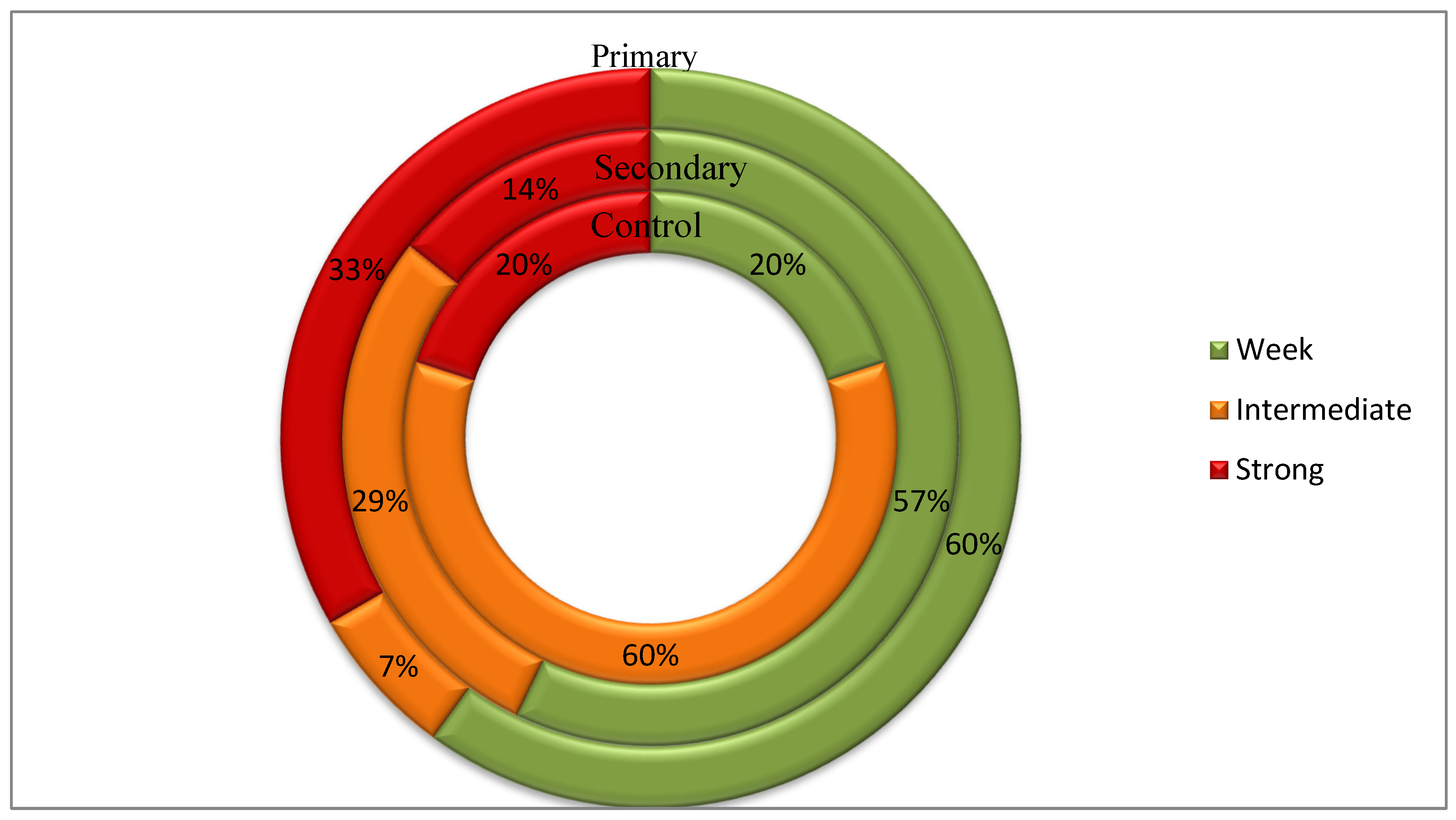
3.3.2. Biofilm Formation Rate

The tested Gram-negative rods (n = 27) were capable of biofilm formation. Of these, 52% produced weak biofilms, 22% moderate biofilms, and 26% strong biofilms (Figure 3). The highest proportion of strong biofilm-producing strains was found in patients with primary CRS (33%), whereas from the secondary CRS group, only 14% of strains formed a strong biofilm (p-value = 0.412777). All isolated strains belonging to the Enterobacter and Proteus genera produced a strong biofilm, while none of the Escherichia and Citrobacter strains exhibited strong biofilm formation. Only one out of 12 Escherichia coli strains formed a moderate biofilm.

4. Discussion

Numerous sinus microbiota studies used new culture-independent techniques [11,13,22,24,34,35,36]. The studies based on a molecular approach to assess sinonasal microbial communities identified more bacterial species per individual than those identified by standard clinical culture methods. Although a greater number of isolates were identified exclusively using molecular methods, several species undetected by molecular techniques were successfully identified through cultivation [37,38]. Moreover, culture methods remain irreplaceable for practical reasons, including the ability to identify the microorganism at the species level and antimicrobial susceptibility testing for drugs already introduced into treatment and new preparations. In our study, culture-based methods were used, allowing for further analysis.
Another important aspect is selecting the appropriate specimen type for microbiological assessment. Several studies suggested that tissue samples may be more optimal for patients with CRS than the middle nasal meatus swabs. The authors reported that tissues provided less variation between samples than swabs, enabling the isolation of bacteria living in the biofilm and penetrating the mucosa cells [26,39]. Considering the above factors, tissue biopsies were collected from patients in both the CRS and the control groups for our study. The sampling sites for patients with CRS and controls may show niche-specific differences in microbiota, which may be a study limitation. However, we decided to sample the interior turbinate mucosa as a maxillary and ethmoid sinus drainage pathway.
Many authors point out the diversity of the sinus microbiome in both health and disease. Moreover, the diversity of bacteria in a healthy state has a protective effect [25,26,40,41]. For example, Cutibacterium acnes, identified in about 80% of healthy people, maintains a physiological bacterial and fungal balance thanks to the bacteriocin it produces and affects the host’s immune response [42,43,44].
However, it is believed that in contrast to healthy individuals, the sinus microbiota of CRS patients is characterized by significant disturbance, with reduced bacterial diversity and richness [38,45]. Dysbiosis-alterations in the composition and function of the microbiota- has been suggested as one of the mechanisms engaged in CRS pathogenesis [15,25,38,45].
The qualitative composition of microorganisms in patients with CRS is influenced by age, smoking, previous surgery, and prior antibiotic therapy Antibiotic use has a significant impact on the microbiological profile of the sinuses: (1) it reduces the abundance of beneficial or commensal bacteria (e.g., Corynebacterium) that play a protective role; (2) it promotes the selection and proliferation of resistant bacterial strains; (3) it facilitates colonization by opportunistic pathogens (e.g., S. aureus); and (4) it disrupts microbial homeostasis, which may predispose patients to infection recurrence or progression to chronic forms of the disease [46,47,48]. Differences in the quantitative and qualitative composition of the microbiota can also be associated with the severity of CRS, comorbidities, and geographic location.
Ramakrishnan et al. studied the composition of the sinonasal microbiota in healthy individuals and found that the most abundant bacterial phyla were Firmicutes, Proteobacteria, and Actinobacteria. According to some authors, the predominant bacteria at the species level identified in CRS patients include Staphylococcus epidermidis, Staphylococcus aureus, Haemophilus influenzae, Corynebacterium spp., Streptococcus pneumoniae, gram-negative bacteria from the Enterobacterales order, as well as anaerobic species, primarily Cutibacterium and Peptostreptococcus [34,46,49,50,51].
Bassiouni et al. proposed three distinct states of the sinonasal microbiota based on their research findings. In the first state, Corynebacterium species were dominant; in the second, Staphylococcus spp. prevailed, and in the third, the microbiota was dominated by Streptococcus, Haemophilus, Moraxella, and Pseudomonas species. These three microbiota states were observed in healthy individuals and those with sinonasal diseases. However, the authors noted geographical differences: in Europe, Staphylococcus spp.-dominated microbiota was more prevalent, whereas in Asia, the first microbiota type, characterized by a predominance of Corynebacterium spp., was more common [52].
The results of our study showed that the dominant microorganisms isolated from the sinuses of CRS patients were Gram-positive cocci, primarily of the Staphylococcus genus, which is consistent with findings from other studies [48,50,53,54,55]. Among the isolated coagulase-negative staphylococci (CNS), S. epidermidis was the most prevalent. However, since this microorganism was also frequently isolated from patients in the control group, the conclusions we can draw regarding the role of S. epidermidis as a pathogen are limited. Nevertheless, our results confirm the frequent occurrence of CNS, particularly S. epidermidis, in samples taken from patients with CRS, as reported by other authors [26,46,56]. Furthermore, the presence of S. aureus, in both healthy and diseased individuals suggests that certain bacteria may exhibit pathogenic or commensal behavior depending on the specific strain, genetic expression, overall abundance, and interactions with other microorganisms within the mucosal community [34].
An important point of interest is the occurrence of facultatively anaerobic Gram-negative rods in the clinical material collected from patients with CRS. The reported incidence of Enterobacterales rods varies and, depending on the study [35,57,58,59,60]
In our study, Enterobacterales were present in 32% of patients with secondary CRS and 29% with primary CRS, representing 20% and 17% of the total isolates, respectively. The results indicated that the genus richness of the primary CRS group was slightly higher than that of secondary CRS, with E. coli being the dominant species in both groups.
The “overrepresentation” of E. coli in CRS patients was also confirmed using 16S rRNA sequencing by Copeland et al. [61]. Krawczyk et al. also drew attention to Gram-negative rods associated with CRS. The authors described three cases of CRS patients from whom E. coli were isolated [62]. These patients did not respond to antibiotic treatment targeting the classical pathogens commonly associated with sinus infections. The isolated E. coli strains exhibited sensitivity to antibiotics used in the treatment of Gram-negative infections and possessed several virulence factors typically found in strains from urinary tract infections [62].
In our study and reports from other authors, Gram-negative bacilli are rarely isolated from individuals without CRS. Therefore, it is suggested that one of the causes of disease symptoms may be lipopolysaccharide, the main component of the cell wall, which can trigger mucosa inflammation and cause tissue remodeling [23].
Stern et al. isolated Gram-negative rods more frequently from CRSwNP patients than from patients without polyps [50]. Similarly, Chalermwatanachai found large numbers of E. coli in CRSwNP patients with asthma [63].
In a study by Feazel et al., antibiotic use, asthma, and prior surgery influenced the sinus microbiome. Antibiotics and asthma were associated with significant reductions in bacterial diversity and increased abundance of S. aureus, whereas surgery was associated with decreased species richness. Prolonged, repeated antibiotic administration may reduce the complexity of the microbial community and lead to the emergence of a few dominant bacteria. Therefore, this may be one of the reasons for isolating different species of Gram-negative rods from clinical specimens [64].
In our studies, Enterobacterales, especially E. coli, were also isolated from patients with asthma and those who had previously used antibiotics.
Guidelines differ for the use of antibiotics in the treatment of CRS patients [55,65] According to the EPOS2020 steering group [37], there is insufficient evidence that the use of antibiotics in long-term and short-term therapy impacts treatment outcomes in adults with CRS compared with placebo. This group analyzed the results of studies using macrolides (mainly in long-term treatment) and amoxicillin, amoxicillin with clavulanic acid, cefaclor, cefuroxime, ciprofloxacin, and levofloxacin. It should be noted that Gram-negative rods are naturally resistant to macrolides. Given that the occurrence of Gram-negative rods is found in a significant number of patients with CRS, it is advisable to select an antibiotic based on the result of the drug susceptibility test. The Gram-negative rods isolated in our study were 100% sensitive to ciprofloxacin. However, significant percentages of strains were resistant to ampicillin and amoxicillin with clavulanic acid, which effectively precludes the use of these drugs in empirical therapy. In the case of ceftazidime and cefuroxime, resistant strains were isolated only from patients with primary CRS. Kim et al. assessed the trends in drug susceptibility of bacteria isolated from patients with CRS and found high percentages of Klebsiella spp. and Enterobacter spp. strains sensitive to ciprofloxacin (one Klebsiella spp. strain resistant), while the number of ESBL-producing strains increased in the analyzed periods from 0.4% to 7.4% in the case of Klebsiella spp. and from 0% to 3.7% in the case of Enterobacter spp. [60]. In our studies, no ESBL-positive strains were recorded.
In recent years, researchers have identified bacterial biofilm as a significant factor in the pathogenesis of CRS. Biofilms are highly organized, complex bacterial structures enclosed in an extracellular matrix composed of polysaccharides, nucleic acids, and proteins. The matrix provides bacteria with a mechanism to reduce metabolic activity, protecting them from both the host’s defense mechanisms and antibiotics. It is believed that biofilms exacerbate inflammation by activating the immune system by releasing planktonic bacteria and possibly exotoxins [48,53,65]. Moreover, Psaltis et al. described that S. aureus biofilm is a predisposing factor for treatment failure and poorer postoperative outcomes [34].
The presence of bacterial biofilm in sinus mucosa has been observed in 44–92% of CRS patients, depending on the detection method used [12,15,48,66] Dlugaszewska et al. reported that 76.67% (23/30) of CRS patients undergoing FESS had evidence of biofilms on SEM micrographs [32]. The occurrence of bacterial biofilms in CRS patients at a similar frequency has also been confirmed by other authors [28,67,68,69].
The most commonly identified species in biofilms are S. aureus, coagulase-negative staphylococci (CNS), particularly S. epidermidis, S. pneumoniae, P. aeruginosa, H. influenzae, and, less frequently, bacteria from the genera Acinetobacter, Proteus, Citrobacter, Klebsiella, and Enterobacter [12,15,67,70,71,72]. The mechanisms associated with biofilm formation may contribute to microbiota imbalance. The Quorum Sensing (QS) system is used by bacteria residing in biofilms for communication influences, among other things, the spatial differentiation of biomass, antibiotic resistance, and the expression of virulence factors, which may impact disease pathogenesis and mediate the host’s immune response [32,47,66]. Biofilms on the surface of the sinus mucosa may contribute to the persistence of inflammation observed in treatment-resistant CRS [53].
The Gram-negative rods examined in this study also demonstrated the ability to form biofilms under in vitro conditions. Seven out of the 27 tested isolates formed a strong biofilm. These included P. mirabilis (n = 2), E. aerogenes (n = 1), E. cloacae (n = 1), M. morganii (n = 1), S. marcescens (n = 1), and K. oxytoca (n = 1). In contrast, E. coli and Citrobacter spp. exhibited a lower capacity for biofilm formation. However, bacteria in biofilm form are also present in healthy sinuses [32,73]. The presence of biofilm in a group of patients without signs of chronic inflammation suggests that biofilm alone is not responsible for the manifestation of chronic sinusitis. A parallel correlation with other etiopathogenic factors is necessary. However, biofilm in CRS patient’s sinuses is associated with more severe symptoms and poorer outcomes.
Clinicians often emphasize the role of Gram-positive bacteria in CRS; however, our findings also highlight a significant contribution of Gram-negative bacteria, such as Escherichia coli, Klebsiella oxytoca, and Citrobacter spp. The presence of these microorganisms may indicate an atypical course of infection, for example, following prior antibiotic therapy, surgical interventions, or immunocompromised patients. All Gram-negative strains identified in our study demonstrated biofilm-forming capacity, which has important clinical implications: biofilms can lead to recurrent infections and reduced efficacy of standard antibiotic treatments. Such cases often require prolonged therapy and consideration of combination treatment strategies.
The study revealed varying susceptibility of Gram-negative rods to ampicillin, tigecycline, and amoxicillin-clavulanic acid. This underscores the need for (1) individualized antibiotic therapy guided by susceptibility testing (antibiogram), (2) microbiological diagnostics in cases of treatment-resistant CRS, and (3) consideration of combination therapies or localized treatments aimed at biofilm eradication.
The treatment of CRS involving Gram-negative bacteria may necessitate interdisciplinary collaboration between otolaryngologists, microbiologists, and infectious disease specialists. It should be tailored to each patient based on medical history, microbiological findings, and clinical presentation.
  • Future Perspectives
A detailed analysis of the impact of individual microorganisms or groups of bacteria on histopathology, immunology, the presence of disease-related protein components in the mucus, and the clinical picture of CRS may allow the determination of the endotype activated in an individual patient. These data will improve the diagnosis, prognosis, and management strategies.
Furthermore, the integration of microbiological analyses with proteomic investigations (e.g., protein profiling of nasal mucus) and immunological studies (e.g., characterization of the inflammatory response) holds the potential to enhance our understanding of CRS significantly and to lay the groundwork for the development of personalized therapeutic approaches.
Furthermore, given the confirmed presence of bacterial biofilms in CRS, as demonstrated in recent studies, the implementation of novel combination therapies specifically targeting biofilm structures—such as probiotic/prebiotic formulations, biologic agents, and photodynamic therapy—should be considered as promising avenues for treatment.
Traditional culture-based methods may fail to detect the full spectrum of microbial species involved; thus, the application of metagenomic sequencing techniques to characterize the complete microbial profile of the sinonasal environment may offer critical insights into the role of the sinus microbiota in the pathogenesis of CRS.
  • Limitations
  • The samples in our study were obtained from patients with CRS during ESS. The patients with acute rhinosinusitis or exacerbations of CRS were excluded.
  • The sampling site for CRS and control patients was not the same. The anterior ethmoid sinus mucosa was collected from CRS patients, whereas the interior turbinate mucosa was collected from control patients.
  • The study protocol did not include molecular methods, so the results refer only to cultivable bacteria.

5. Conclusions

In summary, this study characterized the sinus bacterial communities in patients with and without chronic rhinosinusitis. Although Gram-positive bacteria were the most frequently isolated species in CRS patients, Gram-negative bacteria–particularly E. coli–also represented a considerable proportion of the isolates obtained during endoscopic sinus surgery. Notably, several Gram-negative rods demonstrated the ability to form biofilms, a factor that may contribute to persistent infection and treatment failure. These findings highlight the importance of performing antimicrobial susceptibility testing to guide the selection of effective, targeted therapies for CRS.

Author Contributions

Conceptualization, J.D. and M.L.; methodology, J.D., M.R. and M.L.; software, J.D., M.R., D.K. and T.F.-R.; validation, M.R. and D.K.; formal analysis, M.R. and D.K.; investigation, M.R., D.K. and T.F.-R. data curation, M.R., D.K. and T.F.-R.; writing—original draft preparation, M.R., D.K., J.D. and M.L.; writing—review and editing, J.D. and M.R.; resources, J.D., M.R. and D.K.; supervision, J.D. and M.R.; project administration, J.D. All authors have read and agreed to the published version of the manuscript.

Funding

This research received no external funding.

Institutional Review Board Statement

The study was conducted in accordance with the Declaration of Helsinki, and approved by the Bioethics Committee of the Karol Marcinkowski University of Medical Sciences in Poznan, protocol number 150/18.

Informed Consent Statement

Informed consent was obtained from all subjects involved in the study.

Data Availability Statement

The raw data supporting the conclusions of this article will be made available by the authors on request.

Conflicts of Interest

The authors declare no conflicts of interest.

Abbreviations

The following abbreviations are used in this manuscript:
CRSChronic rhinosinusitis
ESSEndoscopic sinus surgery
EPOSEuropean Position Paper on Rhinosinusitis and Nasal Polyps
CRSwNPCRS with nasal polyps
CRSsNPCRS without nasal polyps

References

  1. Fokkens, W.J.; Lund, V.J.; Hopkins, C.; Hellings, P.W.; Kern, R.; Reitsma, S.; Toppila-Salmi, S.; Bernal-Sprekelsen, M.; Mullol, J.; Alobid, I.; et al. European Position Paper on Rhinosinusitis and Nasal Polyps 2020. Rhinology 2020, 58, 1–464. [Google Scholar] [CrossRef] [PubMed]
  2. Min, H.K.; Lee, S.; Kim, S.; Son, Y.; Park, J.; Kim, H.J.; Lee, J.; Lee, H.; Smith, L.; Rahmati, M.; et al. Global Incidence and Prevalence of Chronic Rhinosinusitis: A Systematic Review. Clin. Exp. Allergy 2025, 55, 52–66. [Google Scholar] [CrossRef] [PubMed]
  3. de Loos, D.D.; Lourijsen, E.S.; Wildeman, M.A.M.; Freling, N.J.M.; Wolvers, M.D.J.; Reitsma, S.; Fokkes, W.J. Prevalence of chronic rhinosinusitis in the general population based on sinus radiology and symptomatology. J. Allergy Clin. Immunol. 2019, 143, 1207–1214. [Google Scholar] [CrossRef]
  4. DeConde, A.S.; Bodner, T.E.; Mace, J.C.; Smith, T.L. Response shift in quality of life after endoscopic sinus surgery for chronic rhinosinusitis. JAMA Otolaryngol. Head Neck Surg. 2014, 140, 712–719. [Google Scholar]
  5. Wahid, N.W.; Smith, R.; Clark, A.; Salam, M.; Philpott, C.M. The socioeconomic cost of chronic rhinosinusitis study. Rhinology 2020, 58, 112–125. [Google Scholar] [CrossRef]
  6. Rudmik, L. Economics of Chronic Rhinosinusitis. Curr. Allergy Asthma Rep. 2017, 17, 20. [Google Scholar] [CrossRef]
  7. Bachert, C.; Marple, B.; Schlosser, R.J.; Hopkins, C.; Schleimer, R.P.; Lambrecht, B.N.; Bröker, B.M.; Laidlaw, T.; Song, W.J. Adult chronic rhinosinusitis. Nat. Rev. Dis. Primers 2020, 6, 86. [Google Scholar] [CrossRef]
  8. Kuan, E.C.; Suh, J.D. Systemic and Odontogenic Etiologies in Chronic Rhinosinusitis. Otolaryngol. Clin. N. Am. 2017, 50, 95–111. [Google Scholar] [CrossRef]
  9. Kato, A.; Schleimer, R.P.; Bleier, B.S. Mechanisms and pathogenesis of chronic rhinosinusitis. J. Allergy Clin. Immunol. 2022, 149, 1491–1503. [Google Scholar] [CrossRef]
  10. Stevens, W.W.; Lee, R.J.; Schleimer, R.P.; Cohen, N.A. Chronic rhinosinusitis pathogenesis. J. Allergy Clin. Immunol. 2022, 136, 1442–1453. [Google Scholar] [CrossRef]
  11. Zhao, Y.; Chen, J.; Hao, Y.; Wang, B.; Wang, Y.; Liu, Q.; Zhao, J.; Li, Y.; Wang, P.; Wang, X.; et al. Predicting the recurrence of chronic rhinosinusitis with nasal polyps using nasal microbiota. Allergy 2022, 77, 540–549. [Google Scholar] [CrossRef]
  12. Michalik, M.; Krawczyk, B. Chronic Rhinosinusitis—Microbiological Etiology, Potential Genetic Markers, and Diagnosis. Int. J. Mol. Sci. 2024, 25, 3201. [Google Scholar] [CrossRef] [PubMed]
  13. Ramakrishnana, V.R.; Hauser, L.J.; Frank, D.N. The sinonasal bacterial microbiome in health and disease. Curr. Opin. Otolaryngol. Head Neck Surg. 2016, 24, 20–25. [Google Scholar] [CrossRef]
  14. Orlandi, R.R.; Kingdom, T.T.; Smith, T.L.; Bleier, B.; DeConde, A.; Luong, A.U.; Poetker, D.M.; Soler, Z.; Welch, K.C.; Wise, S.K.; et al. International consensus statement on allergy and rhinology: Rhinosinusitis 2021. Int. Forum Allergy Rhinol. 2021, 11, 211–739. [Google Scholar] [CrossRef]
  15. Sivasubramaniam, R.; Douglas, R. The microbiome and chronic rhinosinusitis. World J. Otorhinolaryngol. Head Neck Surg. 2018, 314, 216–221. [Google Scholar] [CrossRef] [PubMed]
  16. Ramakrishnan, V.R.; Feazel, L.M.; Gitomer, S.A.; Ir, D.; Robertson, C.E.; Frank, D.N. The microbiome of the middle meatus in healthy adults. PLoS ONE 2013, 8, e85507. [Google Scholar] [CrossRef]
  17. Anderson, M.; Stokken, J.; Sanford, T.; Aurora, R.; Sindwani, R. A systematic review of the sinonasal microbiome in chronic rhinosinusitis. Am. J. Rhinol. Allergy 2016, 30, 161–166. [Google Scholar] [CrossRef]
  18. Pattaroni, C.; Marsland, B.J.; Harris, N.L. Early-Life Host-Microbial Interactions and Asthma Development: A Lifelong Impact? Immunol. Rev. 2025, 330, e70019. [Google Scholar] [CrossRef]
  19. Kloepfer, K.M.; Kennedy, J.L. Childhood respiratory viral infections and the microbiome. J. Allergy Clin. Immunol. 2023, 152, 827–834. [Google Scholar] [CrossRef]
  20. Clark, S.E. Commensal bacteria in the upper respiratory tract regulate susceptibility to infection. Curr. Opin. Immunol. 2020, 66, 42–49. [Google Scholar] [CrossRef]
  21. Durack, J.; Christophersen, C.T. Human Respiratory and Gut Microbiomes—Do They Really Contribute to Respiratory Health? Front. Pediatr. 2020, 8, 528. [Google Scholar] [CrossRef]
  22. Liang, Y.; Xie, R.; Xiong, X.; Hu, Z.; Mao, X.; Wang, X.; Zhang, J.; Sun, P.; Yue, Z.; Wang, W.; et al. Alterations of nasal microbiome in eosinophilic chronic rhinosinusitis. J. Allergy Clin. Immunol. 2023, 151, 1286–1295. [Google Scholar] [CrossRef] [PubMed]
  23. Gómez-García, M.; Moreno-Jimenez, E.; Morgado, N.; García-Sánchez, A.; Gil-Melcón, M.; Pérez-Pazos, J.; Estravís, M.; Isidoro-García, M.; Dávila, I.; Sanz, C. The Role of the Gut and Airway Microbiota in Chronic Rhinosinusitis with Nasal Polyps: A Systematic Review. Int. J. Mol. Sci. 2024, 25, 8223. [Google Scholar] [CrossRef] [PubMed]
  24. Lu, Y.T.; Wang, S.H.; Liou, M.L.; Shen, T.A.; Lu, Y.C.; Hsin, C.H.; Yang, S.F.; Chen, Y.Y.; Chang, T.H. Microbiota Dysbiosis in Fungal Rhinosinusitis. J. Clin. Med. 2019, 8, 1973. [Google Scholar] [CrossRef]
  25. Ccami-Bernal, F.; Barriga-Chambi, F.; Ortiz-Benique, Z.N.; Ferrary, E.; Torres, R. Variability of the Microbiota in Chronic Rhinosinusitis: A Scoping Review. Am. Acad. Otolaryngol.–Head Neck Surg. Found. 2024, 8, 70029. [Google Scholar] [CrossRef]
  26. Koeller, K.; Herlemann, D.P.R.; Schuldt, T.; Ovari, A.; Guder, E.; Podbielski, A.; Kreikemeyer, B.; Olzowy, B. Microbiome and Culture Based Analysis of Chronic Rhinosinusitis Compared to Healthy Sinus Mucosa. Front. Microbiol. 2018, 9, 643. [Google Scholar] [CrossRef] [PubMed]
  27. Wei, H.Z.; Li, Y.C.; Wang, X.D.; Lu, X.X.; Hu, C.H.; He, S.; Liu, X. The microbiology of chronic rhinosinusitis with and without nasal polyps. Eur. Arch. Otorhinolaryngol. 2018, 275, 1439–1447. [Google Scholar] [CrossRef]
  28. Vanderpool, E.J.; Rumbaugh, K.P. Host-microbe interactions in chronic rhinosinusitis biofilms and models for investigation. Biofilm 2023, 29, 100160. [Google Scholar] [CrossRef]
  29. Szaleniec, J.; Gibała, A.; Hartwich, P.; Hydzik-Sobocińska, K.; Konior, M.; Gosiewski, T.; Szaleniec, M. Challenging the gold standard: Methods of sampling for microbial culture in patients with chronic rhinosinusitis. Eur. Arch. Otorhinolaryngol. 2021, 278, 4795–4803. [Google Scholar] [CrossRef] [PubMed] [PubMed Central]
  30. Vandepitte, J.; Verhaegen, J.; Engbaek, K.; Rohner, P.; Piot, P.; Heuck, C.C. Basic Laboratory Procedures in Clinical Bacteriology, 2nd ed.; World Health Organization: Geneva, Switzerland, 2003. [Google Scholar]
  31. European Committee on Antimicrobial Susceptibility Testing. Breakpoint Tables for Interpretation of MICs and Zone Diameters Version 14.0. 2024. Available online: http://www.eucast.org (accessed on 14 October 2024).
  32. Dlugaszewska, J.; Leszczynska, M.; Lenkowski, M.; Tatarska, A.; Pastusiak, T.; Szyfter, W. The pathophysiological role of bacterial biofilms in chronic sinusitis. Eur. Arch. Otorhinolaryngol. 2016, 273, 61989–61994. [Google Scholar] [CrossRef]
  33. Stepanovic, S.; Vukovic, D.; Hola, V.; Di Bonaventura, G.; Djukić, S.; Ćwirković, I.; Ruzicka, F. Quantification of biofilm in microtiter plates: Overview of testing conditions and practical recommendations for assessment of biofilm production by staphylococci. APMIS 2007, 115, 891–899. [Google Scholar] [CrossRef]
  34. Psaltis, A.P.; Mackenzie, B.W.; Cope, E.K.; Ramakrishnan, V.R. Unraveling the role of the microbiome in chronic rhinosinusitis. J. Allergy Clin. Immunol. 2022, 149, 1513–1521. [Google Scholar] [CrossRef]
  35. Chegini, Z.; Shariati, A.; Asghari, A.; Rajaeih, S.; Ghorbani, M.; Jalessi, M.; Mirshekar, M.; Razavi, S. Molecular analysis of dominant paranasal sinus bacteria in patients with and without chronic rhinosinusitis. Arch. Microbiol. 2022, 204, 327. [Google Scholar] [CrossRef] [PubMed]
  36. Lucas, S.K.; Feddema, E.; Boyer, H.C.; Hunter, R.C. Diversity of cystic fibrosis chronic rhinosinusitis microbiota correlates with different pathogen dominance. J. Cyst. Fibros. 2021, 20, 678–681. [Google Scholar] [CrossRef] [PubMed]
  37. Hoggard, M.; Wagner Mackenzie, B.; Jain, R.; Taylor, M.W.; Biswas, K.; Douglas, R.G. Chronic Rhinosinusitis and the Evolving Understanding of Microbial Ecology in Chronic Inflammatory Mucosal Disease. Clin. Microbiol. Rev. 2017, 30, 321–348. [Google Scholar] [CrossRef] [PubMed] [PubMed Central]
  38. Rom, D.; Bassiouni, A.; Eykman, E.; Liu, Z.; Paramasivan, S.; Alvarado, R.; Earls, P.; Psaltis, A.J.; Harvey, R.J. The Association Between Disease Severity and Microbiome in Chronic Rhinosinusitis. Laryngoscope 2019, 129, 1265–1273. [Google Scholar] [CrossRef]
  39. Cho, S.W.; Kim, D.Y.; Choi, S.; Won, S.; Kang, H.R.; Yi, H. Microbiome profiling of uncinate tissue and nasal polyps in patients with chronic rhinosinusitis using swab and tissue biopsy. PLoS ONE 2021, 16, e0249688. [Google Scholar] [CrossRef]
  40. Biswas, K.; Hoggard, M.; Jain, R.; Taylor, M.W.; Douglas, R.G. The nasal microbiota in health and disease: Variation within and between subjects. Front. Microbiol. 2015, 9, 134. [Google Scholar] [CrossRef]
  41. Hoggard, M.; Biswas, K.; Zoing, M.; Wagner Mackenzie, B.; Taylor, M.W.; Douglas, R.G. Evidence of microbiota dysbiosis in chronic rhinosinusitis. Int. Forum Allergy Rhinol. 2017, 7, 230–239. [Google Scholar] [CrossRef]
  42. Psaltis, A.J.; Wormald, P.J. Therapy of Sinonasal Microbiome in CRS: A Critical Approach. Curr. Allergy Asthma Rep. 2017, 17, 59. [Google Scholar] [CrossRef]
  43. Boase, S.; Foreman, A.; Cleland, E.; Tan, L.; Melton-Kreft, R.; Pant, H.; Hu, F.Z.; Ehrlich, G.D.; Wormald, P.J. The microbiome of chronic rhinosinusitis: Culture, molecular diagnostics and biofilm detection. BMC Infect. Dis. 2013, 8, 210. [Google Scholar] [CrossRef]
  44. Morawska-Kochman, M.; Jermakow, K.; Nelke, K.; Zub, K.; Pawlak, W.; Dudek, K.; Bochnia, M. The pH Value as a Factor Modifying Bacterial Colonization of Sinonasal Mucosa in Healthy Persons. Ann. Otol. Rhinol. Laryngol. 2019, 128, 819–828. [Google Scholar] [CrossRef] [PubMed]
  45. Jeican, I.I.; Barbu Tudoran, L.; Florea, A.; Flonta, M.; Trombitas, V.; Apostol, A.; Dumitru, M.; Aluaș, M.; Junie, L.M.; Albu, S. Chronic Rhinosinusitis: MALDI-TOF Mass Spectrometry Microbiological Diagnosis and Electron Microscopy Analysis; Experience of the 2nd Otorhinolaryngology Clinic of Cluj-Napoca, Romania. J. Clin. Med. 2020, 9, 3973. [Google Scholar] [CrossRef]
  46. Ramakrishnan, V.R.; Hauser, L.J.; Feazel, L.M.; Ir, D.; Robertson, C.E.; Frank, D.N. Sinus microbiota varies among chronic rhinosinusitis phenotypes and predicts surgical outcome. J. Allergy Clin. Immunol. 2015, 136, 334–342. [Google Scholar] [CrossRef] [PubMed]
  47. Lee, K.; Pletcher, S.D.; Lynch, S.V.; Goldberg, A.N.; Cope, E.K. Heterogeneity of microbiota dysbiosis in chronic rhinosinusitis: Potential clinical implications and microbial community mechanisms contributing to sinonasal inflammation. Front. Cell. Infect. Microbiol. 2018, 8, 168. [Google Scholar] [CrossRef] [PubMed]
  48. Lee, J.T.; Frank, D.N.; Ramakrishnan, V. Microbiome of the paranasal sinuses: Update and literature review. Am. J. Rhinol. Allergy 2016, 30, 3–16. [Google Scholar] [CrossRef]
  49. Tabet, P.; Endam, L.M.; Boisvert, P.; Boulet, L.P.; Desrosiers, M. Gram-negative bacterial carriage in chronic rhinosinusitis with nasal polyposis is not associated with more severe inflammation. Int. Forum Allergy Rhinol. 2015, 5, 289–293. [Google Scholar] [CrossRef]
  50. Stern, S.; Hadar, T.; Nachalon, Y.; Ben-Zvi, H.; Soudry, E.; Yaniv, E. Bacteriology of the Middle Meatus in Chronic Rhinosinusitis with and without Polyposis. ORL 2016, 78, 223–231. [Google Scholar] [CrossRef]
  51. Saibene, A.M.; Vassena, C.; Pipolo, C.; Trimboli, M.; De Vecchi, E.; Felisati, G.; Drago, L. Odontogenic and rhinogenic chronic sinusitis: A modern microbiological comparison. Int. Forum Allergy Rhinol. 2016, 6, 41–45. [Google Scholar] [CrossRef]
  52. Bassiouni, A.; Paramasivan, S.; Shiffer, A.; Dillon, M.R.; Cope, E.K.; Cooksley, C.; Ramezanpour, M.; Moraitis, S.; Ali, M.J.; Bleier, B.S.; et al. Microbiotyping the sinonasal microbiome. Front. Cell. Infect. Microbiol. 2020, 10, 137. [Google Scholar] [CrossRef]
  53. Lam, K.; Schleimer, R.; Kern, R.C. The Etiology and Pathogenesis of Chronic Rhinosinusitis: A Review of Current Hypotheses. Curr. Allergy Asthma Rep. 2015, 15, 41. [Google Scholar] [CrossRef] [PubMed]
  54. Leszczyńska, J.; Stryjewska-Makuch, G.; Ścierski, W.; Lisowska, G. Bacterial Flora of the Nose and Paranasal Sinuses Among Patients Over 65 Years Old with Chronic Rhinosinusitis Who Underwent Endoscopic Sinus Surgery. Clin. Interv. Aging 2020, 14, 207–215. [Google Scholar] [CrossRef]
  55. Fokkens, W.J.; Lund, V.J.; Mullol, J.; Bachert, C.; Alobid, I.; Baroody, F.; Cohen, N.; Cervin, A.; Douglas, R.; Gevaert, P.; et al. EPOS 2012: European position paper on rhinosinusitis and nasal polyps 2012. A summary for otorhinolaryngologists. Rhinology 2012, 50, 1–12. [Google Scholar] [CrossRef] [PubMed]
  56. Huntley, K.S.; Raber, J.; Fine, L.; Bernstein, J.A. Influence of the Microbiome on Chronic Rhinosinusitis with and Without Polyps: An Evolving Discussion. Front. Allergy 2021, 1, 737086. [Google Scholar] [CrossRef] [PubMed]
  57. Li, Y.T.; Huang, S.S.; Ma, J.H.; Hsieh, B.H.; Tsou, Y.A.; Lin, C.D.; Tai, C.J.; Shih, L.C. Bacteriology of Different Phenotypes of Chronic Rhinosinusitis. Laryngoscope 2024, 134, 1071–1076. [Google Scholar] [CrossRef]
  58. Wagner Mackenzie, B.; Dassi, C.; Vivekanandan, A.; Zoing, M.; Douglas, R.D.; Biswas, K. Longitudinal analysis of sinus microbiota post endoscopic surgery in patients with cystic fibrosis and chronic rhinosinusitis: A pilot study. Respir. Res. 2021, 22, 106. [Google Scholar] [CrossRef]
  59. Gan, W.; Zhang, H.; Yang, F.; Liu, S.; Liu, F.; Meng, J. The influence of nasal bacterial microbiome diversity on the pathogenesis and prognosis of chronic rhinosinusitis patients with polyps. Eur. Arch. Otorhinolaryngol. 2021, 278, 1075–1088. [Google Scholar] [CrossRef]
  60. Kim, D.; Assiri, A.M.; Kim, J.H. Recent Trends in Bacteriology of Adult Patients with Chronic Rhinosinusitis. J. Clin. Med. 2019, 8, 1889. [Google Scholar] [CrossRef]
  61. Copeland, E.; Leonard, K.; Carney, R.; Kong, J.; Forer, M.; Naidoo, Y.; Oliver, B.G.G.; Seymour, J.R.; Woodcock, S.; Burke, C.M.; et al. Chronic Rhinosinusitis: Potential Role of Microbial Dysbiosis and Recommendations for Sampling Sites. Front. Cell. Infect. Microbiol. 2018, 8, 57. [Google Scholar] [CrossRef]
  62. Krawczyk, B.; Michalik, M.; Fordon, M.; Wysocka, M.; Samet, A.; Nowicki, B. Escherichia coli Strains with Virulent Factors Typical for Uropathogens were Isolated from Sinuses from Patients with Chronic Rhinosinusitis-Case Report. Pathogens 2020, 25, 318. [Google Scholar] [CrossRef]
  63. Chalermwatanachai, T.; Vilchez-Vargas, R.; Holtappels, G.; Lacoere, T.; Jáuregui, R.; Kerckhof, F.M.; Pieper, D.H.; Van de Wiele, T.; Vaneechoutte, M.; Van Zele, T.; et al. Chronic rhinosinusitis with nasal polyps is characterized by dysbacteriosis of the nasal microbiota. Sci. Rep. 2018, 8, 7926. [Google Scholar] [CrossRef]
  64. Feazel, L.M.; Robertson, C.E.; Ramakrishnan, V.R.; Frank, D.N. Microbiome complexity and Staphylococcus aureus in chronic rhinosinusitis. Laryngoscope 2012, 122, 467–472. [Google Scholar] [CrossRef]
  65. Smith, S.S.; Kim, R.; Douglas, R. Is there a role for antibiotics in the treatment of chronic rhinosinusitis? J. Allergy Clin. Immunol. 2022, 149, 1504–1512. [Google Scholar] [CrossRef] [PubMed]
  66. Ratajczak, M.; Kamińska, D.; Nowak-Malczewska, D.M.; Schneider, A.; Długaszewska, J. Relationship between antibiotic resistance, biofilm formation, genes coding virulence factors and source of origin of Pseudomonas aeruginosa clinical strains. Ann. Agric. Environ. Med. 2021, 28, 306–313. [Google Scholar] [CrossRef] [PubMed]
  67. Sabino, H.A.C.; Valera, F.C.P.; Santos, D.V.; Fantucci, M.Z.; Titoneli, C.C.; Martinez, R.; Anselmo-Lima, W.T.; Tamashiro, E. Biofilm and Planktonic Antibiotic Resistance in Patients with Acute Exacerbation of Chronic Rhinosinusitis. Front. Cell. Infect. Microbiol. 2022, 17, 813076. [Google Scholar] [CrossRef] [PubMed]
  68. Manciula, L.G.; Jeican, I.I.; Tudoran, L.B.; Albu, S. Biofilms and inflammation in patients with chronic rhinosinusitis. Med. Pharm. Rep. 2020, 93, 374–383. [Google Scholar] [CrossRef]
  69. Vlad, D.; Dutu, A.; Apostol, A.; Trombitas, V.; Mihalca, A.; Albu, S. The Effect of Spray Cryotherapy on Microbial Biofilms in Chronic Rhinosinusitis. Curr. Infect. Dis. Rep. 2018, 13, 41. [Google Scholar] [CrossRef]
  70. Di Luca, M.; Navari, E.; Esin, S.; Menichini, M.; Barnini, S.; Trampuz, A.; Casani, A.; Batoni, G. Detection of Biofilms in Biopsies from Chronic Rhinosinusitis Patients: In Vitro Biofilm Forming Ability and Antimicrobial Susceptibility Testing in Biofilm Mode of Growth of Isolated Bacteria. Adv. Exp. Med. Biol. 2018, 1057, 1–27. [Google Scholar] [CrossRef]
  71. Foreman, A.; Boase, S.; Psaltis, A.; Wormald, P.J. Role of bacterial and fungal biofilms in chronic rhinosinusitis. Curr. Allergy Asthma Rep. 2012, 12, 127–135. [Google Scholar] [CrossRef]
  72. Ferguson, B.J.; Stolz, D.B. Demonstration of biofilm in human bacterial chronic rhinosinusitis. Am. J. Rhinol. 2005, 19, 452–457. [Google Scholar] [CrossRef]
  73. Bezerra, T.F.; Padua, F.G.; Gebrim, E.M.; Saldiva, P.H.; Voegels, R.L. Biofilms in chronic rhinosinusitis with nasal polyps. Otolaryngol. Head Neck Surg. 2011, 144, 612–616. [Google Scholar] [CrossRef] [PubMed]
Figure 1. EPOS Division of Primary CRS Subtypes based on Fokkens, 2020 [1]. AFRS-allergic fungal rhinosinusitis; CCAD-central compartment allergic disease; CRSwNP-chronic rhinosinusitis with nasal polyps; eCRS-eosinophilic CRS.
Applsci 15 06108 g001
Figure 2. Antimicrobial susceptibility of tested Gram-negative bacteria.
Applsci 15 06108 g002
Figure 3. Classification of the strength of biofilm production.
Applsci 15 06108 g003
Table 1. Patients characteristics.
Control Group
(n = 47)
Patients with
Primary CRS
(n = 52)
Patients with Secondary CRS
(n = 22)
Mean age384543
Male/female30/1729/2312/10
(63.8%/36.2%)(55.8%/44.2%)(54.5%/45.5%)
Asthma4 (8.5%)16 (30.8%)1 (4.5%)
Inhalant allergy12 (25.5%)9 (17.3%)8 (35.5%)
Hypertension6 (12.8%)8 (15.4%)3 (13.6%)
Cancer1 (2.1%)1 (1.9%)0 (0.0%)
Diabetes3 (6.4%)1 (1.9%)6 (27.3%)
Steroid use6 (12.8%)13 (25%)9 (40.9%)
Smoking7 (14.9%)8 (15.45%)4 (18.2%)
Antibiotics6 (12.8%)7 (13.5%)3 (13.6%)
Sinus irrigation3 (6.4%)10 (19.2%)7 (31.8%)
Contact with
chemicals17 (36.2%)12 (23.1%)5 (22.7%)
Facial trauma6 (12.8%)2 (3.8%)1 (4.5%)
Sinus surgery1 (2.1%)16 (30.8%)6 (27.3%)
Nose surgery10 (21.3%)7 (13.5%)9 (40.9%)
Table 2. Microbiota of patients with CRS.
BacteriaPrimary CRS
(n = 90 Strains)
Secondary CRS
(n = 35 Strains)
Control
(n = 73 Strains)
p-Value *
Anaerobes (obligate)
Cutibacterium acnes2 (2.2%)1 (2.9%)2 (2.7%)0.9690
Peptostreptococcus spp.1 (1.1%)-1 (1.4%)0.7942
Fusobacterium spp.1 (1.1%)--0.5471
Mixed anaerobic component
of the URT microbiota
4 (4.4%)2 (5.7%)6 (8.2%)0.6012
Gram-positive Aerobes
Staphylococcus epidermidis33 (36.7%)13 (37.1%)16 (22.0%)0.0931
Staphylococcus aureus14 (15.6%)4 (11.4%)6 (8.2%)0.3578
CNS8 (8.9%)2 (5.7%)3 (4.1%)0.4605
Corynebacterium spp.3 (3.3%)-4 (5.5%)0.3495
Streptococcus pneumoniae1 (1.1%)1 (2.9%)-0.3775
Streptococcus spp.3 (3.3%)2 (5.7%)1 (1.4%)0.4559
Enterococcus faecalis--1 (1.4%)0.4229
Enterococcus spp.2 (2.2%)--0.2975
Gram-negative Aerobes
Escherichia coli7 (7.8%)4 (11.4%)1 (1.4%)0.0797
Klebsiella oxytoca2 (2.2%)1 (2.9%)-0.3971
Serratia marcescens1 (1.1%)--0.5471
Citrobacter koseri-1 (2.9%)3 (4.1%)0.1662
Citrobacter freundii-1 (2.9%)-0.0963
Proteus mirabilis1 (1.1%)-1 (1.4%)0.7942
Enterobacter cloacae2 (2.2%)--0.2975
Morganella morgani2 (2.2%)--0.2975
Mixed aerobic component
of the URT microbiota
3 (3.3%)3 (8.5%)28 (38.4%)0.00000001
URT-Upper Respiratory Tract, CNS–Coagulase-Negative Staphylococci other than S. epidermidis. * χ2 Pearsona with Yates correction.
Disclaimer/Publisher’s Note: The statements, opinions and data contained in all publications are solely those of the individual author(s) and contributor(s) and not of MDPI and/or the editor(s). MDPI and/or the editor(s) disclaim responsibility for any injury to people or property resulting from any ideas, methods, instructions or products referred to in the content.

