Article

Breast Cancer Detection Using a High-Performance Ultra-Wideband Vivaldi Antenna in a Radar-Based Microwave Breast Cancer Imaging Technique

by
Şahin Yıldız
* and
Muhammed Bahaddin Kurt
Electrical and Electronics Engineering Department, Faculty of Engineering, Dicle University, 21280 Diyarbakır, Turkey
*
Author to whom correspondence should be addressed.
Appl. Sci. 2025, 15(11), 6015; https://doi.org/10.3390/app15116015
Submission received: 19 April 2025 / Revised: 22 May 2025 / Accepted: 23 May 2025 / Published: 27 May 2025
(This article belongs to the Section Electrical, Electronics and Communications Engineering)

Abstract

:
In this study, a novel improved ultra-wideband (UWB) antipodal Vivaldi antenna suitable for breast cancer detection via microwave imaging was designed. The antenna was made more directional by adding three pairs of nestings to the antenna fins by adding elliptical patches. The frequency operating range of the proposed antenna is UWB 3.6–13 GHz, its directivity is 11 dB, and its gain is 9.27 dB. The antenna is designed with FR4 dielectric material and dimensions of 34.6 mm × 33 mm × 1.6 mm. It was demonstrated that the bandwidth, gain, and directivity of the proposed antenna meet the requirements for UWB radar applications. The Vivaldi antenna was tested on an imaging system developed using the CST Microwave Studio (CST MWS) program. In CST MWS, a hemispherical heterogeneous breast model with a radius of 50 mm was created and a spherical tumor with a diameter of 0.9 mm was placed inside. A Gaussian pulse was sent through Vivaldi antennas and the scattered signals were collected. Then, adaptive Wiener filter and image formation algorithm delay-multiply-sum (DMAS) steps were applied to the reflected signals. Using these steps, the tumor in the breast model was scanned at high resolution. In the simulation application, the tumor in the heterogeneous phantom was detected and imaged in the correct position. A monostatic radar-based system was implemented for scanning a breast phantom in the prone position in an experimental setting. For experimental measurements, homogeneous (fat and tumor) and heterogeneous (skin, fat, glandular, and tumor) breast phantoms were produced according to the electrical properties of the tissues. The phantoms were designed as hemispherical with a diameter of 100 mm. A spherical tumor tissue with a diameter of 16 mm was placed in the phantoms produced in the experimental environment. The dynamic range of the VNA device used allowed us to image a 16 mm diameter tumor in the experimental setting. The developed microwave imaging system shows that it is suitable for the early-stage detection of breast cancer by scanning the tumor in the correct location in breast phantoms.

1. Introduction

Breast cancer is the most common type of cancer in women and is responsible for the second highest number of deaths worldwide [1]. In general, breast cancer accounts for 30 percent of cancers. Breast cancer causes 15 percent of cancer-related deaths [2].
The World Health Organization’s goal is to prevent 2.5 million deaths worldwide between 2020 and 2040 by reducing the number of global breast cancer deaths by 2.5% per year. Thus, it is predicted that the number of deaths from breast cancer in women under the age of 70 will decrease by 25% by 2030 and by 40% by 2040. The most important aspect to achieve these goals is early diagnosis [3]. Early diagnosis of cancer is the main factor in reducing deaths caused by breast cancer [4,5]. There are traditional imaging methods used in the early diagnosis of breast cancer. It is important to screen patients with these methods and detect cancer at an early stage [6].
Imaging methods such as ultrasound, positron emission tomography (PET), mammography, and magnetic resonance imaging (MRI) are important imaging tools in cancer detection [7,8,9,10]. Besides the advantages of these imaging techniques, there are also various disadvantages.
Mammography imaging is the most commonly used method (two screenings per year) and is good at detecting mammary gland calcification and is not suitable for people with high gland density. Screening is performed by compressing the breast and has a carcinogenic effect [11,12]. Ultrasound, which is a painless imaging technique, does not have high resolution and is not suitable for small masses [13]. MRI imaging is expensive and time-consuming and has high resolution. It is also not suitable for those who are sensitive to contrast agents [14,15]. Although PET imaging is good for detecting cancer stages, it is not suitable for screening because of its limited ability to detect tumors with low metabolic activity. In addition, radioactive tracers are used in the screening process [16,17]. Microwave imaging has emerged as a promising alternative for breast tumor diagnosis to overcome the disadvantages of traditional imaging techniques [12]. Microwave imaging is an imaging technique that is cheap, painless, suitable for rapid screening, does not have the effect of ionizing radiation, and does not require contrast agents [11,12,16,18,19].
Microwave imaging detects differences in the dielectric properties of healthy and cancerous tissues in the breast [13,20,21,22,23]. It uses non-ionizing electromagnetic waves to examine tumor detection based on scattering. This has led to the development of microwave imaging techniques and has gained importance in recent years [24].
The dielectric properties of tumor tissues in the microwave spectrum significantly change their conductivity compared to normal tissue, and the tumor can be detected. The frequencies of biological tissues are not the same due to differences in water content [25,26,27]. Since there are frequency differences, benign and malignant tumors can be classified structurally. This can enable the detection of cancer in the initial stage (maximum tumor size of 20 mm) [28]. The structures of the tumor in stages can be listed as follows: tumor shape, edges, surface texture, and depth. Malignant tumors show features such as irregular and asymmetric shape, blurred borders, and rough and complex surface due to microlobules. Benign tumors show features such as spherical, oval, or regular shape, compactness, and smoother surface [29]. These features can help categorize the tumor in microwave imaging.
The radar-based microwave breast cancer imaging (RMWBI) technique involves solving a nonlinear inverse scattering problem to reveal the electrical properties of tissues [30]. Extensive patient studies have been conducted with RMWBI since the 1990s [31,32]. The purpose of RMWBI is to detect and localize tumors. In microwave hyperthermia tumor detection, dipole antennas focus energy on the tumor using electromagnetic waves. The ambient temperature is isolated and the dipole antennas are fixed to the tissues in the breast model and heat is given, and the tissues are raised above normal temperature levels. Thus, tumor detection is performed by creating thermal models [33,34,35]. The RMWBI system is non-contact and does not require an invasive procedure for measurement. It also does not require heat insulation in the measurement environment. In the RMWBI system, the antennas used as sensors send pulse signals towards the target (breast phantom) and collect the reflected signals back. In these systems, different antenna types are used according to need. These are resonant, patch, bowtie, monopole, dipole, Vivaldi, and other antenna types [29,36,37,38,39]. Among these antenna types, the Vivaldi antenna stands out with its advantages [40].
Vivaldi antennas are ideal for RMWBI since they provide high gain, bandwidth, high directivity, and efficient impedance matching [41,42]. Compared to other techniques such as patch or dipole antennas, the Vivaldi antenna shows superior performance in terms of optimum accuracy and tissue penetration [43,44,45]. Vivaldi antenna has been used as a sensor in many studies in the literature [40,46,47,48,49,50,51,52,53]. Various materials such as epoxy-FR4, polycarbonate, polyimide, rubber, and silicone are used as substrates in Vivaldi antenna design. The use of the FR4 substrate is ideal for the design of Vivaldi antennas due to the desired wide bandwidth, impedance matching, stable radiation values, high gain, good directivity, compact size, cheapness, and easy availability [54,55,56].
All of the signals received back from the target with the RMWBI system are signals reflected from breast tissues and other environmental noise. In order to obtain a tumor response, it is necessary to eliminate the unwanted portions of the signals reflected back from the breast. The most difficult part in the filtering of the reflected signals is the elimination of noise [57]. For this purpose, filtering techniques are used to obtain tumor response [58,59]. The adaptive Wiener filter has been proposed as an effective algorithm to remove noise and this method has been used in studies in the literature [53,57,60,61,62]. In addition to filtering algorithms, image formation algorithms are also very important in tumor detection. Algorithms such as rotation subtraction, signal filtered merging, singular value decomposition, and hybrid approaches have been proposed, but their performance also depends on anatomical features [63,64,65,66]. DAS and DMAS are the reference image formation algorithms for RMWBI systems [67,68]. The DMAS imaging algorithm is an extended version of DAS with higher contrast resolution [69,70]. The DMAS algorithm provides high resolution, making it ideal for tumor detection [53,67].
In the RMWBI technique, breast phantom tissues to be used as target objects must be designed in accordance with the electrical properties of the real breast. The design of artificial breast phantom tissues in simulation and experimental environments according to appropriate electrical properties has been presented in scientific studies [71,72,73]. The study conducted by Lazebnik et al. continues to be the main reference due to its electrical properties close to real breast tissues and the long life of the breast phantom [53,72]. There are studies in the literature that use breast phantom for breast tumor detection [48,50,53,74,75,76,77,78,79].
The radiation performance of Vivaldi antennas can be improved by changing some of their geometrical properties. The antenna geometry was optimized using particle swarm optimization and the gain and directivity of the Vivaldi antenna were increased by adding side slots [48,80]. Double petal was used to reduce the low frequency [49]. When the antenna dimensions were calculated according to the FR4 based transmission line model, approximately 60 mm × 55 mm was obtained. In this study, the antenna dimensions were reduced by approximately 65.4% and produced as 34.6 mm × 33 mm. In order to prevent antenna performance loss during the size reduction process, slots were dug into the antenna. A low-cost and compact UWB antenna operating in the 3.60–13 GHz range has been developed. The antenna design was carried out using the CST MWS simulation program. The Vivaldi antenna is designed with linear phase response and group delay. The designed Vivaldi antenna has been implemented in hardware. Many laboratory measurements were carried out using the hardware-generated Vivaldi antenna and Aritsu MS2028C VNA.
A heterogeneous breast phantom with a tumor was designed using the CST MWS simulation program. Two heterogeneous phantoms with a radius of 50 mm were created from healthy and tumor tissues placed under a 2 mm thick skin layer for the breast model. A spherical tumor with a diameter of 0.9 mm was placed inside the heterogeneous phantom. Tests were performed on the heterogeneous breast phantom with the tumor designed in the Vivaldi antenna simulation environment with features such as real gain and directionality. A Gaussian signal was sent to the heterogeneous breast phantom with an antenna and the reflected signals were collected. Tumor responses were obtained by calibrating the unwanted parts of the collected signals. Tumor images were constructed as an energy map in the X–Y and X–Z planes using the DMAS beam forming algorithm [80].
Homogeneous and heterogeneous breast phantoms with tumors were produced in the laboratory environment using materials suitable for the real breast structure. These phantoms were automatically scanned in the measurement setup by designing a 3D device in the prone position. A 16 mm diameter tumor was detected and imaged in homogeneous and heterogeneous breast phantoms. It was shown that the measurements made with the proposed antenna in both simulation and experimental environments are suitable for the early-stage detection of breast cancer.

2. Materials and Methods
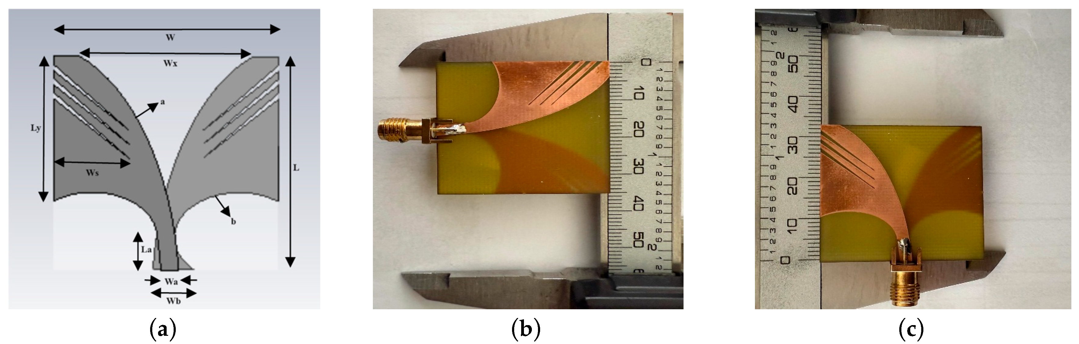
Antenna Design

FR4 material is used in the proposed Vivaldi antenna design, and this antenna has a loss tangent value of 0.002 and a dielectric constant of 4.4. Of note, 0.035 micron copper was used on both surfaces of the FR4 material. A 50 Ω impedance female coaxial connector is used as the feeding port in the FR4 material. The FR4 thickness is 1.6 mm. Antenna design and simulation were performed using CST MWS. The antenna size was reduced to 34.6 mm × 33 mm using this program. The Vivaldi antenna was manufactured using a printed circuit board machine.
The exponential equation used for antenna design calculates the edge taper of the antenna as follows [81]:
y = C1eRx + C2
Here, R is the angle ratio y skew. Using the geometric parameters of the antenna, C1 and C2 can be calculated:
C 1 = y 2 y 1 e R x 2 e R x 1
C 2     y 1 e R x 2 y 2 e R x 1 e R x 2 e R x 1
The first points of the curves in the antenna are y 1 and x 1 ; the last points are y 2 and x 2 .
The geometric configurations of the Vivaldi antenna are shown in Table 1.
As seen in Figure 1, the Vivaldi antenna was designed with 3-slot pairs. The antenna operates between 3.60 and 13 GHz frequency. The measurement results for parameter S11 of the proposed Vivaldi antenna are as shown in Figure 2.
The S11 parameter represents the frequency operating range of the antennas [82]. The low values of the return loss curves indicate that the antenna is operating stably [83,84]. As shown in Figure 2, the curves in the operating band range dropped to –19, –20.5, and –23 dB levels. This shows that the antenna is stable.
The maximum real gain of the realized Vivaldi antenna at 9.5 GHz frequency is 9.1 dB, the gain is 9.27, and the directivity is 11 dB. Figure 3 shows the gain, real gain, and directivity results.
The relationship between the radiation directivity of the frequency points in the antenna operating band and the object (electrical properties of the breast tissue) was investigated. The simulation results for the radiation patterns for the E plane of the Vivaldi antenna at 4.5 GHz, 7 GHz, 9 GHz, and 10.5 GHz are shown in Figure 4. The Vivaldi antenna provides a directional radiation pattern in the simulation process without the breast phantom. A wider beam was observed without the breast model. It is shown that the majority of the power at the upper frequencies of the proposed directional antenna is radiated towards the object (breast model).
A comparison of the proposed antenna with some previously reported antennas is given in Table 2.
As a result, a new opposite-polarity Vivaldi antenna design was realized in this study. Table 2 presents the comparison of Vivaldi antenna studies and the antenna designed within the scope of this study. The proposed antenna stands out with its small size, high bandwidth, high gain, and directivity when compared to other Vivaldi antennas in the literature.

3. Simulation Environment and RMWBI System

3.1. Breast Model

Several types of tissue make up the female breasts. They consist of tissue types such as muscle, ligament, glandular, lymphatic, fat, nipple, and skin. At microwave frequencies, breast tissues have different electrical properties. The theoretical basis for detecting breast cancer with RMWBI is based on the high contrast between the dielectric constant and conductivity between healthy tissue and cancerous tissue. Figure 5 shows the dielectric properties of healthy and tumor tissues obtained in the study [91].
The measurement values corresponding to the tissues of the phantom model used during the measurement in the simulation environment are shown in Table 3 [92]. As seen in Table 3, the dielectric constant of tumor breast tissue is greater than that of healthy breast tissue. This is because tumor tissues contain more water and are more active than healthy tissues.
A heterogeneous breast phantom was created in the simulation environment using tissues appropriate to real breast tissue. The heterogeneous breast phantom was 100 mm in diameter and its outer part was covered with skin tissue. Fat, glandular tissue, and a 0.9 mm diameter tumor were added under the skin part. Finally, a heterogeneous phantom model was created by adding a nipple onto the skin. The heterogeneous breast phantom created in Figure 6 is shown in the X–Y and Y–Z planes. Figure 7 shows the 12 different measurement positions of the Vivaldi antenna and the breast model.
With the RMWBI technique, data are obtained using two different methods. This includes monostatic and multistatic measurement methods [93]. In multistatic measurement, antennas are fixedly placed around the breast phantom. Each antenna in the array transmits signals sequentially while the other antennas collect the reflected signals. Therefore, it is a complex and time-consuming measurement method [94]. However, the proposed monostatic measurement method is a simple, fast, and effective method used for the detection of breast cancer. It also offers high data rates to find the tumor using DMAS [95].

3.2. RMWBI System and Visualization in the Simulation Environment

For the detection of breast tumors, the source signal of the transmission is Gaussian waves and the backscattered signal from the target is received and recorded. The recorded signals are preprocessed to remove artifacts from system components such as skin and tissues as well as antenna coupling signals [77]. Other signals that are greater than the amplitude of the tumor response are then suppressed and filtered. Thus, a low-amplitude tumor response is obtained.
We developed an RMWBI system on CST MWS with a Vivaldi antenna. The antenna is used to transmit the signal and collect the reflected signal. The proposed Vivaldi antenna is rotated in twelve different positions around the created tumorous heterogeneous breast phantom. The measurement taken from the tumor-free heterogeneous phantom was used for the calibration process. This is the subtraction method stage in which the signals scattered from healthy breast tissues are subtracted from the total number of signal scattered from the tumorous breast and recorded on the receiving antenna. The most dominant part here is the signals emitted from the skin. The signal reflected from the tumor is large compared to its amplitude. It is very difficult to find a tumor response without removing the dominant parts because it will result in heavy noise in the tumor response due to the dominant signals. The subtraction method is expressed in Equation (4):
STY = STSH
In Equation (4), ST represents the signal in each channel, SH represents the signal scattered from the tumor-free breast, and STY represents the tumor response signal obtained as a result of filtering. An image enhancement method using Gaussian band-pass filtering is proposed for this imaging system. The proposed method is typically used to eliminate short-pulse signal averaging artifact effects [96]. The Hilbert transform was used to integrate a modulated Gaussian pulse of the incident wave into a radar-based microwave imaging system. A peak is formed at the center of the integrated signal, and the maximum peaks of this tumor point are collected at a focal point, and the location of the tumor is displayed.
In order to obtain a smooth image from the Gaussian signal, which is the tumor response, the envelope of the signal is taken with the Hilbert transform. This transformation is expressed in Equation (5):
H [ s ( t ) ] = 1 Π + s ( Γ ) s ( t Γ ) d Γ
S A ( t ) = s ( t ) + j h [ s ( t ) ]
S A ( t ) = A ( t ) e j ω ( t )
Here, SA(t) is the analytical signal, s(t) is the filtered real signal, H{s(t)} is the complex composition, and Equation (7) shows the time envelope.
The DMAS focusing method was used for tumor detection using the UWB microwave imaging technique. In DMAS, using the finite difference method, signals reflected from the target are shifted in the time domain, doubled, and summed to create a focal spot. The difference multiplication operation was performed according to DAS. The DMAS focusing algorithm is shown in Equation (8):
I ( Γ 0 ) = 0 T m = 1 M 1 j = m + 1 M x m r m r 0 . x j ( r j r 0 ) 2 d t
Here, for each pixel, τm represents the delay time, xm represents the tumor response, and the energy of the pixel energy at distance Γ is expressed as I( Γ 0 ).
I( Γ ) is the energy of pixels at location Γ (x, y, z). Here, v is the wave velocity, Δt is the time steps, and di is the distance between the pixel and the antenna. Time delay (Equation (9)), distance (Equation (10)), speed of the wave (Equation (11)) are shown below respectively:
Γ n ( Γ ) = 2 v Δ t d i
d i = ( x x i ) 2 + ( y y i ) 2 + ( z z i ) 2 v
v = c ε r
Here, εr represents the relative permittivity of the medium and c represents the speed of light.
The signals sent from the antenna to the phantom and reflected back are shown in Figure 8a and Figure 8b, respectively.
The tumor response was obtained by applying filtering and DMAS imaging steps to the reflected signals.
A small tumor with a diameter of 0.9 mm was dropped into the heterogeneous breast phantom in the simulation environment as shown in Figure 6. As seen in Figure 9, energy profiles of the heterogeneous breast model were created by applying signal processing steps.
As shown in Figure 9a, the structured image of the location of the 0.9 mm diameter tumor in the heterogeneous breast phantom in the X–Y plane (top view) was obtained. As shown in Figure 9b, the image of the location of the tumor in the X–Z plane (side view) was obtained. As a result, the location of the tumor placed in the heterogeneous breast model was displayed in the correct position.

4. Results and Discussion

A 3D scanning device was built that allows one to send signals to the phantom and receive the reflected signals. A simple mechanism consisting of a 50 mm radius hemispherical plastic container suitable for scanning in the prone position was set up. The setup was completed by leaving the breast phantom in the plastic cap. Homogeneous and heterogeneous breast phantoms were prepared for measurements in the experimental setting. The homogeneous phantom consists of fat and a tumor. The heterogeneous breast phantom consists of skin, fat, glands, and a tumor.
The proposed Vivaldi antenna was produced for the receiver and transmitter sensors to be used in the experimental environment. In experimental environments, tumor detection capacity depends significantly on the characteristics of the VNA device used. The dynamic range of the device is of great importance in this regard. Therefore, tumor detection capacity is not as high as in the simulation environment. In experimental settings, it is difficult to detect tumors in phantoms containing glandular tissue because this tissue weakens the tumor response [53]. Therefore, heterogeneous and homogeneous phantoms were used for tumor detection. The procedure for preparing homogeneous and heterogeneous breast phantoms with the tumors that we used was employed [72,97]. Unlike this procedure, in this study, powdered bovine gelatin and “Fairy” washing detergent with the same properties were used as surfactants [53]. The quantities of components required to construct the breast phantoms are shown in Table 4.
The diameter of the phantoms was 100 mm, and a tumor with a diameter of 16 mm was placed inside the phantom. Since the dynamic range of the VNA used in the experimental setting was not high enough to detect very small tumors (maximum 83 dB), we kept the tumor size large in stage-one breast cancer (the tumor size in stage 1 can be a maximum of 20 mm). During the production phase of the phantoms, a tumor was first created and allowed to solidify. After the tumor was created in the homogeneous phantom, an oily phantom was made into a plastic bowl and left inside the tumor. In the heterogeneous phantom, skin tissue (2 mm) was first made into a plastic bowl, and after it solidified, 35% fat tissue was added. Finally, the phantom was produced by adding glandular tissue and the tumor (16 mm diameter). All phantoms were left to solidify for 2 days. The phantoms were removed from the mold after solidification. In the measurements, a 16 mm tumor was detected in homogeneous and heterogeneous phantoms. Figure 10 shows the phantom construction stages.
In the proposed radar system, the Vivaldi antenna is used as a transmitter and receiver. The radar system is shown in Figure 11. Using the VNA’s time domain feature, a Gaussian signal was automatically sent and the reflected signals were recorded. The signals recorded in the VNA were transferred to a computer for the signal processing stage. The filtering technique specified in Equation (4) in the previous section was not used in the signal processing stage. Since it is not possible to measure breasts with and without tumors at the same time, they are not suitable for real applications. An adaptive Wiener filter was used as the filtering technique. The signals collected with this filtering technique were calibrated [59]. After the phantoms were produced, a monostatic radar-based imaging system was set up to detect the tumor inside the phantom, as shown in Figure 11.
The S11 parameter response of the heterogeneous phantom with and without a tumor in VNA with the established system is shown in Figure 12.
As a result of all signal processing stages, images of tumors located in heterogeneous and homogeneous breast were obtained. Structured images of the tumor located in the homogeneous and heterogeneous phantoms are shown in Figure 13. In Figure 13 (a-homogeneous and b-heterogeneous), the location of the tumor in the breast phantoms was obtained in the correct location in the X–Y plane (top view). In addition, in Figure 12 (c-homogeneous and d-heterogeneous), the image of the tumor in the X–Z (side view) plane was obtained in the correct location. It is shown that the RMWBI system developed using the proposed Vivaldi antenna can detect and scan small tumors in the breast at the correct location.
Since the dynamic range of the VNA device used in the experimental setting was not high enough to detect very small tumors, tumor detection in the homogeneous phantom resulted in a less noisy image compared to the heterogeneous phantom. Additionally, the glandular tissue found in the heterogeneous phantom weakens the tumor response. Tumor sizes detected in studies in the literature are shown in Table 5.

5. Conclusions

In this study, we designed and fabricated three pairs of nests made by adding elliptical patches to the fins of a novel UWB antipodal Vivaldi antenna suitable for breast cancer detection via microwave imaging. The small, compact size of the designed antenna enables the created system to be integrated into the circuit for the breast cancer detection prototype. For the performance testing of the proposed imaging system, an imaging system was created in the CST MWS simulation program. A very small spherical tumor with a diameter of 0.9 mm was placed into the heterogeneous breast model created in the simulation program and the image of the tumor’s location was determined in the correct location. After the success achieved in the simulation application, the microwave breast cancer detection system was established for experimental measurement. For this purpose, we produced an opposite-polarity Vivaldi antenna, with homogeneous and heterogeneous breast phantoms. We left spherical tumors with a diameter of 16 mm inside the breast phantoms produced in the experimental environment. A scanner produced using a 3D printer was then used to measure the breast phantoms. The detection of the tumor in the homogeneous and heterogeneous phantoms was successfully observed with the designed RMWBI method. In the experimental setting, the tumor detection capacity depends significantly on the dynamic range of the VNA device used. Therefore, the tumor detection capacity is not as high as in the simulation setting. In the experimental setting, it is difficult to detect tumors in phantoms containing glandular tissue. This tissue weakens the tumor response. The dynamic range of the VNA device used allowed us to image a tumor with a diameter of 16 mm. Thus, the 16 mm diameter tumor was detected in the correct location in the reconstructed images. The results obtained in our study show that the proposed antenna can be used for radar-based microwave breast cancer detection applications of the imaging system. The proposed system is a good candidate for future clinical studies and is a low-cost system.

Author Contributions

Conceptualization, Ş.Y. and M.B.K.; methodology, Ş.Y. and M.B.K.; validation, Ş.Y. and M.B.K.; formal analysis, Ş.Y. and M.B.K.; investigation, Ş.Y. and M.B.K.; resources, Ş.Y.; data curation, Ş.Y.; writing—original draft preparation, Ş.Y.; writing—review & editing, Ş.Y. and M.B.K.; visualization, Ş.Y.; consultancy, M.B.K. All authors have read and agreed to the published version of the manuscript.

Funding

This research received no external funding.

Institutional Review Board Statement

Not applicable.

Informed Consent Statement

Not applicable.

Data Availability Statement

The original contributions presented in the study are included in the article, further inquiries can be directed to the corresponding author.

Acknowledgments

Experimental environment studies were carried out at Dicle University Science and Technology Application and Research Center.

Conflicts of Interest

The authors declare no conflicts of interest.

References

  1. Chhikara, B.S.; Parang, K. Global Cancer Statistics 2022: The trends projection analysis. Chem. Biol. Lett. 2023, 10, 451. [Google Scholar]
  2. American Cancer Society. Available online: https://www.cancer.org/research/cancer-facts-statistics/all-cancer-facts-figures/2023-cancer-facts-figures.html (accessed on 12 January 2023).
  3. World Health Organization. Available online: https://www.who.int/news-room/fact-sheets/detail/breast-cancer (accessed on 13 March 2024).
  4. Mittal, P.; Singh, S.; Singh, A.; Singh, I.K. Current advances in drug delivery systems for treatment of Triple negative breast cancer (TNBC). Chem. Biol. Lett. 2020, 7, 1–12. [Google Scholar]
  5. Chaurasia, A.S.; Shankhwar, A.K.; Singh, A. Design and analysis of the Flexible Antenna behavior for Microwave Imaging of breast cancer. J. Integr. Sci. Technol. 2024, 12, 727. [Google Scholar] [CrossRef]
  6. Aldhaeebi, M.A.; Almoneef, T.; Bamatraf, S.; Aldhaibain, A.O.; Bakhalah, O.; Alhdad, S.; Bakhalah, S.; Saleem, M.K. Near-field metasurface sensor for an early-stage breast cancer detection. Sens. Int. 2025, 6, 100305. [Google Scholar] [CrossRef]
  7. Pomerantz, B.J. Imaging and Interventional Radiology for Cancer Surgical Oncology for the General Surgeon. Issue Surg. Clin. 2020, 100, 499. [Google Scholar]
  8. Kaiser, C.G.; Dietzel, M.; Vag, T.; Rübenthaler, J.; Froelich, M.F.; Tollens, F. Impact of specificity on cost-effectiveness of screening women at high risk of breast cancer with magnetic resonance imaging, mammography and ultrasound. Eur. J. Radiol. 2021, 137, 109576. [Google Scholar] [CrossRef] [PubMed]
  9. Hernández, M.L.; Osorio, S.; Florez, K.; Ospino, A.; Díaz, G.M. Abbreviated magnetic resonance imaging in breast cancer: A systematic review of literature. Eur. J. Radiol. Open 2021, 8, 100307. [Google Scholar] [CrossRef]
  10. Sueoka, S.; Sasada, S.; Masumoto, N.; Emi, A.; Kadoya, T.; Okada, M. Performance of dedicated breast positron emission tomography in the detection of small and low-grade breast cancer. Breast Cancer Res. Treat. 2021, 187, 125–133. [Google Scholar] [CrossRef]
  11. Aldhaeebi, M.A.; Alzoubi, K.; Almoneef, T.S.; Bamatraf, S.M.; Attia, H.; Ramahi, O.M. Review of microwaves techniques for breast cancer detection. Sensors 2020, 20, 2390. [Google Scholar] [CrossRef]
  12. AlSawaftah, N.; El-Abed, S.; Dhou, S.; Zakaria, A. Microwave imaging for early breast cancer detection: Current state, challenges, and future directions. J. Imaging 2022, 8, 123. [Google Scholar] [CrossRef]
  13. Elsheakh, D.N.; Mohamed, R.A.; Fahmy, O.M.; Ezzat, K.; Eldamak, A.R. Complete breast cancer detection and monitoring system by using microwave textile based antenna sensors. Biosensors 2023, 13, 87. [Google Scholar] [CrossRef] [PubMed]
  14. Adachi, M.; Nakagawa, T.; Fujioka, T.; Mori, M.; Kubota, K.; Oda, G.; Kikkawa, T. Feasibility of portable microwave imaging device for breast cancer detection. Diagnostics 2021, 12, 27. [Google Scholar] [CrossRef] [PubMed]
  15. Chiarelli, A.M.; Blackmore, K.M.; Muradali, D.; Done, S.J.; Majpruz, V.; Weerasinghe, A.; Mirea, L.; Eisen, A.; Rabeneck, L.; Warner, E. Performance Measures of Magnetic Resonance Imaging Plus Mammography in the High Risk Ontario Breast Screening Program. J. Natl. Cancer Inst. 2020, 112, 136–144. [Google Scholar] [CrossRef]
  16. Saeidi, T.; Mahmood, S.N.; Saleh, S.; Timmons, N.; Al-Gburi, A.J.A.; Razzaz, F. Ultra-Wideband (UWB) Antennas for Breast Cancer Detection with Microwave Imaging: A Review. Results Eng. 2025, 25, 104167. [Google Scholar] [CrossRef]
  17. Crișan, G.; Moldovean-Cioroianu, N.S.; Timaru, D.G.; Andrieș, G.; Căinap, C.; Chiș, V. Radiopharmaceuticals for PET and SPECT imaging: A literature review over the last decade. Int. J. Mol. Sci. 2022, 23, 5023. [Google Scholar] [CrossRef]
  18. Bhargava, D.; Rattanadecho, P.; Jiamjiroch, K. Microwave imaging for breast cancer detection-a comprehensive review. Eng. Sci. 2024, 30, 1116. [Google Scholar] [CrossRef]
  19. He, Z.; Chen, Z.; Tan, M.; Elingarami, S.; Liu, Y.; Li, T. A review on methods for diagnosis of breast cancer cells and tissues. Cell Prolif. 2020, 53, e12822. [Google Scholar] [CrossRef]
  20. Kirtania, S.G.; Elger, A.W.; Hasan, M.R.; Wisniewska, A.; Sekhar, K.; Karacolak, T.; Sekhar, P.K. Flexible Antennas: A Review. Micromachines 2020, 11, 847. [Google Scholar] [CrossRef]
  21. Singh, G.; Kaur, J. Skin and Brain Implantable Inset-Fed Antenna at ISM Band for Wireless Biotelemetry Applications. Microwave. Microw. Opt. Technol. Lett. 2020, 63, 510–515. [Google Scholar]
  22. Mahmood, S.N.; Ishak, A.J.; Saeidi, T.; Soh, A.C.; Jalal, A.; Imran, M.A.; Abbasi, Q.H. Full Ground Ultra-Wideband Wearable Textile Antenna for Breast Cancer and Wireless Body Area Network Applications. Micromachines 2021, 12, 322. [Google Scholar] [CrossRef]
  23. Arezoomandan, M.; Mohanna, S.; Bakhtiari Shahri, A. Compact Ultra-Wideband Arch Shaped Wide-Slot Antenna with High fidelity Factor and Bandwidth Enhancement for Microwave Imaging of Breast Cancer. Int. J. Ind. Electron. Control Optim. 2025, 205–215. Available online: https://ieco.usb.ac.ir/article_9146.html (accessed on 1 April 2025).
  24. Lalitha, K.; Manjula, J. Non-invasive microwave head imaging to detect tumors and to estimate their size and location. Phys. Med. 2022, 13, 100047. [Google Scholar] [CrossRef]
  25. Wang, L.; Peng, H.; Ma, J. Microwave breast imaging techniques and measurement systems. In New Perspectives in Breast Imaging; IntechOpen Limited: London, UK, 2017; p. 73. [Google Scholar]
  26. Alwan, M.; Sadek, S.; Katbay, Z. Investigation of tumor using an antenna scanning system. In Proceedings of the 2014 Mediterranean Microwave Symposium (MMS2014), Marrakech, Morocco, 12–14 December 2014; pp. 1–4. [Google Scholar]
  27. Lazebnik, M.; Watkins, C.B.; Hagness, S.C.; Booske, J.H.; Popovic, D.; McCartney, L.; Ogilvie, T. The dielectric properties of normal and malignant breast tissue at microwave frequencies: Analysis, conclusions, and implications from the Wisconsin/Calgary study. In Proceedings of the 2007 IEEE Antennas and Propagation Society International Symposium, Honolulu, HI, USA, 9–15 June 2007; pp. 2172–2175. [Google Scholar]
  28. Akram, M.; Iqbal, M.; Daniyal, M.; Khan, A.U. Awareness and current knowledge of breast cancer. Biol. Res. 2017, 50, 33. [Google Scholar] [CrossRef] [PubMed]
  29. Guliato, D.; Rangayyan, R.M.; Carvalho, J.D.; Santiago, S. Polygonal modeling of contours of breast tumors with the preservation of spicules. IEEE Trans. Biomed. Eng. 2008, 55, 14–20. [Google Scholar] [CrossRef]
  30. Naghibi, A.; Attari, A.R. Near-field radar-based microwave imaging for breast cancer detection: A study on resolution and image quality. IEEE Trans. Antennas Propag. 2020, 69, 1670–1680. [Google Scholar] [CrossRef]
  31. Liang, H.; Xing, L.; Lin, J. Application and algorithm of ground-penetrating radar for plant root detection: A review. Sensors 2020, 20, 2836. [Google Scholar] [CrossRef]
  32. Mohammed, B.J.; Abbosh, A.M.; Mustafa, S.; Ireland, D. Microwave system for head imaging. IEEE Trans. Instrum. Meas. 2013, 63, 117–123. [Google Scholar] [CrossRef]
  33. Nizam-Uddin, N.; Abdulkawi, W.M.; Elshafiey, I.; Sheta, A.F.A. Towards an efficient system for hyperthermia treatment of breast tumors. Biomed. Signal Process. Control 2022, 71, 103084. [Google Scholar] [CrossRef]
  34. Gas, P.; Miaskowski, A.; Subramanian, M. In silico study on tumor-size-dependent thermal profiles inside an anthropomorphic female breast phantom subjected to multi-dipole antenna array. Int. J. Mol. Sci. 2020, 21, 8597. [Google Scholar] [CrossRef]
  35. Miaskowski, A.; Gas, P. Numerical estimation of SAR and temperature distributions inside differently shaped female breast tumors during radio-frequency ablation. Materials 2022, 16, 223. [Google Scholar] [CrossRef]
  36. Álvarez Sánchez-Bayuela, D.; Ghavami, N.; Romero Castellano, C.; Bigotti, A.; Badia, M.; Papini, L.; Raspa, G.; Palomba, G.; Ghavami, M.; Loretoni, R.; et al. A multicentric, single arm, prospective, stratified clinical investigation to confirm MammoWave’s ability in breast lesions detection. Diagnostics 2023, 13, 2100. [Google Scholar] [CrossRef] [PubMed]
  37. Singh, K.; Dhayal, M.; Dwivedi, S. Breast cancer detection by terahertz UWB microstrip patch antenna loaded with 6x6 SRR array. IETE J. Res. 2024, 70, 5295–5310. [Google Scholar] [CrossRef]
  38. Moll, J.; Slanina, T.; Stindl, J.; Maetz, T.; Nguyen, D.H.; Krozer, V. Temperature-induced contrast enhancement for radar-based breast tumor detection at k-band using tissue mimicking phantoms. IEEE J. Electromagn. R F Microw. Med. Biol. 2023, 7, 251–257. [Google Scholar] [CrossRef]
  39. Asok, A.O.; Shukoor, M.A.; Dey, S. Breast cancer detection with metamerial enabled monopole antennas using microwave imaging. In Proceedings of the 2022 IEEE International Conference on Emerging Electronics (ICEE), Bangalore, India, 11–14 December 2022; pp. 1–4. [Google Scholar]
  40. Bhaij, A.; Haddad, A.; Jouali, R.; Sabri, K.; Aoutoul, M. Design and optimization of a vivaldi antenna for early cancer detection: Sar and current density analysis. Int. J. Tech. Phys. Probl. Eng. 2024, 16, 85–92. [Google Scholar]
  41. Lazaro, A.; Villarino, R.; Girbau, D. Design of tapered slot Vivaldi antenna for UWB breast cancer detection. Microw. Opt. Technol. Lett. 2011, 53, 639–643. [Google Scholar] [CrossRef]
  42. Abbak, M.; Çayören, M.; Akduman, İ. Microwave breast phantom measurements with a cavity-backed Vivaldi antenna. IET Microw. Antennas Propag. 2014, 8, 1127–1133. [Google Scholar] [CrossRef]
  43. Salimi, M.; Gheitarani Sehrigh, S.; Rajebi, S. Design and analysis of Microstrip Patch Antenna for hyperthermia applications in breast cancer. Int. J. Tech. Phys. Probl. Eng. 2019, 41, 71–76. [Google Scholar]
  44. Rajput, J.L.; Nandgaonkar, A.B.; Nalbalwar, S.L.; Wagh, A.E.; Huilgol, N.G. Performance evaluation of compact rectangular microstrip antenna for breast hyperthermia. Int. J. Tech. Phys. Probl. Eng. 2021, 48, 87–94. [Google Scholar]
  45. Aldhaeebi, M.A.; Almoneef, T.S. Dipole array sensor for microwave breast cancer detection. IEEE Access 2023, 11, 91375–91384. [Google Scholar] [CrossRef]
  46. Hossain, A.; Pancrazio, S.; Kelley, T.; Pham, A.V. A Compact and Low-Profile High-Gain Multilayer Vivaldi Antenna Based on Gradient Metasurface Superstrates. IEEE Antennas Wirel. Propag. Lett. 2025, 1–5. [Google Scholar] [CrossRef]
  47. Harel, J.; Himdi, M.; Lafond, O.; Clauzier, O.; Vivares, O. A compact Vivaldi-shaped array using antipodal Vivaldi antennas to stabilize the high-frequency radiation pattern. IEEE Access 2025, 13, 29983–29993. [Google Scholar] [CrossRef]
  48. Islam, M.T.; Mahmud, M.Z.; Misran, N.; Takada, J.; Cho, M. Microwave breast phantom measurement system with compact side slotted directional antenna. IEEE Access 2017, 5, 5321–5330. [Google Scholar] [CrossRef]
  49. Natarajan, R.; George, J.V.; Kanagasabai, M.; Lawrance, L.; Moorthy, B. Modified antipodal Vivaldi antenna for ultra-wideband communications. IET Microw. Antennas Propag. 2016, 10, 401–405. [Google Scholar] [CrossRef]
  50. Islam, M.T.; Mahmud, M.Z.; Islam, M.T.; Kibria, S.; Samsuzzaman, M. A low cost and portable microwave imaging system for breast tumor detection using UWB directional antenna array. Sci. Rep. 2019, 9, 15491. [Google Scholar] [CrossRef]
  51. Katbay, Z.; Le, R.M.; Olleik, C.; Pérennec, A.; Sadek, S.; Lababidi, R. Bi-Static Time Domain Study for Microwave Breast Imaging. In Proceedings of the 2018 18th Mediterranean Microwave Symposium (MMS), Istanbul, Turkey, 31 October–2 November 2018; pp. 411–414. [Google Scholar]
  52. Samsuzzaman, M.; Islam, M.T.; Shovon, A.A.S.; Faruque, R.I. A 16-modified antipodal Vivaldi antenna array for microwave-based breast tumor imaging applications. Microw. Opt. Technol. Lett. 2019, 61, 2110–2118. [Google Scholar] [CrossRef]
  53. Özmen, H.; KURT, M.B. Radar-based microwave breast cancer detection system with a high-performanceultrawide band antipodal Vivaldi antenna. Turk. J. Electr. Eng. Comput. Sci. 2021, 29, 2326–2345. [Google Scholar] [CrossRef]
  54. Christydass, S.; Navaneethakrishnan, G.; Palanisamy, R.; Saleel, C.A.; Arputharaj, B.S. Split ring resonators and composite FR4 substrate for analysis and design of tri-band monopole antenna. Appl. Phys. Lett. 2024, 125, 183903. [Google Scholar] [CrossRef]
  55. Ali, S.Z.; Ahsan, K.; ul Khairi, D.; Alhalabi, W.; Anwar, M.S. Advancements in FR4 dielectric analysis: Free space approach and measurement validation. PLoS ONE 2024, 19, e0305614. [Google Scholar] [CrossRef]
  56. Kwe, N.B.; Yadav, V.; Kumar, M.; Savilov, S.V.; Yahya, M.Z.A.; Singh, S.K. A comparative study of dielectric substrate materials effects on the performance of microstrip patch antenna for 5G/6G application. J. Mater. Sci. Mater. Electron. 2024, 35, 1617. [Google Scholar] [CrossRef]
  57. Wang, J.; Zhang, M.; Bai, Y.; Xu, H.; Fan, Y. Distance compensation-based dual adaptive artifact removal algorithm in microwave breast tumor imaging system. Biomed. Signal Process. Control 2024, 88, 105598. [Google Scholar] [CrossRef]
  58. Fear, E.; Li, X.; Hagness, S.; Stuchly, M. Confocal microwave imaging for breast cancer detection: Localization of tumors in three dimensions. IEEE Trans. Biomed. Eng. 2002, 49, 812–822. [Google Scholar] [CrossRef] [PubMed]
  59. Bond, E.J.; Li, X.; Hagness, S.C.; Van, V.B.D. Microwave imaging via spacetime beamforming for early detection of breast cancer. IEEE Trans. Antennas Propag. 2003, 51, 1690–1705. [Google Scholar] [CrossRef]
  60. Ranjitha, K.V.; Pushphavathi, T.P. Analysis on improved Gaussian-Wiener filtering technique and GLCM based feature extraction for breast cancer diagnosis. Procedia Comput. Sci. 2024, 235, 2857–2866. [Google Scholar] [CrossRef]
  61. Saini, R. Modified weiner filter for improving quality of MRI image for detection of diseases. Int. J. Adv. Res. Comput. Sci. 2017, 8, 64. [Google Scholar] [CrossRef]
  62. Chakraborty, R.; Bairagi, A.; Samui, P.; Das, S. Noise removal from digital mammogram. Eur. J. Pharm. Med. Res. 2019, 6, 312–319. [Google Scholar]
  63. Liu, G.; Xiao, X.; Song, H.; Lu, M.; Kikkawa, T. An adaptive window-based hybrid artifact removal method for ultra-wide band imaging enhancement of early breast cancer detection. Biomed. Signal Process. Control 2021, 70, 102980. [Google Scholar] [CrossRef]
  64. Ruvio, G.; Solimene, R.; Cuccaro, A.; Gaetano, D.; Browne, J.E.; Ammann, M.J. Breast cancer detection using interferometric MUSIC: Experimental and numerical assessment. Med. Phys. 2014, 41, 103101. [Google Scholar] [CrossRef]
  65. Felício, J.M.; Bioucas-Dias, J.M.; Costa, J.R.; Fernandes, C.A. Microwave Breast Imaging using a Dry Setup. IEEE Trans. Comput. Imaging 2020, 6, 167–180. [Google Scholar] [CrossRef]
  66. Elahi, M.A.; Shahzad, A.; Glavin, M.; Jones, E.; O’Halloran, M. Hybrid Artifact Removal for Confocal Microwave Breast Imaging. IEEE Antennas Wirel. Propag. Lett. 2014, 13, 149–152. [Google Scholar] [CrossRef]
  67. Pato, M.; Eleutério, R.; Conceição, R.C.; Godinho, D.M. Evaluating the performance of algorithms in axillary microwave imaging towards improved breast cancer staging. Sensors 2023, 23, 1496. [Google Scholar] [CrossRef]
  68. Reimer, T.; Solis-Nepote, M.; Pistorius, S. The Application of an Iterative Structure to the Delay-and-Sum and the Delay-Multiply-and-Sum Beamformers in Breast Microwave Imaging. Diagnostics 2020, 10, 411. [Google Scholar] [CrossRef] [PubMed]
  69. KaramFard, S.S.; Asl, B.M. Fast delay-multiply-and-sum beamformer: Application to confocal microwave imaging. IEEE Antennas Wirel. Propag. Lett. 2019, 19, 14–18. [Google Scholar] [CrossRef]
  70. Islam, M.T.; Samsuzzaman, M.; Kibria, S.; Islam, M.T. Experimental breast phantoms for estimation of breast tumor using microwave imaging systems. IEEE Access 2018, 6, 78587–78597. [Google Scholar] [CrossRef]
  71. Woodard, H.Q.; White, D.R. The composition of body tissues. Br. J. Radiol. 1986, 59, 1209–1218. [Google Scholar] [CrossRef]
  72. Lazebnik, M.; Madsen, E.; Frank, G.; Hagness, S. Tissue-mimicking phantom materials for narrowband and ultrawideband microwave applications. Phys. Med. Biol. 2005, 50, 4245–4258. [Google Scholar] [CrossRef]
  73. Zhang, H.; Arslan, T.; Flynn, B. A single antenna-based microwave system for breast cancer detection: Experimental results. In Proceedings of the 2013 Loughborough Antennas & Propagation Conference (LAPC), Loughborough, UK, 11–12 November 2013; pp. 477–481. [Google Scholar]
  74. Tiang, S.S.; Hathal, M.S.; Anwar, N.N.; Ain, M.F.; Abdullah, M.Z. Development of a Compact Wide-Slot Antenna for Early Stage Breast Cancer Detection Featuring Circular Array Full-View Geometry. Int. J. Antennas Propag. 2014, 2014, 309321. [Google Scholar] [CrossRef]
  75. Sasikala, S.; Karthika, K.; Arunkumar, S.; Anusha, K.; Adithya, S.; Al-Gburi, A.J.A. Design and analysis of a low-profile tapered slot UWB Vivaldi antenna for breast cancer diagnosis. Prog. Electromagn. Res. M 2024, 124, 43–51. [Google Scholar] [CrossRef]
  76. Qashlan, A.M.; Aldhaheri, R.W.; Alharbi, K.H. A Modified Compact Flexible Vivaldi Antenna Array Design for Microwave Breast Cancer Detection. Appl. Sci. 2022, 12, 4908. [Google Scholar] [CrossRef]
  77. Slimi, M.; Jmai, B.; Dinis, H.; Gharsallah, A.; Mendes, P.M. Metamaterial vivaldi antenna array for breast cancer detection. Sensors 2022, 22, 3945. [Google Scholar] [CrossRef]
  78. Saleh, S.; Saeidi, T.; Timmons, N. Simple compact UWB Vivaldi antenna arrays for breast cancer detection. Telecom 2024, 5, 312–332. [Google Scholar] [CrossRef]
  79. Zhang, P.; Zhang, W.X.; Tang, S.J. Improved tapered slot-sine antennas loaded by grating. J. Electromagn. Waves Appl. 2009, 23, 1039–1048. [Google Scholar] [CrossRef]
  80. Lim, H.B.; Nhung, N.T.T.; Li, E.P.; Thang, N.D. Confocal microwave imaging for breast cancer detection: Delay-multiplyand-sum image reconstruction algorithm. IEEE Trans. Biomed. Eng. 2008, 55, 1697–1704. [Google Scholar] [PubMed]
  81. Zhou, B.; Li, H.; Zou, X.; Cui, T.J. Broadband and high-gain planar Vivaldi antennas based on inhomogeneous anisotropic zero-index metamaterials. Prog. Electromagn. Res. 2011, 120, 235–247. [Google Scholar] [CrossRef]
  82. Liu, Y.; Drew, M.G.; Li, H.; Liu, Y. A theoretical analysis of the relationships shown from the general experimental results of scattering parameters s 11 and s 21—Exemplified by the film of BaFe12−iCeiO19/polypyrene with i = 0.2, 0.4, 0.6. J. Microw. Power Electromagn. Energy 2021, 55, 197–218. [Google Scholar]
  83. Liu, Y.; Drew, M.G.; Li, H.; Liu, Y. An experimental and theoretical investigation into methods concerned with “reflection loss” for microwave absorbing materials. Mater. Chem. Phys. 2020, 243, 122624. [Google Scholar] [CrossRef]
  84. Sun, J.; Wang, L.; Yang, Q.; Shen, Y.; Zhang, X. Preparation of copper-cobalt-nickel ferrite/graphene oxide/polyaniline composite and its applications in microwave absorption coating. Prog. Org. Coat. 2020, 141, 105552. [Google Scholar] [CrossRef]
  85. Pandey, G.K.; Meshram, M.K. A printed high gain UWB vivaldi antenna design using tapered corrugation and gratingelements. Int. J. RF Microw. Comput.-Aided Eng. 2015, 25, 610–618. [Google Scholar] [CrossRef]
  86. Fei, P.; Jiao, Y.; Hu, W.; Zhang, F. A miniaturized antipodal Vivaldi antenna with improved radiation characteristics. IEEE Antennas Wirel. Propag. Lett. 2011, 10, 127–130. [Google Scholar]
  87. Pandey, G.; Verma, H.; Meshram, M. Compact antipodal Vivaldi antenna for UWB applications. Electron. Lett. 2015, 51, 308–310. [Google Scholar] [CrossRef]
  88. Chamaani, S.; Mirtaheri, S.A.; Abrishamian, M.S. Improvement of time and frequency domain performance of antipodal Vivaldi antenna using multi-objective particle swarm optimization. IEEE Trans. Antennas Propag. 2011, 59, 1738–1742. [Google Scholar] [CrossRef]
  89. Furat, A.; Pascal, F. A customized reduced size antipodal Vivaldi antenna used in wireless baseband transmission for short-range communication. AEU-Int. J. Electron. Commun. 2016, 70, 1684–1691. [Google Scholar]
  90. Tasnim, F.; Jannat, F.; Kibria, S.; Alam, M.S.; Ahsan, T.; Alam, T.; Islam, M.T. Electromagnetic performances analysis of a microwave imaging system (MIS) for breast tumor detection. In Proceedings of the 2018 International Conference on Innovations in Science, Engineering and Technology (ICISET), Chittagong, Bangladesh, 27–28 October 2018; pp. 442–446. [Google Scholar]
  91. Bidhendi, H.K.; Jafari, H.M.; Genov, R. Ultra-wideband imaging systems for breast cancer detection. In Ultra-Wideband and 60 GHz Communications for Biomedical Applications; Springer: Boston, MA, USA, 2014; pp. 83–103. [Google Scholar]
  92. Aydın, E.A.; Aydın, A.; Sarıbaş, G. Designs of three different octagonal-shaped antennas for microwave-based breast cancer detection. J. Instrum. 2023, 18, P09013. [Google Scholar] [CrossRef]
  93. Kaur, G.; Kaur, A. Breast tissue tumor detection using “S” parameter analysis with an UWB stacked aperture coupled microstrip patch antenna having a “+” shaped defected ground structure. Int. J. Microw. Wirel. Technol. 2020, 12, 635–651. [Google Scholar] [CrossRef]
  94. Klemm, M.; Craddock, I.J.; Leendertz, J.A.; Preece, A.; Benjamin, R. Radar-based breast cancer detection using a hemispherical antenna array—Experimental results. IEEE Trans. Antennas Propag. 2009, 57, 1692–1704. [Google Scholar] [CrossRef]
  95. Kaur, G.; Kaur, A. Monostatic radar-based microwave imaging of breast tumor detection using a compact cubical dielectric resonator antenna. Microw. Opt. Technol. Lett. 2021, 63, 196–204. [Google Scholar] [CrossRef]
  96. Kwon, S.; Lee, H.; Lee, S. Image enhancement with Gaussian filtering in time-domain microwave imaging system for breast cancer detection. Electron. Lett. 2016, 52, 342–344. [Google Scholar] [CrossRef]
  97. Porter, E.; Fakhoury, J.; Oprisor, R.; Coates, M.; Popović, M. Improved tissue phantoms for experimental validation of microwave breast cancer detection. In Proceedings of the Fourth European Conference on Antennas and Propagation, Barcelona, Spain, 12–16 April 2010; pp. 1–5. [Google Scholar]
  98. Rahayu, Y.; Hilmi, M.F.; Masdar, H. A novel design rectangular UWB antenna array for microwave breast tumor detection. In Proceedings of the 2019 IEEE 16th International Conference on Quality in Research (QIR): International Symposium on Electrical and Computer Engineering, Padang, Indonesia, 22–24 July 2019; pp. 1–5. [Google Scholar]
  99. Slimi, M.; Jmai, B.; Mendes, P.; Gharsallah, A. Breast cancer detection based on CPW antenna. In Proceedings of the 2019 IEEE 19th Mediterranean Microwave Symposium (MMS), Hammamet, Tunisia, 31 October–2 November 2019; pp. 1–4. [Google Scholar]
  100. Junaid, K.M.; Sheela, J.J.J.; Logeshwaran, M. Design and Analysis of Novel Face Shaped Microstrip Array Antenna of UWB for Early Breast Tumor Detection. In Proceedings of the 2022 Sixth International Conference on I-SMAC (IoT in Social, Mobile, Analytics and Cloud) (I-SMAC), Dharan, Nepal, 10–12 November 2022; pp. 424–429. [Google Scholar]
  101. Amsaveni, A.; Kaiser, A.; Harish, L.; Rashmi, S. Double Layered Hexagonal Slot Antenna for Breast Cancer Detection using Microwave Imaging. In Proceedings of the 2023 2nd International Conference on Advancements in Electrical, Electronics, Communication, Computing and Automation (ICAECA), Coimbatore, India, 16–17 June 2023; pp. 1–5. [Google Scholar]
  102. Aziza, N.; Samad, F. Early Breast Cancer Detection with Microwave Imaging Technique by Using Spiral PIFA Antenna. In Proceedings of the 2021 5th International Conference on Electrical Information and Communication Technology (EICT), Khulna, Bangladesh, 17–19 December 2021; pp. 1–5. [Google Scholar]
  103. Jahan, I.; Kabir, A. Microstrip patch antenna for breast cancer detection. In Proceedings of the 2021 5th International Conference on Electrical Information and Communication Technology (EICT), Khulna, Bangladesh, 17–19 December 2021; pp. 1–6. [Google Scholar]
  104. Dey, G.K.; Nuzhat, T.; Das, K.P.; Ali, F. Microwave Imaging: CPW Fed Microstrip Patch Antenna with Defected Ground Plane Operating in ISM Band for Breast Tumor Detection. In Proceedings of the 2022 27th Asia Pacific Conference on Communications (APCC), Jeju Island, Republic of Korea, 19–21 October 2022; pp. 390–394. [Google Scholar]
  105. Chowdhury, N.A.; Wang, L.; Islam, M.S.; Gu, L.; Kaya, M. Microstrip patch antenna with an inverted T-type notch in the partial ground for breast cancer detections. Comput. Model. Eng. Sci. 2024, 138, 1301–1322. [Google Scholar] [CrossRef]
Figure 1. Vivaldi antenna configuration: (a) geometrical configuration; (b) antenna width (mm); and (c) antenna length (mm).
Applsci 15 06015 g001
Figure 2. S11 reflection coefficient measurement curve of the proposed opposite-polarity Vivaldi antenna.
Applsci 15 06015 g002
Figure 3. Vivaldi antenna performance: (a) real gain; (b) gain; and (c) directivity.
Applsci 15 06015 g003
Figure 4. Radiation patterns of the Vivaldi antenna: (a) 4.5 GHz without a tumor; (b) 7 GHz without a tumor; (c) 9 GHz without a tumor; (d) 10.5 GHz without a tumor; (e) 4.5 GHz with a tumor; (f) 7 GHz with a tumor; (g) 9 GHz with a tumor; (h) 10.5 GHz with a tumor. (The red lines are the radiation directivity and the blue lines are the angle width.)
Applsci 15 06015 g004
Figure 5. Relative permeability of breast tissues.
Applsci 15 06015 g005
Figure 6. CST MWS heterogeneous breast phantom simulation: (a) X–Y plane and (b) X–Z plane.
Applsci 15 06015 g006
Figure 7. Positioning of Vivaldi antennas around the heterogeneous breast phantom.
Applsci 15 06015 g007
Figure 8. Transmitted and reflected signal display: (a) transmitted signal and (b) reflected signal.
Applsci 15 06015 g008
Figure 9. Images of a 0.9 mm diameter tumor in a 100 mm diameter hemispherical heterogeneous breast model (skin: 2 mm; fat: 35%; glandular tissue: 61.2%): (a) X–Y (top view) planes and (b) X–Z (side view) planes.
Applsci 15 06015 g009
Figure 10. Heterogeneous breast phantom production: (a) adding mixtures suitable for the properties of the tissues; (b) skin tissue; (c) tumor tissue; (d) diameter of the tumor; (e) tumor positioned after fat and glandular tissue; and (f) breast phantom left to dry.
Applsci 15 06015 g010
Figure 11. Setup of the imaging system in the experimental environment.
Applsci 15 06015 g011
Figure 12. VNA S11 image: (a) heterogeneous phantom without a tumor and (b) heterogeneous phantom with a tumor.
Applsci 15 06015 g012
Figure 13. Image of the 16 mm tumor placed into the breast phantom: (a) homogeneous phantom X–Y plane; (b) heterogeneous phantom X–Y plane; (c) homogeneous phantom X–Z plane; and (d) heterogeneous phantom X–Z plane.
Applsci 15 06015 g013
Table 1. Geometric configurations of opposite-polarity Vivaldi antennas.
ParameterVivaldi Antenna Value (mm)
W (Antenna width)34.6
L (Antenna length)33
Wx (Spacing width)26.6
Ly (Flap length)22.4
Ws (Slot width)11.7
Wa (Port width)2.75
Wb (Port width)6.25
La (Port length)6
a (Elliptical edge)0.085
b (Circular diameter)11.25
Table 2. Vivaldi antenna studies in the literature.
Ref. No.Size (mm2)MaterialBandwidth (GHz)Gain (dB)Directivity (dB)
[32]90 × 95Rogers RO30101–4--
[40]96.5 × 51.5FR-43.87–4.496.066.44
[48]88 × 75RT/Duroid 58701.54–79.8-
[49]60 × 60FR-42.4–148.1-
[50]51 × 42RT/Duroid 58702.8–77.8-
[51]39.2 × 44RT/Duroid 60103.1–10.6--
[52]40 × 40FR-42.5–117.27.2
[53]36 × 36FR-43.05–12.28.210.5
[74]73.4 × 42FR-45–1010-
[76]45 × 40FR-42.79–16.664.775.84
[77]25 × 20Polyimide flexible4–9.42.334.06
[78]67 × 46Rogers RO30101–85.8-
[79]88 × 44.75Rogers RO4003C2.42–11.528.14-
[80]84 × 48RT/Duroid 58705.3–9.612-
[85]40 × 45FR-42.9–128.2-
[86]48 × 60FR-42.4–1410-
[87]56 × 50FR-42.0–125.2-
[88]96 × 86.2RF352.6–119-
[89]37 × 21FR-43.6–124.5-
[90]60.77 × 34.98FR-43.04–3.3044.44
Proposed34.6 × 33FR-43.6–139.2711
Table 3. Dielectric properties of breast tissues (frequency operating range: 0–12 Ghz).
TissueDielectric Property (εr)Tangent Loss
Skin17.70.930
Oil3.40.160
Glandular160.940
Tumor181.050
Table 4. Proportions of tissues used in the phantom.
MaterialFatGlandSkinTumor
p-toluic acid (g)0. 1330.2530.2940.346
n-propanol (mL)6.9612.7128.6917.0
Deionized water (mL)132.7241.9279.5328.0
200 Bloom gelatin (g)24.3243.2750.0258.67
Formaldehyde (37% by weight) (g)1.532.743.333.72
oil (mL)265.6141.598.68.4
Washing detergent (mL)12.006.795.862.0
Table 5. Tumor detection studies in the literature (tumor size—simulation and experimental setting).
Ref. No.Simulation Study (Tumor Diameter)Breast ModelExperimental Study (Tumor Diameter)Breast Model
[40]5Heterogeneous--
[48]10Heterogeneous30Heterogeneous
[50]--20Heterogeneous
[52]1.8Heterogeneous--
[53]2Heterogeneous20Homogeneous
[73]--25Heterogeneous
[75]6.5Heterogeneous--
[76]5Heterogeneous--
[77]--25Homogeneous
[78]10Heterogeneous--
[90]10Heterogeneous--
[98]8Heterogeneous--
[99]10Heterogeneous--
[100]5Heterogeneous--
[74]5Heterogeneous40Heterogeneous
[101]3Heterogeneous--
[102]6Heterogeneous--
[103]10Heterogeneous--
[104]5Heterogeneous--
[105]5Heterogeneous--
Proposed0.9Heterogeneous16Heterogeneous and Homogeneous
Disclaimer/Publisher’s Note: The statements, opinions and data contained in all publications are solely those of the individual author(s) and contributor(s) and not of MDPI and/or the editor(s). MDPI and/or the editor(s) disclaim responsibility for any injury to people or property resulting from any ideas, methods, instructions or products referred to in the content.

