Systematic Review

Clinical, Microbiological, and Biochemical Outcomes of Hyaluronic Acid in Non-Surgical Periodontal Therapy: A Systematic Review and Meta-Analysis

by
Alessia Pardo
1,
Veronica Magnani
2,*,
Pietro Montagna
1,*,
Andrea Ala
1,
Gabriele Brancato
1,
Federica Melloni
1,
Giorgio Lombardo
1 and
Daniele De Santis
1
1
Dentistry and Maxillo-Facial Surgery Unit, Department of Surgery, Dentistry, Pediatrics and Gynecology (DIPSCOMI), University of Verona, Piazzale L.A. Scuro 10, 37134 Verona, Italy
2
Department of Medicine and Surgery, Center of Dental Medicine, University of Parma, Via Gramsci 14, 43126 Parma, Italy
*
Authors to whom correspondence should be addressed.
Appl. Sci. 2025, 15(11), 5975; https://doi.org/10.3390/app15115975
Submission received: 16 April 2025 / Revised: 14 May 2025 / Accepted: 20 May 2025 / Published: 26 May 2025
(This article belongs to the Section Applied Dentistry and Oral Sciences)

Abstract

:
Aim: This systematic review and meta-analysis evaluated the adjunctive use of hyaluronic acid (HA) in non-surgical periodontal therapy to improve clinical outcomes in patients with moderate-to-severe chronic periodontitis. The aim was to assess its effectiveness in reducing probing pocket depth (PPD), improving clinical attachment level (CAL), and decreasing bleeding on probing (BOP). Methods: Four electronic databases were searched (PubMed, Cochrane Library, Scopus, Web of Science, Grey Literature) for randomized controlled trials (RCTs) published until January 2025 and involving otherwise healthy patients treated with scaling and root planing (SRP) plus HA after at least 12 weeks of follow-up. Risk of bias was assessed using RoB 2.0; data were collected on clinical assessment, microbiological, and biochemical analysis and then analyzed using a fixed-effects or random-effects model considering the high heterogeneity of the included studies, which calls for caution when interpreting the results. Results: Fourteen of the 21 RCTs reviewed underwent a meta-analysis. Compared with SRP alone, the addition of adjunctive HA ≥ 0.8% led to an improvement in PPD, but the reduction in BOP was less consistent. Biochemical markers indicated less inflammation, oxidative stress, and selective antimicrobial activity. Although studies on 0.2% HA are still limited, improvements have been observed in clinical parameters, along with better biochemical and microbiological outcomes in the experimental group compared to the control group. Conclusions: Despite moderate heterogeneity and methodological limitations, the evidence supports the use of HA combined with SRP in periodontal treatment.

1. Introduction

Periodontitis is a complex chronic inflammatory disease that damages dental structures [1]. Clinical hallmarks are progressive destruction of the gums, the periodontal ligament, and the alveolar bone due to an exaggerated inflammatory response to bacterial accumulation [2]. When untreated, periodontitis can lead to tooth instability and potential tooth loss, as well as potential systemic repercussions throughout the body, as evidenced in the literature. Individuals with moderate to severe periodontitis have an established higher risk of developing diabetes and cardiovascular disease, while the effect of periodontitis is related to many other systemic diseases, which, however, require further studies to be ascertained [3]. Although scaling and root planing (SRP) remain the primary non-surgical treatment approach, their effectiveness may vary depending on disease severity and patient factors [3].
The use of additional agents to improve outcome after non-surgical periodontal treatment has gained wider interest [4]. Hyaluronic acid (HA) has emerged as a promising candidate due to its natural occurrence in the human body and its well-documented anti-inflammatory, healing, and moisturizing properties [5]. It has found various applications in dentistry, for example, in the treatment of lichen planus [6], a lesion that can undergo malignant transformation [7], in maxillofacial district surgery [8], in periodontal regenerative surgery [9], and in the non-surgical treatment of periodontal disease [10].
In the management of periodontitis, several trials have evaluated the efficacy of HA as an adjunct to scaling and root planing (SRP). These studies indicate that HA contributes to significant reductions in probing pocket depth and improvements in clinical attachment levels, particularly in moderate to deep periodontal pockets [11].
The clinical benefits are attributed to HA’s ability to modulate inflammation, promote fibroblast proliferation, stimulate angiogenesis, and enhance extracellular matrix remodeling in the periodontal tissues. Its viscoelastic and hygroscopic properties help maintain a moist wound environment, thereby accelerating soft tissue repair and stabilizing the healing site.
HA is currently available in multiple formulations—such as gels, rinses, and injectable solutions—with varying molecular weights and application protocols. High-molecular-weight HA, in particular, has demonstrated superior anti-inflammatory effects, while lower molecular weights may better promote cellular proliferation.
Clinically, HA is often applied subgingivally post-SRP either as a single dose or through repeated applications over several weeks, depending on the formulation. Furthermore, its combination with other antimicrobial or oxygen-releasing agents has shown synergistic effects in disrupting subgingival biofilms, enhancing both its antimicrobial action and the overall periodontal healing response. [12]
With the present systematic review and meta-analysis, we wanted to analyze the efficacy of HA as an adjunct to non-surgical periodontal therapy for reducing probing depth, clinical attachment level, and bleeding on probing in periodontitis patients. The review describes the mechanisms of action of HA, evaluates the current clinical, microbiological and biochemical evidence, and discusses future prospects for periodontal treatment.

2. Materials and Methods

The present meta-analysis and systematic review were conducted according to PRISMA 2020 guidelines [13]. The review was registered in PROSPERO, the international registry for systematic reviews, under the identifier number CRD42024629547.

2.1. Research Question

Does the adjuvant use of HA (hereafter, the intervention) have a positive effect on clinical, microbiological, and biochemical parameters (hereafter, the outcome) compared with conventional scaling and root planing (hereafter, the control) in patients with periodontitis (hereafter, the patient)?

2.2. Search Strategy

Four electronic databases (PubMed, Cochrane Library, Scopus, Web of Science, Grey Literature) were searched for randomized clinical trials (RCTs) and clinical trials published up to January 2025 and using the search terms and Boolean operators (AND, OR, NOT) (Table 1).
If the selected studies also included microbiological and biochemical analysis, those data were included in the systematic review.

2.3. Screening and Selection

Two independent reviewers (VM and AP) screened study titles, abstracts, and full texts. Disagreement over eligibility was resolved through discussion. The search was restricted to RCTs and clinical trials in humans. In detail, the inclusion criteria are as follows: publication date from January 2004 to the present; must be English language; full-text article; split- or full-mouth design; participants with ASA (Physical Status Classification System) I health status and periodontitis; in good health condition; without systemic, organic, or psychiatric diseases; administration of HA in monotherapy as an adjunct to non-surgical periodontal treatment; HA formulations from 0.2% to 1.6% were considered (but only studies from 0.2% to 0.3% and those from 0.8% to 0.9% were included in the meta-analysis in order not to compromise heterogeneity too much); minimum 3-month follow-up. The primary outcome was changes in pocket probing depth (PPD); the secondary outcomes were changes in clinical attachment level (CAL), bleeding on probing (BOP), microbiological, and biochemical parameters.
The PICO elements considered in this review were as follows:
  • P (Population): patients diagnosed with periodontal disease;
  • I (Intervention): adjunctive use of HA gel in support of SRP;
  • C (Comparison): SRP alone;
  • O (Outcome): clinical, microbiological, and biochemical parameters.
The exclusion criteria were as follows: patients with systemic disease and/or substantial functional limitations with one or more moderate to severe diseases (ASA II, III) and patients with systematic disease (ASA II, III) who had received the following: surgical therapy for periodontitis; dental implants; antibiotic prophylaxis; and topical agents active on biofilm (systemic administration of drugs, antibiotics, probiotics, laser, ozone therapy). The following types of studies were excluded: reviews, editorials, systematic reviews, meta-analyses, PhD theses, case reports, in vitro trials, and studies that did not report clinical outcomes.

2.4. Risk of Bias Assessment

Two reviewers (VM and AP) assessed the studies for risk of bias using the RoB 2 tool developed by Sterne et al. (2019) [14]. The RoB 2 tool (a revised Cochrane risk-of-bias tool for randomized trials) can be applied to evaluate various domains and classify them as having an unclear risk, a low risk, or a high risk of bias. The risk of bias was assessed in accordance with the Cochrane Risk of Bias 2.0 tool. Judgements for each domain were made for each included study according to the official recommendations [14]. The domains are as follows: random sequence generation, allocation concealment, blinding of participants and personnel, blinding of outcome assessment, incomplete outcome data, selective reporting, and other relevant factors. A study was classified as having a high risk of bias if a domain was rated as high risk, an unclear risk of bias if a domain was unclear and none were high risk, and a low risk of bias if all domains were rated as low risk.

2.5. Data Extraction

The data were entered on an Excel spreadsheet (Microsoft 365, Version 2503) including the following: main author, year of publication, study design, country, sample size, patient mean age and sex, test and control interventions, follow-up, outcomes. Where available, means and standard deviations were recorded. For missing standard deviations, the methods outlined in the Cochrane Handbook were applied [15]. No contact was made with the authors of the included studies. In case of missing data, they were excluded from the quantitative summary and reported in the characteristics table. If a study provided the mean and confidence interval, the standard deviation was calculated.

2.6. Data Analysis

Of the twenty-one articles reviewed, fourteen were selected for inclusion in the meta-analysis, and their inclusion in the statistical analysis was based on the availability of data regarding clinical parameters, the percentage of HA used, the application of HA in non-surgical periodontal therapy, and the presence of follow-up data. A meta-analysis was performed using Review Manager Web [15] and the inverse variance method and a fixed-effects or random-effects model to compute 95% confidence intervals (CI). The level of heterogeneity was assessed according to the Cochrane Handbook guidelines: low (0–40%), moderate (30–60%), considerable (50–90%), and high (75–100%) [16].

2.7. Grading the Body of Evidence

Quality of evidence was assessed using GRADEpro GDT (Grading of Recommendations, Assessment, Development and Evaluation and the Guideline Development Tool) [17]. Two reviewers (VM and AP) evaluated the evidence according to risk of bias, inconsistency, indirectness, imprecision, publication bias, effect size, potential confounding, and dose–response relationship. Disagreement between the reviewers was resolved by further discussion.

3. Results

3.1. Study Flow Chart

The search retrieved 186 records. After filtering for article type (articles only), 134 records were deleted; 11 duplicates were deleted, yielding 21 records for further screening. Ultimately, 14 records were deemed eligible for review by both reviewers. Thirteen studies were RCTs and one was a retrospective analysis.
The study selection process is summarized in Figure 1.

3.2. Study Characteristics

The studies originated from fifteen countries: Kingdom of Saudi Arabia, India, Turkey, Vietnam, Poland, Lebanon, Sweden, Malaysia, Egypt, China, Africa, Switzerland, Lithuania, Italy, and the United States of America (USA). The RCTs compared changes in probing depth (PD) after the use of topical agents as an adjunct to biofilm disruption in non-surgical periodontal treatment. Twelve studies used patient assignment and nine had a split-mouth design.
HA gel 0.2% was used in five studies, HA gel 0.8% in eight, and HA gel 0.3% in one study; two studies compared the application of HA gel 0.2% and 0.8%, one study compared HA gel 0.2% and 0.9% NaCl rinses with only SRP and NaCL rinses, one compared HA gel 1.6% and 0.2%, and one used a crosslinked HA gel baseline. They did not specify the HA concentration (Table 2).

3.3. Microbiological Analysis

Eick et al. [22] analyzed six periodontopathogenic bacterial species. At 6 months after root polishing (SRP), A. actinomycetemcomitans (p = 0.027) and C. rectus (p = 0.008) were more prevalent in the control group. Treponema denticola was decreased in the test (p = 0.043) and in the control group at 3 months (p = 0.043), while C. rectus was decreased in the test group (p = 0.028). P. intermedia (p = 0.043) and P. gingivalis (p = 0.016) were increased in the control group. No significant changes in T. forsythia were noted.
Vajawat et al. [32] found a statistical difference at 12 weeks only for A. actinomycetemcomitans and P. gingivalis, with a greater reduction in the test than in the control site. Polepalle et al. [24] reported a significant reduction in colony-forming units (CFUs) in the test compared with the control site at the 12-week follow-up (p < 0.001). The CFU/mL value decreased from 5.21 × 10⁶ to 2.10 × 10⁵ in the test and from 5.12 × 10⁶ to 3.55 × 10⁵ in the control site.
Nguyen et al. [30] observed a significant reduction in T. forsythia after the use of 0.2% HA compared with SRP alone, but no differences in other bacterial species at the 6-week follow-up. In contrast, El Emam et al. [36] and Xu et al. [18] reported no statistical difference between the use of 0.2% HA and SRP alone at the 12-week follow-up.

Biochemical Analysis

Al-Shammari et al. [27] measured human beta defensin-2 levels using an enzyme-linked immunosorbent assay. At the test site, human beta defensin-2 (hBD-2) levels were significantly increased over baseline at 6 and 12 weeks (p = 0 for both comparisons). At baseline, there were no significant differences between the test and the control sites (p > 0.05). At 6 weeks, however, the test site showed significantly higher levels of hBD-2 than the control site (p = 0.012). The mean hBD-2 in the test site at 6 and 12 weeks (29.85 and 28.77) was higher than in the control site.
In contrast, the study by Chauhan et al. [21] measured the total leukocyte count (TLC), differential leukocyte count (DLC), and C-reactive protein (CRP). Only one patient tested positive for CRP at baseline and was not included in the final analysis. Systemic and hematologic parameters showed an increase in white blood cell (WBC) count at 24 h and a decrease at 1 month and 3 months in all groups (p < 0.05), with a greater reduction in the test group. Neutrophil, eosinophil, and monocyte counts peaked at 24 h and were decreased at 3 months, with greater reduction in the test group. WBC count decreased initially and then increased at subsequent follow-ups.
Aydinyurt et al. [29] reported a statistically significant decrease in adenosine deaminase (ADA) levels in gingival crevicular fluid (GCF) in the control and the test group. An increase in catalase (CAT) and glutathione (GSH) levels in GCF was observed in both groups at follow-up (p < 0.05). Differences between baseline and week 1, 2, and 4 were statistically significant for ADA, CAT, and GSH levels in GCF in the control and the test group already at week 1. No statistically significant differences in ADA, CAT, and GSH levels between the groups were observed at the other follow-ups.

3.4. Risk of Bias Assessment
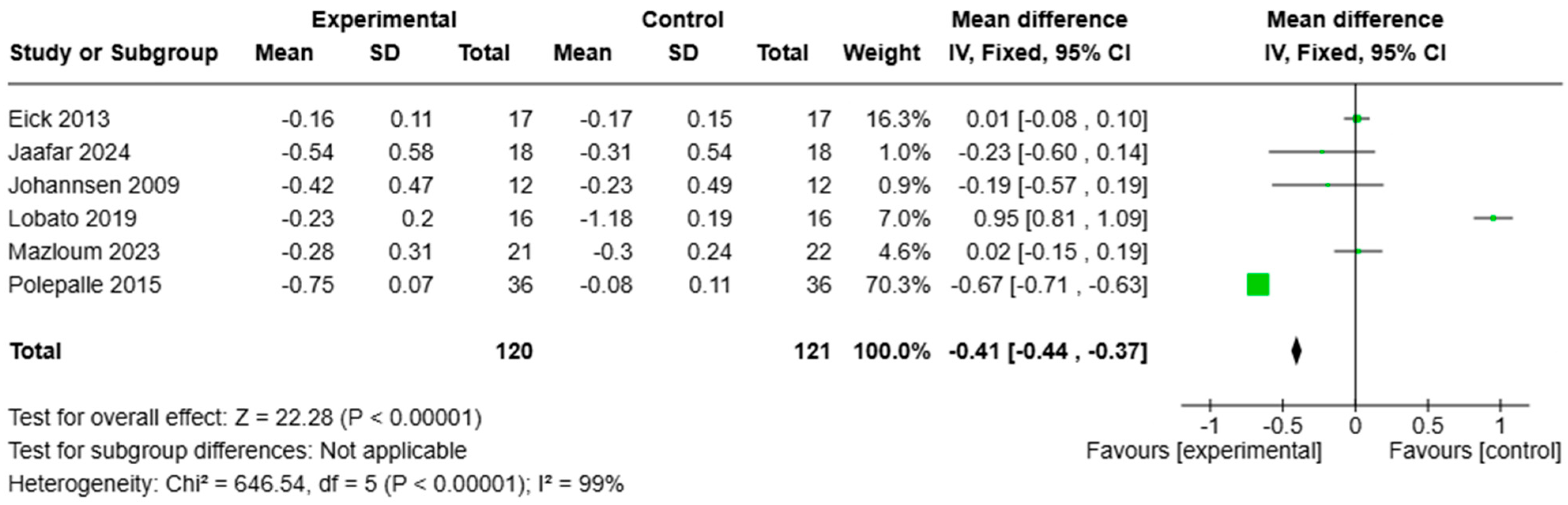
A risk of bias assessment of the RCTs revealed an overall low risk of bias. A total of 15 of the 21 RCTs were classified as low-risk, and 4 had some concerns, while the remaining 2 were high-risk. Concerns arose mainly in the areas of random sequence generation, allocation concealment, and whether the protocol was properly recorded and aligned. The risk of bias assessment is shown in Figure 2.

3.5. Meta-Analysis

Fourteen studies were analyzed quantitatively because they shared similar characteristics such as HA concentration and 12-week follow-up. A meta-analysis was conducted on two subgroups, which were divided into those that used a HA concentration > 0.8% (not including the 1.6%) and those that used a HA concentration < 0.3%. This was performed to compare the effect of HA on reducing probing pocket depth (∆PPD), clinical attachment level (∆CAL), and bleeding on probing (∆BOP) from baseline to 12 week follow-up. The means and standard deviations of the change in PPD, CAL, and BOP from baseline are presented in Figure 3, Figure 4, and Figure 5, respectively.
Baseline scores generally showed no significant differences. The overall analysis yielded the following: [WMD −0.68; 95% CI (−1.14, −0.21) with heterogeneity I2 95%, and p < 0.00001]. The first subgroup analysis (SRP plus HA > 0.8%) showed the following: [WMD −0.73; 95% CI (−1.35, −0.10 with heterogeneity I2 95%, and p < 0.0001]. The second subgroup analysis (SRP plus HA < 0.3%) showed the following: [WMD −0.59; 95% CI (−1.68, 0.50) with heterogeneity I2 95%, and p < 0.0001]. The PPD analysis indicates that the addition of HA 0.8% yields slightly better results, although HA < 0.3% also produces a positive outcome compared to traditional therapy, with a minimal difference between the two concentrations (Figure 3).
For CAL analysis, the baseline scores generally showed no significant differences. Overall analysis yielded [WMD—−124; 95% CI (−1.37, −1.11) with heterogeneity I2 94%, and p < 0.00001]. The first subgroup analysis (SRP plus HA > 0.8%) showed [WMD −132; 95% CI (−1.46, −1.18) with heterogeneity I2 95%, and p < 0.0001]. The second subgroup analysis (SRP plus HA < 0.3%,) showed [WMD −0.69; 95% CI (−1.04, −0.33) with heterogeneity I2 87%, and p = 0.004]
BoP analysis on SRP with HA 0,8% versus SRP showed statistically significant differences with p < 0.00001. Overall analysis showed the following: [WMD −041; 95% CI (−0.44–0.37) with heterogeneity I2 99%, and p < 0.00001] (Figure 5).
The meta-analysis revealed substantial or considerable heterogeneity among the included studies (I2 ranging from 87% to 95%), which considerably limits the reliability and generalizability of the aggregate estimates. Potential sources of heterogeneity may include differences in study design, population characteristics, sample size under investigation, HA concentrations or formulations, outcome measures, and duration of follow-up. Although a random-effects model was applied to account for this variability, the results should be interpreted with caution.

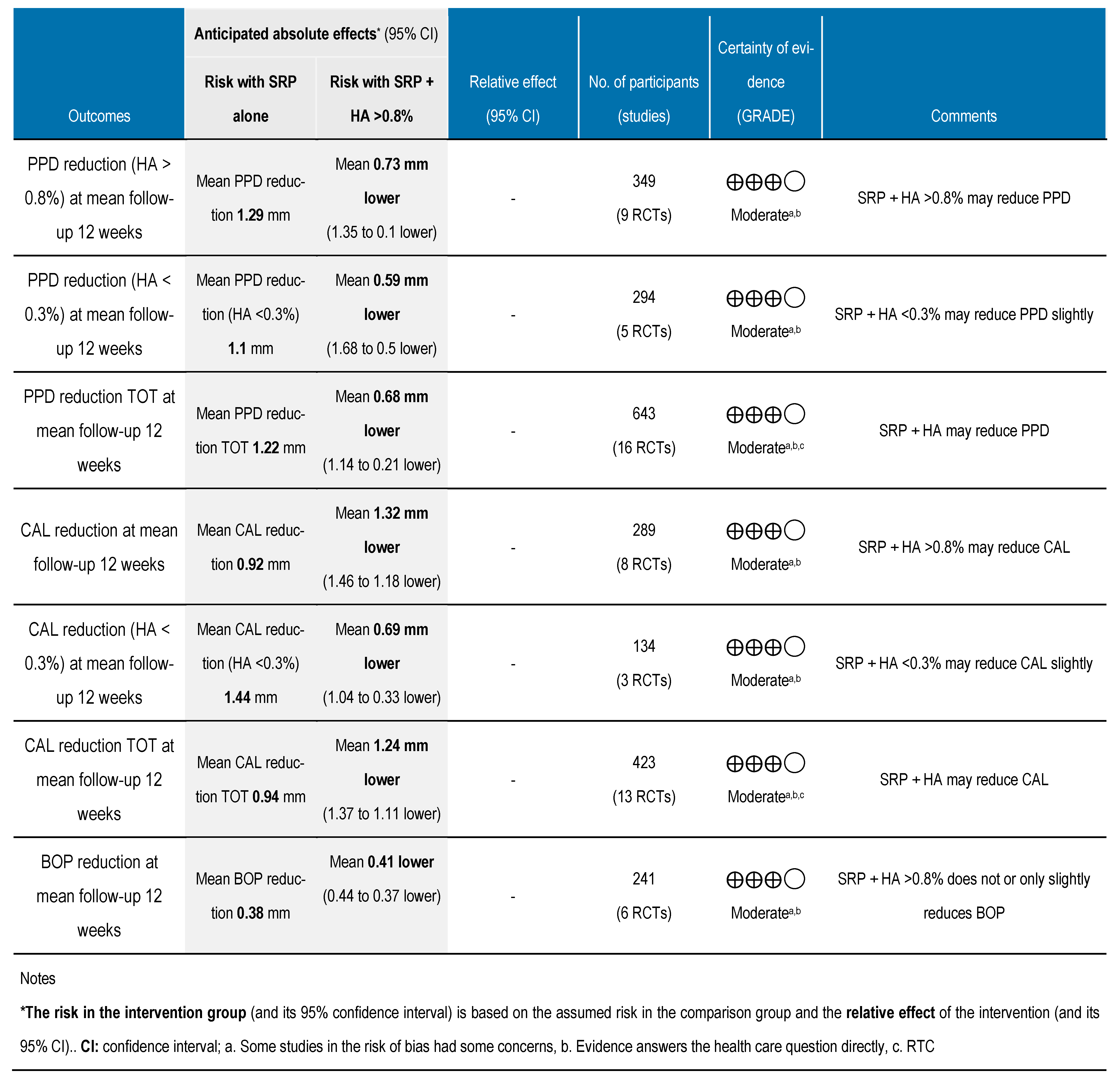
3.6. Grading the Body of Evidence

The certainty of evidence and strength of recommendations were assessed using GRADEpro GDT [17]. The risk of bias ranged from low to serious, and the reporting bias showed some concerns. The analysis suggested moderate certainty in the evidence regarding the additional benefit of HA beyond its application in non-surgical periodontal therapy alone. Its use for enhancing the reduction in probing pocket depth over time may be recommended(Figure 6).

4. Discussion

4.1. Clinical Parameters

The present systematic review discusses the efficacy of topical HA at different concentrations in addition to SRP in non-surgical periodontal therapy for periodontal disease. Non-surgical periodontal therapy and adjunctive therapies have gained increasing attention, with several studies reporting a correlation between periodontitis and systemic disease [39,40]. Non-surgical periodontal treatment seeks to eliminate biofilm through the use of manual, ultrasonic, or sonic devices [3]. Nonetheless, relying on mechanical removal alone may not be sufficient in some clinical scenarios, and the continued presence of periodontopathogens can result in persistent periodontal pockets [41].
Previous research indicates that additional therapy with HA can reduce the need for surgical procedures and improve outcomes for probing pocket depth (PPD) [42]. Three recent systematic reviews [11,43,44] demonstrated the effectiveness of the addition of HA in improving PPD at the 12-week follow-up. A meta-analysis of the PPD parameter, measuring the difference (Δ) between baseline and follow-up, found a statistically significant improvement with the application of HA gel. Nonetheless, due to the high risk of bias and heterogeneity, the need remains for further well-designed RCTs.
In their review, Bertl et al. [45] examined highly heterogeneous studies on HA, its mode of application, and high risk of bias, thus precluding a meta-analysis. For this reason, the application of HA as an adjunct to non-surgical periodontal treatment could not be recommended. There was a statistically significant improvement in PD, CAL, and BOP at test sites in the HA treatment group at both < 0.3% HA and > 0.8% HA. An evaluation of the clinical parameters showed no statistically significant differences in PD between the two concentrations; however, a slightly higher efficacy was noted for treatment with HA > 0.8% than for HA < 0.3%. There was a statistically significant improvement in BOP at the test sites compared with the control sites. The reason for the difference may have been more for the effect of SRP than for the bacteriostatic and non-bactericidal activity of HA, however. A statistically significant difference in efficacy was found for CAL after treatment with HA > 0.8%.
Iorio-Siciliano et al. [38] reported a statistically significant difference in PD and CAL and an acceleration of healing after treatment with HA. Their data suggested a more conspicuous difference between the test and the control group at 12 weeks than at the second follow-up at 24 weeks. Similarly, Polepalle et al. [24] reported improvement in PD, CAL, and BOP, underscoring the beneficial properties of HA in reducing inflammation and accelerating healing. Finally, Olszewska-Czyz et al. [31] reported an improvement in CAL and PD, suggesting the anti-inflammatory action and periodontal regeneration of 1.6% HA.

4.2. Microbiological and Biochemical Analysis

The effect of HA on pathogenic bacteria and the persistence of some species after periodontal treatment led to a decrease in some cases and an increase in others, suggesting the influence of multiple clinical factors. Since its effect appears to be primarily inhibitory rather than eliminative, its use should be reserved as an adjunct to conventional treatment, as demonstrated in the study by Pirnazar et al. [46], where a bacteriostatic but not bactericidal effect of HA was reported. For example, A. actinomycetemcomitans was sensitive to medium and high concentrations, with consistent growth inhibition, and P. gingivalis proliferation was reduced, suggesting the potential of HA to limit the spread of pathogenic bacteria. Furthermore, slight growth inhibition of S. mutans was noted, indicating that HA may modulate the microbial environment in the periodontal setting.
Biomarkers of inflammation, immune response (increase in beta-defensin 2 expression in treated sites), systemic parameters (e.g., leukocyte count), and C-reactive protein (progressive reduction in inflammation) were measured. Aydinyurt et al. [29] analyzed the levels of enzymes and antioxidants in gingival crevicular fluid and observed a significant reduction in adenosine deaminase (ADA), suggesting a potential decrease in inflammation. In addition, catalase (CAT) and glutathione (GSH) levels were increased, indicating improved antioxidant response. The greater improvement initially noted in the test group than in the controls diminished over time, however.

4.3. Formulation of HA and Its Clinical Impact

Different formulations of hyaluronic acid (HA), such as cross-linked versus linear forms, may influence clinical outcomes due to variations in bioavailability, viscosity, and degradation rates.
The studies included in this review often lacked detailed reporting on the type of HA used, limiting our ability to draw firm conclusions regarding formulation-specific effects [4,5,6].
From a clinical perspective, HA could be considered as a supplementary agent following scaling and root planing (SRP), particularly in patients with moderate to severe periodontitis or where enhanced soft tissue healing is desired. However, clear clinical guidelines are still lacking, and treatment protocols remain heterogeneous across studies.

4.4. Study Limitations

One of the main limitations of the meta-analysis was the short-term follow-up. A long follow-up period of 6 or 12 months would have facilitated a more accurate assessment of the effects of HA treatment over time and to compare its efficacy with SRP alone. Furthermore, there was considerable heterogeneity (I2 > 90%) across the studies, which impacted the reliability of the results. Such heterogeneity could result from differences in how periodontal cases are defined, as well as in the diverse stages and grades of periodontal disease. This review did not include formal sensitivity analyses to explore sources of heterogeneity, which is a limitation. It is recommended that future systematic reviews, with more homogeneous data and greater availability of study-level variables, include such analyses. A further limitation concerns the BOP parameter, for which a comprehensive analysis could not be performed owing to the few studies that used a HA concentration < 0.3%. Consequently, the meta-analysis included only studies with a HA concentration > 0.8% per cent and precluded the assessment of efficacy in relation to the percentage used and limiting itself to an absolute analysis of the treatment effect.
An important limitation of our meta-analysis is the variability in the classification of periodontal disease among the included studies. Different classification criteria (e.g., mild, moderate, and severe periodontitis) were used, which may have affected the aggregated results. Despite the review and inclusion of studies published by different sources, no funnel plots or Egger tests were conducted to assess publication bias. This might represent a limitation in our meta-analysis, as a publication bias might have influenced the results. Future analyses should include these tests to explore more critically the possibility of publication bias and its influence on the overall results.

4.5. Implications for Future Research

Future studies should aim to standardize the type, concentration, and application protocols of HA to facilitate more robust comparisons. Moreover, longer follow-up durations, better reporting of HA formulations, and subgroup analyses based on disease severity are recommended to clarify the clinical relevance of HA in periodontal therapy [25,26].

5. Conclusions

Despite the limitations of the current review, it can be concluded that, based on the evidence from a 12-week follow-up, the local application of 0.8% HA gel combined with scaling and root planing (SRP) positively impacts periodontal health in patients with moderate-to-severe chronic periodontitis. Although studies on 0.2% HA are still limited in the literature, its use has shown benefits even at smaller dosages. Indeed, clinical parameters such as PD, BOP, and CAL, along with biochemical and microbiological parameters, improved in the experimental group compared to the control group. Considering the available evidence and the methodological limitations of the meta-analysis, the statistical analysis indicates that adjunctive HA may aid in achieving and maintaining periodontal health.

Author Contributions

Conceptualization, A.P. and V.M.; methodology, A.P., V.M., A.A. and G.B.; software, P.M. and F.M.; validation, G.L., A.P. and D.D.S.; formal analysis, V.M., A.A. and G.B.; investigation, A.P. and V.M.; resources, P.M. and F.M.; data curation, A.P., V.M., A.A. and G.B.; writing—original draft preparation, A.P., V.M., A.A. and G.B.; writing—review and editing, A.P., G.L. and D.D.S.; visualization, A.P.; supervision, G.L. and D.D.S.; project administration, A.P. and D.D.S. All authors have read and agreed to the published version of the manuscript.

Funding

This research received no external funding.

Institutional Review Board Statement

The review was registered in PROSPERO, the international registry for systematic reviews, under the identifier number CRD42024629547.

Informed Consent Statement

Not applicable.

Data Availability Statement

The original contributions presented in the study are included in the article, further inquiries can be directed to the corresponding author.

Acknowledgments

The authors have reviewed and edited the output and take full responsibility for the content of this publication.

Conflicts of Interest

The authors declare no conflicts of interest.

Abbreviations

The following abbreviations are used in this manuscript:
RCTRandomized Clinical Trial
NINo Information
PDProbing Depth
PPDPocket Probing Depth
CALClinical Attachment Level
BOPBleeding On Probing
SRPScaling and Root Planing
HAHyaluronic Acid
PIPlaque Index
RALRelative Attachment Level
GCCIGingival Color Change Index
EIBIEastman Interdental Bleeding Index
PBIPapillary Bleeding Index
GIGingival Index
FMPSFull Mouth Plaque Score
GRHGingival Recession level
APIApproximal Plaque Index
RECRecession
MSBIMean Sulcus Bleeding Index
FMPSFull Mouth Plaque Score
FMBSFull Mouth Bleeding Index

References

  1. Nocini, R.; Lippi, G.; Mattiuzzi, C. Periodontal disease: The portrait of an epidemic. J. Public Health Emerg. 2020, 4. ePub ahead of print. [Google Scholar] [CrossRef]
  2. Könönen, E.; Gursoy, M.; Gursoy, U.K. Periodontitis: A multifaceted disease of tooth-supporting tissues. J. Clin. Med. 2019, 8, 1135. [Google Scholar] [CrossRef]
  3. Rabbani, G.M.; Ash, M.M.; Caffesse, R.G. The effectiveness of subgingival scaling and root planing in calculus removal. J. Periodontol. 1981, 52, 119–123. [Google Scholar] [CrossRef] [PubMed]
  4. Pardo, A.; Fiorini, V.; Zangani, A.; Faccioni, P.; Signoriello, A.; Albanese, M.; Lombardo, G. Topical agents in biofilm disaggregation: A systematic review and meta-analysis. J. Clin. Med. 2024, 13, 2179. [Google Scholar] [CrossRef]
  5. Casale, M.; Moffa, A.; Vella, P.; Sabatino, L.; Capuano, F.; Salvinelli, B.; Lopez, M.A.; Carinci, F.; Salvinelli, F. Hyaluronic acid: Perspectives in dentistry. A systematic review. Int. J. Immunopathol. Pharmacol. 2016, 29, 572–582. [Google Scholar] [CrossRef]
  6. Waingade, M.; Medikeri, R.S.; Gaikwad, S. Effectiveness of hyaluronic acid in the management of oral lichen planus: A systematic review and meta-analysis. J. Dent. Anesth. Pain Med. 2022, 22, 405–417. [Google Scholar] [CrossRef]
  7. Zotti, F.; Nocini, R.; Capocasale, G.; Fior, A.; Peretti, M.; Albanese, M. Malignant transformation evidences of oral lichen planus: When the time is of the essence. Oral Oncol. 2020, 104, 104594. [Google Scholar] [CrossRef] [PubMed]
  8. Bertossi, D.; Malchiodi, L.; Albanese, M.; Nocini, R.; Nocini, P. Nonsurgical rhinoplasty with the novel hyaluronic acid filler VYC-25L: Results using a nasal grid approach. Aesthetic Surg. J. 2021, 41, NP512–NP520. [Google Scholar] [CrossRef]
  9. Vela, O.-C.; Boariu, M.; Rusu, D.; Iorio-Siciliano, V.; Sculean, A.; Stratul, S.-I. Clinical and radiographic evaluation of intrabony periodontal defects treated with hyaluronic acid or enamel matrix proteins: A 6-month prospective study. Oral Health Prev. Dent. 2024, 22, 257–270. [Google Scholar]
  10. Megally, A.; Zekeridou, A.; Cancela, J.; Giannopoulou, C.; Mombelli, A. Short ultrasonic debridement with adjunctive low-concentrated hypochlorite/amino acid gel during periodontal maintenance: Randomized clinical trial of 12 months. Clin. Oral Investig. 2020, 24, 201–209. [Google Scholar] [CrossRef]
  11. Eliezer, M.; Imber, J.-C.; Sculean, A.; Pandis, N.; Teich, S. Hyaluronic acid as adjunctive to non-surgical and surgical periodontal therapy: A systematic review and meta-analysis. Clin. Oral Investig. 2019, 23, 3423–3435. [Google Scholar] [CrossRef] [PubMed]
  12. Ramanauskaite, E.; Machiulskiene Visockiene, V.; Shirakata, Y.; Friedmann, A.; Pereckaite, L.; Balciunaite, A.; Dvyliene, U.M.; Vitkauskiene, A.; Baseviciene, N.; Sculean, A. Microbiological effects of sodium hypochlorite/-amino acids and cross-linked hyaluronic acid adjunctive to non-surgical periodontal treatment. Oral Health Prev. Dent. 2024, 22, 171–180. [Google Scholar] [PubMed]
  13. Page, M.J.; McKenzie, J.E.; Bossuyt, P.M.; Boutron, I.; Hoffmann, T.C.; Mulrow, C.D.; Shamseer, L.; Tetzlaff, J.M.; Akl, E.A.; Brennan, S.E.; et al. The PRISMA 2020 statement: An updated guideline for reporting systematic reviews. BMJ 2021, 372, n71. [Google Scholar] [CrossRef] [PubMed]
  14. Sterne, J.A.C.; Savović, J.; Page, M.J.; Elbers, R.G.; Blencowe, N.S.; Boutron, I.; Cates, C.J.; Cheng, H.-Y.; Corbett, M.S.; Eldridge, S.M.; et al. RoB 2: A revised tool for assessing risk of bias in randomised trials. BMJ 2019, 366, l4898. [Google Scholar] [CrossRef]
  15. Higgins, J.P.T.; Thomas, J.; Chandler, J.; Cumpston, M.; Li, T.; Page, M.J.; Welch, V.A. (Eds.) Cochrane Handbook for Systematic Reviews of Interventions version 6.5 (updated August 2024). Cochrane. 2024. Available online: www.training.cochrane.org/handbook (accessed on 19 March 2025).
  16. Pardo, A.; Baccini, F.; De Manzoni, R.; Viviani, M.; Brentaro, S.; Zangani, A.; Faccioni, P.; Luciano, U.; Zuffellato, N.; Signoriello, A.; et al. Air polishing therapy in supportive periodontal treatment: A systematic review. J. Appl. Cosmetol. 2023, 41 (Suppl. S1). [Google Scholar] [CrossRef]
  17. McMaster University and Evidence Prime. GRADEpro GDT: GRADEpro Guideline Development Tool. Available online: https://gradepro.org (accessed on 10 April 2025).
  18. Xu, Y.; Höfling, K.; Fimmers, R.; Frentzen, M.; Jervøe-Storm, P.M. Clinical and microbiological effects of topical subgingival application of hyaluronic acid gel adjunctive to scaling and root planing in the treatment of chronic periodontitis. J. Periodontol. 2004, 75, 1114–1118. [Google Scholar] [CrossRef]
  19. Johannsen, A.; Tellefsen, M.; Wikesjö, U.; Johannsen, G. Local delivery of hyaluronan as an adjunct to scaling and root planing in the treatment of chronic periodontitis. J. Periodontol. 2009, 80, 1493–1497. [Google Scholar] [CrossRef]
  20. Koshal, A.; Bolt, R.; Galgut, P.N. A Comparison in Postoperative Healing of Sites Receiving Non-Surgical Debridement Augmented With and Without a Single Application of Hyaluronan 0.8% Gel. Available online: http://www.takara-net.com (accessed on 10 April 2025).
  21. Chauhan, A.; Bains, V.; Gupta, V.; Singh, G.; Patil, S. Comparative analysis of hyaluronan gel and xanthan-based chlorhexidine gel, as adjunct to scaling and root planing with scaling and root planing alone in the treatment of chronic periodontitis: A preliminary study. Contemp. Clin. Dent. 2013, 4, 54–61. [Google Scholar]
  22. Eick, S.; Renatus, A.; Heinicke, M.; Pfister, W.; Stratul, S.; Jentsch, H. Hyaluronic acid as an adjunct after scaling and root planing: A prospective randomized clinical trial. J. Periodontol. 2013, 84, 941–949. [Google Scholar] [CrossRef]
  23. Rajan, P.; Baramappa, R.; Rao, N.M.; Pavaluri, A.K.; Indeevar, P.; Ur Rahaman, S. Hyaluronic acid as an adjunct to scaling and root planing in chronic periodontitis. A randomized clinical trail. J. Clin. Diagn. Res. 2014, 8, ZC11–ZC14. [Google Scholar] [CrossRef]
  24. Polepalle, T.; Srinivas, M.; Swamy, N.; Aluru, S.; Chakrapani, S.; Chowdary, B.A. Local delivery of hyaluronan 0.8% as an adjunct to scaling and root planing in the treatment of chronic periodontitis: A clinical and microbiological study. J. Indian Soc. Periodontol. 2015, 19, 37–42. [Google Scholar] [CrossRef] [PubMed]
  25. Shah, S.A.; Vijayakar, H.N.; Rodrigues, S.V.; Mehta, C.J.; Mitra, D.K.; Shah, R.A. To compare the effect of the local delivery of hyaluronan as an adjunct to scaling and root planing versus scaling and root planing alone in the treatment of chronic periodontitis. J. Indian Soc. Periodontol. 2016, 20, 549–556. [Google Scholar]
  26. Sharma, V.; Gupta, R.; Dahiya, P.; Kumar, M. Comparative evaluation of coenzyme Q10-based gel and 0.8% hyaluronic acid gel in treatment of chronic periodontitis. J. Indian Soc. Periodontol. 2016, 20, 374–380. [Google Scholar] [CrossRef]
  27. Al-Shammari, N.M.; Shafshak, S.M.; Ali, M.S. Effect of 0.8% hyaluronic acid in conventional treatment of moderate to severe chronic periodontitis. J. Contemp. Dent. Pract. 2018, 19, 527–534. [Google Scholar] [CrossRef] [PubMed]
  28. Lobato, J.C.R.F.; Dos Santos Vilhena, M.A.; Izidoro, C.; Alves, R.C.; Proença, L. Single application of 0.8% hyaluronic acid as a coadjuvant of nonsurgical treatment in nonsmoking patients with periodontitis: A split-mouth, randomized, controlled pilot clinical trial. J. Indian Soc. Periodontol. 2019, 23, 545–548. [Google Scholar] [CrossRef] [PubMed]
  29. Sahin Aydinyurt, H.; Akbal, D.; Altindal, D.; Bozoglan, A.; Seckin Ertugrul, A.; Demir, H. Evaluation of biochemical and clinical effects of hyaluronic acid on non-surgical periodontal treatment: A randomized controlled trial. Ir. J. Med. Sci. 2020; in press. [Google Scholar] [CrossRef]
  30. Nguyen, T.T.; Ho, H.T.; Huynh, N.C.; Dien, V.H.A.; Vo, T.L. Hyaluronic acid 0.2% application enhanced periodontitis treatment in non-surgical phase. J. Stomatol. 2021, 74, 76–83. [Google Scholar] [CrossRef]
  31. Olszewska-Czyz, I.; Kralik, K.; Prpic, J. Biomolecules in dental applications: Randomized, controlled clinical trial evaluating the influence of hyaluronic acid adjunctive therapy on clinical parameters of moderate periodontitis. Biomolecules 2021, 11, 1491. [Google Scholar] [CrossRef]
  32. Vajawat, M.; Rao, D.P.C.; Kumar, G.S.V.; Rajeshwari, K.G.; Hareesha, M.S. Local delivery of hyaluronic acid as an adjunct to scaling and root planing in the treatment of chronic periodontitis in smokers and non-smokers. J. Indian Soc. Periodontol. 2022, 26, 471–477. [Google Scholar] [CrossRef]
  33. Mazloum, T.A.; Amhaz, G.S.; Abdullah, G.H.; Aboelsaad, N.M. Efficiency of hyaluronic acid versus red injectable platelet-rich fibrin (I-PRF) in treatment of stage III periodontitis: Randomized controlled clinical trial. Int. Arab. J. Dent. 2023, 14, 5–12. [Google Scholar] [CrossRef]
  34. Bertl, K.; Vlachou, S.; Pandis, N.; Zampelis, A.; Stavropoulos, A. Repeated local delivery of hyaluronic acid gel as adjunctive treatment of residual pockets in periodontitis patients undergoing supportive periodontal care: A randomized controlled clinical trial. Clin. Oral Investig. 2024, 28, 451–459. [Google Scholar] [CrossRef] [PubMed]
  35. Adib Jaafar, M.; Voo Vui Ching, E.; Nisha Ali, S.; Subramaniam, J.; Ahmad Yaziz, Y. Hyaluronic acid in managing deep residual pockets in stage III–IV periodontitis: A randomized controlled trial. J. Dent. Indones. 2024, 31, 88–95. [Google Scholar] [CrossRef]
  36. El-Emam, E.; Ezzat, O.; Almalahy, H. Efficacy of locally delivered hyaluronic acid gel as an adjunctive to non-surgical management of stage II or stage III periodontitis: A randomized controlled trial with microbiological analysis. Egypt. Dent. J. 2024, 70, 221–233. [Google Scholar] [CrossRef]
  37. Ramanauskaite, E.; Machiulskiene, V.; Dvyliene, U.M.; Eliezer, M.; Sculean, A. Clinical evaluation of a novel combination of sodium hypochlorite/amino acid and cross-linked hyaluronic acid adjunctive to non-surgical periodontal treatment: A case series. Oral Health Prev. Dent. 2023, 21, 279–284. [Google Scholar] [PubMed]
  38. Iorio-Siciliano, V.; Blasi, A.; Mauriello, L.; Salvi, G.E.; Ramaglia, L.; Sculean, A. Non-surgical treatment of moderate periodontal intrabony defects with adjunctive cross-linked hyaluronic acid: A single-blinded randomized controlled clinical trial. J. Clin. Periodontol. 2024; in press. [Google Scholar] [CrossRef]
  39. Pardo, A.; Barilli, A.; Signoriello, A.; Gualtieri, M.; Brancato, G.; Colapinto, G.; Lombardo, G.; Albanese, M. Oral health conditions and hygiene procedures in patients with Parkinson’s disease: A systematic review. Explor. Med. 2024, 4, 852–869. [Google Scholar] [CrossRef]
  40. Nocini, R.; Favaloro, E.J.; Sanchis-Gomar, F.; Lippi, G. Periodontitis, coronary heart disease and myocardial infarction: Treat one, benefit all. Blood Coagul. Fibrinolysis 2020, 31, 339–345. [Google Scholar] [CrossRef]
  41. Polizzi, E.; Tetè, G. Manual vs mechanical oral hygiene procedures: Has the role of the dental hygienist in phase 2 post-lockdown really changed? Oral Health Prev. Dent. 2020, 18, 1031–1037. [Google Scholar]
  42. Sanz, M.; Herrera, D.; Kebschull, M.; Chapple, I.; Jepsen, S.; Berglund, T.; Sculean, A.; Tonetti, M.S. Evidence-based clinical practice guideline for treatment of stage I–III periodontitis. J. Evid. Based Dent. Pract. 2021, 21, 101638. [Google Scholar]
  43. Shirbhate, U.; Bajaj, P. Injectable and self-invigorating hydrogel applications in dentistry and periodontal regeneration: A literature review. Cureus 2022, 14, e29248. [Google Scholar] [CrossRef]
  44. Gegout, P.-Y.; Stutz, C.; Huck, O. Gels as adjuvant to non-surgical periodontal therapy: A systematic review and meta-analysis. Heliyon 2023, 9, e17789. [Google Scholar] [CrossRef] [PubMed]
  45. Bertl, K.; Bruckmann, C.; Isberg, P.; Klinge, B.; Gotfredsen, K.; Stavropoulos, A. Hyaluronan in non-surgical and surgical periodontal therapy: A systematic review. J. Clin. Periodontol. 2015, 42, 236–247. [Google Scholar] [CrossRef] [PubMed]
  46. Pirnazar, P.; Wolinsky, L.; Nachnani, S.; Haake, S.; Pilloni, A.; Bernard, G.W. Bacteriostatic effects of hyaluronic acid. J. Periodontol. 1999, 70, 370–374. [Google Scholar] [CrossRef] [PubMed]
Figure 1. PRISMA Flow Chart.
Applsci 15 05975 g001
Figure 2. RoB 2 tool (risk of bias assessment) for RCTs [18,19,20,21,22,23,24,25,26,27,28,29,30,31,32,33,34,35,36,37,38].
Applsci 15 05975 g002
Figure 3. Forest plot of the meta-analysis of PPD [18,19,20,22,23,24,25,28,32,33,34,35,36].
Applsci 15 05975 g003
Figure 4. Forest plot of the meta-analysis of CAL [18,19,22,23,24,28,32,33,35,36].
Applsci 15 05975 g004
Figure 5. Forest plot of the meta-analysis of BOP [19,22,24,28,33,35].
Applsci 15 05975 g005
Figure 6. Assessment of quality of evidence for hyaluronic acid therapy.
Applsci 15 05975 g006
Table 1. Research string for databases.
DatabaseSearch TermsBoolean OperatorsNotes
PubMed, Cochrane Library, Scopus, Web of Science(“Periodontitis”[MeSH] OR non-surgical periodontal therapy OR periodontal disease) AND (“Hyaluronic Acid”[MeSH] OR hyaluronan OR hyaluronate)AND, ORUp to January 2025
PubMed, Cochrane Library, Scopus, Web of Science(“Hyaluronic Acid”[MeSH] OR hyaluro*) AND (“Periodontal Diseases”[MeSH] OR periodont*) AND non-surgicalAND, ORUp to January 2025
PubMed, Cochrane Library, Scopus, Web of Science(“Hyaluronic Acid” OR hyaluro*) AND (Periodontitis OR periodont*) AND non-surgicalAND, ORUp to January 2025
Table 2. Study characteristics.
First Author and YearStudy DesignStudy DesignCountrySample Size and SexMean Age/Rang
(Years)
Test and Control
Interventions
Follow-UpOutcome
Xu 2004 [18]RCTSplit-mouthChina20 (11 M, 9 F)48.6 1 SRP
2 SRP + 1 mL 0.2% HA gel
6 and 12 wksBOP, CAL, PD
Johannsen 2009 [19]RCTSplit-mouthSweden12 (sex not stated)42–631 SRP
2 SRP + 0.2 mL + 0.8% HA gel
12 wksBOP, PI, CAL, PD
Koshal 2012 [20]RCTRandom assignmentAfrica52 (sex not stated)/1 SRP + placebo
2 SRP + HA
12 wksGI, PD
Chauhan 2013 [21]RCT with biochemical analysisRandom assignmentIndia11 (sex not stated)30–651 SRP
2 SRP + HA gel
3 SRP + HA gel + CHX gel
6 and 12 wksPD, CAL, PI
Eick 2013 [22]RCT with microbiological analysisRandom assignmentSwitzerland34 (sex not stated)41–72 1 SRP
2 SRP + 0.8% HA gel in office + 0.2% HA gel at home
12, 24, 36 wksBOP, PI, CAL, PD
Rajan 2014 [23]RCTSplit-mouthIndia33 (15 M, 18 F)NI1 SRP
2 SRP + 0.2% HA gel
4 and 12 wksGI, PI, BOP, PPD, CAL
Polepalle 2015 [24]RCTSplit-mouthIndia18 (11 M, 7 F)30–60
45
1 SRP
2 SRP + 0.2 mL 0.8% HA gel
4 and 12 wksPI, BOP, PPD, CAL, CFU
Shah
2016 [25]
RCTSplit-mouthIndia9 (sex not stated)30–60
45
1 SRP
2 SRP + 0.8% HA gel + periodontal dressing
4 and 12 wksGI, PI, PPD, RAL
Sharma 2016 [26]RCTSplit-mouthIndia24 (sex not stated)25–55
40
1 SRP + d coenzyme Q gel
2 SRP + 0.8% HA gel
3 SRP alone
1, 2, 6 wksPI, EIBI, GCCI, PD, CAL
Al-Shammari 2018 [27]RCT with biochemical analysisSplit-mouthKingdom of Saudi Arabia24 (14 F, 10 M) 24–57
40.5
1 SRP alone
2 SRP + 0.8% HA
6 and 12 wksPI, GI, PBI, PPD, CAL, human beta defensin-2 (hBD-2) expression
Lobato 2019 [28]RCTSplit-mouthIndia1641–68 54.51 SRP
2 SRP + 0.8% HA
6 and 12 wksFMPS, GI, BOP, PD CAL
Aydinyurt 2020 [29]RCT with biochemical analysisRandom assignmentTurkey96 (40 M, 56 F) 22–55
38.5
1 SRP + saline irrigation
2 SRP + 0.2% HA gel
3 SRP + 0.2% HA hydrogel mouth rinse
4 SRP + 0.2% HA hydrogel mouth rinse + 0.2% HA gel
1, 2, 4 wksPI, GI, BOP, PD, GRH, CAL, biochemicals (ADA, CAT, GSH)
Nguyen 2021 [30]RCT with microbiological analysisSplit-mouthVietnam34 ni1 SRP + 0.9% NaCl solution rinse
2 SRP + 0.9% NaCl solution rinse + 1 mL 0.2% HA gel
1, 2, 3, 6 wksPI, GI, PPD, CAL, BOP
Olszewska 2021 [31]RCTRandom assignmentPoland100 (51 F, 49 M)25–65
45
1 SRP + HA gel (1.6% reticular HA + 0.2% HA natural)
2 SRP alone
12 wksAPI, BOP, PPD, CAL
Vajawat 2022 [32]RCT with microbiological analysisSplit-mouthIndia2435–65
50
1: SRP + placebo
2: SRP + 0.8% HA gel
4, 12 wksCAL, PD, BI, GI, PI
Mazloum 2023 [33]RCTRandom assignmentLebanon75 20–60
40
1 SRP + 0.8% HA gel
2 SRP + with red i-PRF
3 SRP alone
4, 8, 12 wksCAL, PD, PI, GI, BOP
Bertl
2024 [34]
RCTRandom assignmentSweden56 (37 F, 19 M)35–75
55
1 SRP + 0.3% HA gel
2 SRP + saline solution
12, 24, 36, 48 wksPPD, PI, GI, BOP, CAL
Jaafar 2024 [35]RCTRandom assignmentMalaysia36 (25 F, 11 M)
30–60
45
1 SRP + 0.8% HA gel
2 SRP alone
12 wksPI, PD, CAL, BOP, REC
El Emam 2024 [36]RCTRandom assignmentEgypt28 (14 M, 14 F)28–40
34
1 SRP + local 0.2% HA gel
2 SRP alone
12 wksPI, BI, CAL, MSBI, PD
Ramanaus
kaite 2023 [37]
RCTRandom assignmentLithuania2130–721 SRP alone
2 SRP+ crosslinked HA gel baseline
12 and 24 wksPD, CAL, BOP, PI
Iorio-Siciliano 2025 [38]RCTRandom assignmentItaly38 (14 M, 24 F)Test 49.3 ±11.6
Control 50.8 ± 10.8
1 SRP
2 SRP + HA
4, 12, 24 wksPD, FMPS, FMBS, CAL
Abbreviations: wk(s), week(s); RCT, randomized clinical trial; NI, no information; PD, probing depth; PPD, pocket probing depth; CAL, clinical attachment level; BOP, bleeding on probing; SRP, scaling and root planing; HA, hyaluronic acid; PI, Plaque Index; RAL, relative attachment level; GCCI, Gingival Color Change Index; EIBI, Eastman Interdental Bleeding Index; PBI, Papillary Bleeding Index; GI, Gingival Index; FMPS, Full Mouth Plaque Score; GRH, gingival recession level; API, Approximal Plaque Index; REC, recession; CFUs, colony-forming units; MSBI, Mean Sulcus Bleeding Index; FMPS, Full Mouth Plaque Score; FMBS, Full Mouth Bleeding Index.
Disclaimer/Publisher’s Note: The statements, opinions and data contained in all publications are solely those of the individual author(s) and contributor(s) and not of MDPI and/or the editor(s). MDPI and/or the editor(s) disclaim responsibility for any injury to people or property resulting from any ideas, methods, instructions or products referred to in the content.

