Article

Kinetic Responses to Acute Blood Flow Restriction Exposure in Young Physically Active Women During Isometric Mid-Thigh Pull

by
Sebastián Aliste-Flores
1,2,
Luis Javier Chirosa-Ríos
1,
Ignacio Chirosa-Ríos
1 and
Daniel Jerez-Mayorga
1,3,*
1
Department of Physical Education and Sports, Faculty of Sport Sciences, University of Granada, 18071 Granada, Spain
2
Escuela de Ciencias del Deporte y Actividad Física, Facultad de Salud, Universidad Santo Tomás, Santiago 8370003, Chile
3
Exercise and Rehabilitation Sciences Institute, Faculty of Rehabilitation Sciences, Universidad Andres Bello, Santiago 7560913, Chile
*
Author to whom correspondence should be addressed.
Appl. Sci. 2025, 15(11), 5866; https://doi.org/10.3390/app15115866
Submission received: 10 April 2025 / Revised: 16 May 2025 / Accepted: 21 May 2025 / Published: 23 May 2025

Abstract

:
The application of blood flow restriction (BFR) during resistance exercise enhances muscular adaptations under low-load conditions. However, its acute effects on explosive neuromuscular performance, particularly on kinetic variables such as the rate of force development (RFD), impulse, and peak force remain poorly understood in women. Twenty-five participants underwent randomized sessions under three occlusion conditions (0%, 40%, and 80% limb occlusion pressure), followed by isometric mid-thigh pull (IMTP) assessments at five time points (pre-exercise; post-exercise; and 5, 10, and 15 min post-exercise). Peak force, impulse, and RFD were analyzed across early (0–50 ms), mid (51–150 ms), and late (151–250 ms) time intervals. BFR did not result in statistically significant alterations in RFD or isometric force production at any time or pressure. These findings indicate that acute BFR application, even when volitional fatigue is induced, does not substantially impair neuromuscular function in isometric settings. These findings indicate that acute exposure to BFR, even under fatiguing conditions, does not substantially impair isometric force production or explosive performance in young physically active women. These results support the task-specific and temporally dependent nature of neuromuscular responses to BFR, highlighting the need for population-specific approaches in BFR programming.

1. Introduction

In sports, being fast is not just an advantage; it is often the defining factor between success and failure. The ability to rapidly generate force underlies performance in explosive tasks and is quantified by the rate of force development (RFD), which is a key marker of neuromuscular capacity [1,2]. Assessments such as the isometric mid-thigh pull (IMTP) are widely used to evaluate neuromuscular capacity and monitor training adaptations via force–time analysis [3,4,5]. The RFD is driven by neuromuscular factors, such as motor unit recruitment, firing frequency, synchronization, and type II muscle fiber composition [6,7]. Tendon stiffness aids force transmission and reduces electromechanical delay, whereas intramuscular coordination enhances RFD efficiency [8,9]. High-velocity and maximal-intent resistance training improves neuromuscular properties and the RFD [10]. Although blood flow restriction training (BFRT) has been extensively studied in relation to hypertrophy and dynamic strength, its impact on the RFD under isometric conditions—particularly in female populations—remains poorly understood. Women remain underrepresented in neuromuscular research, despite evidence showing sex-based differences in fatigue resistance, motor unit behavior, and fiber-type distribution [11,12]. Moreover, recent studies have yielded inconsistent findings regarding the effects of BFR on neuromuscular fatigue and performance, with outcomes varying based on training status, occlusion pressure, and task type (dynamic vs. isometric) [5,13,14]. While BFR has shown benefits in both untrained and clinical populations, responses may differ across demographic groups and loading configurations, highlighting the need for population- and task-specific studies [15,16].
Neuromuscular fatigue, resulting from central (impaired neural drive) and peripheral (excitation–contraction disruption) mechanisms, diminishes the peak force (PF), rate of force development (RFD), and impulse (I) during maximal isometric contraction [5,13]. While these effects are well documented in men, research on young active women remains limited despite sex-based differences in fatigue resistance, motor unit behavior, and muscle fiber composition [14,15]. Women may exhibit greater reliance on central fatigue mechanisms and demonstrate distinct kinetic impairments, particularly during explosive tasks [16,17]. Recent computational modeling and biomechanical analyses have also highlighted the influence of fatigue on joint loading and force modulation strategies, which may be relevant when interpreting sex-specific neuromuscular responses [18]. Studies utilizing the isometric mid-thigh pull (IMTP) have predominantly focused on men, leaving the fatigue–RFD relationship in women insufficiently explored [19,20].
BFRT involves the application of external pressure to restrict venous return during low-load resistance exercise, thereby promoting hypertrophy and strength adaptations similar to those observed after high-load training [11,21]. However, BFRT accelerates neuromuscular fatigue owing to ischemia, metabolite accumulation, and increased motor unit recruitment [22,23]. Although BFRT responses have been well studied in men, sex-specific fatigue patterns, particularly in young active women, remain underexplored [24,25]. Additionally, studies have shown that responses to BFR vary depending on factors such as occlusion pressure, training status, and whether the task is dynamic or isometric [16,17,25]. These inconsistencies underscore the need for task- and population-specific analyses to understand BFR’s true impact on neuromuscular performance, particularly under acute conditions.
The isometric mid-thigh pull (IMTP) is a valid and reliable tool for assessing maximal isometric strength and rate of force development (RFD), with intraclass correlation coefficients (ICCs) exceeding 0.90 for peak force and RFD measures [5]. Its accuracy is attributed to standardized positioning, minimal technical demand, and reduced movement variability compared with dynamic tests [19]. The IMTP peak force correlates well with performance in weightlifting, sprinting, and jumping, whereas RFD metrics reflect neuromuscular attributes crucial to explosive performance [10,26]. The test incorporates diverse muscle groups, revealing enhanced activation of the gluteus maximus, gluteus medius, and hamstrings during the unilateral stance compared to the bilateral stance [27]. Furthermore, unilateral execution elicits substantial engagement of the quadriceps femoris muscle [28]. However, differences in the knee and hip angles can affect the outcomes, and RFD measurements show higher variability and require familiarization trials and strict data processing [19]. This study utilized a validated instrument known as a functional electromechanical dynamometer (FEMD). The device facilitates precise adjustment of individual limb dimensions while enabling efficient examination of multiple participants in a reliable and valid fashion [29,30,31,32].
The present investigation aimed to analyze how varying degrees of occlusion influence RFD kinetics during IMTP performance after fatiguing single-joint action in young women. We hypothesized that higher occlusion levels (specifically 80% LOP) would lead to greater peak force production and enhanced rate of force development (RFD) across early, mid, and late intervals compared to lower occlusion levels and the non-occluded condition

2. Materials and Methods

2.1. Study Design

This randomized, repeated-measures, cross-sectional study investigated the acute effects of blood flow restriction (BFR) on isometric force production and rate of force development (RFD). Each participant completed four laboratory sessions over two weeks separated by 48-h intervals to minimize residual fatigue. All sessions were conducted at the same time of day to control for circadian variation. A within-subject design was applied to compare three BFR conditions (0%, 40%, and 80% limb occlusion pressure [LOP]), enhancing statistical power and controlling inter-individual variability.

2.2. Participants

Twenty-five physically active female participants (mean age: 27.37 ± 10.02 years; see Table 1) volunteered for the study. Inclusion criteria were (1) female sex, (2) age ≥ 18 years, (3) participation in ≥3 training sessions per week, (4) ≥6 months of resistance training experience, and (5) clearance for BFR training based on the Blood Flow Restriction Prescreening Questionnaire [33]. Exclusion criteria included a history of vascular disorders within the past 12 months or resting blood pressure > 160/80 mmHg. All participants maintained their usual training during the study and provided written informed consent. Ethical approval was granted by the Scientific Ethics Committee of the University of Granada (approval No. 3950/CEIH/2024), and the study complied with the Declaration of Helsinki (2013).

2.3. Procedures

Participants completed one familiarization session and three randomized experimental sessions, each corresponding to a different BFR condition (0%, 40%, or 80% LOP). LOP was determined individually at rest in the supine position using a vascular Doppler probe (Occlusion Cuff®, Belfast, Ireland) placed over the tibial artery. The cuff was inflated gradually until the arterial pulse signal was no longer detected; this value was recorded as the LOP. Pneumatic cuffs were inflated manually during testing to either 40% or 80% of this individualized value using a mobile app control system.
Each session began with a 5-min warm-up on a cycle ergometer. A functional electromechanical dynamometer (FEMD; MyoQuality®, Granada, Spain) was used for all testing procedures to ensure reproducibility and precise load control. Participants first performed a maximal isometric knee extension test (IKE) to determine their maximum voluntary isometric force (MVIF). Then, three sets of dynamic seated knee extensions were completed, starting at 40% of the individual MVIF. Load increased automatically in 10% MVIF increments on each repetition until volitional failure. The progression was managed using the incremental resistance mode of the MyoQuality® electromechanical dynamometer, which allowed consistent and individualized loading based on each participant’s pre-established MVIF. One-minute rest intervals were provided between sets.
Immediately after cuff removal, participants performed an isometric mid-thigh pull (IMTP), followed by additional trials at 5, 10, and 15 min post-exercise. IMTP testing consisted of a 5-s maximal pull from a standardized position with hip and knee angles between 125° and 145°. Consistent foot and hand positions were ensured using the FEMD setup. Figure 1 illustrates the session protocol.

2.4. Measurements

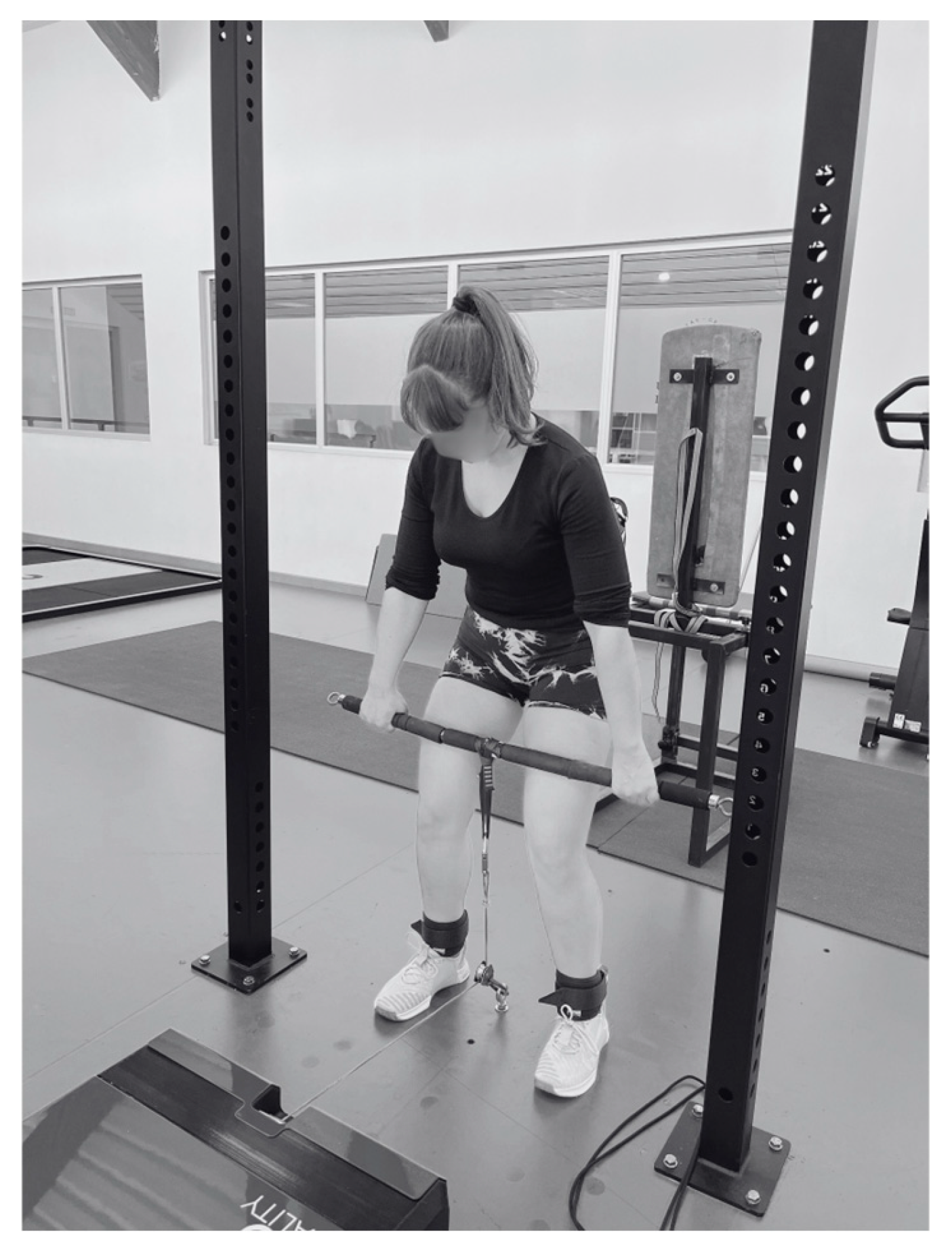
During familiarization, LOP was measured and confirmed with a Doppler probe in supine position. For all experimental sessions, occlusion pressures were set at 0%, 40%, or 80% of each LOP of participants. The IMTP was performed with participants gripping a wire-linked bar at mid-thigh height (Figure 2), with cable length individually adjusted to ensure proper body alignment and eliminate vertical displacement during testing.
The FEMD recorded force and displacement data using a 12-bit load cell and a 2500 ppr encoder powered by a 2000 W motor. Data were sampled at 1000 Hz. The built-in MyoQuality® software (version 2.6.2) applied a uniform internal filtering algorithm before data export. Output was provided in both kilograms and Newtons, though all calculations were performed using SI units (N, N·s, N/s). Kilogram values were retained only for interpretability.
Before each maximal IMTP, participants performed two submaximal familiarization trials (50% and 75% effort). Force data were exported in 10 ms intervals. Peak force (PF) and impulse (IP) were extracted as the maximum instantaneous and cumulative force, respectively. RFD was calculated across three intervals: 0–50 ms, 51–150 ms, and 151–250 ms. Force onset was defined as the first non-zero data point in the filtered force–time signal.

2.5. Data Analysis

All statistical analyses were conducted using JASP (version 0.19.1), and figures were created in GraphPad Prism (version 10.3.1). Data are presented as mean ± standard deviation (SD). Normality was tested with the Shapiro–Wilk test. A repeated-measures ANOVA was used to evaluate within-subject differences across conditions (0%, 40%, 80% LOP) and across time (PRE, POST, 5, 10, and 15 min). RFD data were analyzed by time window (0–50 ms, 51–150 ms, and 151–250 ms). Post hoc comparisons were adjusted using Holm’s correction. Statistical significance was set at p ≤ 0.05. Effect sizes were classified as trivial (d < 0.2), small (0.2 ≤ d < 0.5), moderate (0.5 ≤ d < 0.8), or large (d ≥ 0.8).

2.6. Availability of Data and Materials

All raw data, MATLAB R2024a (version 24.1.0) scripts, and experimental protocols are available from the corresponding author upon request. No generative AI tools were used in the design, data collection, or interpretation of this study. Minor grammar corrections were assisted by AI-based editing tools.

3. Results

3.1. Isometric Force Outcomes

Table 2 presents the descriptive and inferential statistics for the peak force (PF) and impulse (IP) across five time points (PRE, POST, 5 min, 10 min, and 15 min) and three limb occlusion pressure (LOP) conditions (0%, 40%, and 80%). The values are expressed as mean ± SD in kilograms (kg) and Newtons (Nw) for PF and Newton-seconds (Nw·s) for IP.
A significant main effect of time was found for the PF (F (4, 116) = 3.07, p = 0.02, η2 = 0.11) and a marginal effect for the IP (F (4, 116) = 2.41, p = 0.05, η2 = 0.09) under the 0% LOP condition. Post hoc analysis showed a non-significant decrease in the PF from PRE to POST (mean difference = 58.53 Nw, p = 0.07), followed by a return to baseline at 10 min (mean difference PRE–10 min = −4.22 Nw, p = 1.00). The largest decline was observed from POST to 10 min (–62.75 Nw), which reached statistical significance after Holm correction (p = 0.05). Effect sizes for PF comparisons were small to moderate, with the PRE–POST contrast showing a Cohen’s d = 0.35 and a 95% CI of [−0.21, 0.91].
For the IP, the PRE–POST comparison showed a statistically significant reduction (mean difference = 304.29 Nw·s, p = 0.04), with a small effect size (Cohen’s d = 0.28). All subsequent comparisons (e.g., POST–10 min, POST–15 min) were not significant (p > 0.05), with effect sizes remaining small (Cohen’s d ≤ 0.28). No significant time effects were observed for the PF or IP under the 40% or 80% LOP conditions (p > 0.05). Due to minimal fluctuations, no post hoc tests were conducted in these conditions.

3.2. Rate of Force Development (RFD)

In the early RFD window (0–50 ms), a significant main effect of time was observed (F (4, 116) = 2.82, p = 0.03, ω2 = 0.01; see Table 3), although the effect size was small. Post hoc comparisons indicated a reduction in the RFD from PRE to 5 MIN (mean difference = 40.1 N/s, p = 0.06), with a moderate-to-large effect size: Cohen’s d = 0.98, 95% CI [0.39, 1.56]. All other comparisons were not statistically significant after Holm correction (p > 0.18). No significant interaction between time and group was found (p = 0.14), and between-group differences were negligible (F (2, 84) = 0.14, p = 0.87).
In the intermediate RFD window (51–150 ms), significant main effects or interactions were observed (p > 0.03). The post hoc analysis did not yield any statistically significant results among the interactions. However, the PRE–POST comparison showed a large effect size: Cohen’s d = 1.52, 95% CI [0.89, 2.15].
In the late RFD window (151–250 ms), the main effect of time was significant (F (4, 116) = 3.80, p > 0.01, ω   = 0.01). The following post hoc analysis revealed significant temporal reductions following the fatiguing protocol. Specifically, the PRE–POST comparison demonstrated a statistically significant decrease in the RFD (mean difference = 88.09 N/s, p = 0.01), with a small-to-moderate effect size (Cohen’s d = 0.37, 95% CI [0.04, 0.69]). Similarly, significant declines were observed at 5 min (p = 0.02, d = 0.33, 95% CI [0.04, 0.66]), while the PRE–10 min post-exercise and PRE–15 MIN comparison did not reach significance (p = 1.0). These findings suggest that the late phase of force development is transiently impaired immediately after exercise, with partial recovery occurring by 10 min. The consistent reduction across early time points—despite modest effect sizes—supports the sensitivity of the 151–250 ms interval to fatigue-related neuromuscular alterations.
Post hoc comparisons revealed no statistically significant differences between occlusion conditions (0%, 40%, and 80% LOP) across most recovery time points for the early (0–50 ms) and mid (51–150 ms) RFD intervals. Although several contrasts presented large numerical differences—particularly those involving the 80% LOP condition—none reached significance after Holm correction (all p Holm > 0.05).
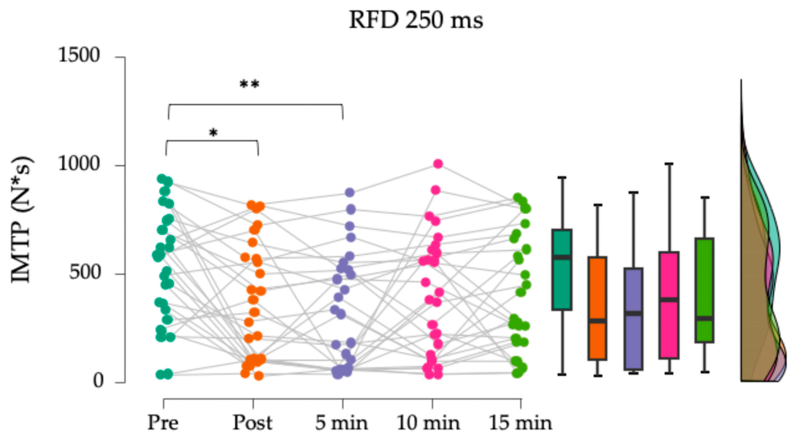
In the 151–250 ms interval, however, a significant within-group difference was observed under the 80% LOP condition (Figure 3), with a marked reduction in RFD from pre-exercise to 5 min post-exercise (Δ = 204.8 N/s, p = 0.003). No other pairwise comparisons in this interval were statistically significant.

4. Discussion

This study analyzed how varying degrees of occlusion influence RFD kinetics during IMTP performance after fatiguing single-joint action in young women. Contrary to our initial hypothesis, RFD kinetics remained largely unaffected across conditions, with only modest time-dependent changes observed. Specifically, early-phase RFD (0–50 ms) and late-phase RFD (151–250 ms) kinetics demonstrated statistically significant main effects of time; however, post hoc analysis revealed only transient reductions at POST and 5 min, without consistent differences between occlusion groups. The mid-phase RFD (51–150 ms) showed no significant changes. Interestingly, the traditional force metrics, peak force (PF) and impulse (IP), declined significantly only in the control condition (0% LOP), whereas no significant reductions were observed under 40% or 80% LOP. These results suggest a protective trend under BFR conditions, potentially attenuating the magnitude of fatigue observed in the non-occluded condition. However, this interpretation remains speculative, as no physiological markers such as electromyography or muscle oxygenation were measured to confirm the mechanism. This nuanced outcome challenges assumptions about BFR’s impact on explosive force performance and highlights the need for context-specific interpretation of BFR-induced fatigue.
The analysis of RFD kinetics across distinct temporal windows revealed differential sensitivity to acute neuromuscular fatigue under blood flow restriction (BFR). Early-phase RFD (0–50 ms) kinetics appeared to be relatively stable, which may reflect a preserved capacity for rapid motor unit discharge during the initial contraction phase despite transient ischemia. This could indicate that the neuromuscular mechanisms responsible for immediate force generation are either less affected by metabolic stress or compensated for by an increased central drive. Similarly, the mid-phase RFD (51–150 ms) demonstrated minimal disruption across conditions, aligning with prior research suggesting that intermediate phases of force development are less responsive to acute fatigue, particularly in isometric contexts [7,34]. These intervals may represent a transition zone in which neither neural nor mechanical determinants are sufficiently challenged to elicit measurable decrements. Moreover, isometric tasks elicit less systemic and mechanical stress than dynamic movements, potentially blunting observable fatigue effects across intermediate timeframes [13].
Notably, the 151–250 ms window revealed a significant intragroup reduction in the 80% LOP condition, suggesting an acute fatigue response specific to this late phase of force production. Although 250 ms represents only a fraction of the 5-s maximal effort, the result likely reflects the neuromuscular consequences of high-pressure BFR rather than contraction duration per se. Elevated occlusion pressures have been shown to increase metabolite accumulation and restrict oxygen delivery, accelerating peripheral fatigue and impairing muscle contractility [35]. In parallel, high-pressure BFR may alter motor unit recruitment dynamics by increasing reliance on higher-threshold units and disrupting firing patterns [25]. These mechanisms can impair the rapid force output in the later phases of contraction, even without visible decrements in the peak force. Given that this was the only statistically significant comparison across all time windows, it underscores the importance of analyzing the RFD temporally and considering the distinct physiological responses elicited by high-pressure BFR. Together, these findings underscore the importance of considering the temporal dimension of the RFD when evaluating the acute effects of BFR, as not all phases respond uniformly to ischemic or neuromuscular perturbations.
Our study employed a progressive single-joint knee extension protocol until failure, starting at 40% of the maximal voluntary isometric force (MVIF), with incremental 10% MVIF increases applied on each repetition until volitional failure. While previous research suggests that low-load resistance training with blood flow restriction (BFR) can elicit neuromuscular adaptations comparable to high-load training, the evidence remains inconsistent, with some studies reporting minimal acute or long-term effects [34,35,36,37]. Despite the progressive loading, our findings indicate no significant alterations in RFD kinetics, suggesting that the imposed physiological conditions were insufficient to acutely influence rapid force production. Although higher occlusion pressures have been associated with increased neuromuscular activation and fatigue-induced performance decline [38,39], our results suggest that the applied load and occlusion levels did not reach the threshold necessary to induce meaningful changes in early-phase force production.
The decision to employ a single-joint task was based on the need to isolate quadriceps fatigue and reduce the influence of inter-joint coordination variability typically present in multi-joint movements. This provided a controlled stimulus prior to IMTP testing. However, this design limits the ecological validity of the results, as multi-joint tasks are more functionally representative of athletic performance. Future studies should explore whether similar kinetic responses to BFR occur when fatigue is induced through compound exercises such as squats or deadlifts. These considerations raise important questions regarding protocol design and the specificity of BFR responses across task types and populations. While this study focused on young, recreationally active women, future research should explore variations in training status, fiber-type composition, and hormonal background to determine their moderating role in BFR efficacy.
Previous studies have demonstrated that the RFD is highly sensitive to neuromuscular fatigue, motor unit recruitment, and tendon stiffness adaptations [40,41,42]. Given the known impact of BFR on neuromuscular activation, it was expected that ischemic stress would acutely influence RFD kinetics [35,43]. However, the findings of this study contrast with prior evidence suggesting that BFR enhances explosive force production, particularly under low-load dynamic conditions [43,44]. The absence of statistically significant differences within and between subjects across the 50, 150, and 250 ms time windows suggests that brief exposure to blood flow restriction (BFR) may not acutely alter motor unit recruitment patterns or force transmission efficiency in an isometric context, which is consistent with the findings of Korkmaz [45].
A critical consideration when interpreting these results is the recovery trajectory of neuromuscular performance after BFR application. Although dynamic tasks frequently exhibit immediate fatigue-induced decrements followed by post-occlusion hyperemia and neuromuscular potentiation [46], the present findings suggest that these effects may not translate to isometric force production. The absence of significant within-subject effects across all time windows suggests that potential BFR-induced neuromuscular adjustments, such as increased motor unit recruitment or altered tendon compliance, did not occur within the analyzed recovery period [23,38]. The observed differences may be attributed to variations in metabolic stress between isometric and dynamic contractions or to the exercise selection in this study. While previous studies have shown that blood flow restriction induces greater lactate accumulation and muscle swelling during dynamic tasks compared to sustained isometric contractions [47,48,49], the present study focused exclusively on kinetic outcomes without incorporating physiological or metabolic markers.
Another plausible explanation lies in the post-BFR recovery trajectory. Studies have shown that dynamic tasks subjected to BFR often exhibit transient performance decrements followed by enhanced muscle perfusion and potentiation post-deflation [50,51]. However, our isometric task appears to bypass this biphasic response, likely due to the absence of post-activation potentiation, which is more typically observed after dynamic tasks involving greater mechanical output and metabolic cost. Although prior research has shown that BFR can enhance neuromuscular performance through mechanisms such as metabolite-induced potentiation, these effects have been predominantly observed in protocols using repeated concentric–eccentric actions or multi-joint exercises, especially in male and female participants [25,45]. In contrast, isometric conditions limit muscle pump activation, hyperemic responses, and neural recruitment variability—factors that may be essential to trigger such potentiation. Our findings therefore suggest that acute BFR exposure under isometric conditions does not replicate the performance enhancements seen in dynamic tasks, potentially due to reduced metabolic stress, the absence of stretch–shortening cycles, or lower central excitability. This divergence highlights the importance of task-specific interpretation and reinforces the need to consider sex, contraction type, and occlusion parameters when evaluating BFR outcomes. The metabolic demands of isometric exercise alone may be insufficient to elicit post-activation potentiation or significant RFD enhancement.
From a practical standpoint, the findings suggest that acute application of BFR does not significantly impact RFD during isometric tasks in young women. Although BFR has been extensively used to enhance hypertrophic and strength adaptations at low loads [11,12,38,50], its acute effects on explosive force production under isometric conditions remain unclear. Given that improvements in RFD are a determinant of rapid force production, these findings raise important considerations regarding the specific contexts in which BFR may be most effective. Further investigations should examine the potential of extended BFR exposure and the load components of the exercise protocol to stimulate isometric force production and facilitate neuromuscular adaptations over time, moving beyond the current focus on acute responses.
Several limitations should be acknowledged. First, although the isometric mid-thigh pull (IMTP) is a well-established and reliable tool for assessing isometric force production and rate of force development (RFD), it does not replicate the biomechanical demands of dynamic or functional tasks. Most athletic and daily activities involve rapid force generation under conditions that require joint displacement, stretch–shortening cycles, and movement-specific coordination. Therefore, caution is warranted when extrapolating the present findings to movement-based performance contexts, as the mechanical and neural requirements differ substantially. Second, while the study specifically focused on young physically active women, it did not account for individual differences in pre-session fatigue or recovery status, which may have influenced neuromuscular performance variability. Third, although different levels of blood flow restriction were employed, the duration of occlusion exposure varied slightly between participants, as each individual reached volitional failure at different times during the knee extension protocol. However, the overall training structure—number of sets and intensity progression—was standardized across subjects. While we did not implement multiple protocols with varying session volumes, doing so would have introduced confounding factors inconsistent with the controlled within-subject design of the present study.
Another relevant methodological limitation is the absence of electromyographic (EMG) data, which would have enabled a more detailed analysis of motor unit recruitment strategies and neuromuscular activation patterns under BFR conditions. Future research should incorporate surface EMG measurement to complement kinetic assessments and enhance the understanding of neuromuscular responses across different occlusion levels. Lastly, no physiological or perceptual measures—such as blood lactate concentration, muscle oxygenation (e.g., via near-infrared spectroscopy), or rating of perceived exertion (RPE)—were included in the protocol. While previous studies have shown that BFR induces greater lactate accumulation and muscle swelling during dynamic tasks compared to sustained isometric contractions, the present investigation prioritized mechanical output variables. Integrating such physiological and perceptual metrics in future studies would contribute to a more comprehensive understanding of acute fatigue and recovery responses under BFR, particularly in female populations.
In summary, acute BFR application did not significantly alter RFD kinetics during isometric mid-thigh pull assessments in young women. These findings suggest that under the tested conditions, BFR does not impair explosive isometric performance. These challenge assumptions were drawn from dynamic protocols and highlight the importance of task-specific evaluation. From an applied perspective, BFR may be a viable strategy for maintaining neuromuscular function under low-load conditions, although future studies should examine its chronic effects and adaptability to other tasks and populations.

Author Contributions

Conceptualization, S.A.-F. and D.J.-M.; methodology, S.A.-F. and D.J.-M.; formal analysis, S.A.-F. and D.J.-M.; investigation, S.A.-F.; data curation, S.A.-F.; visualization, S.A.-F.; writing—original draft preparation, S.A.-F.; writing—review and editing, S.A.-F. and D.J.-M.; supervision, D.J.-M., L.J.C.-R. and I.C.-R. All authors have read and agreed to the published version of the manuscript.

Funding

This work was supported by Grant A.SEJ.227.UGR23 funded by Consejería de Universidad, Investigación e Innovación and by ERDF Andalusia Program 2021–2027.

Institutional Review Board Statement

The study was conducted in accordance with the Declaration of Helsinki and approved by the Scientific Ethics Committee of the University of Granada, Spain (Approval Code: 3950/CEIH/2024).

Informed Consent Statement

Informed consent was obtained from all subjects involved in the study. Participants also consented to the publication of anonymized data. No identifiable images or personal data are included in this publication.

Data Availability Statement

The data that support the findings of this study are available from the corresponding author upon reasonable request. Access is restricted due to ethical considerations related to participant confidentiality.

Acknowledgments

The study was conducted as part of the doctoral thesis of Sebastián Aliste-Flores, enrolled in the Doctoral Program in Biomedicine, within the research line of Physical Activity and Sports at the Faculty of Sports Sciences, University of Granada. The authors would like to thank Hundred Performance and Rehab (Granada, Spain) for facilitating the use of AirBands® during testing and the IDAFISAD-642 research group for their institutional support throughout the project.

Conflicts of Interest

The authors declare no conflicts of interest.

Abbreviations

The following abbreviations are used in this manuscript:
BFRBlood Flow Restriction
FEMDFunctional Electromechanical Dynamometer
IMTPIsometric Mid-Thigh Pull
IPImpulse
IRBInstitutional Review Board
IKEIsometric Knee Extension
LOPLimb Occlusion Pressure
MVIFMaximum Voluntary Isometric Force
PFPeak Force
RFDRate of Force Development
SDStandard Deviation

References

  1. Maestroni, L.; Read, P.; Bishop, C.; Papadopoulos, K.; Suchomel, T.J.; Comfort, P.; Turner, A. The Benefits of Strength Training on Musculoskeletal System Health: Practical Applications for Interdisciplinary Care. Sports Med. 2020, 50, 1431–1450. [Google Scholar] [CrossRef] [PubMed]
  2. Suchomel, T.J.; Nimphius, S.; Stone, M.H. The Importance of Muscular Strength in Athletic Performance. Sports Med. 2018, 48, 765–785. [Google Scholar] [CrossRef] [PubMed]
  3. Haff, G.G.; Carlock, J.M.; Hartman, M.J.; Kilgore, J.L.; Kawamori, N.; Jackson, J.R.; Morris, R.T.; Sands, W.A.; Stone, M.H. Force-Time Curve Characteristics of Dynamic and Isometric Muscle Actions of Elite Women Olympic Weightlifters. J. Strength Cond. Res. 2005, 19, 741–748. [Google Scholar] [CrossRef]
  4. Dos’Santos, T.; Thomas, C.; Comfort, P.; Jones, P.A. Between-Session Reliability of Isometric Mid-Thigh Pull Kinetics and Maximal Power Clean Performance in Male Youth Athletes. J. Strength Cond. Res. 2018, 32, 336–344. [Google Scholar] [CrossRef]
  5. Lum, D.; Haff, G.G.; Barbosa, T.M. The Relationship between Isometric Force-Time Characteristics and Dynamic Performance: A Systematic Review. Sports 2020, 8, 63. [Google Scholar] [CrossRef]
  6. Aagaard, P.; Simonsen, E.B.; Andersen, J.L.; Magnusson, P.; Dyhre-Poulsen, P. Increased Rate of Force Development and Neural Drive of Human Skeletal Muscle Following Resistance Training. J. Appl. Physiol. 2002, 93, 1318–1326. [Google Scholar] [CrossRef]
  7. Maffiuletti, N.A.; Aagaard, P.; Blazevich, A.J.; Folland, J.P.; Tillin, N.A.; Duchateau, J. Rate of Force Development: Physiological and Methodological Considerations. Eur. J. Appl. Physiol. 2016, 116, 1091–1116. [Google Scholar] [CrossRef]
  8. Bojsen-Møller, J.; Magnusson, S.P.; Rasmussen, L.R.; Kjaer, M.; Aagaard, P. Muscle Performance during Maximal Isometric and Dynamic Contractions Is Influenced by the Stiffness of the Tendinous Structures. J. Appl. Physiol. 2005, 99, 986–994. [Google Scholar] [CrossRef]
  9. Dideriksen, J.L.; Del Vecchio, A.; Farina, D. Neural and Muscular Determinants of Maximal Rate of Force Development. J. Neurophysiol. 2020, 123, 149–157. [Google Scholar] [CrossRef]
  10. Taber, C.; Bellon, C.; Abbott, H.; Bingham, G.E. Roles of Maximal Strength and Rate of Force Development in Maximizing Muscular Power. Strength Cond. J. 2016, 38, 71–78. [Google Scholar] [CrossRef]
  11. Lulic-Kuryllo, T.; Inglis, J.G. Sex Differences in Motor Unit Behaviour: A Review. J. Electromyogr. Kinesiol. 2022, 66, 102689. [Google Scholar] [CrossRef] [PubMed]
  12. HUNTER, S.K. The Relevance of Sex Differences in Performance Fatigability. Med. Sci. Sports Exerc. 2016, 48, 2247–2256. [Google Scholar] [CrossRef] [PubMed]
  13. Enoka, R.M.; Duchateau, J. Translating Fatigue to Human Performance. Med. Sci. Sports Exerc. 2016, 48, 2228–2238. [Google Scholar] [CrossRef]
  14. Hunter, S.K. Sex Differences in Fatigability of Dynamic Contractions. Exp. Physiol. 2016, 101, 250–255. [Google Scholar] [CrossRef]
  15. Ansdell, P.; Brownstein, C.G.; Škarabot, J.; Hicks, K.M.; Howatson, G.; Thomas, K.; Hunter, S.K.; Goodall, S. Sex Differences in Fatigability and Recovery Relative to the Intensity–Duration Relationship. J. Physiol. 2019, 597, 5577–5595. [Google Scholar] [CrossRef] [PubMed]
  16. Ansdell, P.; Thomas, K.; Howatson, G.; Hunter, S.; Goodall, S. Contraction Intensity and Sex Differences in Knee-Extensor Fatigability. J. Electromyogr. Kinesiol. 2017, 37, 68–74. [Google Scholar] [CrossRef]
  17. Giuriato, G.; Romanelli, M.G.; Bartolini, D.; Vernillo, G.; Pedrinolla, A.; Moro, T.; Franchi, M.; Locatelli, E.; Andani, M.E.; Laginestra, F.G.; et al. Sex Differences in Neuromuscular and Biological Determinants of Isometric Maximal Force. Acta Physiol. 2024, 240, e14118. [Google Scholar] [CrossRef]
  18. Xu, D.; Zhou, H.; Quan, W.; Gusztav, F.; Wang, M.; Baker, J.S.; Gu, Y. Accurately and Effectively Predict the ACL Force: Utilizing Biomechanical Landing Pattern before and after-Fatigue. Comput. Methods Programs Biomed. 2023, 241, 107761. [Google Scholar] [CrossRef]
  19. Merrigan, J.J.; Dabbs, N.C.; Jones, M.T. Isometric Mid-Thigh Pull Kinetics: Sex Differences and Response to Whole-Body Vibration. J. Strength Cond. Res. 2020, 34, 2407–2411. [Google Scholar] [CrossRef]
  20. Comfort, P.; Dos’Santos, T.; Beckham, G.K.; Stone, M.H.; Guppy, S.N.; Haff, G.G. Standardization and Methodological Considerations for the Isometric Midthigh Pull. Strength Cond. J. 2019, 41, 57–79. [Google Scholar] [CrossRef]
  21. Scott, B.R.; Loenneke, J.P.; Slattery, K.M.; Dascombe, B.J. Exercise with Blood Flow Restriction: An Updated Evidence-Based Approach for Enhanced Muscular Development. Sports Med. 2015, 45, 313–325. [Google Scholar] [CrossRef] [PubMed]
  22. Lixandrão, M.E.; Ugrinowitsch, C.; Berton, R.; Vechin, F.C.; Conceição, M.S.; Damas, F.; Libardi, C.A.; Roschel, H. Magnitude of Muscle Strength and Mass Adaptations Between High-Load Resistance Training Versus Low-Load Resistance Training Associated with Blood-Flow Restriction: A Systematic Review and Meta-Analysis. Sports Med. 2018, 48, 361–378. [Google Scholar] [CrossRef]
  23. Willis, S.J.; Alvarez, L.; Borrani, F.; Millet, G.P. Oxygenation Time Course and Neuromuscular Fatigue during Repeated Cycling Sprints with Bilateral Blood Flow Restriction. Physiol. Rep. 2018, 6, e13872. [Google Scholar] [CrossRef]
  24. Fatela, P.; Mendonca, G.V.; Veloso, A.P.; Avela, J.; Mil-Homens, P. Blood Flow Restriction Alters Motor Unit Behavior During Resistance Exercise. Int. J. Sports Med. 2019, 40, 555–562. [Google Scholar] [CrossRef]
  25. Mendonca, G.V.; Borges, A.; Teodósio, C.; Matos, P.; Correia, J.; Vila-Chã, C.; Mil-Homens, P.; Pezarat-Correia, P. Muscle Fatigue in Response to Low-Load Blood Flow-Restricted Elbow-Flexion Exercise: Are There Any Sex Differences? Eur. J. Appl. Physiol. 2018, 118, 2089–2096. [Google Scholar] [CrossRef] [PubMed]
  26. Proppe, C.; Aldeghi, T.; Rivera, P. Neuromuscular Responses to Failure vs Non-Failure During Blood Flow Restriction Training in Untrained Females. Int. J. Exerc. Sci. 2023, 16, 293–303. [Google Scholar] [CrossRef] [PubMed]
  27. Cormie, P.; McGuigan, M.R.; Newton, R.U. Developing Maximal Neuromuscular Power. Sports Med. 2011, 41, 125–146. [Google Scholar] [CrossRef]
  28. Kuki, S.; Konishi, Y.; Okudaira, M.; Yoshida, T.; Exell, T.; Tanigawa, S. Asymmetry of Force Generation and Neuromuscular Activity during Multi-Joint Isometric Exercise. J. Phys. Fit Sports Med. 2019, 8, 37–44. [Google Scholar] [CrossRef]
  29. Kuki, S.; Yoshida, T.; Okudaira, M.; Konishi, Y.; Ohyama-Byun, K.; Tanigawa, S. Force Generation and Neuromuscular Activity in Multi-Joint Isometric Exercises: Comparison between Unilateral and Bilateral Stance. J. Phys. Fit Sports Med. 2018, 7, 289–296. [Google Scholar] [CrossRef]
  30. del-Cuerpo, I.; Jerez-Mayorga, D.; Delgado-Floody, P.; Morenas-Aguilar, M.D.; Chirosa-Ríos, L.J. Test–Retest Reliability of the Functional Electromechanical Dynamometer for Squat Exercise. Int. J. Environ. Res. Public Health 2023, 20, 1289. [Google Scholar] [CrossRef]
  31. Martinez-Garcia, D.; Rodriguez-Perea, A.; Barboza, P.; Ulloa-Díaz, D.; Jerez-Mayorga, D.; Chirosa, I.; Chirosa Ríos, L.J. Reliability of a Standing Isokinetic Shoulder Rotators Strength Test Using a Functional Electromechanical Dynamometer: Effects of Velocity. PeerJ 2020, 8, e9951. [Google Scholar] [CrossRef]
  32. Rodriguez-Perea, A.; Chirosa Ríos, L.J.; Martinez-Garcia, D.; Ulloa-Díaz, D.; Guede Rojas, F.; Jerez-Mayorga, D.; Chirosa Rios, I.J. Reliability of Isometric and Isokinetic Trunk Flexor Strength Using a Functional Electromechanical Dynamometer. PeerJ 2019, 7, e7883. [Google Scholar] [CrossRef] [PubMed]
  33. Rodriguez-Perea, Á.; Jerez-Mayorga, D.; García-Ramos, A.; Martínez-García, D.; Chirosa Ríos, L.J. Reliability and Concurrent Validity of a Functional Electromechanical Dynamometer Device for the Assessment of Movement Velocity. Proc. Inst. Mech. Eng. Part J. Sport Eng. Technol. 2021, 235, 176–181. [Google Scholar] [CrossRef]
  34. Australian Institute of Sport (AIS). Blood Flow Restriction Training Guidelines. In Australian Government. Available online: https://www.ais.gov.au/position_statements/best_practice_content/blood-flow-restriction-training-guidelines (accessed on 20 January 2024).
  35. D’Emanuele, S.; Maffiuletti, N.A.; Tarperi, C.; Rainoldi, A.; Schena, F.; Boccia, G. Rate of Force Development as an Indicator of Neuromuscular Fatigue: A Scoping Review. Front. Hum. Neurosci. 2021, 15, 701916. [Google Scholar] [CrossRef] [PubMed]
  36. Lauber, B.; König, D.; Gollhofer, A.; Centner, C. Isometric Blood Flow Restriction Exercise: Acute Physiological and Neuromuscular Responses. BMC Sports Sci. Med. Rehabil. 2021, 13, 12. [Google Scholar] [CrossRef]
  37. Centner, C.; Lauber, B. A Systematic Review and Meta-Analysis on Neural Adaptations Following Blood Flow Restriction Training: What We Know and What We Don’t Know. Front. Physiol. 2020, 11, 887. [Google Scholar] [CrossRef] [PubMed]
  38. Suga, T.; Okita, K.; Morita, N.; Yokota, T.; Hirabayashi, K.; Horiuchi, M.; Takada, S.; Omokawa, M.; Kinugawa, S.; Tsutsui, H. Dose Effect on Intramuscular Metabolic Stress during Low-Intensity Resistance Exercise with Blood Flow Restriction. J. Appl. Physiol. 2010, 108, 1563–1567. [Google Scholar] [CrossRef]
  39. Cook, S.B.; Scott, B.R.; Hayes, K.L.; Murphy, B.G. Neuromuscular Adaptations to Low-Load Blood Flow Restricted Resistance Training. J. Sports Sci. Med. 2018, 17, 66–73. [Google Scholar]
  40. Fatela, P.; Reis, J.F.; Mendonca, G.V.; Freitas, T.; Valamatos, M.J.; Avela, J.; Mil-Homens, P. Acute Neuromuscular Adaptations in Response to Low-Intensity Blood-Flow Restricted Exercise and High-Intensity Resistance Exercise: Are There Any Differences? J. Strength Cond. Res. 2018, 32, 902–910. [Google Scholar] [CrossRef]
  41. Jessee, M.B.; Buckner, S.L.; Mouser, J.G.; Mattocks, K.T.; Dankel, S.J.; Abe, T.; Bell, Z.W.; Bentley, J.P.; Loenneke, J.P. Muscle Adaptations to High-Load Training and Very Low-Load Training With and Without Blood Flow Restriction. Front. Physiol. 2018, 9, 1448. [Google Scholar] [CrossRef]
  42. Wernbom, M.; Järrebring, R.; Andreasson, M.A.; Augustsson, J. Acute Effects of Blood Flow Restriction on Muscle Activity and Endurance During Fatiguing Dynamic Knee Extensions at Low Load. J. Strength Cond. Res. 2009, 23, 2389–2395. [Google Scholar] [CrossRef] [PubMed]
  43. Mota, J.A.; Gerstner, G.R.; Giuliani, H.K. Motor Unit Properties of Rapid Force Development during Explosive Contractions. J. Physiol. 2019, 597, 2335–2336. [Google Scholar] [CrossRef]
  44. Mornas, A.; Racinais, S.; Brocherie, F.; Alhammoud, M.; Hager, R.; Desmedt, Y.; Guilhem, G. Faster Early Rate of Force Development in Warmer Muscle: An in Vivo Exploration of Fascicle Dynamics and Muscle-Tendon Mechanical Properties. Am. J. Physiol. Regul. Integr. Comp. Physiol. 2022, 323, R123–R132. [Google Scholar] [CrossRef] [PubMed]
  45. Mckee, J.R.; Girard, O.; Peiffer, J.J.; Hiscock, D.J.; De Marco, K.; Scott, B.R. Repeated-Sprint Training With Blood-Flow Restriction Improves Repeated-Sprint Ability Similarly to Unrestricted Training at Reduced External Loads. Int. J. Sports Physiol. Perform. 2024, 19, 257–264. [Google Scholar] [CrossRef]
  46. Sun, D.; Yang, T. Semi-Squat Exercises with Varying Levels of Arterial Occlusion Pressure during Blood Flow Restriction Training Induce a Post-Activation Performance Enhancement and Improve Vertical Height Jump in Female Football Players. J. Sports Sci. Med. 2023, 22, 212–225. [Google Scholar] [CrossRef] [PubMed]
  47. Castilla-López, C.; Romero-Franco, N. Low-Load Strength Resistance Training with Blood Flow Restriction Compared with High-Load Strength Resistance Training on Performance of Professional Soccer Players: A Randomized Controlled Trial. J. Sports Med. Phys. Fitness 2023, 63, 1146–1154. [Google Scholar] [CrossRef]
  48. Korkmaz Dayican, D.; Ulker Eksi, B.; Yigit, S.; Utku Umut, G.; Ozyurek, B.; Yilmaz, H.E.; Akinci, B. Immediate Effects of High-Intensity Blood Flow Restriction Training on Muscle Performance and Muscle Soreness. Res. Q. Exerc. Sport 2024, 96, 213–222. [Google Scholar] [CrossRef]
  49. Sánchez-Valdepeñas, J.; Cornejo-Daza, P.J.; Rodiles-Guerrero, L.; Páez-Maldonado, J.A.; Sánchez-Moreno, M.; Bachero-Mena, B.; Saez de Villarreal, E.; Pareja-Blanco, F. Acute Responses to Different Velocity Loss Thresholds during Squat Exercise with Blood-Flow Restriction in Strength-Trained Men. Sports 2024, 12, 171. [Google Scholar] [CrossRef]
  50. De Queiros, V.S.; Rolnick, N.; Sabag, A.; Wilde, P.; Peçanha, T.; Aniceto, R.R.; Rocha, R.F.C.; Delgado, D.Z.; Cabral, B.G.d.A.T.; Dantas, P.M.S. Effect of High-Intensity Interval Exercise versus Continuous Low-Intensity Aerobic Exercise with Blood Flow Restriction on Psychophysiological Responses: A Randomized Crossover Study. J. Sports Sci. Med. 2024, 23, 114–125. [Google Scholar] [CrossRef]
  51. Rivera, P.M.; Proppe, C.E.; Gonzalez-Rojas, D.; Wizenberg, A.; Hill, E.C. Effects of Load Matched Isokinetic versus Isotonic Blood Flow Restricted Exercise on Neuromuscular and Muscle Function. Eur. J. Sport Sci. 2023, 23, 1629–1637. [Google Scholar] [CrossRef]
Figure 1. Session protocol scheme.
Applsci 15 05866 g001
Figure 2. Wire-linked bar positioned to mid-thigh height.
Applsci 15 05866 g002
Figure 3. Force behavior across time in 151–250 rate of force development (RFD) window. *: p < 0.01. **: p < 0.02.
Applsci 15 05866 g003
Table 1. Descriptive Characteristics of the Sample.
VariableMean (SD)
Age (years)27.37 (10.02)
Weight (kg)63.84 (12.45)
Height (cm)164.23 (5.99)
BMI (kg/m2)23.52 (3.75)
LOP (mmHg)215.83 (26.94)
40% LOP (mmHg)86.33 (10.77)
80% LOP (mmHg)172.66 (21.55)
LOP: limb occlusion pressure; BMI: body mass index.
Table 2. Summary statistics and repeated-measures ANOVA for IMTP performance across time points and LOP conditions.
LOP
Condition
VariablePREPOST5 min10 min15 minANOVA
(F (4, 116))
pES
40%PF
(kg)
100.85 (23.02)101.29 (24.04)103.57 (25.95)102.82 (28.76)105.39 (29.57)0.900.432 a0.04
PF
(Nw)
989.30 (225.86)993.68 (235.85)1016.00 (254.53)1008.70 (282.17)1033 (290.02)0.900.432 a0.04
IP
(Nw)
4386.36 (1200.61)4202.66 (1002.80)4326.52 (977.46)4348.86 (1161.56)4404.46 (1121.59)0.760.490 a0.03
80%PF
(kg)
99.26 (15.43)104.13 (19.35)104.43 (21.04)103.33 (22.87)105.02 (21.10)1.700.1590.08
PF
(Nw)
973.77 (151.34)1021.49 (189.79)1024.42 (206.39)1013.70 (224.34)1030.24 (207.01)1.700.1590.08
IP
(Nw)
4316.26 (833.05)4308.13 (764.27)4314.78 (876.33)4488.44 (1043.68)4433.14 (853.72)0.710.5830.04
0%PF
(kg)
103.98 (22.07)98.01 (22.43)102.29 (24.69)104.41 (26.54)99.82 (26.65)3.070.020 *0.11
PF
(Nw)
1020.05 (216.54)961.52 (220.07)1003.45 (242.23)1024.27 (260.33)979.19 (261.46)3.070.020 *0.11
IP
(Nw)
4477.08 (1001.31)4172.79 (1084.39)4344.06 (1056.19)4414.64 (1167.51)4325.49 (1152.71)2.410.048 *0.09
Note: Values are mean (SD). PF: peak force; IP: impulse; LOP: limb occlusion pressure; IMTP: isometric mid-thigh pull; Nw: Newtons. p values with asterisk (*) indicate statistical significance (p < 0.05). ES: size effect. a: Greenhouse–Geisser correction applied.
Table 3. Summary of descriptive and post hoc analyses for RFD time windows.
Time WindowEffect/
Comparison
Statisticp ValuePost Hoc **ES95% CI
0–50 msMain effect of timeF (4, 116) = 2.82p = 0.032 *-0.01
PRE–POSTt = 2.38 p = 0.1770.350.08, 0.79
PRE–5 MINt = 2.83 p = 0.0580.980.39, 1.56
PRE–10 MINt = 2.17 p = 0.2610.28−0.10, 0.66
PRE–15 MINt = 1.62 p = 0.7560.22−0.17, 0.62
51–150 msMain effect of timeF (4, 116) = 2.96p > 0.026 *-0.01
PRE–POSTt = 2.65 p = 0.0880.360.89, 2.15
PRE–5 MINt = 2.68 p = 0.0880.33−0.03, 0.69
PRE–10 MINt = 2.06 p = 0.3380.29−0.12, 0.69
PRE–15 MINt = 1.58 p = 0.8240.20−0.17, 0.57
151–250 msMain effect of timeF (4, 116) = 3.80p = 0.009 *-0.01
PRE–POSTt = 3.14 p = 0.040 *0.370.04, 0.69
PRE–5 MINt = 2.92 p = 0.004 *0.330.04, 0.66
PRE–10 MINt = 2.74 p = 1.0000.360.03, 0.68
PRE–15 MINt = 2.22 p = 1.0000.25−0.07, 0.57
RFD = rate of force development. ms: miliseconds. *: statistically significant. **: Holm correction applied for multiple comparisons.
Disclaimer/Publisher’s Note: The statements, opinions and data contained in all publications are solely those of the individual author(s) and contributor(s) and not of MDPI and/or the editor(s). MDPI and/or the editor(s) disclaim responsibility for any injury to people or property resulting from any ideas, methods, instructions or products referred to in the content.