Article

Transcranial Doppler-Based Neurofeedback to Improve Hemispheric Lateralization

Department of Neurosciences “Rita Levi Montalcini”, University of Turin, Corso Raffaello 30, 10125 Torino, Italy
*
Author to whom correspondence should be addressed.
Appl. Sci. 2025, 15(10), 5763; https://doi.org/10.3390/app15105763
Submission received: 21 April 2025 / Revised: 13 May 2025 / Accepted: 16 May 2025 / Published: 21 May 2025

Abstract

:
Functional transcranial Doppler (fTCD) ultrasound can detect cerebral blood flow lateralization to the left/right hemisphere during different tasks. This study aims to test the effectiveness of neurofeedback in improving the individual capacity to lateralize blood flow with mental activity. Bilateral monitoring of blood velocity (CBV) in the middle cerebral arteries was performed in 14 subjects engaged in 15 min of training, followed by a 15 min test in each of four sessions. A ball, displayed on a screen, moved right or left, according to the current right/left difference in normalized CBVs, thus providing a visual neurofeedback of lateralization. The subjects were invited to control the left/right movement of the depicted ball by appropriately orienting their mental activity, freely exploring different strategies. These attempts were completely free and unsupervised during training, while during the test, the subjects were required to follow randomized left/right cues lasting 35 s. Performance was assessed using receiver operating characteristic (ROC) analysis. With training, responses to left and right cues diverged more rapidly and consistently. Accuracy improved significantly from 0.51 to 0.65, and the area under the ROC increased from 0.55 to 0.69. These results demonstrate the effectiveness of neurofeedback in improving lateralization capacity, with implications for the development of fTCD-based brain–computer interfaces.

1. Introduction

Functional brain lateralization is a well-established principle in humans and other vertebrates [1]. It is a crucial concept for understanding how cognitive and motor activities are organized within the cerebral hemispheres. This phenomenon is particularly important for evaluating lateralized functions, such as symbolic communication, perception–action coupling, emotions, and decision-making processes [2]. Brain asymmetry displays significant implications and has been extensively studied through functional neuroimaging techniques for stroke recovery [3,4], neurological disorders [5,6,7,8], and applications in brain–computer interfaces (BCIs) and rehabilitative treatments [9,10,11].
Despite growing interest in lateralization in clinical and cognitive neuroscience, training its modulation through neurofeedback methodologies remains underexplored. Neurofeedback is a cognitive training technique that allows subjects to voluntarily modulate their brain activity through real-time feedback of a given indicator of mental activity. The underlying hypothesis is that this training enhances the cognitive abilities associated with the targeted brain activity by inducing neuroplastic changes in brain structures, affecting the strength and number of synaptic connections, as well as white matter myelination and gray matter volume [12]. Although plasticity is greatest before adolescence, it persists throughout life and underlies brain development and learning [13].
Neurofeedback has found applications in the treatment of many physical and mental disorders, as well as in the field of brain–computer interfaces (BCI) [14,15,16]. In the latter case, it may be used to train both healthy individuals and patients to improve their control over external devices or to interact with virtual environments, making BCI technologies more responsive to the user’s cognitive and emotional state [15].
Electroencephalography (EEG) stands out as the primary tool for capturing real-time electrical patterns in the brain, from which different parameters can be extracted and used as feedback signals, most commonly in visual or auditory form [17,18]. Beside EEG, hemodynamic correlates of brain activity, such as changes in cerebral blood flow and oxygenation generated by neurovascular coupling [19], can also be detected and used for neurofeedback. Several studies have demonstrated this possibility using functional magnetic resonance imaging (fMRI) [20,21,22,23] and more recently, near-infrared spectroscopy (fNIRS) [24,25].
Compared to the weak and noisy EEG signals, hemodynamic measurements may offer a more robust detection of brain activation, although at a lower time resolution and requiring expensive equipment and complicated experimental procedures (particularly fMRI). In this respect, functional transcranial Doppler (fTCD) ultrasound, allowing for the continuous and bilateral measurement of blood velocity from the major cerebral arteries, offers an interesting alternative due to its non-invasiveness, robustness, portability, and possibility of real-time monitoring, while requiring a relatively low-cost instrumentation and simple experimental setup. Although characterized by low spatial resolution, it has often been employed to detect the lateralization of brain functions, particularly regarding language [26,27,28,29,30], and has also been recently proposed for BCI applications [31,32].
Surprisingly, fTCD-based neurofeedback has been poorly investigated. However, it can be hypothesized that training over multiple sessions could lead the subject to increase the capacity to lateralize brain activity and cerebral blood flow to one hemisphere or the other. Moreover, self-training with neurofeedback allows the subjects to develop their own mental strategies, possibly achieving faster and larger hemodynamic changes, with implications in the fields of rehabilitation and BCIs [17].
To our knowledge, only two studies have explored the use of transcranial Doppler (TCD) ultrasound in neurofeedback paradigms, both focusing on modulating cerebral blood flow velocity (CBFV) within single arteries [33,34]. However, these studies did not exploit interhemispheric differences, nor did they provide a direct measure of lateralization as feedback. In contrast, the present study introduces a novel neurofeedback strategy based on the difference in CBFV between the left and right MCAs, allowing participants to actively modulate hemispheric dominance. This lateralized feedback design represents a significant methodological advancement, as it aligns with established principles of hemispheric specialization and opens the door to individualized, task-independent neurofeedback protocols. In fact, our approach is not constrained to predefined lateralizing tasks, thus potentially enhancing both user engagement and adaptability. By capturing interhemispheric dynamics, this method offers a promising foundation for more targeted applications in cognitive modulation and neurorehabilitation.
A technical description of the adopted neurofeedback system has recently been presented [35]. The aim of the present study was to demonstrate, in a group of healthy subjects, the efficacy of neurofeedback in improving their lateralization capacity over four subsequent sessions, each including a free training period and a performance test. The improvements were assessed by quantifying the progressive differentiation of hemodynamic responses to left and right lateralization attempts, as well as the area under the curve of receiver operating curves (ROC) and the accuracy of a simple off-line classification of left/right responses. In view of prospective applications in the field of brain–computer interfaces (BCI) the possible benefit of a three-class classification (left/right/null) of responses was also tested.

2. Materials and Methods

A total of 14 healthy subjects were recruited in the study (6 men, 8 women; mean age 22.3 ± 2.5). All participants were free from neurological or psychiatric disorders, were not taking any medication, and had no known medical conditions. Exclusion criteria included physical disabilities, cerebrovascular disease, and visual impairments. The subjects were also required to abstain from caffeine and smoking for at least 6 h before each experimental session. All participants provided written informed consent and were aware of the purpose and design of the project. The study was conducted in accordance with the Declaration of Helsinki, and the protocol was approved by the Ethics Committee of the University of Turin (protocol number 219859, April 2021).

2.1. Instrumentation

The description of a TCD-based neurofeedback system is well explained by Rabbito et al. [35]. Briefly, it includes the following:
  • A transcranial Doppler (TCD) ultrasound to measure the cerebral blood velocity in the left and right middle cerebral arteries (MCAs), and the two signals are made available as analog output;
  • The signals are digitally sampled (100 Hz) by an Arduino component and transmitted to a PC;
  • A lateralization index is then calculated and used to provide real-time visual feedback to the user, as described below.

2.2. Feedback

The feedback is based on the measurement of blood flow velocity in the two middle cerebral arteries (MCA), obtained from transcranial Doppler (TCD). The raw TCD data were downsampled to 20 Hz using a moving average filter to reduce noise. Specifically, on the screen, the subject visualizes the movement of a ball that can only move horizontally to the right or to the left. This movement is proportional to an index called the lateralization index (LI), which represents the difference between the blood flow velocity detected from the right (CBFVR) and the left (CBFVL) arteries, normalized to their respective basal value, as expressed by the following formula:
L I t = C B F V R t C B F V R b C B F V L t C B F V L b   × 100 ,
The b subscript indicates the basal value obtained from the time average of the signal over a 10 s resting interval at the beginning of the recording. The feedback thus reflects the flow imbalance between the hemispheres; specifically, an increase in LI (positive values) corresponds to a larger flow to the right hemisphere (greater CBFVR) and results in a shift of the ball to the right. Conversely, a decrease, resulting in negative LI values, indicates an increase in flow in the left MCA (to the left hemisphere). In quantitative terms, as an example, an increase in LI(t) equal to 5 means that the difference between CBFVR is larger than CBFVL by about 5% of the resting value. It is also important to clarify that when referring to the right or left hemisphere, it is here intended as the territory supplied by the respective middle cerebral artery.

2.3. Task

The participants were first introduced to the concept that certain brain activities can be lateralized, that increases in brain metabolism are generally matched by corresponding increases in blood flow, and therefore, that LI may detect lateralization of the brain activity. They were then instructed about the meaning of the left/right movement of the ball as visual feedback of LI and about the fact that such movement would happen with a delay of several seconds with respect to the mental task, due to the slowness of the hemodynamic response [36,37]. Based on this understanding, participants were provided with suggestions regarding mental tasks known to produce preferential activation of one hemisphere over the other, as summarized in Table 1. However, they were also encouraged to explore other mental activities and identify the best strategy for them to control the movement of the ball.

2.4. Experimental Protocol

The experiments took place in a quiet and darkened room, with subjects seated on a chair and the screen positioned at a 1 m distance.

2.4.1. Session: Training and Test

The protocol consisted of four sessions conducted on separate days, with a minimum of 24 h between each session. During each session, participants completed both a training phase and a testing phase.
After the participants familiarized themselves with the neurofeedback system, they were invited to relax and clear their minds for two minutes before starting with the training phase. During the training phase, which lasted up to 15 min, unless interrupted earlier by the subject, the participants were asked to learn to control the left/right movement of the ball by freely engaging in different mental activities, as detailed above. A 15 min duration of the training phase was established, based on preliminary investigations, as a trade-off between the necessity to maintain engagement and to minimize mental weariness. Participants could request to end the training phase earlier, if they wished. This phase was essential for learning and becoming accustomed to the neurofeedback, allowing the subjects to discover the most effective mental strategies for directing the ball in one direction or another, as well as for understanding the physiological delay between the onset of the mental task and its possible hemodynamic effect and the presentation of the visual feedback.
After a brief rest period of about 5 min, participants proceeded to the testing phase. In this phase, they were instructed to direct the feedback ball toward the right or the left side of the screen and maintain this displacement for 35 s, according to the indication of a visual cue displayed on the screen (see Figure 1), and then to rest for 15 s (blank screen). This task was repeated 12 times, with an equal number of trials for each direction (6 right, 6 left), presented in a randomized and fully counterbalanced order to avoid potential order or learning effects. The rest periods were designed to help the participants to clear their minds and prepare for the next task.
Before the start of both phases, participants were instructed to remain relaxed and avoid any sudden movement. During this time, baseline data were collected for the subsequent normalization of the signals.

2.4.2. Questionnaire

In order to gather qualitative feedback from participants regarding their experience with the neurofeedback training, a form was provided at the end of the first session. Specifically, participants were asked whether and how they were able to achieve control of the ball and to describe the adopted mental strategies. They were also asked to indicate which strategy they found most effective, whether the feedback was helpful in reaching the target, or to explain why it did not work. Additional questions addressed their level of effort in achieving the goal, the usefulness of the training phase prior to the test, and whether they were satisfied with the duration of the training and test phases. Finally, the participants were encouraged to offer any additional comments and suggestions. This form served as a valuable tool for gathering insights into individual differences in neurofeedback usage and effectiveness.

2.5. Data Analysis and Statistics

The analysis of hemodynamic signals was limited to the test session and oriented to investigate the potential for LI to predict the left/right mental task performed by the subject and to assess whether this capacity was improved by neurofeedback over the four experimental sessions. This aim was approached in different ways. Data analysis was automatically implemented through Matlab (version: R2024b, MathWorks Inc., Natick, MA, USA, https://www.mathworks.com) scripts, which ensures an objective processing workflow. Statistical analysis was conducted using R (version: 4.4.1, R Foundation for Statistical Computing, Vienna, Austria, https://www.r-project.org).

2.5.1. Assessment of the Differentiation of Responses to Left/Right Tasks

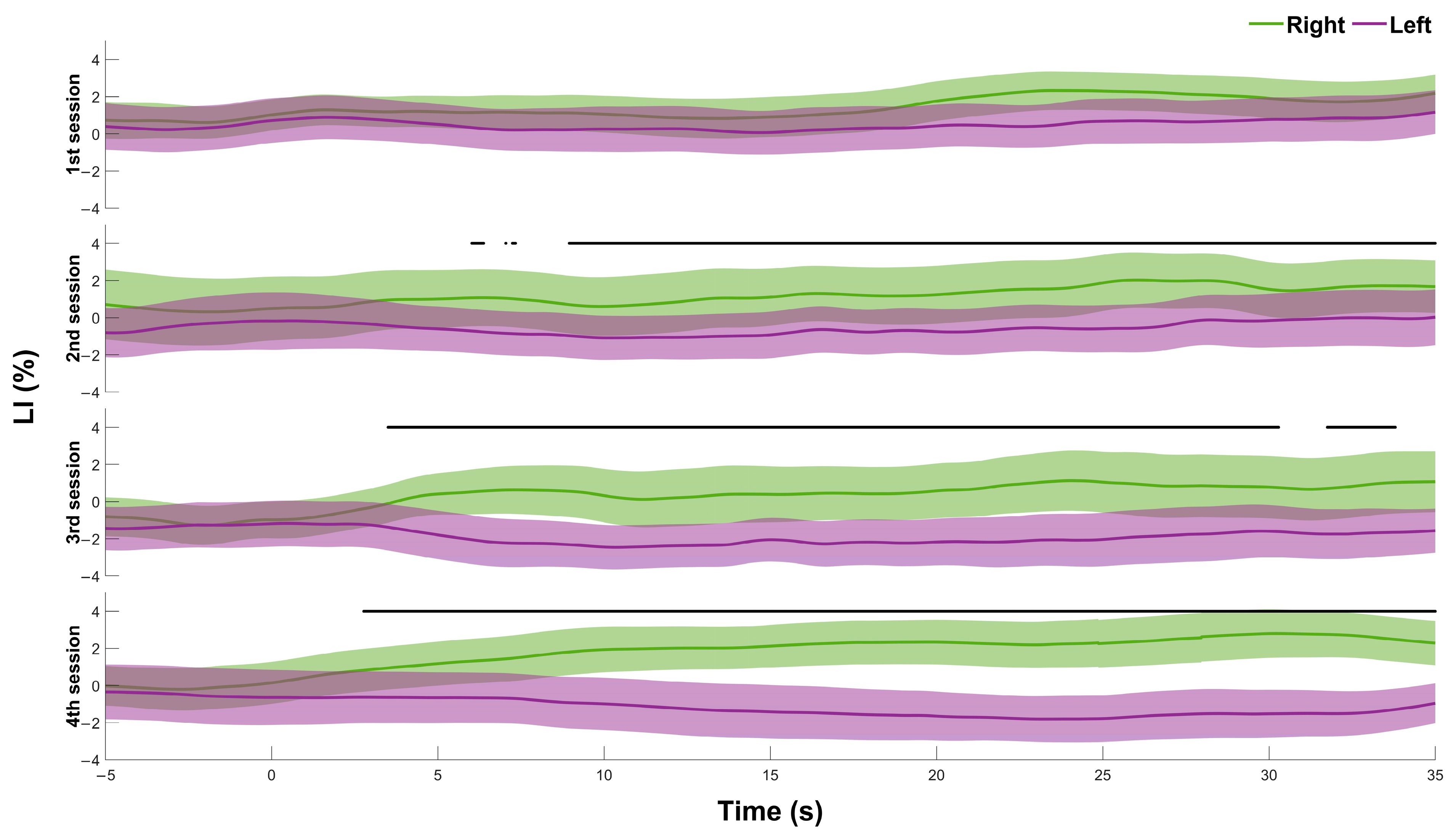
The time course of LI in response to right and left cues was obtained for the different sessions and subjects by separately averaging the six right and left tasks performed by each subject. Before averaging, the tracings were aligned in time with respect to the time of cue presentation (t = 0). A time window of 40 s was adopted, which includes 5 s of baseline (before cue presentation) and the whole 35 s duration of the mental task. To assess the lateralization at the group level, we computed session-level values for each subject. Specifically, for each session, we calculated the average LI response to six right and six left tasks separately; then, the average time value was computed over the whole task duration (35 s). A two-way repeated-measures ANOVA (or non-parametric Friedman test, in case of non-normality data) was conducted with the following factors: lateralization (right vs. left) and session (session 1–4). Post hoc comparisons were corrected using Bonferroni adjustment. After obtaining individual average traces for each condition and subject, we also computed the grand average (mean ± standard deviation) across all subjects to generate the plots shown in Figure 2. To analyze the temporal aspects of the differentiation, grand-average curves were also compared by a Wilcoxon signed-rank test (paired samples) at each time point (14 paired values, one per subject, for left vs. right conditions). The level of significance was set at p < 0.05.

2.5.2. Classification

As usual in BCI application, the ability of the subject to control the ball movement was quantified in terms of accuracy and information transfer rate (ITR). Accuracy was then calculated as the number of correct responses divided by the total number of tasks (12 per session), averaged over all subjects in a given session. On this basis and considering that a binary (left/right) symbol could be transmitted every 50 s (15 s rest + 35 s task), ITR could also be calculated [42].

Binary Classification (Single Threshold)

The subject response to the presented cue was classified as “left” or “right”, depending on whether the change in LI was below or above a defined threshold, respectively. Each classification was then deemed true or false, respectively, depending on whether or not it matched the cue presented in that task. The change in LI was calculated as the difference between the time average calculated over the full 35 s interval of the task and the pre-cue interval (average over the last 5 s before cue presentation). The performance of the classification was then quantified via the construction of a receiver operating characteristic (ROC) curve and calculation of the area under the curve (AUC), which were then used to assess potential improvements across sessions. The accuracy of the classification with the optimal threshold was also calculated as the number of true responses divided by the total number of tasks. The optimal threshold was determined using Youden’s index (J = sensitivity + specificity − 1), identifying the threshold that maximized this value, i.e., the index was computed across all possible thresholds along the ROC curve, and the threshold yielding the highest J was selected.
The significance of the improvement in accuracy over the four sessions was assessed via a repeated-measures ANOVA. Prior to the analysis, the assumption of normality was evaluated. When this assumption was violated, the non-parametric Friedman test was used instead. In cases where statistical significance (p < 0.05) was found, post hoc comparisons were performed using the Bonferroni correction method.

Three-Class Classification (Two Threshold)

Considering a prospective application in the field of brain–computer interface (BCI), the possibility of a three-class classification was investigated to test whether it could improve the performance of the decoder. A two-threshold classification approach was implemented, which defines three classes, i.e., left (LI < lower threshold), right (LI > upper threshold), and non-classified (lower threshold ≤ LI ≤ upper threshold).
To this aim, the lower threshold was varied between the minimum and maximum observed change in LI, and the upper threshold was varied between the current lower threshold and the maximum LI change. Accuracy, ITR, and the number of unclassified responses were calculated for each given pair of thresholds. Accuracy was defined as the ratio of valid responses, (thus, excluding the non-classified types). For ITR assessment, the symbol transfer rate was calculated as the inverse of the duration of each task, including the rest period (1/(15 + 35 s) = 0.02 symbols/s = 1.2 symbols/min). In this way, the loss of time associated with unclassified responses is taken into account as it impacts the overall ITR. This analysis is only performed for the data collected in the 4th session.

3. Results

All recruited subjects enjoyed participating in the study and easily completed the four sessions (see answers to the questionnaire below), and only on a few occasions did they respond that they were anticipating the end of the training phase.

3.1. Differentiation of Responses to Left/Right Tasks

A first intuitive representation of the effectiveness of neurofeedback can be observed in Figure 2, reporting the average time course of LI response to right and left cues in the different sessions. It can be observed that in the first session, the two curves substantially overlap, indicating poor capacity to control the movement of the ball. However, in subsequent sessions, the two curves become significantly different (p < 0.01) from each other for a progressively increased length of time. The response to right/left cues becomes increasingly higher/lower than 0, and the separation between the two curves takes place at a progressively lower latency from cue presentation, reaching a value as low as 2.7 s in the fourth session. Additionally, only in the final session, the lateralization index (LI) remains consistently positive (right cue) or negative (left cue) throughout the task, and the difference between the two curves is maintained statistically significantly, with continuity, until the end of the task.
The repeated-measures ANOVA revealed a significant main effect of lateralization (p < 0.01), indicating an overall difference between right and left hemisphere responses. Additionally, there was a significant interaction between lateralization and session (p = 0.023), indicating that the degree of lateralization changed across sessions. Post hoc comparisons showed no significant difference between right and left responses in Session 1, while significant differences emerged in the following sessions (p < 0.01 in all of them).

3.2. Binary Classification

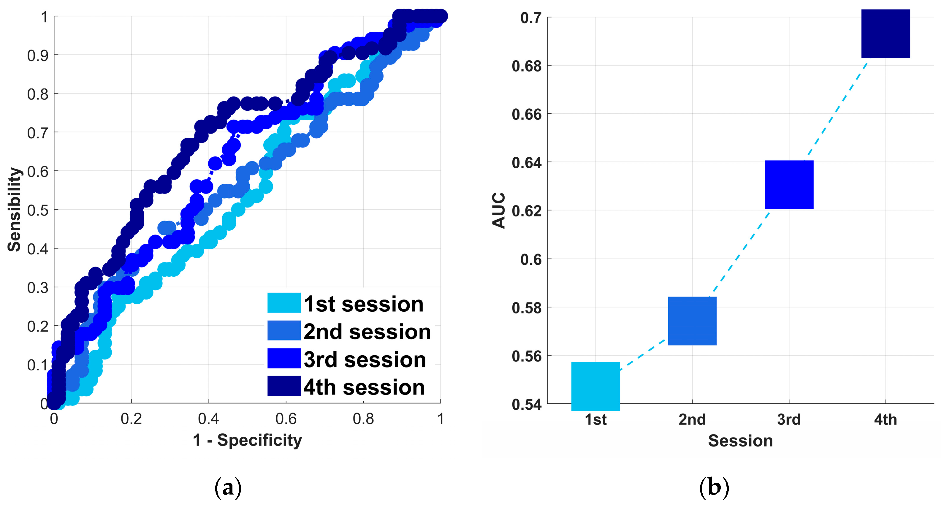
The optimal threshold for classification of the LI response to right/left tasks was investigated using the ROC curves, presented in Figure 3a, with the different colors referring to the different sessions. It can be observed that the classification performance improved progressively over time, as denoted by the increased convexity of the curves and increased AUC (Figure 3b), which again reflect improved subject control over the position of the ball.
The accuracy of the left/right classification when adopting the optimal threshold was distinctly analyzed in all subjects. The results presented in Figure 4 show the inter-individual variability across the four sessions. It can be observed that there is a tendency towards improvement (yellow-white color) with increased training.
On average, the accuracy exhibited a significant dependence on session and was significantly increased in the fourth session compared to the first (p < 0.05), achieving a value of 0.65 and a corresponding ITR of 0.084 bit/min (Table 2).

3.3. Three-Class Classification

As explained in the Methods section, a three-class classification was attempted to test whether it could lead to improved performance of the decoder. The results are graphically presented in Figure 5. Figure 5a indicates that accuracy is maximized when the two thresholds are about t1 = −6 and t2 = +6, which, however, means that when LI variation falls within this range, the response is “unclassified”. However, Figure 5b shows that much lower thresholds are necessary to maximize ITR (t1 at about 0 and t2 between 2 and 4). This is explained by the fact that the increase in accuracy is accompanied by an increased number of unclassified responses (Figure 5c), which lowers the overall performance. However, with this approach, it was possible to improve the performance with respect to binary classification, achieving an ITR of 0.11 instead of the 0.084 obtained by the binary classifier.

3.4. Results from the Questionnaire

The questionnaire was administered only at the end of the first session to assess the appropriateness of the experimental setup. Most participants reported that the duration of both the training and testing phases was adequate, except for three individuals, who found the training phase too long. As a result, from the second session onward, participants were given the option to end the training phase earlier, if they wished.
All participants stated that they put significant effort into achieving the objectives. They found the training period useful and appreciated the ball as a visual feedback tool, which they considered helpful for monitoring their progress.
When asked whether they believed they had achieved good control of lateralization, three participants expressed confidence in their capacity, while the remaining participants stated that they tried but were not fully successful in lateralizing. This outcome is consistent with the results, presenting on average poor performance in the first session. Interestingly, the participants used various strategies to enhance lateralization. For right-hemisphere activation, they often combined emotional, cognitive, and motor approaches, often focusing on personal memories, emotions, or music, particularly instrumental melodies such as classical music or film scores. Relaxation techniques, including meditative states or clearing the mind, were also common. On the motor side, participants imagined left-sided movements like swimming, punching, or using the left hand, with some of them emphasizing the intention rather than visualization of these actions.
For left-hemisphere activation, participants relied on linguistic, logical, and motor tasks linked to the right side of the body. These included imagining movements like playing sports or performing exercises with the right side, as well as engaging in cognitive tasks such as mental arithmetic, counting in foreign languages, or naming objects. Linguistic activities like tongue twisters, proverbs, and reciting texts were also frequently employed. Many participants reported that combining different approaches, like mixing mental calculations with imagined actions, was particularly effective for stimulating left-hemisphere activity.

4. Discussion

In this work, we proposed a neurofeedback system based on hemispheric lateralization, utilizing transcranial Doppler technology, implemented with cost-effective and portable components. While the system can operate on different cerebral arteries, this study was conducted on the middle cerebral arteries, as they supply the cognitive and motor regions of the brain.
The results indicate that neurofeedback training over four sessions effectively improves the individual capacity to voluntarily orient blood flow to the right or left side of the brain, as documented by increasing separation of average LI responses to left/right mental efforts, as well as by increased accuracy and AUC of ROC curves related to the binary classification of the responses. As a side investigation, we verified that excluding “doubtful” responses could further increase the performance of the decoding, according to a three-class classification. Finally, a questionnaire administered at the end of the first session revealed that the subjects successfully experimented with new, original mental strategies to achieve the goal of driving the ball.
To the best of our knowledge, only two studies have investigated the use of TCD to implement neurofeedback systems. In both studies, the focus was on either increasing or decreasing blood flow in MCAs [33] or anterior cerebral arteries (ACAs) [34]. Duschek et al. [33] divided participants into two separate groups tasked with increasing or decreasing CBFV. Without a prior training phase, they asked participants to modify flow velocity during eight sessions, each consisting of six extended neurofeedback trials lasting four minutes. Significant success was observed from the second session on, for increasing flow, and from the seventh session on, for decreasing it [33]. Similarly, Rey et al. [34] presented a neurofeedback system tested on two groups using different protocols, one longer (2 min tasks, repeated six times across four sessions) and one shorter (30 s tasks, repeated five times across three sessions). They reported a progressively increasing success rate with training and better performance with shorter-lasting tasks. However, the paper mainly presented, as a proof of concept, a description of the necessary hardware to achieve the real-time acquisition and processing of blood velocity signals and presentation of a visual feedback to the subject, with no statistical analysis to support the experimental findings, due to the low sample size [34]. Notably, in both studies, a single blood velocity signal related to one given artery, or to the average of left and right arteries, was provided as visual feedback; therefore, the possible difference between the two signals was not assessed. In this respect, the present study introduces a novel approach, i.e., by adopting as visual feedback the difference in CBFVs collected from the two sides, the user is trained to unbalance the blood flow to the left or right hemisphere.
The idea of measuring the lateralization of brain function by assessing hemispheric flow imbalance dates back to the 1970s [43] and was later been implemented with different methodologies. Besides fMRI and more recently, fNIRS, lateralization has been frequently investigated by TCD, e.g., for assessing the lateralization of speech and language functions [30,44].
In addition, the TCD-detected lateralization of brain functions has been employed for implementing BCI applications [45,46,47]. A first attempt was made by Myrden et al. [47], who investigated the possibility to discriminate a word task (mental generation of a sequence of words, all starting with the same given letter), expected to lateralize blood flow to the left side, from a mental rotation task (determination of whether two presented, differently-oriented objects are the same object or mirror images), expected to induce a bilateral flow increase. A similar approach (lateralizing vs. non-lateralizing task) was later used to operate a keyboard [46]. In a subsequent study from the same group, TCD was employed to achieve a mean ITR of 1.08 bits/min using a real-time three-class BCI [31]. Furthermore, their approach was limited to the following specific tasks for lateralization: verbal fluency for lateralization to the left hemisphere and motor imagery of fist clenching or 3D shape tracing for the right hemisphere [31].
In these and other TCD-BCI studies, subjects have always been instructed to follow specific tasks for lateralization. Surprisingly, the idea of letting the subjects explore different strategies, possibly improving the performance of the BCIs by specifically training this capacity with a neurofeedback approach, was not previously tested. More specifically, only a few suggestions were provided about how to interact with the visual feedback, and the subjects were encouraged to freely explore different mental tasks, looking for the most effective strategies to control blood flow lateralization. Interestingly, similar neurofeedback studies based on fNIRS methodologies evidenced that free and spontaneous neurofeedback interaction may yield better results compared to following rigid instructions regarding which mental task to use [25]. Unfortunately, the present study was not designed to investigate individual variability in performance, as evidenced in Figure 4. An interesting aspect to address in future studies may be the identification of the determinants of the varying lateralization ability achieved by the different subjects, possibly including the mental strategy adopted, the motivation, the stability of cerebral hemodynamics, the fatigue accumulated during training, etc.
In the present study, a free, unsupervised neurofeedback training lasting 15 min, followed by an assessment test implemented in each of the four sessions. The improvements observed across sessions demonstrate the effectiveness of the training. In addition, the increasing trend in the performance indicators, AUC, accuracy, and ITR, did not exhibit a plateau, suggesting that further benefits could be achieved by increasing the number of training sessions. Moreover, as indicated by the time course of the average responses to left and right cues (Figure 2), neurofeedback training also progressively shortens the time required to significantly separate the two sets, which suggests that training would also increase BCI performance by allowing for a progressively shorter duration of the tasks.
In agreement with Myrden et al. [47], we observed that a three-class performs slightly better than a two-class classification. In our case, this confirmed the hypothesis that discarding the weakest, i.e., the most “doubtful” responses, improves the ITR, even though discarding certain trials reduces the number of valid trials in the overall time dedicated to the experiment. In fact, as graphically represented in Figure 5, the highest ITR is obtained as the best trade-off between increasing the accuracy of the selection and limiting the number of discarded trials.
It can be observed that the ITR obtained in the present study is extremely low. However, the assessment of the ITR was only meant to quantify the improvement across sessions or between two- and three-class decoding. In fact, a very simple signal processing was adopted here, and large time intervals were dedicated to tasks and rest. More elaborate processing and classification techniques have been proposed and tested in several studies [33,48], and shorter time intervals [47] can be adopted to achieve higher ITR, which is adequate for BCI applications. In addition, to enhance the performance of TCD-based BCIs, integrating additional modalities has proven effective. For example, combining TCD with EEG to capture neuronal activity improved classification accuracy but increased computational complexity [49]. Similarly, Faress et al. combined TCD with fNIRS to enhance spatial resolution and accuracy [50]. Artificial intelligence methods have also been explored to optimize classification algorithms and improve system robustness [51]. These approaches could be evaluated in future implementations of the proposed system.

5. Conclusions and Limitation

In conclusion, the proposed neurofeedback system proved effective in enabling subjects to lateralize cerebral blood flow toward a specific hemisphere, thus controlling the movement of a ball on the screen. While the system exhibits limitations regarding spatial resolution when compared to techniques like EEG, fMRI, and fNIRS, it offers significant advantages in regards to ease of use, robustness, and portability. Unlike EEG and fNIRS, it avoids issues such as noise and crosstalk, making it a practical solution requiring minimal time to locate the arteries and position the equipment on the subject. Moreover, TCD is a cost-effective alternative to these methods, combining economic accessibility with consistent performance.
However, this study exhibits certain limitations. For example, the feedback questionnaire was administered only after the first session. Future improvements should focus on refining the interface and signal classification methods to enhance communication speed and accuracy. Finally, no control group (e.g., with no training) could be included, since the same visual feedback was also used in the test. Future studies are necessary to compare the efficacy of the present neurofeedback to that of other training modalities.
This work represents a preliminary step toward the implementation of a BCI, aiming to allow subjects to identify the mental strategies that work best for them. Beyond BCIs, this type of neurofeedback has potential applications in post-stroke neurorehabilitation, promoting cerebral plasticity, as suggested by recent literature [12,52]. Additionally, the system could be adapted to target other cerebral arteries, offering flexibility for various applications.
Overall, this study lays the groundwork for future advancements in neurofeedback and neurorehabilitation, demonstrating the utility of TCD-based systems, providing real-time hemispheric lateralization and user-friendly implementation.

Author Contributions

Conceptualization, R.R. and S.R.; methodology, R.R. and S.R.; software, R.R.; validation, R.R.; formal analysis, R.R. and L.E.; investigation, R.R. and S.R; data curation, R.R.; writing—original draft preparation, R.R.; writing—review and editing, R.R., L.E., C.G. and S.R.; supervision, S.R. and C.G.; project administration, R.R. and S.R. All authors have read and agreed to the published version of the manuscript.

Funding

This research received no external funding.

Institutional Review Board Statement

The study was conducted in accordance with the Declaration of Helsinki and approved by the Bioethics Committee of the University of Turin (protocol number 219859, April 2021).

Informed Consent Statement

Informed consent was obtained from all subjects involved in the study.

Data Availability Statement

The data that support the findings of this study are available from the corresponding author upon reasonable request.

Acknowledgments

R.R. is a doctoral student in complex systems for quantitative biomedicine-recipient PON innovation, scholarship code DOT13NVIW3 n.2.

Conflicts of Interest

The authors declare no conflict of interest.

Abbreviations

The following abbreviations are used in this manuscript:
BCIbrain–computer interface
EEGelectroencephalogram
fMRIfunctional magnetic resonance imaging
fNIRSfunctional near-Infrared spectroscopy
fTCDfunctional transcranial Doppler ultrasound
MCAmiddle cerebral artery
LIlateralization index
CBFVcerebral blood flow velocity
ITRinformation transfer rate
ROCreceiver operating characteristic
AUCarea under curve

References

  1. Rogers, L.J. A Matter of Degree: Strength of Brain Asymmetry and Behaviour. Symmetry 2017, 9, 57. [Google Scholar] [CrossRef]
  2. Karolis, V.R.; Corbetta, M.; Thiebaut de Schotten, M. The Architecture of Functional Lateralisation and Its Relationship to Callosal Connectivity in the Human Brain. Nat. Commun. 2019, 10, 1417. [Google Scholar] [CrossRef]
  3. Hara, Y. Brain Plasticity and Rehabilitation in Stroke Patients. J. Nippon. Med. Sch. 2015, 82, 4–13. [Google Scholar] [CrossRef]
  4. Bartolomeo, P.; Thiebaut de Schotten, M. Let Thy Left Brain Know What Thy Right Brain Doeth: Inter-Hemispheric Compensation of Functional Deficits After Brain Damage. Neuropsychologia 2016, 93, 407–412. [Google Scholar] [CrossRef]
  5. Alqadah, A.; Hsieh, Y.-W.; Morrissey, Z.D.; Chuang, C.-F. Asymmetric Development of the Nervous System. Dev. Dyn. 2018, 247, 124–137. [Google Scholar] [CrossRef]
  6. Ocklenburg, S.; Mundorf, A.; Gerrits, R.; Karlsson, E.M.; Papadatou-Pastou, M.; Vingerhoets, G. Clinical Implications of Brain Asymmetries. Nat. Rev. Neurol. 2024, 20, 383–394. [Google Scholar] [CrossRef]
  7. Bishop, D.V.M. Cerebral Asymmetry and Language Development: Cause, Correlate, or Consequence? Science 2013, 340, 1230531. [Google Scholar] [CrossRef] [PubMed]
  8. Rey, M.; Dellatolas, G.; Bancaud, J.; Talairach, J. Hemispheric Lateralization of Motor and Speech Functions After Early Brain Lesion: Study of 73 Epileptic Patients with Intracarotid Amytal Test. Neuropsychologia 1988, 26, 167–172. [Google Scholar] [CrossRef] [PubMed]
  9. Young, B.M.; Nigogosyan, Z.; Walton, L.M.; Song, J.; Nair, V.A.; Grogan, S.W.; Tyler, M.E.; Edwards, D.F.; Caldera, K.; Sattin, J.A.; et al. Changes in Functional Brain Organization and Behavioral Correlations after Rehabilitative Therapy Using a Brain-Computer Interface. Front. Neuroeng. 2014, 7, 26. [Google Scholar] [CrossRef]
  10. Ramos-Murguialday, A.; Broetz, D.; Rea, M.; Läer, L.; Yilmaz, O.; Brasil, F.L.; Liberati, G.; Curado, M.R.; Garcia- Cossio, E.; Vyziotis, A.; et al. Brain-Machine Interface in Chronic Stroke Rehabilitation: A Controlled Study. Ann. Neurol. 2013, 74, 100–108. [Google Scholar] [CrossRef]
  11. Rodríguez-García, M.E.; Carino-Escobar, R.I.; Carrillo-Mora, P.; Hernandez-Arenas, C.; Ramirez-Nava, A.G.; Pacheco-Gallegos, M.d.R.; Valdés-Cristerna, R.; Cantillo-Negrete, J. Neuroplasticity Changes in Cortical Activity, Grey Matter, and White Matter of Stroke Patients After Upper Extremity Motor Rehabilitation via a Brain–Computer Interface Therapy Program. J. Neural Eng. 2025, 22, 026025. [Google Scholar] [CrossRef] [PubMed]
  12. Loriette, C.; Ziane, C.; Ben Hamed, S. Neurofeedback for Cognitive Enhancement and Intervention and Brain Plasticity. Rev. Neurol. 2021, 177, 1133–1144. [Google Scholar] [CrossRef] [PubMed]
  13. Bengtsson, S.L.; Nagy, Z.; Skare, S.; Forsman, L.; Forssberg, H.; Ullén, F. Extensive Piano Practicing Has Regionally Specific Effects on White Matter Development. Nat. Neurosci. 2005, 8, 1148–1150. [Google Scholar] [CrossRef] [PubMed]
  14. Orndorff-Plunkett, F.; Singh, F.; Aragón, O.R.; Pineda, J.A. Assessing the Effectiveness of Neurofeedback Training in the Context of Clinical and Social Neuroscience. Brain Sci. 2017, 7, 95. [Google Scholar] [CrossRef]
  15. Huster, R.J.; Mokom, Z.N.; Enriquez-Geppert, S.; Herrmann, C.S. Brain-Computer Interfaces for EEG Neurofeedback: Peculiarities and Solutions. Int. J. Psychophysiol. 2014, 91, 36–45. [Google Scholar] [CrossRef]
  16. Birbaumer, N.; Ramos Murguialday, A.; Weber, C.; Montoya, P. Chapter 8 Neurofeedback and Brain–Computer Interface: Clinical Applications. In International Review of Neurobiology; Academic Press: Cambridge, MA, USA, 2009; Volume 86, pp. 107–117. [Google Scholar]
  17. Enriquez-Geppert, S.; Huster, R.J.; Herrmann, C.S. EEG-Neurofeedback as a Tool to Modulate Cognition and Behavior: A Review Tutorial. Front. Hum. Neurosci. 2017, 11, 51. [Google Scholar] [CrossRef]
  18. Gruzelier, J.H. EEG-Neurofeedback for Optimising Performance. III: A Review of Methodological and Theoretical Considerations. Neurosci. Biobehav. Rev. 2014, 44, 159–182. [Google Scholar] [CrossRef]
  19. Lia, A.; Di Spiezio, A.; Speggiorin, M.; Zonta, M. Two Decades of Astrocytes in Neurovascular Coupling. Front. Netw. Physiol. 2023, 3, 1162757. [Google Scholar] [CrossRef]
  20. Rota, G.; Sitaram, R.; Veit, R.; Erb, M.; Weiskopf, N.; Dogil, G.; Birbaumer, N. Self-Regulation of Regional Cortical Activity Using Real-Time fMRI: The Right Inferior Frontal Gyrus and Linguistic Processing. Hum. Brain Mapp. 2008, 30, 1605–1614. [Google Scholar] [CrossRef]
  21. Moll, J.; Weingartner, J.H.; Bado, P.; Basilio, R.; Sato, J.R.; Melo, B.R.; Bramati, I.E.; de Oliveira-Souza, R.; Zahn, R. Voluntary Enhancement of Neural Signatures of Affiliative Emotion Using fMRI Neurofeedback. PLoS ONE 2014, 9, e97343. [Google Scholar] [CrossRef]
  22. Thibault, R.T.; MacPherson, A.; Lifshitz, M.; Roth, R.R.; Raz, A. Neurofeedback with fMRI: A Critical Systematic Review. NeuroImage 2018, 172, 786–807. [Google Scholar] [CrossRef] [PubMed]
  23. Caria, A.; Veit, R.; Sitaram, R.; Lotze, M.; Weiskopf, N.; Grodd, W.; Birbaumer, N. Regulation of Anterior Insular Cortex Activity Using Real-Time fMRI. Neuroimage 2007, 35, 1238–1246. [Google Scholar] [CrossRef]
  24. Lapborisuth, P.; Zhang, X.; Noah, A.; Hirsch, J. Neurofeedback-Based Functional Near-Infrared Spectroscopy Upregulates Motor Cortex Activity in Imagined Motor Tasks. Neurophotonics 2017, 4, 021107. [Google Scholar] [CrossRef] [PubMed]
  25. Kohl, S.H.; Mehler, D.M.A.; Lührs, M.; Thibault, R.T.; Konrad, K.; Sorger, B. The Potential of Functional Near-Infrared Spectroscopy-Based Neurofeedback—A Systematic Review and Recommendations for Best Practice. Front. Neurosci. 2020, 14, 594. [Google Scholar] [CrossRef] [PubMed]
  26. Gutierrez-Sigut, E.; Daws, R.; Payne, H.; Blott, J.; Marshall, C.; MacSweeney, M. Language Lateralization of Hearing Native Signers: A Functional Transcranial Doppler Sonography (fTCD) Study of Speech and Sign Production. Brain Lang. 2015, 151, 23–34. [Google Scholar] [CrossRef]
  27. Silvestrini, M.; Cupini, L.M.; Matteis, M.; Troisi, E.; Caltagirone, C. Bilateral Simultaneous Assessment of Cerebral Flow Velocity During Mental Activity. J. Cereb. Blood Flow Metab. 1994, 14, 643–648. [Google Scholar] [CrossRef]
  28. Badcock, N.A.; Groen, M.A. What Can Functional Transcranial Doppler Ultrasonography Tell Us About Spoken Language Understanding? Lang. Cogn. Neurosci. 2017, 32, 818–828. [Google Scholar] [CrossRef]
  29. Deppe, M.; Knecht, S.; Papke, K.; Lohmann, H.; Fleischer, H.; Heindel, W.; Ringelstein, E.B.; Henningsen, H. Assessment of Hemispheric Language Lateralization: A Comparison Between fMRI and fTCD. J. Cereb. Blood Flow Metab. 2000, 20, 263–268. [Google Scholar] [CrossRef]
  30. Deppe, M.; Ringelstein, E.B.; Knecht, S. The Investigation of Functional Brain Lateralization by Transcranial Doppler Sonography. NeuroImage 2004, 21, 1124–1146. [Google Scholar] [CrossRef]
  31. Goyal, A.; Samadani, A.-A.; Guerguerian, A.-M.; Chau, T. An Online Three-Class Transcranial Doppler Ultrasound Brain Computer Interface. Neurosci. Res. 2016, 107, 47–56. [Google Scholar] [CrossRef]
  32. Martini, M.L.; Oermann, E.K.; Opie, N.L.; Panov, F.; Oxley, T.; Yaeger, K. Sensor Modalities for Brain-Computer Interface Technology: A Comprehensive Literature Review. Neurosurgery 2020, 86, E108. [Google Scholar] [CrossRef] [PubMed]
  33. Duschek, S.; Schuepbach, D.; Doll, A.; Werner, N.S.; Reyes del Paso, G.A. Self-Regulation of Cerebral Blood Flow by Means of Transcranial Doppler Sonography Biofeedback. Ann. Behav. Med. 2011, 41, 235–242. [Google Scholar] [CrossRef] [PubMed]
  34. Rey, B.; Rodríguez, A.; Lloréns-Bufort, E.; Tembl, J.; Muñoz, M.Á.; Montoya, P.; Herrero-Bosch, V.; Monzo, J.M. Design and Validation of an FPGA-Based Configurable Transcranial Doppler Neurofeedback System for Chronic Pain Patients. Sensors 2018, 18, 2278. [Google Scholar] [CrossRef]
  35. Rabbito, R.; Cinanni, A.; Bussi, L.; Guiot, C.; Roatta, S. A Neuro-Feedback Prototype Based on Transcranial Doppler Ultrasound for Brain Computer Interface Applications. In Proceedings of the 2024 46th Annual International Conference of the IEEE Engineering in Medicine and Biology Society (EMBC), Orlando, FL, USA, 15–19 July 2024; pp. 1–5. [Google Scholar]
  36. Phillips, A.A.; Chan, F.H.; Zheng, M.M.Z.; Krassioukov, A.V.; Ainslie, P.N. Neurovascular Coupling in Humans: Physiology, Methodological Advances and Clinical Implications. J. Cereb. Blood Flow Metab. 2016, 36, 647–664. [Google Scholar] [CrossRef]
  37. Rabbito, R.; Guiot, C.; Roatta, S. Functional Transcranial Doppler (fTCD) Investigation of Brain Lateralization Following Visual Stimuli. In Proceedings of the 2023 45th Annual International Conference of the IEEE Engineering in Medicine & Biology Society (EMBC), Sydney, Australia, 24–27 July 2023; pp. 1–4. [Google Scholar]
  38. Serrien, D.J.; O’Regan, L. Word Perception and Upper-Lower Visual Field Asymmetries. Brain Cogn. 2025, 186, 106294. [Google Scholar] [CrossRef]
  39. Lin, T.-K.; Ryu, S.-J.; Hsu, P.-W. Interhemispheric Comparisons of Cerebral Blood Flow Velocity Changes During Mental Tasks with Transcranial Doppler Sonography. J. Ultrasound Med. 2009, 28, 1487–1492. [Google Scholar] [CrossRef] [PubMed]
  40. Rogers, L.J. Brain Lateralization and Cognitive Capacity. Animals 2021, 11, 1996. [Google Scholar] [CrossRef]
  41. Salinet, A.S.M.; Panerai, R.B.; Robinson, T.G. Effects of Active, Passive and Motor Imagery Paradigms on Cerebral and Peripheral Hemodynamics in Older Volunteers: A Functional TCD Study. Ultrasound Med. Biol. 2012, 38, 997–1003. [Google Scholar] [CrossRef] [PubMed]
  42. Obermaier, B.; Neuper, C.; Guger, C.; Pfurtscheller, G. Information Transfer Rate in a Five-Classes Brain- Computer Interface. IEEE Trans. Neural Syst. Rehabil. Eng. 2001, 9, 283–288. [Google Scholar] [CrossRef]
  43. Olesen, J. Contralateral focal increase of cerebral blood flow in man during arm work. Brain 1971, 94, 635–646. [Google Scholar] [CrossRef]
  44. Knecht, S.; Deppe, M.; Ringelstein, E.B.; Wirtz, M.; Lohmann, H.; Dräger, B.; Huber, T.; Henningsen, H. Reproducibility of Functional Transcranial Doppler Sonography in Determining Hemispheric Language Lateralization. Stroke 1998, 29, 1155–1159. [Google Scholar] [CrossRef] [PubMed]
  45. Khalaf, A.; Sybeldon, M.; Sejdic, E.; Akcakaya, M. A Brain-Computer Interface Based on Functional Transcranial Doppler Ultrasound Using Wavelet Transform and Support Vector Machines. J. Neurosci. Methods 2018, 293, 174–182. [Google Scholar] [CrossRef]
  46. Lu, J.; Mamun, K.A.; Chau, T. Online Transcranial Doppler Ultrasonographic Control of an Onscreen Keyboard. Front. Hum. Neurosci. 2014, 8, 199. [Google Scholar] [CrossRef] [PubMed]
  47. Myrden, A.J.B.; Kushki, A.; Sejdić, E.; Guerguerian, A.-M.; Chau, T. A Brain-Computer Interface Based on Bilateral Transcranial Doppler Ultrasound. PLoS ONE 2011, 6, e24170. [Google Scholar] [CrossRef] [PubMed]
  48. Islam, A.; Ahmed, E.; Islam, A.; Lu, J.; Sarkar, F.; Mamun, K.A. Decoding Human Brain States Using Transcranial Doppler Ultrasonography. In Proceedings of the 2015 International Conference on Electrical Engineering and Information Communication Technology (ICEEICT), Savar, Bangladesh, 21–23 May 2015; pp. 1–6. [Google Scholar] [CrossRef]
  49. Khalaf, A.; Sejdic, E.; Akcakaya, M. A Novel Motor Imagery Hybrid Brain Computer Interface Using EEG and Functional Transcranial Doppler Ultrasound. J. Neurosci. Methods 2019, 313, 44–53. [Google Scholar] [CrossRef]
  50. Faress, A.; Chau, T. Towards a Multimodal Brain–Computer Interface: Combining fNIRS and fTCD Measurements To Enable Higher Classification Accuracy. NeuroImage 2013, 77, 186–194. [Google Scholar] [CrossRef]
  51. Gan, L.; Yin, X.; Huang, J.; Jia, B.; Gan, L.; Yin, X.; Huang, J.; Jia, B. Transcranial Doppler Analysis Based on Computer and Artificial Intelligence for Acute Cerebrovascular Disease. Math. Biosci. Eng. 2023, 20, 1695–1715. [Google Scholar] [CrossRef]
  52. Le Franc, S.; Herrera Altamira, G.; Guillen, M.; Butet, S.; Fleck, S.; Lécuyer, A.; Bougrain, L.; Bonan, I. Toward an Adapted Neurofeedback for Post-Stroke Motor Rehabilitation: State of the Art and Perspectives. Front. Hum. Neurosci. 2022, 16, 917909. [Google Scholar] [CrossRef]
Figure 1. TCD-based neurofeedback.
Applsci 15 05763 g001
Figure 2. Time course of the lateralization index (LI) across the different sessions in response to right (green) and left (violet) cues. The black line indicates the significant difference between the two curves at each time point. Curves represent the average, and the colored band indicates the standard deviation for the subjects (n = 14). Note that the separation between curves progressively increases in subsequent sessions.
Applsci 15 05763 g002
Figure 3. (a) Receiver operating characteristic (ROC) curves related to the binary (left/right) classification of the responses from all subjects in each session; (b) value of the area under the curve (AUC) for ROC in each session, as an indicator of the effectiveness of left/right discrimination. The different colors indicate the different sessions.
Applsci 15 05763 g003
Figure 4. Individual accuracy for the different subjects across the four sessions. The level of accuracy is color-coded, according to the legend shown on the right. Note the variability in behavior for the different subjects, as well as a general trend towards light colors, with increasing session number.
Applsci 15 05763 g004
Figure 5. Results of the three-class classification based on adopting two thresholds for the lateralization index (LI). For each pair of thresholds, t1 and t2 varied through the maximum and minimum values of the LI, and the accuracy (a), information transfer rate (ITR) (b), and number of unclassified responses (c) are calculated.
Applsci 15 05763 g005
Table 1. Mental activities associated with hemispheric activation.
Left HemispheresRight Hemispheres
Word generation [30,38]Spatial orientation [27]
Mental calculation [39]Evoking strong emotions, listening to melodies, and meditation [40]
Movements of the right upper and lower limbs [41]Motor movements of the left side of the body [41]
Table 2. Accuracy and information transfer rate (ITR) of all subjects across each session.
Session #1234
Accuracy0.510.500.600.65
ITR (bit/min)0.000120.0100.0360.084
Disclaimer/Publisher’s Note: The statements, opinions and data contained in all publications are solely those of the individual author(s) and contributor(s) and not of MDPI and/or the editor(s). MDPI and/or the editor(s) disclaim responsibility for any injury to people or property resulting from any ideas, methods, instructions or products referred to in the content.