Article

Biological and Behavioural Effects of Bisphenol A (BPA) Exposure: An In Vivo Study in Drosophila melanogaster

by
Isabel Gaivão
1,2,*,
Rita António Santos
2,3,
Tetiana V. Morozova
4 and
Volodymyr V. Tkach
5,*
1
Animal and Veterinary Research Center (CECAV), University of Trás-os-Montes and Alto Douro (UTAD), Quinta de Prados, 5001-801 Vila Real, Portugal
2
Department of Genetics and Biotechnology (DGB–ECVA), University of Trás-os-Montes and Alto Douro (UTAD), Quinta de Prados, 5001-801 Vila Real, Portugal
3
Biology of Human Reproduction and Infertility Research Group, Center for Neuroscience and Cell Biology, University of Coimbra (CNC-UC), 3000-548 Coimbra, Portugal
4
Ecology and Environmental Protection Department, National Transport University, Omelianovych-Pavlenko Str. 1, 01001 Kyiv, Ukraine
5
General and Material Chemistry Department, Chernivtsi National University, Kotsiubynsky Str. 12, 58001 Chernivtsi, Ukraine
*
Authors to whom correspondence should be addressed.
Appl. Sci. 2025, 15(10), 5588; https://doi.org/10.3390/app15105588 (registering DOI)
Submission received: 12 February 2025 / Revised: 7 April 2025 / Accepted: 13 May 2025 / Published: 16 May 2025
(This article belongs to the Section Biomedical Engineering)

Abstract

:
Bisphenol A (BPA) is one of the most produced compounds worldwide. It acts as an endocrine disruptor and can cause adverse effects in the body, even at low-dose exposures. By interacting with estrogen receptors, it can play an important role in the pathogenesis of several endocrine diseases, such as infertility, hormone-dependent tumours and various metabolic disorders. Exposure in humans, especially early in life, is of particular concern, since it can have a big impact on an individual’s development and growth. The objective of this study was to evaluate, in vivo, the genotoxicity of BPA and its effects on longevity, prolificacy and behaviour in Drosophila melanogaster. To evaluate the biological and behavioural effects, flies were crossed and subjected to different concentrations of BPA (0.5 mM, 1 mM, 2 mM, 5 mM and 10 mM) in Drosophila instant “Carolina” medium hydrated with the BPA solution for 3 days, and then transferred to a non-treated medium, where they continued to lay eggs. This procedure was repeated to obtain the F2 generation. To evaluate genotoxicity, the somatic mutation and recombination test (SMART) and Comet assays were performed. In these cases, higher concentrations of BPA were chosen (1 mM, 10 mM, 20 mM and 50 mM). The results showed that this compound caused changes in longevity and prolificacy, and that these changes also affected subsequent generations. They also showed that BPA affected Drosophila’s behaviour and social interaction, but at the exposure levels investigated here, it did not cause significant genotoxic effects.

1. Introduction

Bisphenols (Figure 1) consist of a group of compounds used mainly in polyester plastics, including polycarbonates, fast-drying epoxy resin adhesives and anticorrosion coatings [1,2,3,4,5]. They are also used in odontology for implant fixation, as well as in cosmetical and pharmaceutical formulation packages.
These compounds are used mostly in polycondensation polymers, which degrade while heated [6,7,8,9,10]. This degradation splits the polymer molecules into monomers, which may penetrate the food or water stored in the polyester ducts or vessels. The main toxic action of bisphenols, principally A, F and S, consists of endocrine disruptions, mainly xenoestrogenic disruptions; bisphenols act as agonists or antagonists through endocrine receptor-dependent signalling pathways, therefore leading to several endocrine diseases, such as male and female infertility, precocious puberty, various metabolic disorders and hormone-dependent tumours. They also have an active effect on the neuroendocrine system by interfering with neuronal differentiation, the growth and formation of synapses and the affected individual’s subsequent behaviour.
The most frequent produced compound among the class of bisphenols is bisphenol A (BPA), which is currently one of the most produced compounds worldwide [10,11,12]. It is often found in food and cosmetic packages and active forms, dental materials, eyeglass lenses, health equipment and thermal paper. It is also considered a food contact material (FCM), which means that it is used in the manufacture of materials that come into direct contact with food, such as plastic packaging, kitchenware, inner coatings of cans and jar caps [13,14,15]. Although it partially degrades, it accumulates in the environment, becoming extremely toxic to aquatic organisms [16,17,18]. A recent European Food Safety Authority (EFSA) investigation, dated 2023 and supported by the European Commission, has shown high levels of bisphenol A intoxication in tested people in some EU member countries, including Portugal [10].
Bisphenol A contamination and human intoxication generally occur from exposure to contaminated air (inhalation of contaminated dust, for example), water, effluents and dust, or through the consumption of contaminated food due to leakage of BPA from its containers [19,20,21]. Infants, young children and pregnant or lactating women represent the highest-risk groups with regard to BPA exposure [22,23].
Several inconsistent findings can be found in the literature for this compound, including controversial data concerning a safe level of human exposure to BPA [24,25]. In 2002, the Scientific Committee on Toxicity, Ecotoxicity and the Environment (CSTEE) concluded that BPA presented a low potential for bioaccumulation, was not corrosive, was not mutagenic nor genotoxic, was not carcinogenic and was a non-inducer of oxidative or membrane damage [26,27]. However, during its last re-evaluation, the EFSA reduced the tolerable daily dose (TDI) from 50 µg/kg of body weight/day to 4 μg/kg of body weight/day [10].
Even though a lot of studies have been conducted to evaluate the toxic effects of BPA in several model organism species (both vertebrate and invertebrate), there is still a lack of studies that consider the possible transgenerational effects of endocrine disruptors such as bisphenol A. With this in mind, for this study, we decided to perform a transgenerational analysis of the toxicity of this compound in Drosophila melanogaster. This species has become one of the most popular model organisms used in studies of most biological phenomena, including population studies, since it allows the study of several generations in a short period. Its high prolificacy also makes statistical analysis easy and reliable [28,29,30,31,32].
Invertebrate endocrine systems are composed primarily of neuroendocrine components, with true endocrine glands working in a similar way to vertebrate glands [33,34,35]. Insect hormones regulate specific functions, such as moulting and metamorphosis, yolk synthesis, diuresis, mobilization of fuel for flight, polyphenism and diapause. In Drosophila, one of the major hormonal systems involves ecdysteroids, hormones that are mainly responsible for moulting. Molecular studies have demonstrated that ecdysteroid receptors are homologous to the steroid hormone receptors of vertebrates, and that they share a set of conserved features, which suggests that EDCs that are capable of binding to steroid hormone receptors can also bind to ecdysteroid receptors of invertebrates, and possibly interfere with the development, fertility and longevity of exposed individuals [36,37]. In fact, BPA has been shown to exhibit anti-ecdysteroid activity in the water flea Daphnia magna, causing alterations in the intermoult period and interfering with embryonic development [38,39,40,41,42]. However, other studies in the literature present controversial results regarding species’ sensitivity to BPA, showing that, at different relevant concentrations, the effect of BPA differs considerably, even between related species, and that there remains a lack of clarity regarding the precise mode of endocrine action.
Assessing the potential effects of endocrine-disrupting compounds for which the action mechanisms are still not well known in the investigated species requires an approach that relies on more general criteria, such as behaviour, reproduction and other biological characteristics, as well as relatively high concentrations, which allow for the identification of molecular and cellular effects that may remain undetectable at lower exposure levels [43]. Therefore, in this study, we evaluated, in vivo, BPA’s effect on Drosophila’s longevity, prolificacy, development time (all hormone-mediated processes) and behaviour after both acute and chronic exposure. In addition, since this organism has emerged as one of the most effective tools in genotoxicity studies [44], and only a few studies have assessed the genotoxicity of EDCs in invertebrates, even though this is one of the most important endpoints in chemical toxicity evaluation and environmental risk assessment [45,46,47], another objective of this study was to verify whether bisphenol A exhibited any genotoxicity in this model organism.

2. Materials and Methods

2.1. Drosophila Stocks
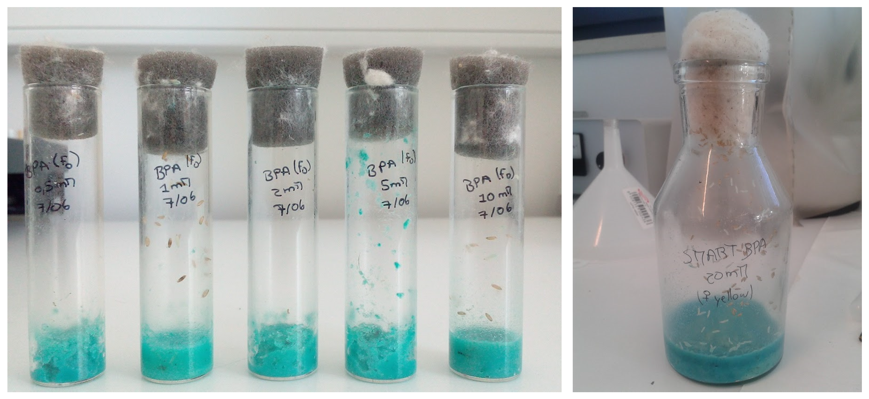
For this study, the D. melanogaster Oregon-K (OK) strain (a gift from Professor Maria Sierra, Oviedo University, Spain) was selected because, among the various strains available for this test, OK was the one that was most sensitive in other studies. The white- and yellow-marker mutations for the white gene were used [48]. Flies were maintained at UTAD, in glass bottles containing a standard medium for Drosophila (100 g sucrose, 100 g yeast, 12 g agar, 5.4 g NaCl, 5 mL propionic acid and 1 L distilled water, prepared at 90 °C and deposited into vials before solidification), at 24 ± 1 °C and approximately 60% humidity. When necessary, they were anesthetized by etherisation. Representative images of some of the experimental groups are provided in Figure 2.

2.2. BPA Treatment

Sterile, purified and pathogen-free BPA (4,4′-Isopropylidenediphenol) was supplied as dry white pills (Acros Organics, Geel, The Netherlands) with purity > 96%, according to the manufacturer’s information.
BPA was first dissolved in a small volume of DMSO, and then H2O was added to prepare the stock solution (DMSO was carefully kept at 0.1% or lower throughout the experiments). For each treatment, this solution was diluted and mixed with “Carolina Drosophila Medium Formula 4-24®” (Carolina Biological Supply Company 2016, Burlington, NC, Canada).
For the longevity, prolificacy and development time analysis, 5 concentrations of BPA were tested (0.5, 1, 2, 5 and 10 mM). These concentrations were chosen based on two previous studies where it was assumed that 1 mM of BPA corresponds, in flies, to the approximate human lowest-observed-adverse-effect level (LOAEL) of 50 mg/kg body weight/day recommended by the U.S. Environmental Protection Agency (EPA) [48,49,50]. The selected concentration range, although not environmentally relevant at first glance, was designed to examine dose–response patterns and potential thresholds of effect, and to dissect mechanistic aspects of toxicity. High-dose exposure is a standard practice in model organism toxicology studies to simulate chronic or cumulative stress scenarios. All assays were performed in duplicate and, for the negative control group, a solution of 0.1% DMSO was added to the instant “Carolina” medium. After the treatment, all flies were kept in a non-treated standard medium. To avoid possible contamination of BPA from plastic material, only glass material was used in this study.
Two types of treatment were used: acute, where we evaluated the effects of BPA exposure over a short period of time (72 h); and chronic, where the individuals were exposed for a longer period (in this case, throughout larval development). Exposure times were kept consistent in all experiments. For the acute treatment, 10 couples (2–5 days old) were mated in vials containing either non-treated (control group) or treated instant “Carolina” medium, and were allowed to feed and lay eggs. After 72 h, the flies were transferred to new vials containing a non-treated standard medium, where they continued to lay eggs. F1 subjects born in the non-treated (F1-N) medium were then used to evaluate the effects of parental BPA acute exposure in the offspring. The F1 progeny born in the treated (F1-T) medium was exposed to BPA throughout the larval development, and was used to evaluate the effects of chronic treatment after parental acute treatment. After hatching, 10 couples of the F1-T group were transferred into new vials containing BPA-treated medium, and the treatment process was repeated to obtain the F2 progeny. This transgenerational study allowed us to evaluate, in the offspring, the effects of acute and/or chronic treatment of the parental generation (F1-N and F2-TN respectively), the effects of acute treatment (F0), the effects of chronic treatment throughout larval development (F1-T) and the effects of consecutive chronic treatments (F2-TT). A scheme of this procedure is depicted in Figure 3A. For the behavioural assay, only the F1-T generation was used (Figure 3B).
As for the genotoxicity assays (SMART and Comet), heterozygous individuals were treated with 1, 10, 20 and 50 mM of BPA. Higher concentrations were used in these assays to evaluate the sensitivity of the flies to the compound and to try to establish a possible concentration–effect relationship. We also considered the fact that there may be cases where organisms are exposed to higher-than-average doses, depending on the ecosystem. Again, all assays were performed in duplicate (Figure 3C).

2.3. Longevity Assay

For each experimental group, between 11 and 30 individuals were used. Longevity was assessed through regular observations of Drosophila mortality (2 to 3 times per week), and, in order to maintain the best conditions, flies were transferred weekly to new vials. The difference in specimen number can be explained by the fact that in some conditions, and particularly in the F2 generation, the number of individuals born was lower than desired. However, in these cases, the number of individuals used in the remaining concentrations was adapted and reduced to diminish this discrepancy and mitigate possible effects on statistical analysis.

2.4. Prolificacy Assay

Prolificacy was evaluated as the total F1 hatched per condition; therefore, the adults that emerged from each experimental group were counted and compared in each treatment. This process was repeated until no more adults were emerging. The larval development time was also evaluated; for this, the days until emergence were counted.

2.5. Behavioural Assay

Flies from the F1-T group were used. A few days after emerging, 10 males (n = 10) were isolated and placed in a 5.2 cm diameter circular arena, where they were left to settle for 30 min. For each concentration, a 90 s video was recorded. Video analysis was performed using Ctrax® Version 0.5.8., a Drosophila tracking software, which assessed the walking speed and activity/distance travelled by each specimen (Figure 4). Individual behaviour and social interaction were evaluated through visual analysis. The exclusive use of male individuals was aimed at avoiding any courtship behaviour, which would interfere with the results.

2.6. Genotoxicity Evaluation

2.6.1. SMART w/w+ Assay

This assay evaluates genetic damage induction in somatic cells during larval exposure. Using genetic markers phenotypically observed in adult tissues, it detects loss of heterozygosity (LOH) in heterozygous individuals, which may be due to point mutations, deletions, non-disjunction or homologous mitotic recombination [51,52]. The eye w/w+ SMART assay was chosen, and the X-chromosome “white” gene (w) was used as a recessive marker to monitor, in wild-type eyes, the presence of white clones in the ommatidia. The assay was essentially performed as described previously by Marcos, Sierra and Gaivão [53]. For each concentration tested (1, 10, 20, 50 mM), as well as for the control, 50 w/w females and 30 w+/Y males, or 50 w+/w+ and 30 w/Y males, were crossed in instant “Carolina” medium hydrated with BPA solution for 3 days to obtain heterozygous progeny. After they had emerged, the adult flies were counted and isolated. Since both types of mating were used, and because the male progeny of the w/w female + w+/Y male crossing had white eyes, only the females were used. The eyes were scored as they hatched, based on the number of affected ommatidia, on a scale of 0 to >8, at 400× magnification, with a binocular magnifier (WILD M3Z, Heerbrugg, Switzerland) and two-foci optical fibre. At least 300 eyes per concentration were scored. It was not possible to calculate the recombination effect, which would be obtained by the difference between the number of spots found in females and the number found in males, since the latter could not be caused by homologous recombination.

2.6.2. Comet Assay

The Comet assay is a sensitive, fast and versatile genotoxic test, capable of detecting DNA breaks in a single cell, which are still within the range of what a cell can repair. In Drosophila, the most used cell types are neuroblasts, midgut cells and haemocytes [54]. Neuroblasts were used in this study, and the assay was performed based on the method described by Sierra et al., 2014 [55], with some modifications. For each BPA concentration tested (1, 10, 20, 50 mM), as well as for the control, neuroblasts from 3 larvae were isolated, macerated and centrifuged in Ringer solution for 5 min at 268× g. After removing the supernatant, 140 µL of 1% low-melting-point (LMP) agarose (Gibco, Thermo Fisher Scientific, Waltham, MA, USA) at 37 °C was added to each microtube. Two 70 µL drops of this solution were placed in a slide precoated with 1.5% normal-melting-point agarose (NMP; Sigma-Aldrich, Lisbon, Portugal), and each drop was covered with an 18 × 18 mm coverslip, spreading the solution. The slides were stored at 4 °C for 5 min and, after agarose solidification, the coverslips were removed. The slides were then immersed in cold, fresh lysis buffer (2.5 M NaCl, 0.1 M EDTA, 10 mM Tris-base, 1% Triton X-100, pH 10), for 2 h at 4 °C, in order for lysis to occur, after which they were placed in an electrophoresis chamber with their frozen ends facing the cathode and without empty spaces among them. The electrophoresis chamber was filled with an electrophoresis buffer (0.3 M NaOH, 1 mM EDTA pH 12.6) until the microgel slides were covered, and kept at 4 °C for 30 min. The next step was electrophoresis, in the dark, at 0.8 V/cm and 300 mA for 30 min at 4 °C (Cleaver Scientific Ltd., Warwickshire, UK). In the end, the slides were neutralized with PBS 1x and distilled water (at 4 °C, 10 min each), left to air-dry, and posteriorly stained with the blue-fluorescent DNA stain DAPI (1 µg/mL, 20 µL in each gel). A total of 100 cells per gel/condition were visually scored with a fluorescence microscope (Nikon Eclipse E400, Nikon Corporation, Tokyo, Japan), and DNA damage was quantified by visual classification of nucleoids into five comet classes, according to tail intensity and length, from 0 (no tail) to 4 (almost all DNA in tail).

2.7. Statistical Analysis

Statistical analysis was carried out using the IBM® SPSS® software version 26.0 for Windows (SPSS Inc., Chicago, IL, USA). To evaluate longevity, the online application for survival analysis OASIS 2 [56] was used. The log-rank test was initially used to test the significance of the mean lifespan between each concentration and the control, and then Fisher’s exact test was used to compare the maximal lifespan at specific time points [57,58,59,60].
A double-decision chi-square test (χ2) [61]) was used to evaluate the effects on prolificacy and the genotoxic effects obtained in the SMART w/w+ assay. In the latter, a calculated m (multiplication factor) was used to facilitate response classification as positive (+), weakly positive (w+), inconclusive (i) or negative (−). Finally, Student’s t-test for independent samples [62] was used for the larval development analysis and the Comet assay analysis.
All graphs were constructed using GraphPadTM Prism 6 (GraphPad Software Inc., San Diego, CA, USA), and the significance level was set at 0.05 (p-value = 0.05).

3. Results

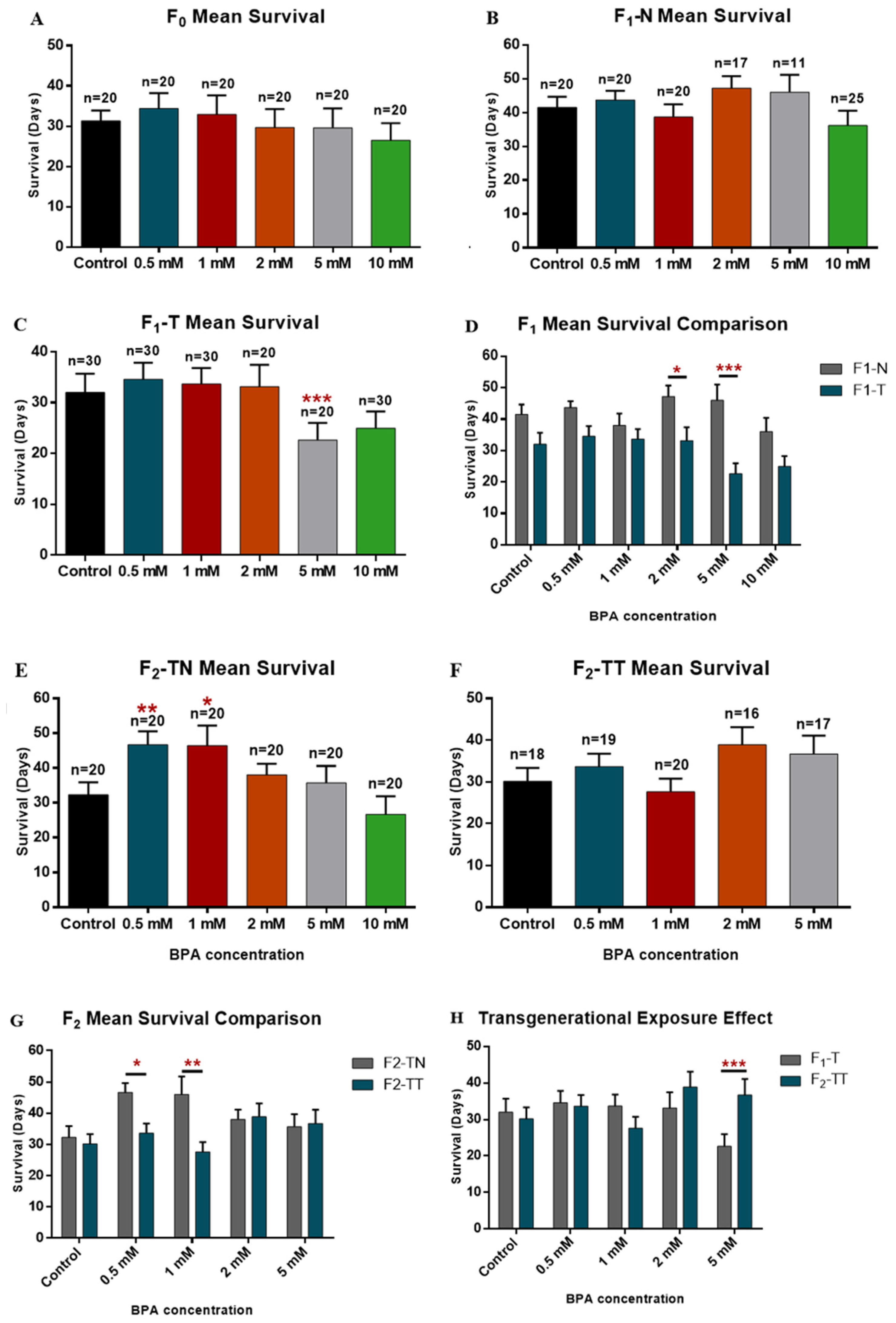
3.1. Effect on Longevity

The parental generation was first exposed to BPA for 72 h to evaluate the effect of acute exposure to the compound, but although a slight decrease was observed at 10 mM, the results showed no significant changes in the mean survival of the subjects (Figure 5A). Humans, however, are continuously exposed to BPA, so chronic treatment throughout larval development was applied in the following two generations. The longevity of non-treated individuals, but whose parents underwent either acute (F1-N) or chronic treatment (F2-TN), was also evaluated (Figure 5). Even though there were no significant differences observed in the F1-N group when comparing the mean lifespan for each concentration and the control, Fisher’s exact test showed a significant change in the flies’ age when their mortality reached 25% at 10 mM (Table 1). These results suggest that acute exposure of the parents may have affected the offspring’s survival, even though they were not exposed to the compound. In the treated subjects of the F1-T, a significant decrease in mean survival was observed at 5 mM, under which conditions longevity did not exceed 23 days (Figure 5C). A significant decrease was also found in maximal longevity when the mortality reached 50% and 75%. At 10 mM, a significant change in survival was also observed when the mortality reached 75% (Table 1).
When comparing F1-N with F1-T, we could see that, although the average survival of the F1-T generation was always lower than that of F1-N, both were affected, mainly at 2 mM and 5 mM, under which conditions the differences were significant (Figure 5D). Furthermore, at 5 mM, the differences were also significant when comparing the maximal longevity at 25%, 50% and 75% mortality. Fisher’s exact test even showed significant changes at 25% mortality for the 0.5, 2 and 10 mM concentrations, and at 75% for the 10 mM concentration (Table 2). We can thus conclude that chronic exposure to BPA during larval development exhibits greater toxicity than acute exposure, and that, since the most significant alterations in maximal longevity were observed in the early stages of life, this suggests that BPA may have a more relevant role in earlier deaths, and that greater emphasis should be placed on these deaths in future studies.
Regarding the F2 generation, a significant increase in the mean survival of F2-TN individuals was observed at the two lower concentrations. Significant changes were also observed at 25% mortality at the 10 mM concentration and at 50% mortality at the 0.5 mM (Figure 5E/Table 1). In F2-TT, the statistical analysis showed that the observed variations in mean survival were not significant. However, the maximal longevity obtained at 5 mM was shown to be significantly different from that obtained for the control at 50% mortality (Table 1). Treatment with 10 mM BPA proved to be toxic because no subjects were born at this concentration, and as such, longevity could not be assessed.
Comparing the mean survival of both F2 generation groups, significant differences were observed at 0.5 and 1 mM (Figure 5G). Fisher’s exact test also showed a significant difference at these concentrations when mortality reached 50% and, for the 1 mM concentration, at 75% as well (Table 2). Since both controls showed similar values and the F2-TN generation did not undergo any treatment, the observed variations in this group may have been caused by the parental chronic treatment, which also affected the offspring’s longevity.
The effect of chronic exposure in successive generations was also evaluated by comparing the average longevity of F1-T with that of F2-TT. We found that, in F2-TT, the variation between concentrations was more pronounced than the one obtained in F1-T, and that at 5 mM, the difference in mean survival was very significant (Figure 5H). Significant differences were also observed in mortality at 50% and 75% for the 1 mM and 5 mM concentrations (Table 2). Non-monotonic dose–response curves (NMDRCs) were obtained in both generations, which is common in studies involving EDCs such as BPA [43,44,45,46]. These results suggest that chronic exposure to BPA during successive generations increasingly affects the survival of individuals, leading to increasing variation in their longevity.

3.2. Effect on Prolificacy

After acute parental exposure (72 h), a slight but non-significant variation in the number of emerged adults was observed in the standardized medium (F1-N; Figure 6A). In the treated medium (F1-T), where the individuals remained exposed to BPA throughout larval development, a significant decrease was observed at 2 and 5 mM. At 10 mM, a slight increase in prolificacy was observed; however, the number of emerged adults was still significantly different from the control (Figure 6B). This suggests that acute treatment of the progenitors, by itself, does not have a great influence on prolificacy, whereas chronic treatment during larval development can significantly, and not monotonically, affect the number of emerged adults. The results obtained in the two media were also compared (Figure 6C). A significant difference was observed between the number of emerged adults in both controls, as well as at 1 and 5 mM. This variation in the controls may have been due to a difference in medium humidity or contamination of the standard medium, since they were performed simultaneously.
In the F2 generation, greater variation in both media was observed. In F2-TN, the prolificacy varied non-monotonically with an increase in concentration, and these changes were significant at 0.5 and, especially, at 1 and 5 mM, when compared to the control (Figure 6D). Since only their progenitors (F1-T) were exposed chronically to BPA, these differences show, once again, that chronic exposure of the progenitors causes transgenerational effects. In the F2-TT generation, the changes were even more severe, with a sudden increase in prolificacy observed at 0.5 mM, followed by a progressive decrease with an increase in concentration. These variations were very significant at all concentrations except for the 2 mM concentration, where the number of emerged adults was closer to the control (Figure 6E). This suggests that a new chronic exposure in this generation, after parental treatment, can cause an accumulated toxic effect. When comparing the results obtained in both media, there were still extremely significant differences at 2 and 5 mM, which evidences, once again, the impact of chronic exposure to BPA on the larval development of successive generations (Figure 6F).
The results for the larval development time analysis showed that, at low concentrations, BPA caused the development of D. melanogaster to be slightly faster, and that at higher concentrations, this acceleration was reversed and led to a delay in development. These differences, however, were not significant (Supplementary Figure S1).

3.3. Effects on Behaviour

The results obtained in this study showed no significant alterations in walking speed; however, an increase in flying activity at 0.5, 1 and 2 mM (with the highest activity peak) was observed, followed by a decrease at the highest concentrations. We can thus assume that, although the velocity remained constant at all concentrations, the distance travelled by the individuals varied non-monotonically.
Regarding general behaviour and social interaction between the flies, we verified that from the 1 mM concentration up, flies began to exhibit abnormal behaviour. Individually, there was an increase in repetitive movements, such as cleaning, and the appearance of random movements. Socially, it was observed that in the groups treated with 1 mM or more, flies collided with each other several times and maintained greater proximity to each other than was observed in the control.

3.4. Genotoxic Effect
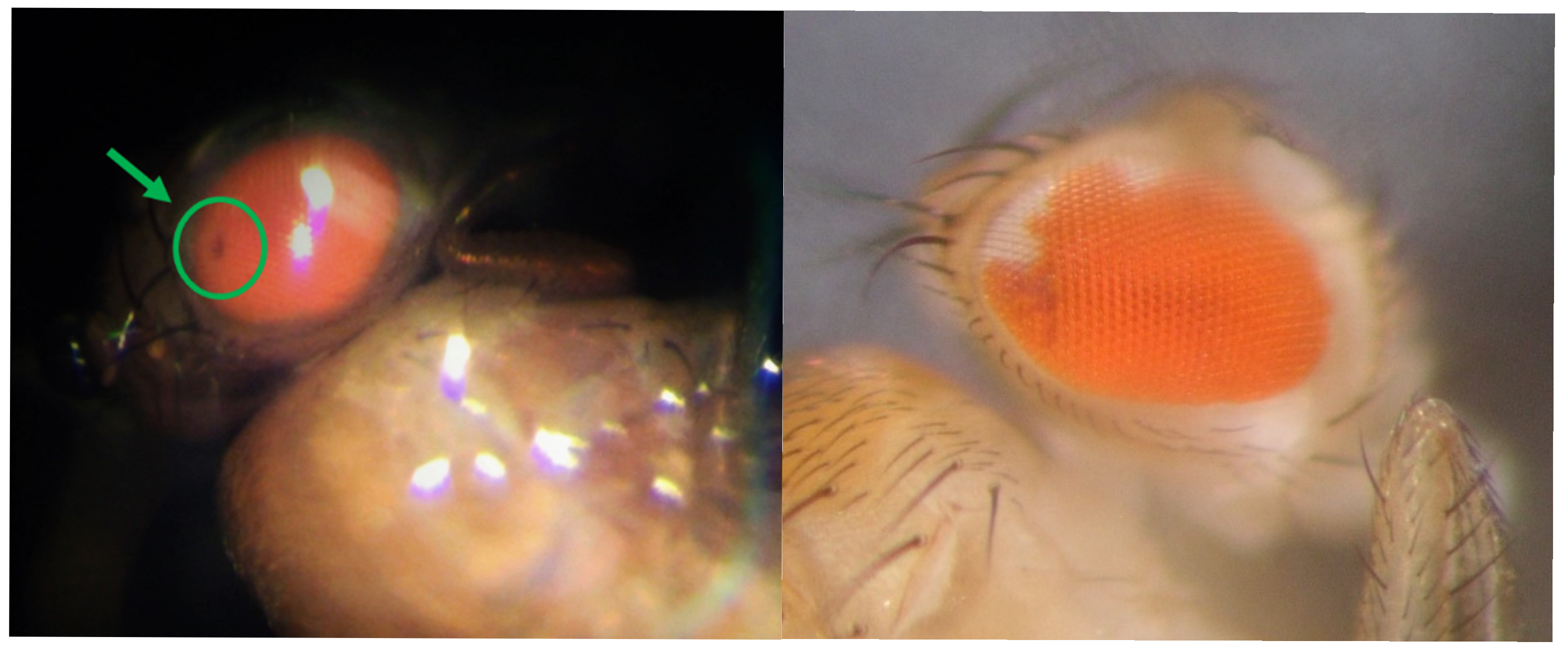
3.4.1. SMART Assay

The SMART assay (Figure 7) showed an increase in spot proportion, proportional to the concentration, at all concentrations, except for 50 mM. Statistical analysis using the double-decision χ2 test and a calculated m showed a negative effect at 10 mM, showed a weakly positive effect at 20 mM, and was inconclusive at 1 and 50 mM (Table 3). These results suggest that BPA may cause genetic damage in somatic cells at very high exposure doses, but the fact that the results were inconclusive for the 1 mM concentration, which corresponds, in flies, to the human LOAEL, made it necessary to perform additional testing to evaluate the potential genotoxic effect at environmentally relevant doses.

3.4.2. Comet Assay

The Comet assay was performed to complement the results obtained by the SMART assay. It was performed in duplicate, and 100 cells per gel/condition were visually scored each time. After fluorescence microscopy analysis of the slides, the results obtained showed an increase in basal damage at all concentrations when compared with the control. However, statistical analysis performed using Student’s t-test for independent samples later revealed that these changes were indeed significant for all concentrations, except for the 1 mM concentration (Figure 8). The results from both genotoxic assays thus seem to prove that BPA is potentially genotoxic, but only causes genetic damage in Drosophila cells at environmentally unrealistic exposure doses, way above the LOAEL.

4. Discussion

BPA is currently recognized as an endocrine disruptor. EDCs are defined by the World Health Organization (WHO) as “an exogenous substance or mixture that alters function(s) of the endocrine system and consequently causes adverse health effects in an intact organism, or its progeny, or (sub)populations” [5,6,7,8,9,10,63,64,65]. They can interfere with hormonal function in several ways: by affecting hormone biosynthesis directly, or by affecting their metabolism, transport and/or action mechanism. They can also act on a genomic and epigenomic level, altering gene expression or genomic imprinting. A lot of EDCs are thus known to cause alterations in fetal development and the typical feedback mechanisms of the endocrine system, often with transgenerational effects [11,12,13,14,15,66,67,68,69]. Chemicals such as BPA are nowadays a major concern worldwide, mainly because they may cause bioaccumulation and reproductive toxicity.
Several groups of investigators have concluded, from studies in several animals, that BPA can cause disorders in the development of various organs of the body, such as the male and female reproductive tract, the prostate, the mammary gland, tissues and organs involved in metabolism, and even the brain (causing neurobehavioral changes). Human studies have also shown an association between exposure to BPA and a variety of diseases, including various types of cancer, infertility problems (low counts and quality of sperm) and worsening of asthma, among others [68]. In this study, Drosophila melanogaster was used as a model organism to evaluate the biological, behavioural and genotoxic effects caused by exposure to BPA and explore its potential mechanisms of action. Our results showed that, when it comes to longevity, BPA caused no significant changes in mean survival after acute exposure. However, individuals are continuously exposed to BPA, so chronic treatment was applied in the following two generations. We then observed that chronic exposure to BPA during larval development exhibited greater toxicity than acute exposure, having a greater impact on maximal longevity in the early stages of life. This suggests that BPA may have a more relevant effect on earlier deaths, and that it is on those deaths that greater emphasis should be placed in future studies. The results also show that parental chronic treatments can also affect offspring’s longevity, and that chronic exposure to BPA during successive generations increasingly affects the survival of individuals, leading to increasing variations in their longevity. Non-monotonic dose–response curves (NMDRCs) were obtained in both generations, which is common in studies involving EDCs such as BPA. In fact, according to Laura N Vandenberg [69], NMDRCs are common in the BPA literature, occurring in more than 20% of all experiments and in at least one endpoint in more than 30% of all studies. Similar conclusions regarding the ability of EDCs to produce NMDRCs have been made by the World Health Organization, the United Nations Environment Programme, the Endocrine Society, and several other scientists [11,12,13,14,15,69]. In a study by Dong Zhou et al. [70] the nematode Caenorhabditis elegans was used to evaluate several parameters, including lifespan, after a chronic exposure to BPA (10 days, from L4 larvae to day 10 adults) in a long-term, low-concentration experiment. Their results, contrary to ours, showed no significant effects following exposure, which could have been due to the fact that the concentrations they used ranged between 0.0001 and 10 µM, concentrations much lower than the ones used in this study.
As for the effects on prolificacy, the obtained results suggest, as expected, that acute treatment of the progenitors, by itself, does not have a great influence on prolificacy, whereas chronic treatment during larval development can significantly, and not monotonically, affect the number of emerged adults. It was also observed that chronic exposure of successive generations caused transgenerational effects and an accumulated toxic effect. No significant alterations were observed in the development time of the individuals, which is interesting, since ecdysterone antagonists such as BPA should delay, or at least alter, this process. Another study was conducted by Atli et al. [71] with the objective of evaluating the effect of BPA exposure on Drosophila fecundity. Although their results showed a decrease in the number of laid eggs for all the tested concentrations, the dose–response curve was also non-monotonic. In Atli’s study, however, only the effects of acute exposure were assessed, and the possible transgenerational effects were not considered. Nevertheless, the comparison between both studies allows us to conclude that the exposure of individuals to BPA during larval development has harmful effects on their fecundity and the number of emerged adults in the offspring. Weiner and colleagues [72] also performed a study in which they evaluated the effect of BPA exposure during larval development on larval growth and early pupariation of D. melanogaster. In that study, however, all tested concentrations caused early pupariation and, consequently, faster development of the flies. Other studies carried out in invertebrates have demonstrated that BPA was responsible for the induction of metamorphosis in Capitella capitata larvae, and that exposure of ramshorn snails (Marisa cornuarietis) induced a hyperfeminization syndrome at all concentrations tested. [73,74]
Another of the evaluated parameters was the behaviour of the individuals. The use of Drosophila as a model organism in behavioural studies has been growing in recent years, and it has already been used in several studies on neurodegenerative diseases, such as Alzheimer’s or Parkinson’s disease [75]. The results of this study showed no significant alterations in walking speed, although the distance travelled by the individuals varied non-monotonically. From the 1 mM concentration up, flies began to exhibit abnormal behaviour, with an increase in random and repetitive (cleaning) movements. Flies also maintained greater proximity to each other than was observed in the control, colliding several times. These results can be compared to the ones obtained by Kulbir Kaur [76]. In that study, only the concentrations of 0.5 and 1 mM were used, but the result of the walking speed analysis was similar. However, in Kaur’s study, a decrease in the travelled distance at 1 mM, as well as a non-significant variation at 0.5 mM, were observed. As for social interaction, the results obtained by Kaur et al. [76] were, once again, similar to ours, with an increase in repetitive behaviour and alterations in the interaction between flies observed in both studies. Kaur and his colleagues suggested that these changes in individual and group behaviour may be related to the ones observed in diseases such as autism and attention deficit hyperactivity disorder (ADHD), highlighting the potential role of BPA in those diseases. In fact, several studies have already shown an association between BPA and modifications in the dopaminergic and NMDAergic systems, as well as the development of an ADHD phenotype in several model organisms, thus raising questions about its role in childhood human diseases [77,78,79]. Although epidemiological data for the human species are scarce, a study by Shruti Tewar found evidence that higher urinary BPA concentrations were associated with ADHD in U.S. children, and that these associations were stronger in boys than in girls [80].
As for the last objective of this study, the results from the SMART and Comet assays showed that BPA exposure only causes genetic damage in Drosophila cells at environmentally unrealistic exposure doses, with no genotoxic effects at the concentration that corresponds, in flies, to the human LOAEL (1 mM). Nevertheless, the use of these higher concentrations allowed us to establish a concentration–effect relationship, since a significant increase in genetic damage with increasing concentration was observed.
Besides BPA, the SMART assay has already been used in a study evaluating the genotoxicity of bisphenol A-glycidyl methacrylate (BisGMA), a monomeric resin used in dentistry [81]. In that study, however, the results were negative for all concentrations used, which may be because those concentrations were inferior to the ones used in this work. Another explanation provided by the authors to justify the obtained results was the fact that the dimension and inflexibility of BisGMA could render it difficult for it to access the nucleus via nuclear pores.
No similar studies using the Comet assay in Drosophila are available to enable a direct comparison of the results; however, there are studies involving other model organisms, such as the invertebrate midge Chironomus riparius [82,83,84]. In that study, the authors observed an increase in the extent of DNA damage in fourth-instar larvae, as compared to control solvent-exposed individuals, in both concentrations tested (0.5 and 3 mg/L). Interestingly, they tested the effect of two different exposure times (24 h and 96 h), and found that the extent of DNA damage at 0.5 mg/L decreased with exposure time, possibly due to the activation of DNA-repairing mechanisms under prolonged exposure to BPA. That conclusion may thus explain the fact that, in our study, no significant increase in DNA damage was observed at the lowest concentration (1 mM), since the treatment we used was administered until the third stage of larval development.
The present study involved relatively high (millimolar) concentration ranges of BPA, varying from 0.5 to 50 mmol/L. This was chosen to assess sublethal and acute toxic effects in D. melanogaster, according to the experimental toxicology approaches generally used in invertebrates [85,86,87]. It is also a standard approach in ecogenetical and ecotoxicological essays to detect cellular and molecular effects that may remain undetectable at lower doses. This is particularly important for mechanisms that may exhibit non-linear or threshold-dependent responses.
Although these doses exceed typical environmental or dietary exposure levels, they may be applied in mechanistic studies in order to elicit, and thereby elucidate, physiological, behavioural and developmental responses, which are quantitatively assessed. The use of a wide concentration gradient, including high doses, allows for the identification of toxicity thresholds, the characterization of dose-dependent effects and the investigation of potential compensatory stress-response mechanisms. High concentrations are particularly important when working with Drosophila species, including D. melanogaster, which generally exhibit high tolerance to xenobiotics, due to their rapid metabolism and protective barriers.
Moreover, similar concentrations of BPA have already been used in previous studies involving invertebrate models, including Drosophila, aiming to study endocrine-disrupting activity, reproductive toxicity, oxidative stress and gene-expression alterations. Therefore, the chosen concentration range may be justified within the context of ecotoxicology and exploratory toxicology, being consistent with the goals of assessing the mechanistic impacts of BPA at different intensity levels.
Chen et al. [85] exposed adult D. melanogaster to 0.5 mM BPA solution and observed significant morphological alterations in the midgut, which reduced the survival rate and impaired climbing ability. Oxidative stress was considered to be a key mechanism, justifying BPA-induced gastrointestinal toxicity.
On the other hand, Adesanoye et al. [86] investigated the ameliorative effects of flavonoid antioxidant luteolin on BPA-induced toxicity in D. melanogaster. The results confirmed BPA-induced oxidative stress imbalance and behavioural deficits in flies. On the other hand, luteolin increased the survival rate and augmented antioxidant markers in flies, as well as ameliorating BPA-induced oxidative degeneration. This provides further insight into the antioxidative and chemopreventive properties of luteolin against BPA-induced toxicity.
Sarkar and coworkers [87] investigated the ameliorating effect of tetravalent cerium oxide nanoparticles, which is analogous to that of luteolin [86], and confirmed that BPA induced lipid peroxidation and ROS formation. Also, BPA exposure at millimolar doses induced micronuclei formation and impeded nucleocytoplasmic transport, whereas CeO2 nanoparticles improved dynamic and sexual behaviour abnormalities and ameliorated oxidative stress induced by BPA.
Santos Musachio [88] and coworkers analyzed the effects of BPA on D. melanogaster during the embryonic period. It was shown that a 1 mM dose of BPA had an effect on developmental, behaviouristic and biochemical markers. The pupal eclosion rate decreased. Moreover, the F1 flies also developed behaviouristic changes in sexual, dynamic and social behaviour as a result of BPA’s interference with the dopaminergic system, in a similar manner to the human phenotypes ASD and ADHD, induced by oxidative stress.
Nguyen et al. [89] mention that BPA has been linked to behavioural changes in children, and has been shown to impact neurodevelopmental processes in animal models. BPA in concentrations up to 2 mM induces hyperactivity in larvae, increases grooming in adults and reduces sexual behaviour. Moreover, behavioural changes are highly dependent on the genetic background, suggesting that BPA may elicit a gene–environment interaction with gFmr1, causing fragile X syndrome and autism spectrum disorder.
Begum and coauthors [90] exposed D. melanogaster to concentrations of 0.0035 g/mL, which corresponds to nearly 15.3 mM, and 0.005 g/mL (nearly 21.9 mM). The feeding rate, foraging path, climbing ability and frequency of courtship displays were reduced for both females and males at 15.3 mM, whereas for the higher concentrations, up to 21.9 mM, only treated females, and not males, exhibited significantly reduced climbing behaviour.
A study by Ramadhan and coauthors [91] exposed D. melanogaster to doses up to 2.2 mM, confirming negative geotaxis in the climbing reflex, indicating the negative effect of BPA for motorics in imago, which may also be manifested in higher organisms.
Exposure to other bisphenolic compounds has also been evaluated at millimolar concentrations. For example, in [92], exposure of Drosophila melanogaster to 1 mM bisphenol F (BPF) caused failure in the regulation of neurodevelopmentally important genes, including dFmr1, in FXS-model strains, as well as other genes linked to synapse formation and neuronial protection. This may link the genotoxicity and neurotoxicity of not only BPA, but also BPF, to developmental retardations and autism spectrum disorder.
In [93], Herrero and coworkers exposed the aquatic midge Chironomus riparus to the BPA alternative bisphenol S (BPS) at concentrations up to 500 μg/L, and the BPS exposure caused crucial alterations in genes, including EcR, ERR, E74, cyp18a1, hsp70, hsp40, cyp4g, GPx and GST, related to the ecdotysone pathway. These alterations showed the possibility for BPS to cause crucial developmental changes in an invertebrate model.
In [94], various bisphenols, including BPA, BPS and BPF, were tested on Drosophila melanogaster for their possible genetically induced neurotoxic effects. The concentrations used were up to 12 mM for BPA, whereas those for BPF reached 25 mM. The neurotoxic effect and its nature was highly dependent on the bisphenol composition, and its intensity varied according to BPZ > BPC and BPAF > BPB > BPS > BPAP ≈ BPA ≈ BPF > BPE, which indicates the necessity to re-evaluate the bisphenols mentioned in [94] and other compounds of the group as alternatives to BPA for use in BPA-free-labelled packages.
On the other hand, the use of lower doses of pure BPA and its mixtures with other xenoestrogenic food-contact substances (bisphenol S, tetrabromobisphenol A, bisphenol C2) and synthetic food additives (sucralose, aspartame, saccharine, etc.) may reveal possible synergism in the genotoxic effects of the components of these mixtures. Investigating this is important for the objective of revealing the possible mutagenic effects of two synthetic substances when acting together, which may or may not be manifested by each of them separately.
For example, Branco and Lemos [95] investigated the gene transcription effects in D. melanogaster caused by interaction between bisphenol A and dietary sugar. The investigation revealed an increase in BPA’s effect on Drosophila caused by intake of sugar, which confirms the possibility of BPA interactions with other food components as a possible cause of the genotoxic effects of BPA alongside other ingredients in the human diet.
The work in [96] confirmed that BPA may cause a type 2 diabetes-like condition in Drosophila, and showed that a BPA + high sucrose diet increased the magnitude of disturbance to Malpighian tubules, manifested in fibrosis and malfunction, suggesting enhanced oxidative stress.
On the other hand, antioxidants [97] may reduce BPA toxicity. This work suggests a mechanism of formation of three estrogenic metabolites by BPA which are harmful to oxidative homeostasis in humans. Sulfaminoacids (taurine), carotenoids (lycopene), polyphenolic compounds (curcumin, luteolin, quercetin, naringin and naringenine) and indolic antioxidants (melatonin) were confirmed to inhibit BPA-induced oxidative stress and thereby alleviate its neurotoxic and genotoxic effects, confirming the statements in [86].
Analogously, the work in [98] confirmed that natural products containing polyphenolic compounds may mitigate BPA-induced oxidative stress and alleviate its toxicity. Therefore, the presence of different food components may either enhance BPA toxicity or reduce its poisoning activity.
Therefore, the investigation of low-dose (nano- and picomolar) genotoxicity of bisphenol A, and its mixtures with other food-contact substances and synthetic food additives, on D. melanogaster and other model organisms is planned for our further investigation.

5. Conclusions

The present study aimed to evaluate the biological and behavioural effects of BPA exposure on D. melanogaster, while also trying to determine its possible mechanisms of action. Based on our results, we can conclude that, at the concentrations studied, exposure to BPA causes significant changes in the longevity and proliferation of this model organism, and affects its activity and social interaction, but it does not cause any genotoxic effects on Drosophila cells in vivo.
Although we must take into account the fact that high concentrations were used, and that they may not reflect the environmental concentrations to which these specimens are usually exposed, the rationale behind this approach is that high doses allow for the detection of cellular and molecular effects that may remain undetectable at lower doses, especially when such mechanisms present non-linear or threshold-dependent responses. For instance, the use of these concentrations allowed us to pinpoint specific alterations in Drosophila’s lifespan and reproduction which were passed on to offspring, with aggravated effects associated with the time and duration of exposure. These important transgenerational effects might not have been detected if lower concentrations had been used. Furthermore, we cannot forget that in certain environments (e.g., wastewater, industrial discharges), BPA concentrations can reach significant levels, making our study relevant for scenarios involving localized pollution. Regardless, future research should encompass a broader range of doses, including environmentally relevant concentrations, to provide a more comprehensive risk assessment of BPA.
On another note, the results obtained by this study show that D. melanogaster is indeed susceptible to BPA exposure, and is therefore a good model organism for in vivo toxicity and genotoxicity studies, even when the compound acts as an endocrine disruptor. In future studies, the techniques applied here must be optimized to allow for comparison between the obtained results, and focus should be placed on the still poorly understood epigenetic effects of this compound, which may explain the transgenerational effects observed, and which have already been evaluated in other species. It should also be considered that environmental exposure to these compounds never occurs in isolation, due to the mixtures of compounds present in the environment, which may interact with each other and/or cause cumulative effects on organisms.

Supplementary Materials

The following supporting information can be downloaded at: https://www.mdpi.com/article/10.3390/app15105588/s1, Figure S1: Effect of BPA exposure on the development times of the F1-T and F2-TT individuals. Statistical significance obtained by the t-student test, with significance level set at 0.05.

Author Contributions

Conceptualization, I.G., R.A.S., V.V.T. and T.V.M.; methodology, I.G.; validation, I.G., R.A.S., V.V.T. and T.V.M.; formal analysis, I.G. and R.A.S.; investigation, I.G., R.A.S., V.V.T. and T.V.M.; resources, I.G.; data curation, I.G., R.A.S. and T.V.M.; writing—original draft preparation, I.G., R.A.S., V.V.T. and T.V.M.; writing—review and editing, I.G., R.A.S., V.V.T. and T.V.M.; visualization I.G., R.A.S., V.V.T. and T.V.M.; supervision, I.G.; project administration, I.G.; funding acquisition, I.G. All authors have read and agreed to the published version of the manuscript.

Funding

This work was supported by the CECAV project UID/00772, funded by the Portuguese Foundation for Science and Technology (FCT).

Data Availability Statement

The original contributions presented in this study are included in the article. Further inquiries can be directed to the corresponding authors.

Acknowledgments

The authors would like to thank João Ferreira for his assistance with all the statistical analysis.

Conflicts of Interest

The authors declare no conflicts of interest.

Abbreviations

The following abbreviations are used in this manuscript:
BPABisphenol A
EFSAEuropean Food Safety Authority
DMSODimethylsulfoxide
DAPI4′,6-diamidino-2-phenylindole
SMARTSomatic mutation and recombination test
EDCEndocrine-disrupting chemicals
LOH Loss of heterozygosity
OASIS Online Application for Survival Analysis
EDTA Ethylenediaminetetraacetic acid
CSTEE Scientific Committee on Toxicity, Ecotoxicity and the Environment
BisGMA Bisphenol A-glycidyl methacrylate
LOAEL Lowest-observed-adverse-effect level
ADHD Attention deficit hyperactivity disorder
NMDRC Non-monotonic dose–response curve
WHO World Health Organization

References

  1. Varghese, S.V.; Hall, J.M. Bisphenol A substitutes and obesity: A review of the epidemiology and pathophysiology. Front. Endocrinol. 2023, 14, 1155694. [Google Scholar] [CrossRef] [PubMed]
  2. Tkach, V.V.; Morozova, T.V.; Kushnir, M.V.; de Oliveira, S.C.; Kryvetskyi, V.V.; Kryvetska, I.I.; Kryvetskyi, I.V.; Biryuk, I.G.; Sykyrytska, T.B.; Ivanushko, Y.G.; et al. The Theoretical Description for Bisphenol S and Bisphenol AF Cathodic Determination by Bivalent Chromium, Intercalated into Conducting Polymeric Material. Lett. Appl. NanoBioSci. 2025, 14, 8. [Google Scholar] [CrossRef]
  3. Akash, M.S.H.; Rasheed, S.; Rehman, K.; Imran, M.; Assiri, M.A. Toxicological evaluation of bisphenol analogues: Preventive measures and therapeutic interventions. RSC Adv. 2023, 31, 21613–21628. [Google Scholar] [CrossRef] [PubMed]
  4. Tumu, K.; Vorst, K.; Curtzwiler, G. Endocrine modulating chemicals in food packaging: A review of phthalates and bisphenols. Compr. Rev. Food Sci. Food Saf. 2023, 22, 1337–1359. [Google Scholar] [CrossRef] [PubMed]
  5. Khalili Sadrabad, E.; Hashemi, S.A.; Nadjarzadeh, A.; Askari, E.; Akrami Mohajeri, F.; Ramroudi, F. Bisphenol A release from food and beverage containers—A review. Food Sci. Nutr. 2023, 11, 3718–3728. [Google Scholar] [CrossRef]
  6. Peinado, F.M.; Iribarne-Durán, L.M.; Artacho-Cordón, F. Human Exposure to Bisphenols, Parabens, and Benzophenones, and Its Relationship with the Inflammatory Response: A Systematic Review. Int. J. Mol. Sci. 2023, 24, 7325. [Google Scholar] [CrossRef]
  7. Rybczyńska-Tkaczyk, K.; Skóra, B.; Szychowski, K.A. Toxicity of bisphenol A (BPA) and its derivatives in diverse biological models with the assessment of molecular mechanisms of toxicity. Environ. Sci. Pollut. Res. 2023, 30, 75126–75140. [Google Scholar] [CrossRef]
  8. Moreno-Gómez-Toledano, R.; Delgado-Marín, M.; Cook-Calvete, A.; González-Cucharero, C.; Alcharani, N.; Jiménez-Guirado, B.; Hernandez, I.; Ramirez-Carracedo, R.; Tesoro, L.; Botana, L.; et al. New environmental factors related to diabetes risk in humans: Emerging bisphenols used in synthesis of plastics. World J. Diabetes 2023, 14, 1301–1313. [Google Scholar] [CrossRef]
  9. Dasiman, R.; Nor, N.M.; Eshak, Z.; Mutalip, S.S.M.; Suwandi, N.R.; Bidin, H.; Abas, R. A Systematic Review of the Effects of Diphenol Analogs on Embryonic Development and Cytoskeletal Organization of Zebrafish (Danio rerio) Embryos. Biointerface Res. Appl. Chem. 2023, 13, 75. [Google Scholar] [CrossRef]
  10. EFSA Panel on Food Contact Materials, Enzymes and Processing Aids (CEP); Lambré, C.; Barat Baviera, J.M.; Bolognesi, C.; Chesson, A.; Cocconcelli, P.S.; Crebelli, R.; Gott, D.M.; Grob, K.; Lampi, E.; et al. Re-evaluation of the risks to public health related to the presence of bisphenol A (BPA) in foodstuffs. EFSA J. 2023, 21, e06857. [Google Scholar] [CrossRef]
  11. Abu Hasan, H.; Hafizuddin Muhammad, M.; Kurniawan, S.; Buhari, J.; Abuyezad, O.H. Managing. Managing Bisphenol A Contamination: Advances in Removal Technologies and Future Prospects. Water 2023, 15, 3573. [Google Scholar] [CrossRef]
  12. Charkiewicz, A.E.; Omeljaniuk, W.J.; Niklinski, J. Bisphenol A—What Do We Know? A Global or Local Approach at the Public Health Risk Level. Int. J. Mol. Sci. 2024, 25, 6229. [Google Scholar] [CrossRef] [PubMed]
  13. Neri, I.; Russo, G.; Grumetto, L. Bisphenol A and Its Analogues: From Their Occurrence in Foodstuffs Marketed in Europe to Improved Monitoring Strategies—A Review of Published Literature from 2018 to 2023. Arch. Toxicol. 2024, 98, 2441–2461. [Google Scholar] [CrossRef] [PubMed]
  14. Peters, A.E.; Ford, E.A.; Roman, S.D.; Bromfield, E.G.; Nixon, B.; Pringle, K.G.; Sutherland, J.M. Impact of Bisphenol A and its Alternatives on Oocyte Health: A Scoping Review. Hum. Reprod. Update 2024, 30, 653–691. [Google Scholar] [CrossRef]
  15. Razia, S.; Hadibarata, T.; Lau, S.Y. A Review on Biodegradation of Bisphenol A (BPA) with Bacteria and Fungi Under Laboratory Conditions. Int. Biodeteriorat. Biodegrad. 2024, 195, 105893. [Google Scholar] [CrossRef]
  16. Zaborowska, M.; Wyszkowska, J.; Borowik, A.; Kucharski, J. Bisphenols—A Threat to the Natural Environment. Materials 2023, 16, 6500. [Google Scholar] [CrossRef]
  17. Liu, D.; Kang, G.; Zhang, Y.; Shi, L.; Ma, B.; Zhang, S.; Lu, G. Exploring the Distribution and Fate of Bisphenol A in an Aquatic Microcosm, Combined with a Multimedia Model. Ecotoxicol. Environ. Saf. 2025, 290, 117752. [Google Scholar] [CrossRef]
  18. Paolella, G.; Fabbricino, M.; Lcasci, A.; Sirakov, M.; Pontoni, L. Fate of Bisphenol A in Marine Environment: A Critical Review. Chem. Eng. J. 2024, 495, 153228. [Google Scholar] [CrossRef]
  19. Gupta, A.; Singh, A.; Mishra, V.K. Impact of Bisphenol A in the Environment and its Removal Through Biological Agents: A Review. Environ. Qual. Manag. 2024, 34, e22246. [Google Scholar] [CrossRef]
  20. Tse, H.-T.; Au, C.-K.; Chan, W. Bisphenol A in Disposable Face Masks: A Novel Human Exposure Pathway and Impact on the Aquatic Environment. Chem. Res. Toxicol. 2025, 38, 347–352. [Google Scholar] [CrossRef]
  21. Peter Stein, T. Does Bisphenol A (BPA) Exposure Cause Human Diseases? Biomedicines 2024, 12, 2678. [Google Scholar] [CrossRef] [PubMed]
  22. Ejaredar, M.; Lee, Y.; Roberts, J.D.; Sauve, R.; Dewey, D. Bisphenol A Exposure and Children’s Behavior: A Systematic Review. J. Expo. Sci. Environ. Epidemiol. 2017, 27, 175–183. [Google Scholar] [CrossRef] [PubMed]
  23. Manzoor, F.M.; Tariq, T.; Fatima, B.; Sahar, A.; Tariq, F.; Munir, S.; Khan, S.; Ranjha, M.M.A.N.; Sameen, A.; Zeng, X.-A.; et al. An Insight into Bisphenol A, Food Exposure and Its Adverse Effects on Health: A Review. Front. Nutr. 2022, 9, 1047827. [Google Scholar] [CrossRef] [PubMed]
  24. vom Saal, F.S.; Vandenberg, L. Update on the Health Effectsof Bisphenol A:Overwhelming Evidence of Harm. Endocrinology 2020, 162, bqaa171. [Google Scholar] [CrossRef]
  25. Costa, S.A.; Severo, M.; Correia, D.; Carvalho, C.; Magalhães, V.; Vileia, S.; Cunha, S.; Casal, S.; Lopes, C.; Lopes, C.; et al. Methodological Approaches for the Assessment of Bisphenol A Exposure. Food Res. Int. 2023, 173, 113251. [Google Scholar] [CrossRef]
  26. Opinion of the SCTEE on the Risk Assessment of Bisphenol A, Human Health Part. Available online: https://ec.europa.eu/health/ph_risk/committees/sct/documents/out156_en.pdf (accessed on 6 February 2025).
  27. Kamrin, M.A.; Bisphenol, A. A Scientific Evaluation. MedGenMed 2004, 6, 7. Available online: https://pmc.ncbi.nlm.nih.gov/articles/PMC1435609/ (accessed on 12 May 2025).
  28. Marçal, R.; Pacheco, M.G.; Guilherme, S. Unveiling the Nexus between Parental Exposure to Toxicants and Heritable Spermiotoxicity—Is Life History a Shield or a Shadow? Environ. Toxicol. Pharmacol. 2022, 95, 103955. [Google Scholar] [CrossRef]
  29. Efthimiou, I.; Vlastos, D.; Ioannidou, C.; Tsilimigka, F.; Drosopoulou, E.; Mavragani-Tsipidou, P.; Potsi, F.; Gournis, D.; Antonopoulou, M. Assessment of the genotoxic potential of three novel composite nanomaterials using human lymphocytes and the fruit fly Drosophila melanogaster as model systems. Chem. Eng. J. Adv. 2022, 9, 100230. [Google Scholar] [CrossRef]
  30. Santos-Cruz, L.F.; Ponciano-Gómez, A.; Torres-Gregorio, J.T.; Ramírez-Cruz, B.G.; Vázquez-Gómez, B.; Hernández-Portilla, L.B.; Flores-Ortiz, C.M.; Dueñas-García, I.E.; Heres-Pulido, M.E.; Castañeda-Partida, L.; et al. Zearalenone Does Not Show Genotoxic Effects in the Drosophila melanogaster Wing Spot Test, but It Induces Oxidative Imbalance, Development, and Fecundity Alterations. Toxins 2023, 15, 568. [Google Scholar] [CrossRef]
  31. Turna Demir, F. Protective Effect of Resveratrol Against Genotoxicity Induced by Nano and Bulk Hydroxyapatite in Drosophila melanogaster. J. Toxicol. Environ. Health 2022, 85, 850–865. [Google Scholar] [CrossRef]
  32. Colonna Romano, N.; Marchetti, M.; Marangoni, A.; Leo, L.; Retrosi, D.; Rosato, E.; Fanti, L. Neuronal Progenitors Suffer Genotoxic Stress in the Drosophila Clock Mutant per. Cells 2024, 13, 1944. [Google Scholar] [CrossRef] [PubMed]
  33. Ford, A.; LeBlanc, G. Endocrine Disruption in Invertebrates: A Survey of Research Progress. Environ. Sci. Technol. 2020, 54, 13365–13369. [Google Scholar] [CrossRef] [PubMed]
  34. Orchard, I.; Lange, A.B. The neuroendocrine and endocrine systems in insect—Historical perspective and overview. Mol. Cell. Endocrinol. 2024, 580, 112108. [Google Scholar] [CrossRef] [PubMed]
  35. Morthorst, J.E.; Holbech, H.; de Crozé, N.; Matthiessen, P.; LeBlanc, G.A. Thyroid-like hormone signaling in invertebrates and its potential role in initial screening of thyroid hormone system disrupting chemicals. Int. Environ. Ass. Manag. 2023, 19, 63–82. [Google Scholar] [CrossRef]
  36. Knigge, T.; LeBlanc, G.; Ford, A.T. A Crab Is Not a Fish: Unique Aspects of the Crustacean Endocrine System and Considerations for Endocrine Toxicology. Front. Endocrinol. 2021, 12, 587608. [Google Scholar] [CrossRef]
  37. Crane, M.; Dungey, S.; Lillicrap, A.; Thompson, H.; Weltje, L.; Wheeler, J.R.; Lagadic, L. Commentary: Assessing the endocrine disrupting effects of chemicals on invertebrates in the European Union. Environ. Sci. Eur. 2022, 34, 36. [Google Scholar] [CrossRef]
  38. Sreevidya, C.P.; Manoj Kumar, T.M.; Balakrishnan, S.; Kunjiraman, S.; Sarasan, M.; Magnuson, J.T. Puthumana Establishment of a cell culture from Daphnia magna as an in vitro model for (eco)toxicology assays: Case study using Bisphenol A as a representative cytotoxic and endocrine disrupting chemical. Aquat. Toxicol. 2025, 278, 107173. [Google Scholar] [CrossRef]
  39. Alvares, M.F.; Villar-Argaiz, M.; Vela Soria, F.; Zambrano, A.F.; Medina-Sánchez, A.; Carrillo, P. Thresholds and interactive effects of BPA-gradient and temperature on life history traits of Daphnia magna. Environ. Pollut. 2024, 355, 124186. [Google Scholar] [CrossRef]
  40. Qian, L.; Chen, C.; Guo, L.; Zhang, X.; Zheng, J.; Wang, G.; Zhang, X. Developmental and Reproductive Impacts of Four Bisphenols in Daphnia magna. Int. J. Mol. Sci. 2022, 23, 14561. [Google Scholar] [CrossRef]
  41. Oliveira Pereira, E.A.; Labine, L.M.; Kleywegt, S.; Jobst, K.J.; Simpson, A.J.; Simpson, M.J. Metabolomics Reveals That Bisphenol Pollutants Impair Protein Synthesis-Related Pathways in Daphnia magna. Metabolites 2021, 11, 666. [Google Scholar] [CrossRef]
  42. Wang, Y.; Zhao, T.; Yang, X.; Liu, H. Aquatic toxicity and ecological risk of bisphenol B, and comparison with those of bisphenol A. Hum. Ecol. Risk Assessm. Int. J. 2024, 30, 819–832. [Google Scholar] [CrossRef]
  43. Franko, N.; Kodila, A.; Dolenc, M.S. Adverse Outcomes of the Newly Emerging Bisphenol A Substitutes. Chemosphere 2024, 364, 143147. [Google Scholar] [CrossRef] [PubMed]
  44. Welch, C.; Johnson, E.; Tupikova, A.; Anderson, J.; Tinsley, B.; Newman, J.; Alfareh, A.; Davis, A.; Rodriguez, L.; Visger, C.; et al. Bisphenol A Affects Neurodevelopmental Gene Expression, Cognitive Function and Synaptic Morphology in Drosophila melanogaster. NeuroToxicity 2022, 89, 67–78. [Google Scholar] [CrossRef] [PubMed]
  45. Dueña-Moreno, J.; Mora, A.; Kumar, M.; Meng, X.-Z.; Mahlknecht, J. Worldwide Risk Assessment of Phthalates and Bisphenol A in Humans: The Need for Updating Guidelines. Envir. Int. 2023, 181, 108294. [Google Scholar] [CrossRef]
  46. Mirzaei, G.; Yazdanfar, N.; Shariatifar, N.; Molaee-Aghaee, E.; Sadighara, P. Health Risk Assessment and Determination of Bisphenol A and Aflatoxin M1 in Infant Formula. BMC Nutr. 2025, 11, 6. [Google Scholar] [CrossRef]
  47. Otto, P.A. Elementary Atlas of Drosophila melanogaster Mutations. Genet. Mol. Biol. 2022, 45, e20220211. [Google Scholar] [CrossRef]
  48. Gonzalez-Squalli, E.; Caron, M.; Loppin, B. The White Gene as a Transgenesis Marker for the Cricket Gryllus bimaculatus. G3 2024, 14, jkae235. [Google Scholar] [CrossRef]
  49. Wang, J.; Hong, X.; Liu, W.; Zhang, L.; Yan, S.; Li, Z.; Zha, J. Comprehensive assessment of the safety of bisphenol A and its analogs based on multi-toxicity tests in vitro. J. Hazard. Mater. 2025, 486, 136983. [Google Scholar] [CrossRef]
  50. Sendra, M.; Stampar, M.; Fras, K.; Novoa, B.; Figueras, A.; Zegura, B. Adverse (geno)toxic effects of bisphenol A and its analogues in hepatic 3D cell model. Environ. Int. 2023, 171, 107721. [Google Scholar] [CrossRef]
  51. Jiang, N.; Naz, S.; Ma, Y.; Han, S.K.; Choi, Y.; Nam, H.G.; Lee, S.-J. An Overview of Comet Assay Application for Detecting DNA Damage in Aquatic Animals. Agriculture 2023, 13, 623. [Google Scholar] [CrossRef]
  52. Costea, M.A.; Rosan, C.A.; Laslo, V.; Agud, E.; Purcarea, C.; Vicas, S.I. The Comet Assay as a Sustainable Method for Evaluating the Genotoxicity Caused by the Soluble Fraction Derived from Sewage Sludge on Diverse Cell Types, Including Lymphocytes, Coelomocytes and Allium cepa L. Cells. Sustainability 2024, 16, 457. [Google Scholar] [CrossRef]
  53. Rodríguez, R.; Gaivão, I.; Aguado, L.; Espina, M.; García, J.; Martínez-Camblor, P.; Sierra, L.M. The Comet Assay in Drosophila: A Tool to Study Interactions between DNA Repair Systems in DNA Damage Responses In Vivo and Ex Vivo. Cells 2023, 12, 1979. [Google Scholar] [CrossRef] [PubMed]
  54. Augustyniak, M.; Gladysz, M.; Dziewiecka, M. The Comet assay in insects—Status, prospects and benefits for science. Mut. Res. Rev. Mut. Res. 2016, 767, 67–76. [Google Scholar] [CrossRef] [PubMed]
  55. Gaivão, I.; Sierra, L.M.Z. Drosophila comet assay: Insights, uses and future perspectives. Front. Genet. 2014, 5, 00304. [Google Scholar] [CrossRef]
  56. Yang, J.-S.; Nam, H.-J.; Seo, M.; Han, S.K.; Choi, Y.; Nam, H.G.; Lee, S.-J.; Kim, S. OASIS: Online Application for the Survival Analysis of Lifespan Assays Performed in Aging Research. PLoS ONE 2011, 6, e23535. [Google Scholar] [CrossRef]
  57. Li, M.; Macro, J.; Huggins, B.J.; Mishra, D.; Martin, D.; Kannan, K. Rogina Extended lifespan in female Drosophila melanogaster through late-life calorie restriction. GeroScience 2024, 46, 4017–4035. [Google Scholar] [CrossRef]
  58. Levine, B.J.; Hoffmann, J.M. Microbiome Transplants May Not Improve Health and Longevity in Drosophila melanogaster. Biol. Open 2025, 14, bio061745. [Google Scholar] [CrossRef]
  59. Kristensen, T.N.; Schonherz, A.A.; Rohde, P.D.; Sorensen, J.G.; Loeschke, V. Selection for stress tolerance and longevity in Drosophila melanogaster have strong impacts on microbiome profiles. Sci. Rep. 2024, 14, 17789. [Google Scholar] [CrossRef]
  60. Mosley, O.L.; Villa, J.A.; Kamalakkannan, A.; James, E.; Hoffman, J.; Lyu, Y. Stochasticity in dietary restriction-mediated lifespan outcomes in Drosophila. GeroScience 2025. [Google Scholar] [CrossRef]
  61. Aslam, M.; Albassam, M. Analysis and Allocation of Cancer-Related Genes Using Vague DNA Sequence Data. Front. Genet. 2022, 13, 858005. [Google Scholar] [CrossRef]
  62. Hayashi, M. “Statistical significance” and other important considerations in genotoxicity safety testing. Mut. Res. Gen. Toxicol. Environ. Mutagen. 2023, 888, 503627. [Google Scholar] [CrossRef] [PubMed]
  63. Core, A.C.; La Merrill, M.A.; Partisaul, H.; Sargis, R.M. Endocrine Disrupting Chemicals: Threats to Human Health. Pesticides, Plastics, Forever Chemicals and Beyond; Endocrine Society and IPEN: Austin, TX, USA, 2024. [Google Scholar]
  64. Reincke, M.; Arlt, W.; Damdimopoulou, P.; Kohrle, J.; Bertherat, J. Endocrine disrupting chemicals are a threat to hormone health: A commentary on behalf of the ESE. Nat. Rev. Endocrinol. 2024, 20, 187–188. [Google Scholar] [CrossRef] [PubMed]
  65. Pang, J.; Liu, P.; Yu, X.; Zhang, Z.; Liu, J. The adverse role of endocrine disrupting chemicals in the reproductive system. Front. Endocrinol. 2024, 14, 1324993. [Google Scholar] [CrossRef]
  66. Zhang, Y.; Wang, B.; Sun, W.; Wang, G.; Liu, Z.; Zhang, X.; Ding, J.; Han, Y.; Zhang, H. Paternal exposures to endocrine-disrupting chemicals induce intergenerational epigenetic influences on offspring: A review. Environ. Int. 2024, 187, 108689. [Google Scholar] [CrossRef]
  67. Cortes-Ramirez, S.A.; Ho, S.-M.; Leung, Y.K. Endocrine-Disrupting Chemicals: A Looming Threat to Current and Future Generations. Int. J. Mol. Sci. 2024, 25, 8222. [Google Scholar] [CrossRef]
  68. Pool, K.R.; Gajanayakage, R.H.; Connolly, C.; Blache, D. Ancestral lineages of dietary exposure to an endocrine disrupting chemical drive distinct forms of transgenerational subfertility in an insect model. Sci. Rep. 2024, 14, 18153. [Google Scholar] [CrossRef]
  69. Vandenberg, L.N.; Gerona, R.G.; Kannan, K.; Taylor, J.A.; van Breemen, R.B.; Dickenson, C.A.; Liao, C.; Yuan, Y.; Newbold, R.R.; Padmanabhan, V.; et al. A round robin approach to the analysis of bisphenol a (BPA) in human blood samples. Environ. Health 2014, 13, 25. [Google Scholar] [CrossRef]
  70. Zhou, D.; Yang, J.; Li, H.; Cui, C.; Yu, Y.; Liu, Y.; Lin, K. The chronic toxicity of bisphenol A to Caenorhabditis elegans after long-term exposure at environmentally relevant concentrations. Chemosphere 2016, 154, 546–551. [Google Scholar] [CrossRef]
  71. Atlı, E.; Ünlü, H. Developmental and Reproductive Effects of Bisphenol A Bpa in Drosophila Melanogaster. Hacet. J. Biol. Chem. 2012, 40, 61–68. Available online: https://dergipark.org.tr/en/pub/hjbc/issue/61880/926040 (accessed on 12 May 2025).
  72. Weiner, A.K.; Ramirez, A.; Zintel, T.; Rose, R.W.; Wolff, E.; Parker, A.L.; Bennett, K.; Johndreau, K.; Rachfalski, C.; Zhou, J.; et al. Bisphenol A Affects Larval Growth and Advances the Onset Metamorphosis in Drosophila melanogaster. Ecotoxicol. Environ. Saf. 2014, 101, 7–13. [Google Scholar] [CrossRef]
  73. Kalaiselvan, K.; Padmavathy, P.; Velu, R.; Robinson, J.; Samraj, A.; Ranjeet, K. Evidence of microplastics in the polychaete worm (capitellids—Capitella capitata) (Fabricicus, 1780) along Thoothukudi region. Environ. Monit. Assess. 2024, 196, 556. [Google Scholar] [CrossRef]
  74. Oehlmann, J.; Schulte-Oehlmann, U.; Bachmann, J.; Jeyashakila, R.; Aanand, S.; Kutty, R. Bisphenol A Induces Superfeminization in the Ramshorn Snail Marisa cornuarietis (Gastropoda: Prosobranchia) at Environmentally Relevant Concentrations. Environ. Health Perspect. 2006, 114, 127–133. [Google Scholar] [CrossRef] [PubMed]
  75. Santos Musachio, E.A.; Machado Araújo, S.; Cardoso Bartolotto, V.A.; de Freitas Couto, S.; Dahleh, M.M.M.; Poetini, M.R.; Jardim, E.F.; Barreto Meichtry, L.; Ramborgher, B.P.; Roehrs, R.; et al. Bisphenol A exposure is involved in the development of Parkinson like disease in Drosophila melanogaster. Food Chem. Toxicol. 2020, 137, 111128. [Google Scholar] [CrossRef]
  76. Kaur, K.; Simon, A.F.; Chauhan, V.; Chauhan, A. Effect of bisphenol A on Drosophila melanogaster behavior—A new model for the studies on neurodevelopmental disorders. Behav. Brain Res. 2015, 284, 77–84. [Google Scholar] [CrossRef] [PubMed]
  77. Ji, R.; Cui, M.; Zhou, D.; Pan, X.; Xie, Y.; Wu, X.; Liang, X.L.; Zhang, H.; Song, W. Adulthood bisphenol A exposure induces anxiety in male mice via downregulation of alpha-1D adrenergic receptor in paraventricular thalamus. Ecotoxicol. Environ. Saf. 2023, 262, 115205. [Google Scholar] [CrossRef]
  78. Parrado, A.C.; Salaverry, L.S.; Macchi, R.; Bessone, M.L.; Mangone, F.M.; Castro, M.; Canellada, A.M.; Rey-Roldan, E. Immunomodulatory effect of dopamine in human keratinocytes and macrophages under chronical bisphenol—A exposure conditions. Immunobiology 2023, 228, 152335. [Google Scholar] [CrossRef]
  79. Kaimal, A.; Hooversmith, J.M.; Al Hamsi, M.H.; Holmes, P.V.; MohanKumar, P.S.; MohanKumar, S.M.J. Prenatal Exposure to Bisphenol A and/or Diethylhexyl Phthalate Impacts Brain Monoamine Levels in Rat Offspring. J. Xenobiot. 2024, 14, 1036–1050. [Google Scholar] [CrossRef]
  80. Tewar, S.; Auinger, P.; Braun, J.M.; Lamphear, B.; Yolton, K.; Epstein, J.N.; Ehrlich, S.; Froehlich, T. Association of Bisphenol A exposure and Attention-Deficit/Hyperactivity Disorder in a national sample of U.S. children. Environ. Res. 2016, 150, 112–118. [Google Scholar] [CrossRef]
  81. Rogocka, M.; Lewusz-Butkiewicz, K.; Marek, E.; Mazurek/Mochol, M.; Lagocka, R. The toxicity of Bis-GMA, a basic monomer of the dental composite’s organic matrix—A narrative review. Pomeran J. Life Sci. 2023, 69, 38–43. [Google Scholar] [CrossRef]
  82. Martin-Folgar, R.; Sabroso, C.; Cañas-Portilla, A.I.; Torres-Ruiz, M.; Gonzalez-Caballero, M.C.; Dorado, H.; Velasco, I.; Morelas, M. DNA damage and molecular level effects induced by polystyrene (PS) nanoplastics (NPs) after Chironomus riparius (Diptera) larvae. Chemosphere 2024, 346, 140552. [Google Scholar] [CrossRef]
  83. Karzi, V.; Ozcagli, E.; Tzatzarakis, M.N.; Vakonaki, E.; Fragkiadoulaki, I.; Kalliantasi, A.; Chalkiadaki, C.; Alegakis, A.; Stivaktakis, P.; Karzi, A.; et al. DNA Damage Estimation after Chronic and Combined Exposure to Endocrine Disruptors: An In Vivo Real-Life Risk Simulation Approach. Int. J. Mol. Sci. 2023, 24, 9989. [Google Scholar] [CrossRef] [PubMed]
  84. Morales, M.; de la Fuente, M.; Folgar, R.M. BPA and its analogues (BPS and BPF) modify the expression of genes involved in the endocrine pathway and apoptosis and a multi drug resistance gene of the aquatic midge Chironomus riparius (Diptera). Environ. Pollut. 2020, 265, 114806. [Google Scholar] [CrossRef] [PubMed]
  85. Chen, Z.; Wang, F.; Wen, D.; Mu, R. Exposure to Bisphenol A Induced Oxidative Stress, Cell Death and Impaired Epithelial Homeostasis in the Adult Drosophila melanogaster Midgut. Ecotoxicol. Environ. Saf. 2022, 248, 114285. [Google Scholar] [CrossRef]
  86. Adesanoye, O.A.; Abolaji, O.A.; Faloye, T.R.; Olaoye, H.O.; Adedara, A.O. Luteolin/Supplemented Diets Ameliorates Bisphenol A-Induced Toxicity in Drosophila melanogaster. Food. Chem. Toxicol. 2020, 142, 111478. [Google Scholar] [CrossRef] [PubMed]
  87. Sarkar, A.; Mahendran, T.S.; Meenakshisundaram, A.; Christopher, R.V.; Dan, P.; Sundararajan, V.; Jana, N.; Venkatasubbu, D.; Mohideen, S.S. Role of Cerium Oxide Nanoparticles in Improving Oxidative Stress and Developmental Delays in Drosophila melanogaster as an In Vivo Model for Bisphenol A Toxicity. Chemosphere 2021, 284, 131363. [Google Scholar] [CrossRef]
  88. Santos Musachio, E.A.; de Freitas Couto, S.; Rosula Poetini, M.; Christopher, R.V.; Dan, P.; Sundararajan, V.; Jana, N.; Venkatasubbu, D.; Mohideen, S.S. Bisphenol A Exposure During the Embryonic Period: Insights into Dopamine Relationship and Behavioral Disorders in Drosophila melanogaster. Food. Chem. Toxicol. 2021, 157, 112526. [Google Scholar] [CrossRef]
  89. Nguyen, U.; Tinsley, B.; Sen, Y.; Stein, J.; Palacios, Y.; Ceballos, A.; Welch, C.; Nzenkue, K.; Penn, A.; Murphy, L.; et al. Exposure to Bisphenol A Differentially Impacts Neurodevelopment and Behavior in Drosophila Melanogaster from Distinct Genetic Backgrounds. Neurotoxicology 2021, 82, 146–157. [Google Scholar] [CrossRef]
  90. Begum, M.; Pallab, P.; Das, D.; Ghosh, S. Endocrine-Disrupting Plasticizer Bisphenol A Exposure Causes Changes in Behavioral Attributes in Drosophila melanogaster. Toxicol. Environ. Health Sci. 2020, 12, 237–246. [Google Scholar] [CrossRef]
  91. Ramadhan, M.J.; Kharomah, S.; Kharomah, S.; Zahrah, N.A.; Maghfiroh, H.; Fahmi, M.I.N.; Zubaidah, S.; Fauzi, A. Negative Geotaxis Assay in Three Drosophila Strains Consuming Bisphenol A: Duration and Number of Climbing Successes. IOP Conf. Ser. Earth Environ. Sci. 2021, 1439, 012010. [Google Scholar] [CrossRef]
  92. Fishburn, J.L.A.; Larson, H.L.; Nguyen, A.; Welch, C.; Moore, T.; Penn, A.; Newman, J.; Mangino, A.; Widman, E.; Ghobashy, R.; et al. Bisphenol F Affects Neurodevelopmental Gene Expression, Mushroom Body Development and Behavior in Drosophila melanogaster. Neurotoxicology Teratol. 2024, 102, 107331. [Google Scholar] [CrossRef]
  93. Herrero, O.; Aquilino, M.; Sanchez-Arguello, P.; Planello, R. The BPA-substitute Bisphenol S Alters the Transcription of Gene Related to Endocrine Stress Response and Biotransformation Pathways in the Aquatic Midge Chironomus Riparius (Diptera, Chironomidae). PLoS ONE 2018, 13, e0193387. [Google Scholar] [CrossRef] [PubMed]
  94. Wang, J.; Wu, C.; Zhang, X.; Song, Y.; Wang, B.; Zhang, K.; Sun, M. Developmental Neurotoxic Effects of Bisphenol A and its Derivatives in Drosophila melanogaster. Ecotoxicol. Environ. Saf. 2023, 260, 115098. [Google Scholar] [CrossRef] [PubMed]
  95. Branco, A.T.; Lemos, B. Interaction between Bisphenol A and Dietary Sugar Affects Global Gene Transcription in Drosophila melanogaster. Genom. Data 2022, 2, 308–311. [Google Scholar] [CrossRef] [PubMed]
  96. Rani, L.; Saini, S.; Thakur, R.S.; Dhowdhuri, D.K.; Gautam, N.K. Single and Combined Effect of Bisphenol A with High Sucrose Diet on the Diabetic and Renal Tubular Dysfunction Phenotypes in Drosophila melanogaster. Environ. Toxicol. Pharmacol. 2022, 96, 103977. [Google Scholar] [CrossRef]
  97. Maczka, W.; Grabarczyk, M.; Winska, K. Can Antioxidants Reduce the Toxicity of Bisphenol? Antioxidants 2022, 11, 413. [Google Scholar] [CrossRef]
  98. Sirasanagandla, S.R.; Al-Huseini, I.; Sakr, H.; Moqadass, M.; Das, S.; Juliana, N.; Abu, I.F. Natural Products in Mitigation of Bisphenol A Toxicity: Future Therapeutic Use. Molecules 2022, 27, 5384. [Google Scholar] [CrossRef]
Figure 1. Most commonly used bisphenols.
Applsci 15 05588 g001
Figure 2. Representative images of some of the experimental groups used in this study.
Applsci 15 05588 g002
Figure 3. (A) BPA treatments used to evaluate longevity, prolificacy and development time. Ten couples of flies (n = 20; 2–5 days old) were crossed in a BPA-treated medium and were allowed to feed and lay eggs, first in that medium, and then in a non-treated standard medium. Both the F1-N and F1-T progeny were transferred, after they had hatched, into new vials containing standard or BPA-treated food, respectively. The treatment process was repeated for the F1-T generation to obtain the F2 progeny. (B) The protocol performed to analyze BPA’s effect on Drosophila behaviour using individuals from the F1-T generation obtained in the toxicity analysis. (C) Treatments used to evaluate the genotoxic effects of BPA using the SMART w/w+ and Comet assays.
Applsci 15 05588 g003
Figure 4. A representative image from the behavioural analysis performed using the Ctrax® 0.5.8 software. Different colors identify the trajectory of different individuals.
Applsci 15 05588 g004
Figure 5. The results of the longevity assay. (A) The mean survival obtained for the parental generation after acute exposure to BPA. (B) The mean survival obtained in F1-N after acute parental exposure. (C) The mean survival obtained in F1-T after chronic exposure throughout larval development and acute parental exposure. (D) Comparison between the mean survival obtained in F1-N and F1-T. (E) The mean survival obtained in F2-TN after chronic parental exposure. (F) The mean survival obtained in F2-TT after chronic exposure throughout larval development and chronic parental exposure. (G) Comparison between the mean survival obtained in F2-TN and F2-TT. (H) Comparison between the mean survival obtained in F1-T and F2-TT, to evaluate the effects of chronic exposure to BPA in successive generations. Statistical significance was obtained by the log-rank test, with the significance level set at 0.05. The results are plotted as mean + SEM when compared to the control (n = 20 flies/treatment). Legend: *—p < 0.05; **—p < 0.01; ***—p < 0.005.
Applsci 15 05588 g005
Figure 6. The results of the prolificacy assay. (A) The mean prolificacy obtained in F1-N after acute parental exposure. (B) The mean prolificacy obtained in F1-T after chronic exposure throughout larval development and acute parental exposure. (C) Comparison between the mean prolificacy obtained in F1-N and F1-T. (D) The mean prolificacy obtained in F2-TN after chronic parental exposure. (E) The mean prolificacy obtained in F2-TT after chronic exposure throughout larval development and acute parental exposure. (F) Comparison between the mean prolificacy obtained in F2-TN and F2-TT. Statistical significance was obtained using the χ2 test, with the significance level set at 0.05. The results are plotted as mean + SEM when compared to the control (n = 20 flies/treatment). Legend: *—p < 0.05; **—p < 0.01; ***—p < 0.005.
Applsci 15 05588 g006
Figure 7. Examples of the presence of white clones in the ommatidia observed during the SMART assay, after chronic exposure of the flies to 10 mM of BPA throughout larval development.
Applsci 15 05588 g007
Figure 8. The mean basal damage observed in the Comet assay and its significance for p < 0.05, obtained by Student’s t-test. The results are plotted as mean + SEM when compared to the control. Legend: *—p < 0.05; ***—p < 0.005.
Applsci 15 05588 g008
Table 1. Statistical analysis of the maximal longevity assay for the F0 to F2 generations. Maximal longevity was assessed at several mortality points using Fisher’s exact test. The significance level was set at 0.05. Legend: (ns)—non-significant; *—p < 0.05; **—p < 0.01; ***—p < 0.005.
ConditionStatistics
Fisher’s Exact Test
Maximal Longevity at 25%Maximal Longevity at 50%Maximal Longevity at 75%Maximal Longevity at 90%
F0Control vs. 0.5 mMnsnsnsns
Control vs. 1 mMnsnsnsns
Control vs. 2 mMnsnsnsns
Control vs. 5 mMnsnsnsns
Control vs. 10 mMnsnsnsns
F1-NControl vs. 0.5 mMnsnsnsns
Control vs. 1 mMnsnsnsns
Control vs. 2 mMnsnsnsns
Control vs. 5 mMnsnsnsns
Control vs. 10 mM*nsnsns
F1-TControl vs. 0.5 mMnsnsnsns
Control vs. 1 mMnsnsnsns
Control vs. 2 mMnsnsnsns
Control vs. 5 mMns****ns
Control vs. 10 mMnsns***ns
F2-TNControl vs. 0.5 mMns**nsns
Control vs. 1 mMnsnsnsns
Control vs. 2 mMnsnsnsns
Control vs. 5 mMnsnsnsns
Control vs. 10 mM***nsnsns
F2-TTControl vs. 0.5 mMnsnsnsns
Control vs. 1 mMnsnsnsns
Control vs. 2 mMnsnsnsns
Control vs. 5 mMns*nsns
Control vs. 10 mMnsnsnsns
Table 2. Statistical comparison between the maximal longevity of F1-N and F1-T, F2-TN and F2-TT, and F1-T and F2-TT. Maximal longevity was assessed at several mortality points using Fisher’s exact test. The significance level was set at 0.05 (n = 20 flies/treatment). Legend: (ns)—non-significant; *—p < 0.05; **—p < 0.01; ***—p < 0.005.
ConditionStatistics
Fisher’s Exact Test
Maximal Longevity at 25%Maximal Longevity at 50%Maximal Longevity at 75%Maximal Longevity at 90%
F1-N vs. F1-TControl F1-N vs. Control F1-Tnsnsnsns
0.5 mM F1-N vs. 0.5 mM F1-T*nsnsns
1 mM F1-N vs. 1 mM F1-Tnsnsnsns
2 mM F1-N vs. 2 mM F1-T*nsnsns
5 mM F1-N vs. 5 mM F1-T*****ns
10 mM F1-N vs. 10 mM F1-T***ns**ns
F2-TN vs. F2-TTControl F2-TN vs. Control F2-TTnsnsnsns
0.5 mM F2-TN vs. 0.5 mM F2-TTns***nsns
1 mM F2-TN vs. 1 mM F2-TTns**ns
2 mM F2-TN vs. 2 mM F2-TTnsnsnsns
5 mM F2-TN vs. 5 mM F2-TTnsnsnsns
F1-T vs. F2-TTControl F1-T vs. Control F2-TTnsnsnsns
0.5 mM F1-T vs. 0.5 mM F2-TTnsnsnsns
1 mM F1-T vs. 1 mM F2-TTns**ns
2 mM F1-T vs. 2 mM F2-TTnsnsnsns
5 mM F1-T vs. 5 mM F2-TTns*****ns
Table 3. The results of the SMART w/w+ assay, obtained by the double-decision χ2 test. The number of spots per number of analyzed eyes and the percentage of female eyes with spots observed at each BPA concentration. Legend: (i)—inconclusive; (-)—negative; (w+)—weakly positive; (+)—positive, according to acceptance or rejection of H0 (the condition does not increase the number of spots when compared with the control) and/or HA (the condition induces m (calculated multiplication factor) more spots when compared to the control). The number of analyzed eyes (n) varied between 364 and 748 per treatment.
BPA ConcentrationObserved Spots/Analyzed Eyes (n)Number of Spots per 100 Eyes (%)Results Obtained by X2 Test for Significance
Control 22/7482.941
1 mM25/7103.521i
10 mM 19/3645.220-
20 mM 45/8105.556w+
50 mM 31/7324.235i
Disclaimer/Publisher’s Note: The statements, opinions and data contained in all publications are solely those of the individual author(s) and contributor(s) and not of MDPI and/or the editor(s). MDPI and/or the editor(s) disclaim responsibility for any injury to people or property resulting from any ideas, methods, instructions or products referred to in the content.

