Article

Cross-Sectional Study on Proportions of Type 2 Diabetic Patients Presenting with Oral Candidal Lesions

by
Janitha Weerasinghe
1,
Lahiru Weerasinghe
1,
Vinusika Thirugnanasampanthar
1,
Primali Jayasooriya
1 and
Tommaso Lombardi
2,*
1
Department of Oral Pathology, Faculty of Dental Sciences, University of Peradeniya, Peradeniya 20400, Sri Lanka
2
Unit Oral Medicine and Maxillo-Facial Pathology, Department of Surgery, Service of Maxillofacial and Oral Surgery, University Hospital and Faculty of Medicine, 1205 Geneva, Switzerland
*
Author to whom correspondence should be addressed.
Appl. Sci. 2025, 15(10), 5539; https://doi.org/10.3390/app15105539
Submission received: 15 April 2025 / Revised: 8 May 2025 / Accepted: 10 May 2025 / Published: 15 May 2025
(This article belongs to the Section Applied Dentistry and Oral Sciences)

Abstract

:

Featured Application

This study highlights the significant association between oral candidal lesions and type 2 diabetes mellitus patients, emphasizing the need for routine oral examinations, as all lesions were identified as incidental findings. These findings suggest that males, individuals older than 60 years, and denture wearers are at statistically significantly higher risk of developing oral candidal lesions. Clinically, this work can guide healthcare providers, particularly in diabetic care settings, to integrate oral health assessments into regular check-ups. Early identification of oral candidal lesions can lead to prompt treatment, minimizing potential complications and promoting overall oral health in this at-risk population.

Abstract

This study investigated the proportions and clinicopathological presentations of oral candidal lesions in type 2 diabetes mellitus (DM) patients attending the Diabetic Clinic at Teaching Hospital Peradeniya; Sri Lanka. A descriptive cross-sectional study was conducted on 355 DM patients aged over 18 years who had been receiving treatment for at least one year. Clinical photographs and periodic acid–Schiff (PAS)-stained cytological specimens confirmed diagnoses. Oral candidal lesions were found in 17.6% of patients; with Denture Stomatitis (4.0%), Erythematous Candidiasis (3.4%), Pseudomembranous Candidiasis (3.1%), and Chronic Hyperplastic Candidiasis (2.8%) being the most common types. Notably; all lesions were identified as incidental findings. Erythematous Candidiasis was more frequently noted among individuals older than 60 years (p = 0.041); while Denture Stomatitis was more common with higher glycemic levels (>140 mg/dL) (p = 0.045). Males were significantly more susceptible to oral candidal lesions (p = 0.002); except for Pseudomembranous Candidiasis and Denture Stomatitis; which were more frequent in females. Smoking (p = 0.005) and betel quid chewing (p = 0.008) were also identified as significant risk factors. Binary logistic regression revealed that males (OR = 3.160) and denture wearers (OR = 2.348) had a higher likelihood of developing oral candidal lesions. Despite the relatively low prevalence; routine oral examinations are recommended for early detection and management; ensuring better oral health in this at-risk population.

1. Introduction

Diabetes mellitus (DM) is a group of metabolic disorders characterized by chronic hyperglycemia due to defects in insulin secretion, insulin action, or both (American Diabetes Association, 2013). It is a global health challenge, with the International Diabetes Federation (IDF) reporting that 1 in 10 adults—approximately 537 million people—are living with DM (IDF, 2022). The disease disproportionately affects developing countries, particularly in South Asia, where the prevalence is rising. In Sri Lanka, the prevalence of DM is 9.8%, with an age–sex standardized prevalence of 21.8% [1]. Chronic hyperglycemia leads to long-term complications affecting various organ systems, including the retina, kidneys, nerves, cardiovascular system, and oral cavity [2]. Among the many complications of DM, oral manifestations are often overlooked despite their significant impact on quality of life. DM is associated with an increased risk of periodontal diseases, diabetic sialadenitis, xerostomia, burning mouth syndrome, oral candidiasis, and angular cheilitis [3]. Oral candidiasis is frequently observed in diabetic patients, especially those with poor glycemic control.
Candida species, which normally exist as commensals in the oral cavity, can become opportunistic pathogens under conditions such as immunosuppression, high salivary glucose levels, and reduced salivary flow, leading to the development of various oral lesions [4]. The most commonly isolated species in oral candidiasis is Candida albicans [5]. Candida spp. exhibit several virulence factors that contribute to their pathogenicity, including morphological switching, biofilm formation, adherence mechanisms, and environmental adaptability [6]. When transitioning from the yeast form to the filamentous (hyphal) form, C. albicans gains invasive properties, allowing it to breach mucosal barriers and cause lesions such as Pseudomembranous Candidiasis (PMC), Erythematous Candidiasis (EC), Chronic Hyperplastic Candidiasis (CHC), Median Rhomboid Glossitis (MRG), and Denture Stomatitis (DS) [7]. Studies indicate that the prevalence of oral candidiasis in DM patients ranges from 13.7% to 64%, with PMC being the most common presentation [8,9]. Despite extensive global research on DM-associated oral candidiasis, there is a notable gap in local data from Sri Lanka. To the best of our knowledge, no studies have specifically examined the prevalence and types of oral candidal lesions in Sri Lankan DM patients. This study aimed to address this gap by identifying the proportions of type 2 DM patients presenting with oral candidal lesions. Additionally, this study will raise awareness among affected individuals, allowing for early intervention and appropriate treatment. In this study, periodic acid Schiff (PAS) staining, a cost-effective and reliable method, was used to identify the hyphal form of Candida in oral mucosal lesions [10]. The findings will provide valuable insights for clinical practice, research, and healthcare professional training, ultimately improving the management of oral health in diabetic patients.

2. Materials and Methods

2.1. Study Design, Study Setting, and Sample Selection

A cross-sectional study was conducted to assess the proportions of oral candidal lesions in patients with type 2 DM and to explore the relationship between demographic factors, risk factors, and the development of such lesions. This study was conducted at the Diabetic Clinic of Teaching Hospital (T/H) Peradeniya and the Department of Oral Pathology, Faculty of Dental Sciences, University of Peradeniya. The study sample comprised of type 2 DM patients, both with and without clinically visible oral candidal lesions, attending the Diabetic Clinic at Teaching Hospital (T/H) Peradeniya. These patients were subjected to the following inclusion and exclusion criteria:
Inclusion criteria—Patients aged 18 years or older attending the Diabetic Clinic at T/H Peradeniya.
Exclusion criteria—Patients diagnosed with type 1 DM, type 2 DM patients who had undergone organ transplantation, and type 2 DM patients who were immunocompromised due to other conditions, including malignancies and autoimmune diseases such as systemic lupus erythematosus and rheumatoid arthritis.
Due to lack of data regarding the prevalence of oral mucosal lesions caused by Candida, a proportion of 64%, which is the prevalence of oral candidiasis in DM patients reported by Martinez et al., 2013 [5], was used as the reference value for sample size calculation. The sample size was determined to be 355 using Lamshow’s formula, as seen below.
n = p ( 1 p ) × Z 2 l 2 ;   n = 0.64 ( 1 0.64 ) × 1.962 2 0.05 2
p = 0.64, which is a proportion of candidiasis obtained from the study by Martinez et al., 2013 [5].
Z2= 1.96, which is a standard normal deviate value used to avoid an α error in two-sided tests.
l = 0.05, which is a level of precision.
An interviewer-administered data collection form was used to record the following: demographic details, medical history (duration of DM and the presence of other non-communicable diseases (NCDs)), risk factors (denture wearing, smoking, betel chewing, and alcohol consumption), and glycemic levels. For clinical confirmation, photographs of identified oral lesions were taken and documented.

2.2. Cytological Sample Collection and Staining Protocol with PAS

Once the patients gave consent, they were requested to rinse their mouths for 20 s to remove debris. Thereafter, all oral mucosal surfaces were visualized using a mirror. Once oral lesions suspected to be candidal lesions were observed, sterile cotton buds were placed firmly against the candidal lesions and rotated 4 to 5 times to collect the cytological specimens. Thus, the cytological samples obtained were spread on labeled glass slides, fixed in 95% ethanol for 10 min, and air-dried. Thereafter, slides were stained with periodic acid–Schiff stain (PAS) (Biomarq Life Sciences Private Limited, Hyderabad, India) as mentioned below. Additional swabs were taken from the impression surface of dentures belonging to DS patients.
The PAS staining protocol was as follows: The fixed cytological smears were first hydrated. A 1% periodic acid–Shiff solution was applied and allowed to act for five minutes. Following this, the slides were rinsed under running tap water for three minutes and subsequently washed with distilled water three to five times. Schiff’s reagent was then applied, and the slides were incubated for 10 to 15 min. After incubation, the slides were rinsed under running tap water for three to five minutes until a magenta color developed, indicating the presence of periodic acid–Schiff-positive components. Counterstaining was performed using hematoxylin to allow visualization of the nuclei, and the slides were rinsed again under running tap water until the nuclei appeared blue. The staining was enhanced with a final rinse under running water. Finally, the slides were air-dried and mounted for microscopic examination. Once the staining protocol was completed, the slides were visualized under a light microscope at the scanning power (×4 magnification), and the adequacy of the smear was determined by identifying >50 epithelial cells. Thereafter, under high magnification (×40), magenta-colored fungal hyphae were identified and recorded. However, in most lesions, the yeast form of Candida was also identified; only lesions with hyphae were determined to be attributed to Candida, as it is the pathogenic form capable of producing disease. Appropriate negative (smear from a healthy individual) and positive controls (smear from a diagnosed patient with Pseudomembranous Candidiasis) were used to determine the accuracy of staining and as a reference to identify candidal hyphae.
The methodology is summarized in Figure 1.

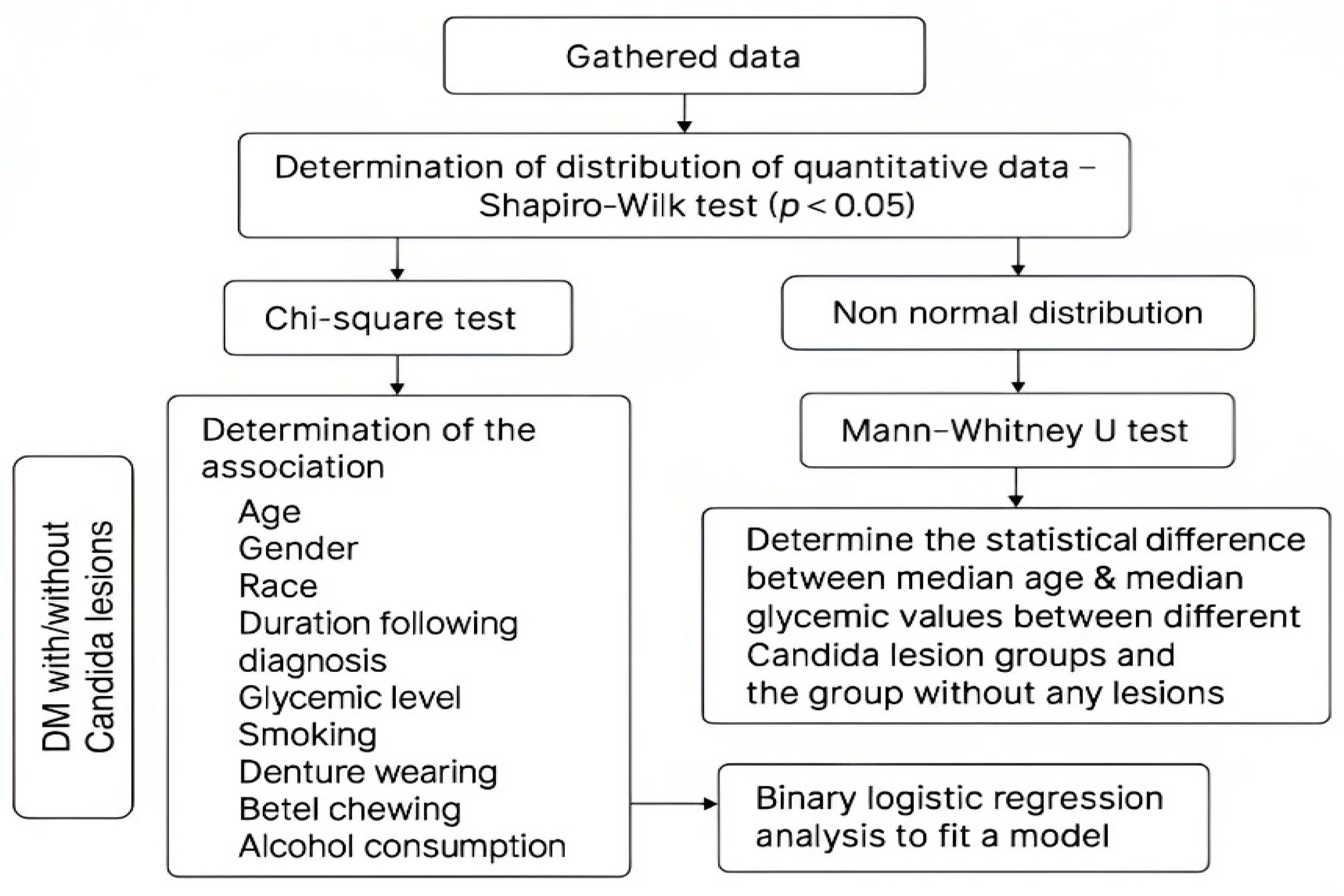
2.3. Statistical Analysis

Data were analyzed by using SPSS (Statistical Package for the Social Sciences). For statistical analysis, the distribution of quantitative data was assessed and found to be non-normally distributed. Consequently, non-parametric tests were employed. The chi-square test was used to evaluate the association between oral candidal lesions and socio-demographic and risk factors. Binary logistic regression was subsequently conducted to further explore these associations. The Mann–Whitney U test was applied to compare quantitative variables across different types of oral candidal lesions. A p-value of <0.05 was considered significant. The rationale and statistical tests employed in this study are summarized in Figure 2.

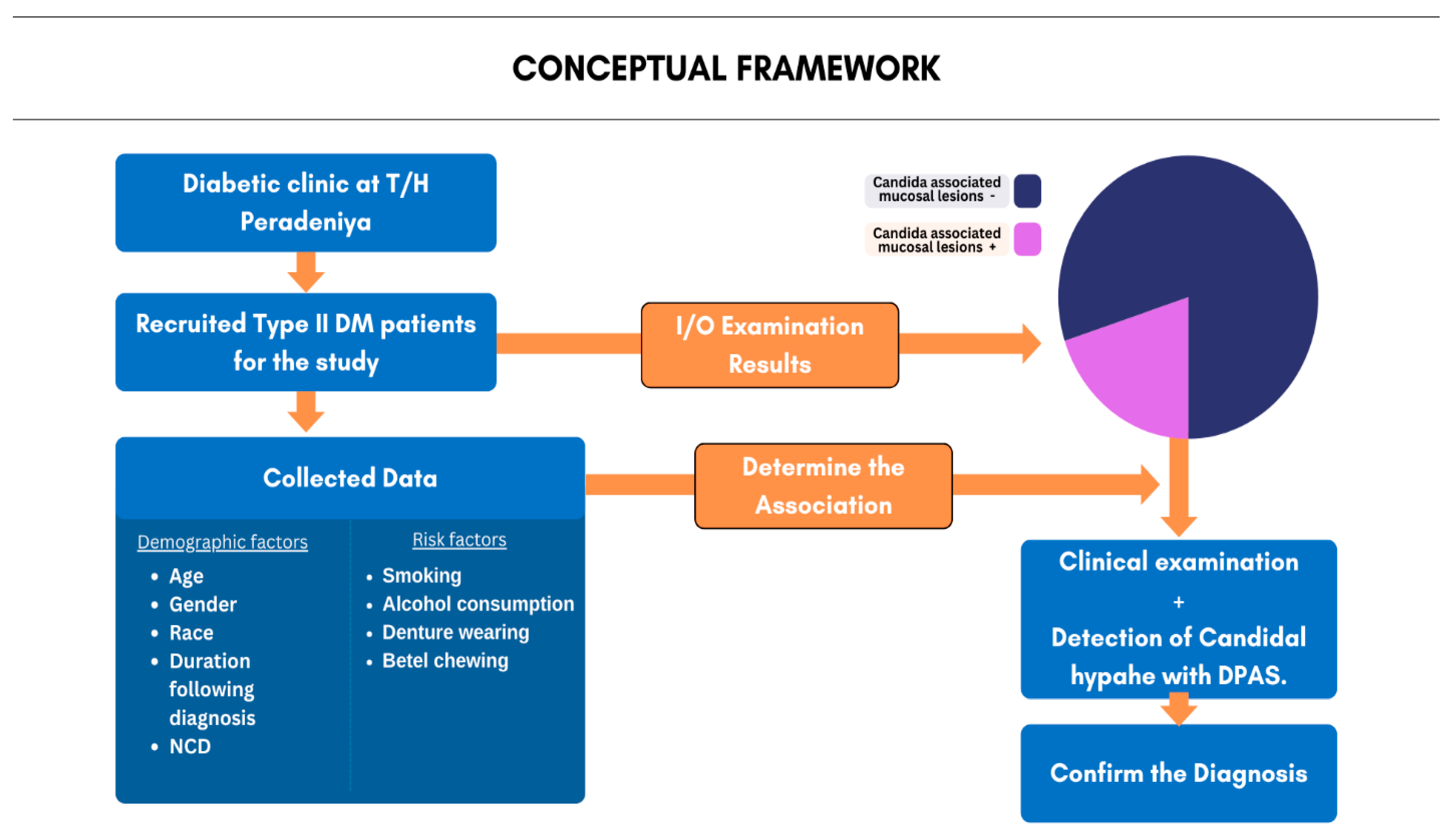
2.4. Methodology Overview

A cross-sectional study was conducted among 355 type 2 DM patients at the Diabetic Clinic, Teaching Hospital Peradeniya. Patients were clinically screened for oral candidal lesions, which were confirmed via exfoliative cytopathology and PAS staining. Demographic data and risk factors were collected using an interviewer-administered form. Non-parametric tests (chi-square and Mann–Whitney U tests) and binary logistic regression were applied using statistical package for the social sciences-26th version (SPSS), IBM Corp, New York, USA, with significance set at p < 0.05.

3. Results

The mean age of the study population was 60.38 years (SD ± 10.36), ranging from 29 to 86 years. The majority (50%) were over 60 years old. There was a female predominance (67.6%), with a male-to-female ratio of 1:2. The ethnic distribution predominantly consisted of Sinhalese (84.1%), aligning with the general population of the Kandy District. Fifty-five percent (55.1%) of participants were diagnosed within the last decade. Additionally, 54.8% were also receiving treatments for hypertension and dyslipidemia, while 26.7% only had type 2 DM. Figure 3 presents the detailed socio-demographic features of the study population.
Candidal lesions were identified in 17.6% of the study population (62/352 patients), with the diagnoses confirmed through identification of fungal hyphae with exfoliative cytology using PAS staining. The most common lesion was Denture Stomatitis (DS) (4%) (Figure 4), followed by Erythematous Candidiasis (EC) (3.4%), Pseudomembranous Candidiasis (PMC) (3.1%) (Figure 5), and Chronic Hyperplastic Candidiasis (CHC) (2.8%) (Table 1). Less frequently identified conditions included Angular Cheilitis (AC) (1.1%), Candida-associated dry mouth (0.6%), and Median Rhomboid Glossitis (MRG) (0.3%), which were excluded from further clinicopathological analysis due to their low prevalence. A noteworthy finding was that all DM patients (100%) were unaware of the existence of their oral lesions, which were detected as incidental findings during this study.
PMC (72.7%) was predominantly found on the dorsum of the tongue, with a slight female predominance (male-to-female ratio of 0.8:1). The median glycemic level in this group was 137 mg/dL. EC had the highest median age (67 years) and median glycemic level (160 mg/dL). Unlike PMC, EC was more prevalent in males (male-to-female ratio of 1:0.7), but similarly, most lesions (58%) were on the dorsum of the tongue. CHC showed higher median glycemic levels (152 mg/dL) than PMC (137 mg/dL) and was mostly located on the buccal mucosa. DS, the most common lesion, was significantly associated with upper denture wearers and was exclusively found on the hard palate (100%). It was more frequent in females (male-to-female ratio of 0.27:1) (Table 1). Patients with EC were significantly older compared to those without candidal lesions (p = 0.041, Mann–Whitney U test). DS patients had significantly higher glycemic levels than those without lesions (p = 0.045, Mann–Whitney U test), highlighting the impact of hyperglycemia on DS occurrence in denture wearers.
According to Table 2, males were significantly more likely to present with oral candidal lesions (27.19%) compared to females (13.44%, p = 0.002, χ2 test). Patients older than 60 years had a higher prevalence (10.51%) of candidal lesions than those younger than 60 years (7.38%); this was not statistically significant (p = 0.153, χ2 test). Higher glycemic levels (>110 mg/dL) were associated with a greater likelihood (13.35%) of candidal lesions compared to those with lower glycemic levels (<110 mg/dL, 4.54%); however, this was not statistically significant. The presence of other NCDs (e.g., hypertension and dyslipidemia) was more common (10.22%) in patients with candidal lesions than in those with DM only (4.26%); this was not statistically significant (p = 0.848, χ2 test). Table 3 presents the association between different risk factors and oral candidal lesions. Smokers had a significantly higher prevalence of candidal lesions (22.2%) compared to non-smokers (9.7%, p = 0.005, χ2 test). Betel chewers were significantly more likely to have candidal lesions (33.33%) than non-chewers (18.33%, p = 0.008, χ2 test). Alcohol consumption was not identified as a significant risk factor for candidal lesions (p = 0.227, χ2 test). Male sex was associated with a 3.16 times higher risk of developing oral candidal lesions (OR = 3.16, 95% CI: 1.713–5.828). Denture wearing significantly increased the risk of developing oral candidal lesions (OR = 2.348, 95% CI: 1.240–4.447).

4. Discussion

This study addresses a significant research gap regarding the prevalence of oral candidal lesions in type 2 DM patients within a specific demographic area. While the general population may also experience such lesions, focusing on DM patients is crucial, as integrating this knowledge into their management protocols can improve clinical outcomes. Our study observed all recognized and previously described oral candidal lesions, including PMC, EC, CHC, DS, AC, MRG, and Candida-associated dry mouth. Surprisingly, only 17.6% (Table 1) of type 2 DM patients exhibited Candida-related mucosal lesions, which is a lower prevalence than expected. An extensive literature review did not identify comparable studies focusing exclusively on oral candidal lesions in DM patients. Instead, our findings were compared with studies examining oral Candida carriage rates, which range from 13.7% to 81% in DM patients [5,11], and studies evaluating specific oral candidal lesions. A Sri Lankan study reported an oral Candida carriage rate of 81%, with 33% of patients having counts exceeding 2000 CFU/mL [11]. Martinez et al. highlighted challenges in diagnosing Candida-induced lesions without the detection of pathogenic hyphae in exfoliative cytology [5], which was a challenge also encountered in our study. A statistically significant male predisposition was observed among patients with oral candidal lesions, aligning with studies conducted in India and Saudi Arabia [12,13]. However, Sampath et al. [11] reported a female predilection for Candida carriage in Sri Lankan DM patients. This discrepancy may be due to the higher prevalence of risk habits such as smoking and betel quid chewing among males in our study sample. Interestingly, previous studies have also indicated a higher proportion of infection-free individuals among males [14], suggesting that risk behaviors rather than gender alone may contribute to the observed differences.
Denture Stomatitis (DS) was the most frequently identified Candida-associated lesion, affecting 4.0% of type 2 DM patients. Among them, 21.42% were males and 78.58% were females. Previous systematic reviews and meta-analyses reported a higher DS prevalence (10–67%) in DM patients [15]. The lower prevalence in our study may be attributed to good denture hygiene practices among participants. However, among patients in whom DS was diagnosed, a notable lack of denture hygiene awareness was observed. Similar female predominance in DS cases has been reported in India [16], whereas Javed et al. found a higher prevalence of Candida albicans carriage in denture-wearing males [17]. A significantly higher median glycemic level was identified in DS patients compared to those without lesions. This aligns with previous studies [18,19] that highlight the correlation between hyperglycemia and increased Candida colonization. Regarding the clinical presentation of DS, our study revealed 21.4% and 78.6% of Newton type 1 and type 2 lesions, respectively. Similar to the previous literature, the majority of DS lesions identified in our study were Newton type 2 lesions. However, in contrast to the available literature, where the majority of Newton type 2 lesions were associated with symptoms, all of our DS patients were asymptomatic, and the lesions were diagnosed as incidental findings [20,21].
Erythematous Candidiasis (EC) was the second most common lesion in our study. The literature suggests that DM patients wearing dentures are at risk of developing EC [22]. Other risk factors include antibiotic and inhaler use [23]; however, such practices were less common among our participants, possibly contributing to the lower EC prevalence. A statistically significant age difference was observed between EC patients and those without lesions (p < 0.05; mean rank with EC: 201.25; mean rank with no lesions: 148.97). While no prior studies have reported this finding, our results suggest a higher likelihood of EC development in elderly DM patients, emphasizing the need for clinician awareness for early diagnosis and management.
There are studies that indicate a high prevalence (38%) of PMC among DM patients with oral candidal lesions [5]. PMC is commonly associated with immunocompromised states, including DM [24]. The relatively lower prevalence in our study may be attributed to effective DM management at the Diabetic Clinic at T/H Peradeniya and the exclusion of DM patients with other immunosuppressive conditions. CHC is a controversial entity due to its potential for malignant transformation [25]. Our study identified a relatively higher proportion of CHC cases in DM patients compared to a previous study [25], likely due to additional risk factors such as smoking and betel quid chewing in our study population. Diagnosing CHC solely based on clinical and cytological findings poses challenges, as distinguishing it from leukoplakia with Candida superinfection requires biopsy confirmation. This limitation may have resulted in an overestimation of CHC cases in our study. Only 1.1% of DM patients in our study presented with AC, which is a significantly lower prevalence than the 14.1% reported in an Indian study [16]. This suggests that DM alone is not a primary risk factor for AC, which is influenced by additional factors such as vitamin deficiencies, reduced occlusal vertical dimension, and bacterial co-infection [26]. Cytological samples from our AC patients confirmed a high bacterial colonization rate, supporting the multifactorial etiology of this lesion.
More than half (53.96%) of oral candidal lesions occurred in patients older than 60 years. The literature suggests that individuals of older age are more susceptible to oral candidiasis [27]. Studies show a significantly higher Candida prevalence in adults aged 56–92 years [28] and an increased colonization rate in diabetic patients over 40 years [29]. The age distribution in our study aligns with these findings, reinforcing the need for heightened clinical vigilance in older DM patients. Studies suggest a positive correlation between glycemic levels and Candida colonization [18]. In our study, 47 out of 63 patients with oral candidal lesions had fasting blood glucose levels exceeding 110 mg/dL. However, other research indicates that glucose concentration alone does not affect Candida albicans’ invasive behavior [30], suggesting that additional risk factors contribute to lesion development.
A statistically significant association between smoking and Candida lesions was observed. Studies in Iran reported a dose-dependent relationship, with increased cigarette consumption correlating with higher oral candidiasis risk [31]. A Sri Lankan study also found a significant association between smoking and oral Candida carriage in DM patients [5]. Smoking-related alterations, such as reduced salivary flow, lowered salivary pH, and decreased immunoglobulin A levels, contribute to increased Candida colonization [31]. A notable association was found between betel quid chewing and Candida-associated lesions. A Thai study reported elevated Candida levels in betel quid chewers [32]. Further research in Bangladesh linked betel chewing to hyperglycemia in DM patients, potentially influencing Candida carriage and lesion development [33]. However, a contrasting study found no significant effect of betel quid chewing on Candida carriage [34].

5. Conclusions

The relatively lower prevalence of oral candidal lesions observed in our study suggests that effective glycemic control and medical management may play a protective role in DM patients. However, the higher risk among males and denture wearers highlights the need for targeted interventions in these subgroups. Early detection and timely management of oral candidal lesions are essential to improving overall health outcomes and quality of life in this vulnerable population. Routine oral health assessments, oral screenings, and oral health education should be incorporated into diabetes management protocols to facilitate early detection and timely treatment of oral candidal lesions in diabetic clinics.

Author Contributions

Conceptualization, J.W., P.J., L.W. and V.T.; methodology, J.W., P.J., L.W. and V.T.; software, J.W., L.W., V.T. and P.J.; formal analysis, P.J.; investigation and resources, L.W., J.W., V.T. and P.J.; data curation, V.T., J.W., L.W. and P.J.; writing—original draft preparation, J.W., L.W., P.J. and V.T.; writing—review and editing, P.J., J.W., L.W., V.T. and T.L.; supervision, V.T., J.W., L.W., P.J. and T.L.; project administration, V.T., J.W., L.W., P.J. and T.L. All authors have read and agreed to the published version of the manuscript.

Funding

This research received no external funding.

Institutional Review Board Statement

Ethical approval was obtained from the Ethics Review Committee (ERC) of the Faculty of Dental Sciences, University of Peradeniya (Approval No. ERC/FDS/UOP/UGR/2023/20). Administrative clearance was granted by the head of the Department of Oral Pathology and the directors of T/H Peradeniya. Written informed consent was obtained from all participants before clinical examination, specimen collection, and photography.

Informed Consent Statement

Informed consent was obtained from all subjects involved in this study.

Data Availability Statement

The dataset of this study is available upon reasonable request from the corresponding authors. Due to ethical restrictions and confidentiality agreements, the data are not publicly accessible.

Conflicts of Interest

The authors declare no conflicts of interest.

References

  1. Rannan-Eliya, R.P.; Wijemunige, N.; Perera, P.; Kapuge, Y.; Gunawardana, N.; Sigera, C.; Jayatissa, R.; Herath, H.M.M.; Gamage, A.; Weerawardena, N.; et al. Prevalence of diabetes and pre-diabetes in Sri Lanka: A new global hotspot–estimates from the Sri Lanka Health and Ageing Survey 2018/2019. BMJ Open Diabetes Res. Care 2023, 11, e003160. [Google Scholar] [CrossRef] [PubMed]
  2. Papatheodorou, K.; Banach, M.; Bekiari, E.; Rizzo, M.; Edmonds, M. Complications of Diabetes 2017. J. Diabetes Res. 2018, 2018, 3086167. [Google Scholar] [CrossRef]
  3. Rohani, B. Oral manifestations in patients with diabetes mellitus. World J. Diabetes. 2019, 10, 485–489. [Google Scholar] [CrossRef]
  4. Rodrigues, C.F.; Rodrigues, M.E.; Henriques, M. Candida sp. Infections in patients with diabetes mellitus. J. Clin. Med. 2019, 8, 76. Available online: https://www.ncbi.nlm.nih.gov/pmc/articles/PMC6352194/ (accessed on 27 September 2023). [CrossRef] [PubMed]
  5. Martinez, R.F.F.; Jaimes-Aveldañez, A.; Hernández-Pérez, F.; Arenas, R.; Miguel, G.F.S. Oral Candida spp carriers: Its prevalence in patients with type 2 diabetes mellitus. An. Bras. Dermatol. 2013, 88, 222–225. [Google Scholar] [CrossRef]
  6. Rodríguez-Archilla, A.; Piedra-Rosales, C. Candida species oral detection and infection in patients with diabetes mellitus: A meta-analysis. Iberoam. J. Med. 2021, 3, 115–121. [Google Scholar] [CrossRef]
  7. Sudbery, P.E. Growth of Candida albicans hyphae. Nat. Rev. Microbiol. 2011, 9, 737–748. [Google Scholar] [CrossRef]
  8. Patil, S.; Rao, R.S.; Majumdar, B.; Anil, S. Clinical appearance of oral Candida infection and therapeutic strategies. Front. Microbiol. 2015, 6, 1391. [Google Scholar] [CrossRef]
  9. Ghabanchi, J.; Andisheh Tadbir, A.; Darafshi, R.; Sadegholvad, M. The prevalence of median rhomboid glossitis in diabetic patients: A case-control study. Iran. Red Crescent. Med. J. 2011, 13, 503–506. [Google Scholar]
  10. Yao, Y.; Shi, L.; Zhang, C.; Sun, H.; Wu, L. Application of fungal fluorescent staining in oral candidiasis: Diagnostic analysis of 228 specimens. BMC Microbiol. 2019, 19, 96. [Google Scholar] [CrossRef]
  11. Sampath, A.; Weerasekera, M.; Dilhari, A.; Gunasekara, C.; Bulugahapitiya, U.; Fernando, N.; Samaranayake, L. Type 2 diabetes mellitus and oral Candida colonization: Analysis of risk factors in a Sri Lankan cohort. Acta Odontol. Scand. 2019, 77, 508–516. [Google Scholar] [CrossRef] [PubMed]
  12. Anita Kumar, S.; Pandya, H. Speciation and antifungal susceptibility testing of Candida isolated from immunocompromised patients of a tertiary care centre in Gujarat, India. J. Pure Appl. Microbiol. 2023, 17, 427–433. [Google Scholar] [CrossRef]
  13. Alrashed, F.A.; Iqbal, M.; Alsubiheen, A.M.; Ahmad, T. Exploring determinants of sex and family history-based disparity in type 2 diabetes mellitus prevalence among clinical patients. BMC Public Health 2024, 24, 682. [Google Scholar] [CrossRef]
  14. Loster, J.E.; Wieczorek, A.; Loster, B.W. Correlation between age and gender in Candida species infections of complete denture wearers: A retrospective analysis. Clin. Interv. Aging 2016, 11, 1707. [Google Scholar] [CrossRef] [PubMed]
  15. Martorano-Fernandes, L.; Dornelas-Figueira, L.M.; Marcello-Machado, R.M.; Silva, R.B.; Magno, M.B.; Maia, L.C.; del Bel Cury, A.A. Oral candidiasis and denture stomatitis in diabetic patients: Systematic review and meta-analysis. Braz. Oral Res. 2020, 34, e113. [Google Scholar] [CrossRef]
  16. Mathew, A.L.; Daniel, M.P.; Cherian, S.A.; Joseph, B.B. Prevalence of oral mucosal lesions among diabetic patients in south Kerala, India. J. Oral Med. Toxicol. 2017, 1, 20–22. [Google Scholar]
  17. Javed, F.; Klingspor, L.; Sundin, U.; Altamash, M.; Klinge, B.; Engström, P.E. Periodontal conditions, oral Candida albicans and salivary proteins in type 2 diabetic subjects with emphasis on gender. BMC Oral Health 2009, 9, 12. [Google Scholar] [CrossRef]
  18. Ganapathy, D.M.; Joseph, S.; Ariga, P.; Selvaraj, A. Evaluation of the influence of blood glucose level on oral candidal colonization in complete denture wearers with type-II diabetes mellitus: An in vivo study. Dent. Res. J. 2013, 10, 87–92. [Google Scholar]
  19. Dorocka-Bobkowska, B.; Zozulinska-Ziolkiewicz, D.; Wierusz-Wysocka, B.; Hedzelek, W.; Szumala-Kakol, A.; Budtz-Jörgensen, E. Candida-associated denture stomatitis in type 2 diabetes mellitus. Diabetes Res. Clin. Pract. 2010, 90, 81–86. [Google Scholar] [CrossRef]
  20. Perić, M.; Miličić, B.; Kuzmanović Pfićer, J.; Živković, R.; Arsić Arsenijević, V. A systematic review of denture stomatitis: Predisposing factors, clinical features, etiology, and global Candida spp. distribution. J. Fungi 2020, 6, 328. [Google Scholar] [CrossRef]
  21. Abuhajar, E.; Ali, K.; Zulfiqar, G.; Al Ansari, K.; Raja, H.Z.; Bishti, S.; Anweigi, L. Management of chronic atrophic candidiasis (denture stomatitis)—A narrative review. J. Fungi 2023, 9, 3029. [Google Scholar] [CrossRef]
  22. Motta-Silva, A.C.; Aleva, N.A.; Chavasco, J.K.; Armond, M.C.; França, J.P.; Pereira, L.J. Erythematous oral candidiasis in patients with controlled type II diabetes mellitus and complete dentures. Mycopathologia 2010, 169, 215–223. [Google Scholar] [CrossRef] [PubMed]
  23. Fukushima, C.; Matsuse, H.; Tomari, S.; Obase, Y.; Miyazaki, Y.; Shimoda, T.; Kohno, S. Oral candidiasis associated with inhaled corticosteroid use: Comparison of fluticasone and beclomethasone. Ann. Allergy Asthma Immunol. 2003, 90, 646–651. [Google Scholar] [CrossRef]
  24. Berberi, A.; Noujeim, Z.; Aoun, G. Epidemiology of oropharyngeal candidiasis in human immunodeficiency virus/acquired immune deficiency syndrome patients and CD4+ counts. J. Int. Oral Health 2015, 7, 20–23. [Google Scholar] [PubMed]
  25. Lorenzo-Pouso, A.I.; Pérez-Jardón, A.; Caponio, V.C.A.; Spirito, F.; Chamorro-Petronacci, C.M.; Álvarez-Calderón-Iglesias, Ó.; Gándara-Vila, P.; Muzio, L.L.; Pérez-Sayáns, M. Oral chronic hyperplastic candidiasis and its potential risk of malignant transformation: A systematic review and prevalence meta-analysis. J. Fungi 2022, 8, 1093. [Google Scholar] [CrossRef] [PubMed]
  26. Federico, J.R.; Basehore, B.M.; Zito, P.M. Angular cheilitis. In StatPearls; StatPearls Publishing: Treasure Island, FL, USA, 2024. Available online: http://www.ncbi.nlm.nih.gov/books/NBK536929/ (accessed on 6 May 2024).
  27. Taylor, M.; Brizuela, M.; Raja, A. Oral candidiasis. In StatPearls; StatPearls Publishing: Treasure Island, FL, USA, 2024. Available online: http://www.ncbi.nlm.nih.gov/books/NBK545282/ (accessed on 6 May 2024).
  28. Zaremba, M.L.; Daniluk, T.; Rozkiewicz, D.; Cylwik-Rokicka, D.; Kierklo, A.; Tokajuk, G.; Dąbrowska, E.; Pawińska, M.; Klimiuk, A.; Stokowska, W.; et al. Incidence rate of Candida species in the oral cavity of middle-aged and elderly subjects. Adv. Med. Sci. 2006, 51 (Suppl. 1), 233–236. [Google Scholar]
  29. Kumar, B.V.; Padshetty, N.S.; Bai, K.Y.; Rao, M.S. Prevalence of Candida in the oral cavity of diabetic subjects. J. Assoc. Physicians India 2005, 53, 599–602. [Google Scholar]
  30. Dornelas Figueira, L.M.; Ricomini Filho, A.P.; da Silva, W.J.; Del Bel Cury, A.A.; Ruiz, K.G.S. Glucose effect on Candida albicans biofilm during tissue invasion. Arch. Oral Biol. 2020, 117, 104728. [Google Scholar] [CrossRef]
  31. Khalili, P.; Movagharipoor, A.; Sardari, F.; Movaghari Pour, F.; Jamali, Z. Oral candidiasis and cigarette, tobacco, alcohol, and opium consumption in Rafsanjan, a region in the southeast of Iran. BMC Oral Health 2023, 23, 262. [Google Scholar] [CrossRef]
  32. Deeiam, K.; Pankam, J.; Sresumatchai, V.; Visedketkan, P.; Jindavech, W.; Rungraungrayabkul, D.; Pimolbutr, K.; Klongnoi, B.; Khovidhunkit, S.-O.P. Presence of Candida and its associated factors in participants attending oral cancer screening in the lower northeastern area of Thailand. BMC Oral Health 2023, 23, 527. [Google Scholar] [CrossRef]
  33. Hasan, Z.; Islam, M.R.; Hasan, M.S.; Sakib, M.M.; Islam, M.S.; Chowdhury, M.B.; Sarker, S.; Islam, M.R.; Hasnat, M.A.; Nath, L.M.; et al. Risk analysis of chewing betel quid among diabetic patients from the northeastern part of Bangladesh. medRxiv. 2020. Available online: https://www.medrxiv.org/content/10.1101/2020.01.15.20017731v1 (accessed on 10 June 2024).
  34. Javed, F.; Yakob, M.; Ahmed, H.B.; Al-Hezaimi, K.; Samaranayake, L.P. Oral Candida carriage among individuals chewing betel-quid with and without tobacco. Oral Surg. Oral Med. Oral Pathol. Oral Radiol. 2013, 116, 427–432. [Google Scholar] [CrossRef] [PubMed]
Figure 1. Outline of the study methodology. Note: T/H = Teaching Hospital; NCD = non-communicable disease.
Applsci 15 05539 g001
Figure 2. The rationale for the use of statistical tests.
Applsci 15 05539 g002
Figure 3. Socio-demographic presentation of the study sample. Note: NCD = non-communicable disease; HTN = hypertension; DL = dyslipidemia.
Applsci 15 05539 g003
Figure 4. Clinical images show Denture Stomatitis on the hard palate of a 54-year-old female patient (a) with poor denture hygiene (b). The lesion was characterized by erythema. The PAS-stained smear reveals a cluster of magenta-colored candidal hyphae, which are non-branching (arrows) in nature, confirming the diagnosis of DS (c).
Applsci 15 05539 g004
Figure 5. Clinical images show a white plaque-like lesion that could be scraped off on the dorsum of the tongue of a 60-year-old female patient (a), who was unaware of the presence of the lesion. PAS staining revealed candidal hyphae (arrows), which confirms the diagnosis of PMC (b).
Applsci 15 05539 g005
Table 1. Clinico-pathological presentation of commonly observed candidal lesions.
LesionsN (%)
[Out of 63 Lesions]
Site* Median Age in YearsGender* Median Glycemic Level (mg/dL)
MaleFemale
PMC11
17.5%
Dorsum of the tongue
Buccal mucosa
8
3
6256137
EC12
19%
Dorsum of the tongue
Buccal mucosa
Hard palate
Soft palate
7
3
1
1
6775160
CHC10
15.9%
Buccal mucosa
Alveolar mucosa
Lip
8
1
1
61.582152
DS14
22.2%
Hard palate1460.5311148
Note: PMC = Pseudomembranous Candidiasis; EC = Erythematous Candidiasis; CHC = Chronic Hyperplastic Candidiasis; DS = Denture Stomatitis. * Due to non-normal distribution of data, median value was used to obtain a meaningful representation instead of mean value.
Table 2. Comparison of clinicopathological presentations of patients with and without candidal lesions.
FeatureDM with Candidal Lesions (n %)DM Without Candidal Lesions (n %)X2 Test
p Value
1. Age in years
≤60
≥61

26 (7.38)
37 (10.51)

148 (42.04)
141 (40.05)
0.153
2. Gender
Male
Female

31 (8.80)
32 (9.09)

83 (23.57)
206 (58.52)
0.002
3. Race
Sinhala
Tamil
Muslim

54 (15.34)
5 (1.42)
4 (1.13)

242 (68.75)
20 (5.68)
27 (7.67)
0.73
4. Duration following diagnosis in years
≤15
≥16

44 (12.5)
19 (5.39)

227 (64.48)
62 (17.61)
0.173
5. Relationship with other NCDs
DL + HTN
Other NCDs present
No other NCDs

36 (10.22)
12 (3.40)
15 (4.26)

157 (44.60)
53 (15.05)
79 (22.44)
0.848
6. Glycemic level (mg/dL)
≤110
≥111

16 (4.54)
47 (13.35)

89 (25.28)
200 (56.81)
0.396
Table 3. Association between risk factors of DM patients and occurrence of oral candidal lesions.
Risk FactorsDM with Candidal Lesions (n%)DM Without Candidal Lesions (n%)X2dfp
1. Denture
Denture wearer
Non-denture wearer

21 (33.33)
42 (66.7)

73 (25.3)
216 (74.7)
1.72310.189
2. Smoking
Present
Absent

14 (33.3)
28 (66.7)

49 (15.8)
261 (84.2)
7.73310.005
3. Betel chewing
Present
Absent

21 (28.4)
53 (71.6)

42 (15.1)
236 (84.9)
7.00410.008
4. Alcohol misuse
Present
Absent

13 (23.6)
42 (76.4)

50 (16.8)
247 (83.2)
1.46110.227
Disclaimer/Publisher’s Note: The statements, opinions and data contained in all publications are solely those of the individual author(s) and contributor(s) and not of MDPI and/or the editor(s). MDPI and/or the editor(s) disclaim responsibility for any injury to people or property resulting from any ideas, methods, instructions or products referred to in the content.

