Article

Does Pilates Breathing Affect the Biceps Brachii Neuromuscular Efficiency During Submaximal Contraction?

by
Denys Batista Campos
,
Maria de Cassia Gomes Souza Macedo
,
Kariny Realino do Rosário Ferreira
,
Arthur Ferreira Esquirio
,
Ana Clara Leal
,
Gabriela Lopes Gama
and
Alexandre Carvalho Barbosa
*
Musculoskeletal Research Group—NIME, Department of Physical Therapy, Federal University of Juiz de Fora, Av. Moacir Paleta 1167, São Pedro, Governador Valadares 35020-360, Minas Gerais, Brazil
*
Author to whom correspondence should be addressed.
Appl. Sci. 2025, 15(10), 5528; https://doi.org/10.3390/app15105528
Submission received: 2 April 2025 / Revised: 6 May 2025 / Accepted: 12 May 2025 / Published: 15 May 2025
(This article belongs to the Special Issue Exercise Physiology and Biomechanics in Human Health: 2nd Edition)

Abstract

:
The Pilates breathing technique is theorized to improve neuromuscular efficiency, but its specific effects on peripheral muscles have not been thoroughly investigated. This study evaluated how Pilates breathing influenced the neuromuscular efficiency of the biceps brachii muscle during submaximal elbow flexion in comparison to regular breathing. Fifty-eight healthy adults without prior experience with the Pilates method of exercise performed concentric and eccentric elbow contractions at 20%, 40%, and 60% of their maximal voluntary isometric contraction under two breathing conditions: the specialized Pilates breathing pattern (executing movements exclusively during expiration) and normal breathing patterns. Muscle activity was measured using surface electromyography, with neuromuscular efficiency quantified as the relationship between muscle electrical activity and force production. The results revealed significantly improved neuromuscular efficiency during Pilates breathing at all tested intensity levels, with the most substantial enhancement observed at 60% of maximal effort. The eccentric phase of movement demonstrated greater efficiency gains compared to the concentric phase. These findings indicate that the distinct breathing pattern used in Pilates can independently enhance neuromuscular performance in the biceps brachii. This study suggests that incorporating Pilates breathing techniques could be beneficial in rehabilitation programs and strength training regimens to optimize both muscle function and movement efficiency. Additional research is recommended to examine the long-term effects and practical applications in clinical and athletic settings.

1. Introduction

The Pilates method of exercise (PME) is widely recognized as an integrative approach to physical conditioning that emphasizes muscle control, posture, and breathing [1]. Together, these elements work to improve strength, flexibility, neuromuscular coordination, and overall body awareness [2]. Originating in the early 20th century, PME was developed as a rehabilitative practice to improve physical function, but it became globally known for its applications in fitness, therapy, and athletic training [3,4]. PME can be practiced on specialized equipment, such as the reformer or Cadillac, or even during mat exercises, offering versatility across diverse populations and settings [5].
PME has six principles: centering, concentration, control, precision, flow, and breathing. These principles not only form the method’s foundation but also provide a biomechanical and neuromuscular framework that supports functional movement patterns [2]. Breathing is emphasized as a vital component of PME, serving to regulate the intra-abdominal pressure, stabilize the core muscles, and enhance mind–body awareness. The Pilates breathing technique combines diaphragmatic and costal breathing, which promotes thoracic expansion while maintaining abdominal engagement [4]. This unique approach to breathing has been linked to improved core stability and muscle excitation, particularly in the deep stabilizing muscles, such as the transverse abdominis, lumbar multifidi, and pelvic floor [6].
The integration of breathing and centering during PME has significant implications for muscle excitation and performance. Studies using electromyography have demonstrated that Pilates-based breathing techniques can enhance neuromuscular efficiency, a parameter that reflects the relationship between neural drive and force production [7]. Neuromuscular efficiency is particularly relevant in resistance training and rehabilitation, as it quantifies the optimization of motor unit recruitment and firing patterns [8]. Efficient muscle function, as evidenced by reduced electromyographic amplitudes during high-force tasks, is indicative of an adaptive neuromuscular system capable of performing tasks with a minimal energetic cost [9]. Additionally, the principles of resistance training underscore the importance of gradually increasing the mechanical load to stimulate neuromuscular adaptations; this concept aligns with the objectives of Pilates training, which often incorporates progressive resistance using springs or body weight [10].
Research on neuromuscular efficiency during concentric and eccentric muscle actions reveals distinct patterns. Eccentric exercises demonstrate significantly lower muscle excitation compared to concentric [11]. Recent investigations have explored the influence of Pilates breathing on specific muscle groups, particularly in the context of upper limb movement [12]. Research on the biceps brachii during concentric and eccentric phases has revealed greater electromyographic activity when Pilates breathing associated with centralization was employed compared to regular breathing techniques. These findings suggest that the coordinated excitation of respiratory and core muscles can enhance the excitability and performance of distal muscles [6]. However, there is a lack of studies isolating the effects of the Pilates breathing on neuromuscular efficiency or examining its impact across different load intensities for a given exercise.
Despite growing interest in the biomechanical and neuromuscular effects of PME, many aspects of its mechanisms remain underexplored—specifically regarding the isolated contribution of Pilates breathing in tasks requiring upper limb coordination. Furthermore, the interaction between load and breathing techniques in eliciting neuromuscular responses warrants further investigation. Therefore, the present study aimed to analyze the effects of Pilates breathing on the neuromuscular efficiency during concentric and eccentric phases of the biceps brachii at varying load levels. By comparing Pilates breathing with regular breathing, this research sought to elucidate the role of breathing techniques in neuromuscular efficiency. These findings have the potential to inform evidence-based practices in rehabilitation, strength training, and performance enhancement.

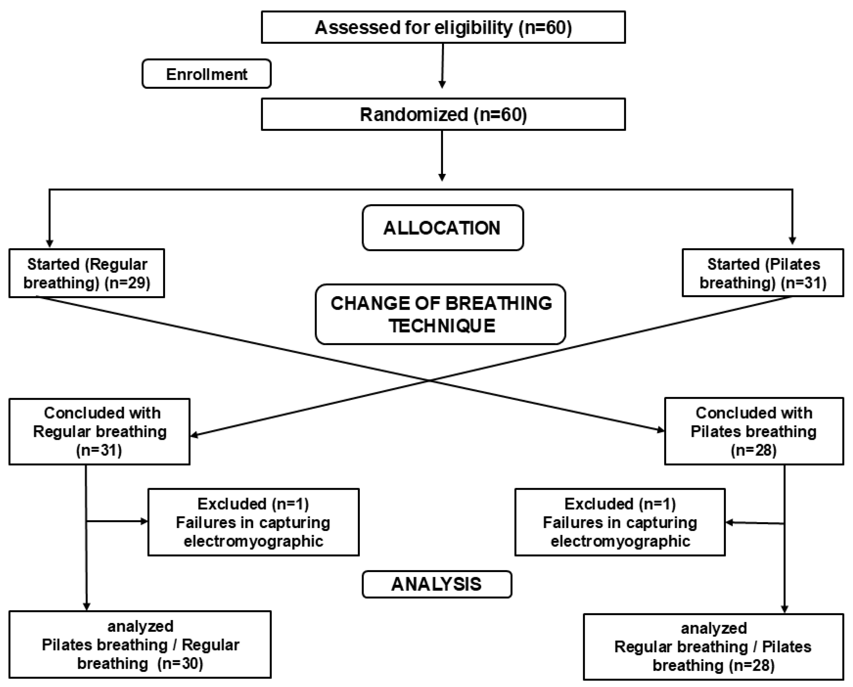
2. Materials and Methods
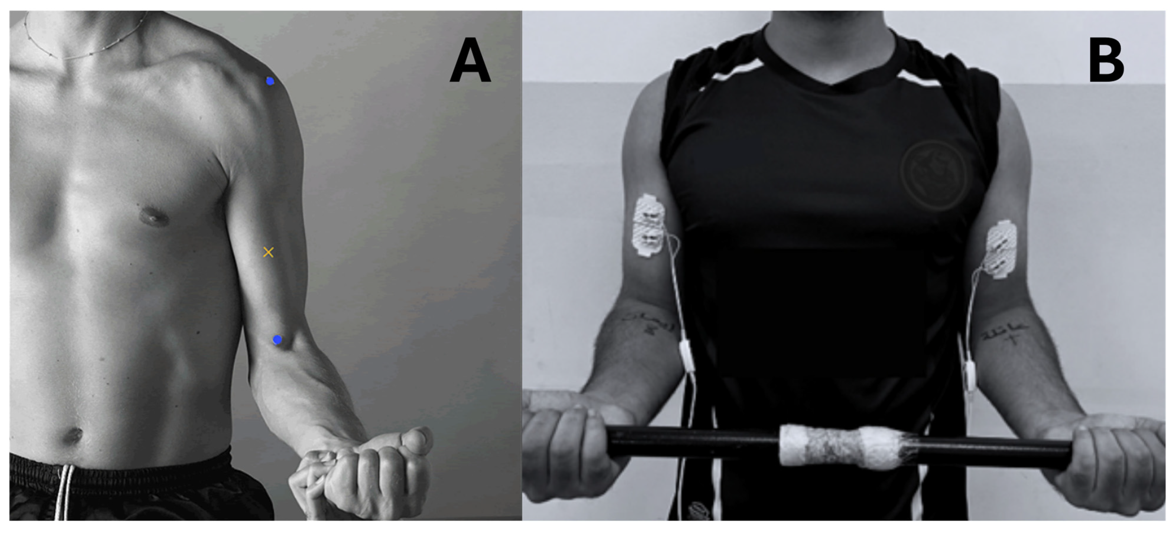
A total of fifty-eight healthy adults from both sexes (18–30 years) joined the present study. The participants were recruited by personal contacts and public invitation through folders from December 2023 to February 2024. The participants’ characteristics are shown in Table 1. The a priori two-tailed sample size calculation was based on a previous study, considering the effect size of 0.48, the alpha level of 5%, and 95% power, returning a total of 32 participants.
Considering sample loss of 30%, 44 participants were needed to reach the sampling power. However, 60 participants were selected and the data of fifty-eight were analyzed, as shown in Figure 1. The inclusion criteria were an age between 18 and 30 years old and having never practiced PME. As exclusion criteria, the participants were required to have no history of severe orthopedic and neurological disorders, cardiovascular disease, or upper limb surgery. This randomized clinical trial was conducted in accordance with the Declaration of Helsinki. The Ethics Committee of the Federal University of Juiz de Fora (number 66768023.1.0000.5147) approved all procedures employed in the present study. The trial was registered in the Brazilian clinical trials registry (number RBR-5q5g4s6). All participants gave written informed consent prior to participation.

2.1. Data Recording

Muscle excitation was measured using a biological signal acquisition module with eight analog channels (MiotecTM Biomedical Equipment, Porto Alegre, RS, Brazil). The conversion of analog to digital signals was performed by an A/D board with a 16-bit resolution input range, sampling frequency of 2 kHz, common rejection module greater than 100 dB, signal/noise ratio less than 03 μV RMS, and impedance of 109 Ω. The electromyographic surface (sEMG) signals were recorded as the root mean square (RMS) in μV and the average frequency in Hz with surface electrodes (20 mm diameter and a center-to-center distance of 20 mm). Prior to the fixation of the electrodes, trichotomy was performed and the skin was cleaned with 70% alcohol. The muscles analyzed by sEMG were as follows: right and left arm long head of biceps brachii. Auto-adhesive surface electrodes were attached to the muscle bellies and positioned parallel to the muscle fibers, according to the techniques described in the sEMG guidelines for Non-Invasive Muscle Assessment (SENIAM); see Figure 2. The volunteers stood with the elbow flexed at 90° and the forearm dorsum horizontally downward. The electrodes were positioned on the line between the medial acromion and the cubital fossa. The sEMG signals were amplified and filtered (10–500 Hz, notch 60 Hz) [13].

2.2. Exercise Procedures

Maximal voluntary isometric contraction
To establish the maximal isometric output (100%), each participant performed 3 maximal voluntary isometric contractions (MVICs) of elbow flexion (with the participant standing with the knee flexed at 20° and elbow flexed at 90°), measured by a laboratory-grade load cell (MiotecTM Biomedical Equipment, Porto Alegre, RS, Brazil; maximum tension–compression = 200 kgf, precision of 0.1 kgf, maximum error of measurement = 0.33%) attached to an acquisition module with eight analog channels (MiotoolTM, MiotecTM Biomedical Equipment, Porto Alegre, RS, Brazil). The laboratory-grade load cell was previously calibrated using 10% (20 kgf) of its maximal tension–compression, according to the manufacturer’s recommendations. Both limbs were simultaneously assessed. The laboratory-grade load cell was anchored to a stable surface, and participants were instructed to exert maximal effort during the isometric test. The maximal force output data were obtained from the average of the three MVICs.

2.3. Experimental Protocol

After 5 min of rest, each participant was then instructed to perform 18 trials (9 for regular and 9 for Pilates breathing) of a complete full dynamic movement (concentric–eccentric) of the elbow combined to a breathing technique at 3 × 20%, 3 × 40%, and 3 × 60% of the flexion MVIC. All participants performed both breathing techniques (Pilates breathing and regular breathing), and the breathing technique order was randomized. The randomization sequence was independently generated using http://www.randomizer.org. The participants who started by performing regular breathing performed Pilates breathing later and vice versa. The order of the loads (20%, 40%, and 60% of the flexion MVIC) was also randomized for each participant using the above-mentioned website. One minute of rest between each load adjustment was allowed.
The regular breathing was performed with a concentric contraction during inspiration and an eccentric contraction during expiration. The Pilates breathing consisted of an initial deep inspiratory phase, followed by moving the elbow through flexion during an expiratory phase, another deep inspiration, and the final eccentric extension during the final expiratory phase. Thus, the elbow was moved only during the expiratory phase. To control the execution timing, all participants performed familiarization before the task, which consisted of demonstrating and teaching the participants how to perform the exercise, along with each breathing technique. For both breathing exercises, the concentric and eccentric timing phases were set at 2 s each, controlled by the rater.

2.4. Data Extraction

The sEMG data were normalized using the maximal voluntary isometric contraction (MVIC). The mean muscle activation of the biceps brachii was subsequently computed for both the concentric and eccentric phases. Data acquisition and offline analysis were performed using the MIOTEC Suite™ software (v. 1.0; MIOTEC™; Biomedical Equipment, Porto Alegre, RS, Brazil). To determine the onset of elbow movement, the concentric–eccentric transition, and the termination of the motion, all trials were video-recorded in synchronization with the sEMG system. Temporal markers corresponding to movement onset, phase transition, and movement end were manually assigned based on the video footage. The EMG signals for each contraction phase were then segmented according to these markers. Neuromuscular efficiency was assessed by calculating the ratio between the mean normalized sEMG activity and the corresponding force level (%MVIC: 20%, 40%, and 60%). This metric was expressed as %sEMG per kilogram-force (%sEMG/kgf). In this context, lower values indicate better neuromuscular efficiency, reflecting the muscle’s ability to generate the required force with reduced electrical activity, suggestive of more optimized neuromuscular performance [9].

2.5. Data Analysis

A descriptive analysis (median, minimum, maximum) was performed. Normality and homogeneity were assessed using the Shapiro–Wilk and Levene tests, respectively. The data subjected to the Shapiro–Wilk test indicated the absence of normality (p < 0.05). Considering that these results violated the assumptions for the use of parametric testing and the sample size, the Friedman nonparametric analysis of variance for repeated measures was used. Additionally, the Durbin–Conover post hoc test for paired contrasts was used, avoiding multiple comparisons. Effect sizes (ES) were calculated using Cohen’s d test. The ES were qualitatively classified as very small (0.01 to 0.19); small (0.20 to 0.49); moderate (0.50 to 0.79); large (0.8 to 1.19); very large (1.2 to 1.99); and huge (>2) [14]. Significance was set at p < 0.05. All statistical analysis was performed using the freeware JAMOVI (the JAMOVI Project, version 1.6.15, retrieved from http://www.jamovi.org).

3. Results

Sixty participants were initially assessed, but data from two participants were excluded due to failure in capturing electromyographic signals. Thus, fifty-eight participants were included in the final analysis. All analyzed participants were right-handed, which may have implications for the generalizability of the results, particularly in studies involving limb dominance or neuromuscular efficiency. The participants’ characteristics are shown in Table 1. The comparison of the neuromuscular efficiency index obtained during the execution of the exercise using regular breathing or Pilates breathing at different load levels is presented in Table 2. The data showed that the neuromuscular efficiency index was lower during Pilates breathing compared to regular breathing; significant differences were found across all tested parameters. Higher loads (40% and 60% of MVIC) showed better neuromuscular efficiency responses. The ES ranged from very small to large in the comparisons. The analyses also showed that the dominant biceps of all participants presented a better efficiency index when compared to the non-dominant side. The ES ranged from very small to moderate in the comparisons. For more details, confidence intervals are provided in the Supplementary Materials (available online).

4. Discussion

The present study compared the neuromuscular efficiency index during concentric and eccentric phases of the biceps brachii action associated with regular breathing and Pilates breathing at varying load levels. The results showed that the neuromuscular efficiency index was lower when movement was associated with Pilates breathing than when it was associated regular breathing. The comparison between these two breathing techniques showed that Pilates breathing resulted in better neuromuscular efficiency of the biceps brachii during concentric and eccentric phases. According to previous studies, this finding can be explained by the better neuromuscular efficiency induced by Pilates breathing, which means that fewer motor units are required to produce the same level of force. Neuromuscular efficiency is calculated considering the amount of neural stimulation and the muscle’s capacity to generate force. Therefore, a muscle that is able to generate greater torque with lower muscle fiber excitation is considered more efficient [15,16].
A study carried out with 10 healthy women [17] comparing the excitation of the biceps brachii when using breathing techniques with a centralization technique showed an increase in biceps brachii motor unit recruitment, suggesting that there is greater muscle excitation when exercise is associated with centralization and breathing techniques from the Pilates method. During this study, the centering technique was associated with Pilates breathing, while the participants performed a biceps brachii isometric contraction during elbow flexion movement. The present study did not associate Pilates breathing with the centralization technique. In addition, the participants performed the biceps brachii isotonic contraction at the same time as the breathing technique. Therefore, these differences may be due to the distinct properties of concentric versus isometric contractions and to concurrent neural influences [17], because, in the previous study, the participants needed to focus their attention on two simultaneous techniques when performing the biceps brachii contraction (breathing and centralization techniques).
Another study evaluated the effects of a centralization technique and Pilates breathing on lower-limb muscle activity during squats [18]. In this study, thirteen adults with some experience in the Pilates method performed three 60° squats under three experimental conditions: (I) normal breathing, (II) abdominal contraction with normal breathing, and (III) abdominal contraction with Pilates breathing. The results showed that squats with abdominal contraction associated with Pilates breathing resulted in increased sEMG for the rectus femoris, biceps femoris, and tibialis anterior muscles during the flexion phase, increasing the movement stability. These findings differ from the results presented in the present study, which showed less muscle excitation when performing isotonic contractions at varying load levels. The better neuromuscular efficiency indices observed in the present study when muscle contraction was associated with Pilates breathing may reflect greater neuromuscular efficiency—despite lower electrical excitation, the muscles were able to perform the proposed exercise with the same load levels. These findings suggest that the practice of Pilates breathing leads to the more efficient recruitment of motor units, which allows force generation and joint stabilization with less neural effort, resulting in greater energy savings during exercise.
The results of the present study are similar to those presented in [19], which included 15 women who practiced Pilates and 15 women who did not. The participants’ right and left multifidus muscles were evaluated with electromyography to estimate the neuromuscular efficiency. The results related that, although no difference in sEMG was observed between the groups, higher values of the peak isometric torque and neuromuscular efficiency were observed in Pilates practitioners, suggesting that the Pilates breathing practice is effective in training spinal muscles and improving the neuromuscular efficiency in women.
A physiological hypothesis that justifies these results is the notion that Pilates breathing increases the volume and oxygenation levels, and it can be used to support any exercise program to provide a physiological environment that is susceptible to better muscle recruitment. This is supported by our findings and the results previously reported [6,18,20]. The principle of Pilates breathing is that the individual controls their breathing by performing specific force movements only during expiration, making it slower and deeper; these actions can lead to cardiometabolic and sympathetic flow changes dependent on breathing patterns [21].
A study with 27 participants aged between 20 and 27 years [22], which compared lower-limb neuromuscular responses to postural disturbances during spontaneous and slow breathing, showed that slow breathing shortened the latency of sEMG in lower-limb muscles during postural disturbances when compared to spontaneous breathing, Thus, slow breathing decreases sympathetic nervous system activity, redistributes blood circulation to the working muscles, and affects the metabolism of skeletal muscle cells and membranes, which can have a direct impact on skeletal muscle performance, such as the contraction velocity and strength. Thus, deep and slow breathing techniques can impact muscle performance.
However, the observed improvements in neuromuscular efficiency may not be universally applicable. As reported in previous studies, lower sEMG activity could also be related to reductions in motor unit recruitment, potentially leading to muscles’ insufficient activation and suboptimal performance, especially in populations with different baseline fitness levels or motor control capabilities [23,24]. However, this explanation does not appear to account for the current findings, as, in the present study, the same participants performed both experimental conditions, moving the same load, minimizing the influence of individual variations on performance.
The present study’s results also showed that the right biceps brachii had a better neuromuscular efficiency index when compared to the left biceps brachii during the concentric and eccentric phases. These results can be explained by studies in the literature, which indicate that the dominant side presents greater efficiency in motor tasks due to neuromuscular factors and biomechanical adaptations [25]. Lateral dominance is the result of greater motor representation and control in the cerebral hemisphere contralateral to the dominant limb, which results in the more refined control of movements and greater precision and muscle strength. Additionally, the dominant limb often experiences greater exposure to everyday activities, leading to greater coordination and motor skill development over time [26]. These results are corroborated by studies that highlight differences in muscle strength and neuromuscular activation in the members of the dominant group rather than the non-dominant one [27]. However, considering the results of the present study, these explanations should be viewed with caution. This is because, although Pilates breathing may promote greater efficiency in specific contexts, further research is needed to better understand these results’ functional relevance in daily and clinical contexts. Additionally, one important point to highlight is that all participants were right-handed, which may have implications for the results’ generalizability, particularly considering right and left hand comparisons. Future studies considering both right-handed and left-handed cases should be considered to confirm the results.
Some limitations should be addressed in this study. The study was conducted with healthy volunteers. Other populations, such as older people, people with neurological disorders, or patients undergoing post-surgical rehabilitation, may have different outcomes. However, studies with healthy populations are important to evaluate the efficacy of PME under ideal conditions, allowing the identification of the basic benefits of the practice before considering adaptations for groups with specific needs. Another limitation of this study is related to the fact that PME was not practiced in association with the contraction of the core muscles. However, the present study aimed to evaluate the effects of PME breathing in isolation. Future research may investigate the effects of the practice of PME alone and associated with the contraction of the core muscles of the upper and lower limbs when performing resistance exercises in isotonic contractions. Finally, no equipment was used to control scapular movement, which can influence biceps brachii activity due to this muscle’s insertion. Thus, it is suggested that future studies investigate the influence of scapular movement in biceps brachii activity during Pilates breathing.

5. Conclusions

The present study demonstrates that Pilates breathing significantly enhances neuromuscular efficiency during concentric and eccentric phases of the biceps brachii compared to regular breathing, particularly at higher load levels (40% and 60% of MVIC). The lower electromyographic activity observed during Pilates breathing suggests better motor unit recruitment. These findings highlight the potential applicability of Pilates breathing in clinical setting for upper-limb rehabilitation and exercise programs for individuals with sarcopenia and muscle weakness. This potential, however, should be evaluated in future studies including these clinical populations.

Supplementary Materials

The following supporting information can be downloaded at: https://www.mdpi.com/article/10.3390/app15105528/s1.

Author Contributions

Conceptualization: D.B.C., A.C.B. and M.d.C.G.S.M.; Methodology: D.B.C., A.C.B. and K.R.d.R.F.; Software: A.F.E. and G.L.G.; Validation: K.R.d.R.F., A.C.L. and A.F.E.; Formal Analysis: D.B.C., M.d.C.G.S.M. and A.C.B.; Investigation: D.B.C., K.R.d.R.F., A.C.L. and G.L.G.; Resources: A.C.B. and M.d.C.G.S.M.; Data Curation: K.R.d.R.F., A.F.E. and G.L.G.; Writing—Original Draft Preparation: D.B.C. and M.d.C.G.S.M.; Writing—Review and Editing: A.C.B., K.R.d.R.F. and A.C.L.; Visualization: A.F.E. and G.L.G.; Supervision: A.C.B.; Project Administration: D.B.C. and A.C.B.; Funding Acquisition: A.C.B. All authors have read and agreed to the published version of the manuscript.

Funding

This study was supported, in part, by the Coordenação de Aperfeiçoamento de Pessoal de Nível Superior—Brasil (CAPES)—Finance Code 001 and by the Fundação de Amparo à Pesquisa de Minas Gerais (FAPEMIG)—number APQ 02040/18. This research was funded by the Federal University of Juiz de Fora.

Institutional Review Board Statement

This randomized clinical trial was conducted in accordance with the Declaration of Helsinki. The Ethics Committee of the Federal University of Juiz de Fora (number 66768023.1.0000.5147, approval date: 5 May 2023) approved all procedures employed in the present study. The trial was registered in the Brazilian clinical trials registry (number RBR-5q5g4s6, approval date: 8 May 2024).

Informed Consent Statement

Informed consent was obtained from all subjects involved in this study.

Data Availability Statement

The data presented in this study are openly available via Mendeley Data at [28].

Conflicts of Interest

The authors declare no conflicts of interest.

Abbreviations

The following abbreviations are used in this manuscript:
PMEPilates Method of Exercise
MVICMaximal Voluntary Isometric Contraction
sEMGElectromyographic Surface

References

  1. Campos, J.L.; Vancini, R.L.; Rodrigues Zanoni, G.; Barbosa De Lira, C.A.; Santos Andrade, M.; Jacon Sarro, K. Effects of Mat Pilates Training and Habitual Physical Activity on Thoracoabdominal Expansion during Quiet and Vital Capacity Breathing in Healthy Women. J. Sports Med. Phys. Fit. 2018, 59, 57–64. [Google Scholar] [CrossRef] [PubMed]
  2. Pereira, M.J.; Mendes, R.; Mendes, R.S.; Martins, F.; Gomes, R.; Gama, J.; Dias, G.; Castro, M.A. Benefits of Pilates in the Elderly Population: A Systematic Review and Meta-Analysis. Eur. J. Investig. Health Psychol. Educ. 2022, 12, 236–268. [Google Scholar] [CrossRef] [PubMed]
  3. Pereira, M.J.; André, A.; Monteiro, M.; Castro, M.A.; Mendes, R.; Martins, F.; Gomes, R.; Vaz, V.; Dias, G. Methodology and Experimental Protocol for Studying Learning and Motor Control in Neuromuscular Structures in Pilates. Healthcare 2024, 12, 229. [Google Scholar] [CrossRef] [PubMed]
  4. Pucci, G.C.M.F.; Neves, E.B.; Saavedra, F.J.F. Effect of pilates method on physical fitness related to health in the elderly: A systematic review. Rev. Bras. Med. Esporte 2019, 25, 76–87. [Google Scholar] [CrossRef]
  5. Byrnes, K.; Wu, P.-J.; Whillier, S. Is Pilates an Effective Rehabilitation Tool? A Systematic Review. J. Bodyw. Mov. Ther. 2018, 22, 192–202. [Google Scholar] [CrossRef]
  6. Barbosa, A.W.C.; Guedes, C.A.; Bonifácio, D.N.; De Fátima Silva, A.; Martins, F.L.M.; Almeida Barbosa, M.C.S. The Pilates Breathing Technique Increases the Electromyographic Amplitude Level of the Deep Abdominal Muscles in Untrained People. J. Bodyw. Mov. Ther. 2015, 19, 57–61. [Google Scholar] [CrossRef]
  7. De Luca, C.J.; Contessa, P. Biomechanical Benefits of the Onion-Skin Motor Unit Control Scheme. J. Biomech. 2015, 48, 195–203. [Google Scholar] [CrossRef]
  8. Hughes, D.C.; Ellefsen, S.; Baar, K. Adaptations to Endurance and Strength Training. Cold Spring Harb. Perspect. Med. 2018, 8, a029769. [Google Scholar] [CrossRef]
  9. Campos, D.B.; Ferreira, I.C.; Souza, M.A.; Amorim, M.; Intelangelo, L.; Silveira-Nunes, G.; Barbosa, A.C. Acceleration Profiles and the Isoinertial Squatting Exercise: Is There a Direct Effect on Concentric–Eccentric Force, Power, and Neuromuscular Efficiency? J. Sport Rehabil. 2021, 30, 646–652. [Google Scholar] [CrossRef]
  10. Panhan, A.C.; Gonçalves, M.; Cardozo, A.C. Electromyographic Activation and Co-Contraction of the Thigh Muscles During Pilates Exercises on the Wunda Chair. J. Chiropr. Med. 2023, 22, 322–327. [Google Scholar] [CrossRef]
  11. Cardoso, E.A.; Neto, F.R.; Martins, W.R.; Bottaro, M.; Carregaro, R.L. Neuromuscular Efficiency of the Knee Joint Muscles in the Early-Phase of Strength Training: Effects of Antagonist’s Muscles Pre-Activation. Motricidade 2018, 14, 24–32. [Google Scholar] [CrossRef]
  12. Alvarenga, G.M.D.; Charkovski, S.A.; Santos, L.K.D.; Silva, M.A.B.D.; Tomaz, G.O.; Gamba, H.R. The Influence of Inspiratory Muscle Training Combined with the Pilates Method on Lung Function in Elderly Women: A Randomized Controlled Trial. Clinics 2018, 73, e356. [Google Scholar] [CrossRef] [PubMed]
  13. Hermens, H.J.; Freriks, B.; Disselhorst-Klug, C.; Rau, G. Development of Recommendations for SEMG Sensors and Sensor Placement Procedures. J. Electromyogr. Kinesiol. 2000, 10, 361–374. [Google Scholar] [CrossRef] [PubMed]
  14. Sawilowsky, S.S. New Effect Size Rules of Thumb. J. Mod. App. Stat. Meth. 2009, 8, 597–599. [Google Scholar] [CrossRef]
  15. Magalhães, I.; Bottaro, M.; Mezzarane, R.A.; Neto, F.R.; Rodrigues, B.A.; Ferreira-Júnior, J.B.; Carregaro, R.L. Kinesiotaping Enhances the Rate of Force Development but Not the Neuromuscular Efficiency of Physically Active Young Men. J. Electromyogr. Kinesiol. 2016, 28, 123–129. [Google Scholar] [CrossRef]
  16. Silva, Y.; Melo, M.; Gomes, L.; Bonezi, A.; Loss, J. Análise da resistência externa e da atividade eletromiográfica do movimento de extensão de quadril realizado segundo o método Pilates. Braz. J. Phys. Ther. 2009, 13, 82–88. [Google Scholar] [CrossRef]
  17. Carvalho Barbosa, A.W.; Martins, F.L.M.; Vitorino, D.F.D.M.; Almeida Barbosa, M.C.S. Immediate Electromyographic Changes of the Biceps Brachii and Upper Rectus Abdominis Muscles Due to the Pilates Centring Technique. J. Bodyw. Mov. Ther. 2013, 17, 385–390. [Google Scholar] [CrossRef]
  18. Barbosa, A.C.; Martins, F.M.; Silva, A.F.; Coelho, A.C.; Intelangelo, L.; Vieira, E.R. Activity of Lower Limb Muscles During Squat With and Without Abdominal Drawing-in and Pilates Breathing. J. Strength. Cond. Res. 2017, 31, 3018–3023. [Google Scholar] [CrossRef]
  19. Panhan, A.C.; Gonçalves, M.; Eltz, G.D.; Villalba, M.M.; Cardozo, A.C.; Bérzin, F. Neuromuscular Efficiency of the Multifidus Muscle in Pilates Practitioners and Non-Practitioners. Complement. Ther. Med. 2018, 40, 61–63. [Google Scholar] [CrossRef]
  20. Marques, N.R.; Morcelli, M.H.; Hallal, C.Z.; Gonçalves, M. EMG Activity of Trunk Stabilizer Muscles during Centering Principle of Pilates Method. J. Bodyw. Mov. Ther. 2013, 17, 185–191. [Google Scholar] [CrossRef]
  21. Siedlecki, P.; Ivanova, T.D.; Shoemaker, J.K.; Garland, S.J. The Effects of Slow Breathing on Postural Muscles during Standing Perturbations in Young Adults. Exp. Brain Res. 2022, 240, 2623–2631. [Google Scholar] [CrossRef] [PubMed]
  22. Russo, M.A.; Santarelli, D.M.; O’Rourke, D. The Physiological Effects of Slow Breathing in the Healthy Human. Breathe 2017, 13, 298–309. [Google Scholar] [CrossRef]
  23. Carroll, T.J.; Riek, S.; Carson, R.G. Neural Adaptations to Resistance Training: Implications for Movement Control. Sports Med. 2001, 31, 829–840. [Google Scholar] [CrossRef] [PubMed]
  24. Kim, S.-T.; Lee, J.-H. The Effects of Pilates Breathing Trainings on Trunk Muscle Activation in Healthy Female Subjects: A Prospective Study. J. Phys. Ther. Sci. 2017, 29, 194–197. [Google Scholar] [CrossRef]
  25. Virgile, A.; Bishop, C. A Narrative Review of Limb Dominance: Task Specificity and the Importance of Fitness Testing. J. Strength. Cond. Res. 2021, 35, 846–858. [Google Scholar] [CrossRef]
  26. Schaffer, J.E.; Sainburg, R.L. Interlimb Differences in Coordination of Unsupported Reaching Movements. Neuroscience 2017, 350, 54–64. [Google Scholar] [CrossRef]
  27. Alkhawaldeh, I.M.; Altarawneh, M. Difference in One-Repetition Maximum and Electromyography between the Dominant and Non-Dominant Arms and Their Relationship to Exercise Onset: An Experimental Study of the Biceps Brachii Muscle. Ann. Appl. Sport Sci. 2023, 11. [Google Scholar] [CrossRef]
  28. Campos, D. Does Pilates Breathing Affect the Biceps Brachii Neuromuscular Efficiency During Submaximal Contraction. Mendeley Data, V1. 2025. Available online: https://data.mendeley.com/datasets/brrd6g8grb/1 (accessed on 2 April 2025). [CrossRef]
Figure 1. Selection of participants and flow diagram.
Applsci 15 05528 g001
Figure 2. (A) Electrode placement recommended by SENIAM for biceps brachii muscle. The × marks the electrodes’ placement. The blue dots mark the medial acromion (superior) and the fossa cubit (inferior). (B) Electrodes positioned on the muscle belly, aligned parallel to the muscle fibers.
Applsci 15 05528 g002
Table 1. Participants’ characteristics.
MaleFemaleAllp Male vs. Female
n (%)20 (32.1%)38 (67.8%)58 (100%)0.008 *
Age, y, mean ± SD23.20 ± 2.6222.6 ± 2.6422.8 ± 2.620.47
Weight, mean ± SD81.0 ± 10.263.3 ± 10.369.0 ± 13.20.07
Height, mean ± SD1.76 ± 0.111.63 ± 0.051.67 ± 0.090.04 *
BMI, mean ± SD26.4 ± 4.1123.7 ± 3.4724.6 ± 3.880.60
Legend: SD = standard deviation; BMI = body mass index. * significant differences assigned.
Table 2. Comparison of the neuromuscular efficiency index (sEMG%/kgf) considering the effects of regular and Pilates breathing techniques during concentric (conc) and eccentric (ecc) phases of the right and left biceps brachii muscle with 20%, 40%, and 60% loads, considering the maximum voluntary isometric contraction (MVIC). Descriptive data given as median (minimum; maximum).
RightLeftRight vs. Left
RegularPilates pESRegular Pilates pESRegularESPilatesES
20% of MVICconc2.9
(0.5–10.5)
3.2
(0.5–19.4)
0.031 *0.51
moderate
4.3
(0.9–19.2)
3.5
(0.9–13.2)
0.003 *−0.71
moderate
<0.001 *0.62
moderate
0.9380.16
very small
ecc2.6
(0.3–11.5)
2.6
(0.3–7.8)
0.03 *−0.47
small
2.9
(0.5–14.4)
2.7
(0.6–14.2)
0.036 *−0.5
moderate
0.6380.16
very small
0.660.15
very small
40% of MVICconc2.6
(0.3–13.6)
2.4
(0.8–10)
0.008 *0.92
large
4
(1.0–13.7)
3.4
(0.8–17.9)
0.002 *0.91
large
<0.001 *0.67
moderate
< 0.001 *0.62
moderate
ecc2.5
(0.2–10.8)
2.3
(0.7–8.2)
0.07 *0.95
large
2.7
(0.4–11.5)
2.4
(0.7–13.1)
0.03 *0.94
large
<0.001 *0.13
very small
0.5790.08
very small
60% of MVICconc2.7
(0.7–8.7)
2.5
(0.5–10)
<0.001 *−0.7
moderate
3.8
(1.0–22.4)
3.4
(0.9–15.3)
0.023 *−0.59
moderate
<0.001 *0.68
moderate
<0.001 *0.63
moderate
ecc2.6
(0.5–7.8)
2.2
(0.4–11.3)
0.019 *−0.45
small
2.4
(0.9–17.3)
2.2
(0.6–16.1)
0.03 *−0.65
moderate
0.7870.14
very small
0.660.13
very small
Legend: conc = concentric; ecc = eccentric; Right = right biceps brachii muscle; Left = left biceps brachii muscle; ES = effect size; * significant differences assigned.
Disclaimer/Publisher’s Note: The statements, opinions and data contained in all publications are solely those of the individual author(s) and contributor(s) and not of MDPI and/or the editor(s). MDPI and/or the editor(s) disclaim responsibility for any injury to people or property resulting from any ideas, methods, instructions or products referred to in the content.

