

Recent Progress and Challenges Regarding Magnetite-Based Nanoparticles for Targeted Drug Delivery


Abstract
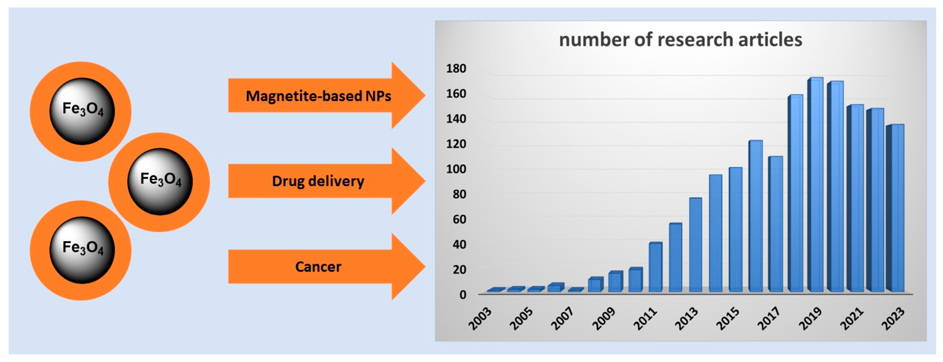
1. Introduction
1.1. Magnetite Nanoparticles for Biomedical Application
1.2. Assumptions of Targeted Drug Delivery Systems
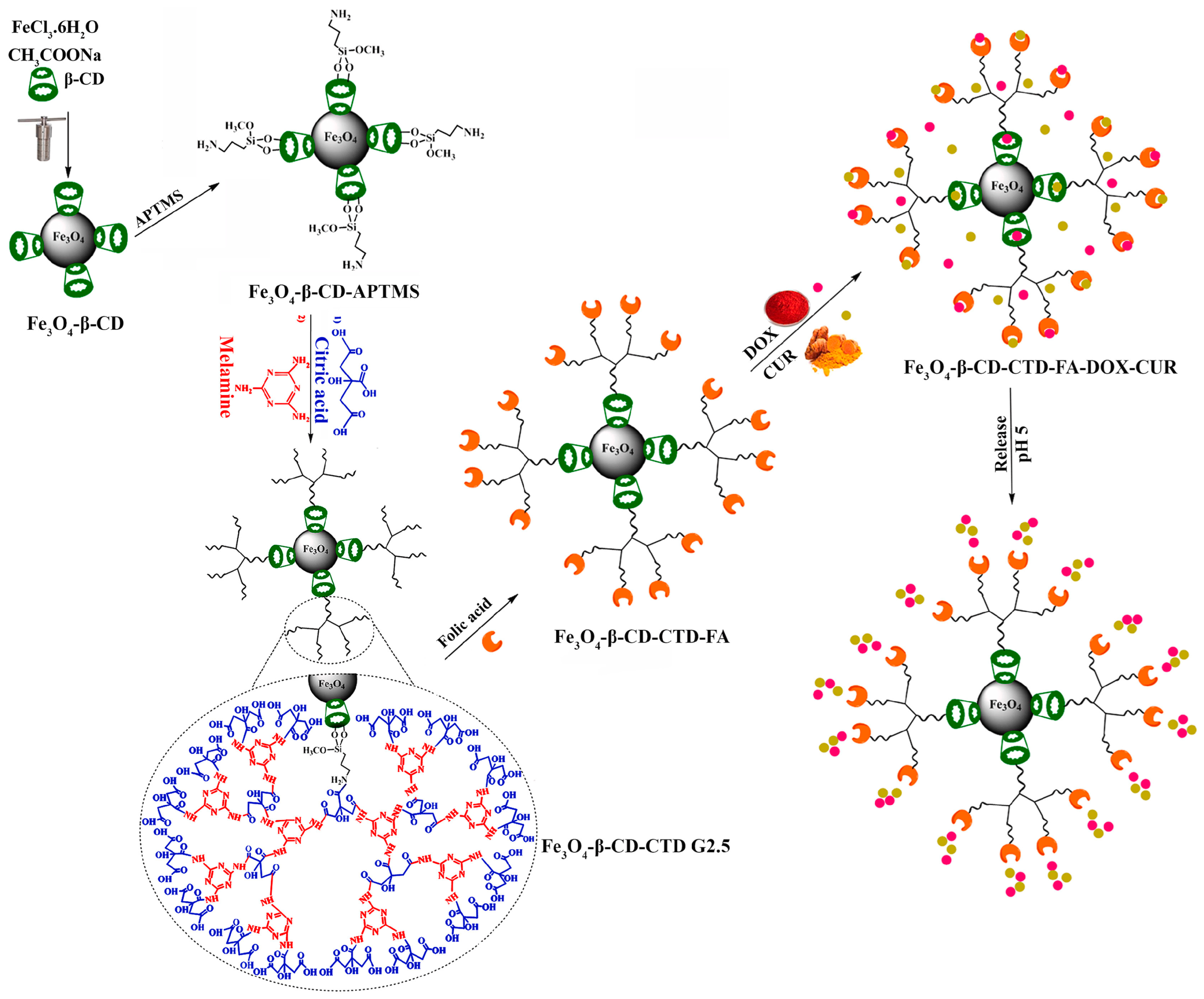
2. New Hybrid Systems for Potential Targeted Delivery

| Components of Magnetite-Based Delivery System | Fabrication/ Characterization | Loaded Drug | Responsiveness * | Reference |
|---|---|---|---|---|
| Methoxy poly(ethylene glycol) Chitosan | Commercial product/ NMR, TEM | DOX | Redox-responsive | [28] |
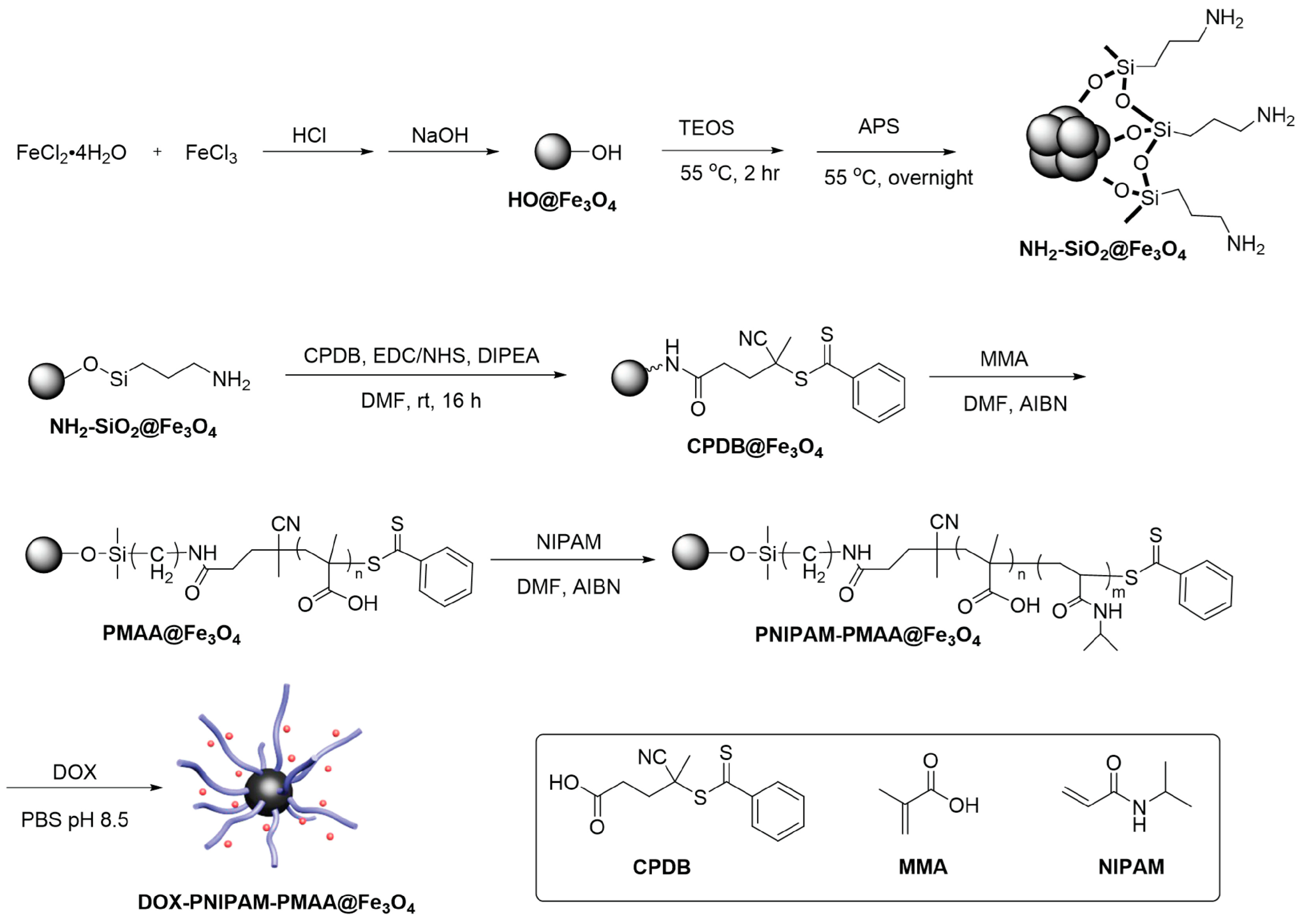
| Poly (methacrylic acid) Poly (N-Isopropylacrylamide) | Coprecipitation/ FTIR, TGA, SEM, DLS, zeta potential | DOX | pH-responsive Thermo-responsive | [29] |
| Poly (acrylic acid/acrylamide) | Coprecipitation/ FTIR, XRD, TEM | DOX | pH-responsive | [30] |
| Polyethylene glycol Graphene quantum dots | Coprecipitation/ FTIR, XRD, TEM, VSM, DLS, TGA, BET | DOX | pH-responsive | [31] |
| Triazine dendrimer Folic acid-derived quantum dots | Coprecipitation/ FTIR, XRD, SEM, EDX, VSM, N2 adsorption–desorption, BET, BJH, zeta potential | DOX | pH-responsive photoluminescent activity | [32] |
| Carbon Pluronic® F-127 | Solution combustion/ FTIR, XRD, TGA, N2 adsorption–desorption, BET, SEM, TEM, EDX, DLS | DOX | pH-responsive | [33] |
| N-(phosphonomethyl)iminodiacetic acid Silica | Coprecipitation/FTIR, TEM, EDX, VSM, zeta potential | DOX | Not available | [34] |
| Poly(ethylene glycol) diacrylate gelatin methyacryloyl | Commercial product/ SEM, EDX, XPS, VSM | DOX GEM | Redox-responsive Thermo-responsive | [36] |
| Silica Silver nanoparticles | Coprecipitation/ FTIR, XRD, TEM, EDX, TGA, N2 adsorption–desorption | EPI | Not available | [41] |
| Graphene oxide Cyanuric chloride | Coprecipitation/ FTIR, TEM, XRD, XPS, AFM | MTX | pH-responsive | [38] |
| Dimethylaminoethyl methacrylate κ-carrageenan | Microwave-induced coprecipitation/FTIR, XRD, TEM, VSM, DLS | 5-FU | pH-responsive Thermo-responsive | [39] |
| Molecularly imprinted polymer Fluorescence | Coprecipitation/FTIR, XRD, SEM, EDX, VSM | DTX | Not available | [40] |
3. Magnetite-Based Nanomaterials for Specific Cancer Treatment
3.1. Breast Cancer Treatment
| Components of Magnetite-Based Delivery System | Cell Model | Characterization of Anticancer Properties | Reference |
|---|---|---|---|
| SBA-15, L-cysteine, PEI, CD, FA | MCF7 | Drug release, cell viability, cellular uptake, biocompatibility | [46] |
| Tragacanth gum, FA | MCF7 | Drug release, cytotoxicity, cellular uptake | [47] |
| Oxidized alginate, cystamine | MCF7 | Interaction with human serum albumin, cytocompatibility, cytotoxicity | [48] |
| Carboxymethyl chitosan, aminated lignosulfonate | MCF7 | Drug release, cytotoxicity | [49] |
| Cationic molecularly imprinted polymer | 4T1 | Drug release, cytotoxicity, hemolysis assay In vivo: tumor induction, tumor volume, histopathological studies | [50] |
| poly(N-isopropyl acrylamide) | MCF7 | Drug release, biocompatibility, cell viability, cellular uptake | [51] |
| Polyvinyl alcohol, polyvinylpyrrolidone, FA, β-estradiol | MCF7 | Drug release, cell viability, cytotoxicity | [52] |
| CaCO3 | MCF7 | Drug release, cytotoxicity | [53] |
| Citric acid | 4T1 MDA MB 468 | Drug release, cytotoxicity | [54] |
| Diatoms | MCF7 | Drug release, biocompatibility, cytotoxicity | [55] |
| Chitosan, graphene oxide | MCF7 | Drug release, cytotoxicity | [59] |
| Polyethylene glycol | 4T1 | Drug release, enzyme-mimic activity, cell viability, apoptosis In vivo: tumor condition | [60] |
| Metal–organic framework | 4T1 | Drug release, cytotoxicity | [61] |
| Silica, metal–organic framework | MCF7 | Drug release, cytotoxicity | [62] |
| Au, cysteamine, aptamer | 4T1 MCF7 | Drug release, cytotoxicity | [63] |
| Hyaluronic acid, L-cysteine | MCF7 | Drug release, cytotoxicity | [64] |
| β-cyclodextrin, glycodendrimer, FA | MDA MB 231 | Drug release, cytotoxicity, cellular uptake, antioxidant activity | [65] |
| Chitosan, zein | MCF7 | Drug release, cytotoxicity, synergistic effect | [66] |
| Graphene quantum dots, FA | MCF7 MG63 | Drug release, cytotoxicity | [67] |
| Polyvinylpyrrolidone, graphene oxide | MDA MB 231 | Drug release, cytotoxicity | [68] |
3.2. Treatment of Other Cancers
| Components of Magnetite-Based Delivery System | Cell Model | Characterization of Anticancer Properties | Loaded Drug/ Targeted Cancer | Reference |
|---|---|---|---|---|
| Dextran, polylactic acid | H22 | Drug release, cytotoxicity In vivo: liver cancer inhibition | DOX/liver | [70] |
| Pluronic F127, polyethyleneimine | HepG2 | Drug release, cellular uptake | DOX/liver | [71] |
| Boron nitride, polyethylene glycol | HepG2 | Drug release, cytotoxicity, cellular uptakeIn vivo: tumor volume, histopathological studies | DOX/liver | [72] |
| Formyl deoxycholic acid, FA | HepG2 | Drug release, blood compatibility, cellular uptake, cytotoxicity, apoptosis staining, Western blotting | DOX/liver | [73] |
| Zn-Al layered double hydroxide | HepG2 | Drug release, cytotoxicity | DOX/liver | [74] |
| polyethylene glycol | HepG2 | Drug release, cytotoxicity | SFB/liver | [75] |
| Polyethylene glycol | HepG2 | Drug release, cytotoxicity, cellular uptake In vivo: under-skin implantation, 2D mapping, content in liver tissue | SFB/liver | [76] |
| Polyvinyl alcohol, Zn-Al layered double hydroxide | HepG2 | Drug release, cytotoxicity | 5-FU/liver | [77] |
| Polyethylene glycol, crocetin | HepG2 | Drug release, cytotoxicity | CRT/liver | [78] |
| Polyethylene glycol, Au, monoclonal antibodies | - | Drug release In vivo: histopathological studies | AVS/liver | [79] |
| Cellulose | HCT116 HT29 | Drug release, cytotoxicity, cell vitality, apoptosis induction, mitochondrial function, magnetic targeting | 5-FU/colon | [80] |
| Polyethylene glycol, poly-ε-caprolactone, FA | HT29 Caco-2 SW480 HCT116 | Drug release, cytotoxicity In vivo: cellular uptake, tumor volume, Western blotting | 5-FU/colon | [81] |
| Chitosan | CT26 | Cell viability | OXA&IRI/colon | [83] |
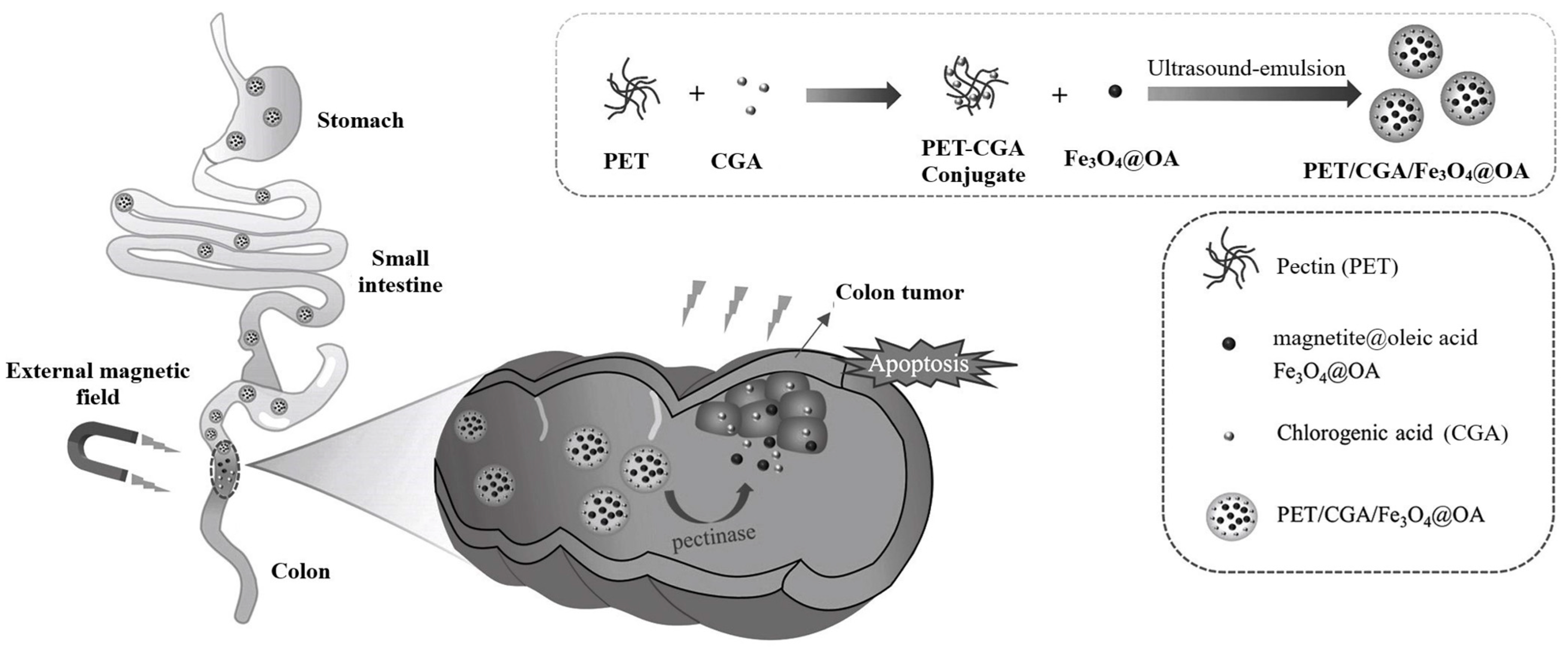
| Oleic acid, pectin | HCT116 | Drug release, cytotoxicity, cellular uptake | CGA/colon | [84] |
| Silica, ZIF-8, chitosan, FA | TC1 | Drug release In vivo: antitumor activity, pronecrotic effect | CPT/cervical | [86] |
| Polyglycerol, FA | HeLa | Drug release, cytotoxicity, cellular uptake | CUR/cervical | [87] |
| Oleic acid, chitosan, FA | HeLa | Drug release, cytotoxicity, cellular uptakeIn vivo: antitumor activity | DOX/cervical | [88] |
| Chitosan, FA | MG63 | Drug release, cell viability, cellular uptake, apoptosis induction | DOX/ osteosarcoma | [89] |
| Bioactive glass | MG63 | Drug release, cell viability, cellular uptake | DOX/ osteosarcoma | [90] |
| Graphene oxide, β-cyclodextrin | Saos2 MG63 | Drug release, cytotoxicity, cellular uptake, apoptosis induction, expression levels | DOX&MLT/ osteosarcoma | [91] |
| β-cyclodextrin | MG63 | Cell viability | PTX/ osteosarcoma | [92] |
| Albumin, perfluorohexane | AGS | Drug release, cytotoxicity, apoptosis induction | CPT/gastric | [93] |
| Chitosan, polyethylene glycol, FA | WEHI164 | Drug release, cytotoxicity, apoptosis induction In vivo: tumor volume | PTX/ fibrosarcoma | [94] |
| Aspartic acid, FA | B16 F1 | Drug release, cytotoxicity | DOX/skin | [95] |
| Chitosan | PC 3 | Drug release, cytotoxicity | TEL/prostate | [96] |
| Poly (lactic-co-glycolic) acid | A 498 | Drug release, cytotoxicity In vivo: acute toxicity | SIL/renal | [97] |
4. Multifunctional Magnetite-Based Nanomaterials
| Components of Magnetite-Based Delivery System | Application in Cancer Therapy * | Loaded Drug/Targeted Cancer | Reference |
|---|---|---|---|
| Tragacanth gum, poly acrylic acid, cystamine, FA | HT | DOX/breast | [98] |
| β-cyclodextrin, poly(2-ethyl-2-oxazoline) | HT | DOX/breast | [99] |
| Poly (lactic-co-glycolic acid), alginate | MHT | DOX/lung | [100] |
| Graphene oxide, maltodextrin, FA | PTT | DOX/liver | [101] |
| Carbon, ZnO, FA | PTT | DOX/liver | [102] |
| Hydroxypropyl cellulose, carboxymethyl chitosan, black phosphorus | PTT | ART/liver | [103] |
| Graphene oxide, β-cyclodextrin, cholic acid, hyaluronic acid | PTT | CPT/liver | [104] |
| Graphene oxide, hydroxypropyl cellulose | PTT | PTX/breast | [105] |
| liposome | PTT | BFL/breast | [106] |
| Gelatin, CuS, FA | PTT | CUR/breast | [107] |
| Poly(ethylene)glycol-poly(β-amino esters), dipalmitoyl phosphatidylcholine | MHT, PTT | DOX/breast | [108] |
| Poly(N-isopropylacrylamide-acrylic acid-(ethylene glycol) methacrylate), herceptin | MRI | DOX/breast | [109] |
| Carbon quantum dots | MRI | DOX/breast | [110] |
| Poly(glycidyl methacrylate-polyethylene glycol), salep dialdehyde | MRI | DOX/breast | [111] |
| β-cyclodextrin, Pep42 | MRI | DOX/breast | [112] |
| Poly (ε-caprolactone), chitosan | MRI | GEM/breast | [113] |
| Polyvinylpyrrolidone | MRI, MHT | DOX/osteosarcoma | [114] |
| Punica granatum fruit peel extract | MRI, MHT | 5-FU/colorectal | [115] |
| Gold nanoparticles | MRI, PTT | DOX/cervical | [116] |
| Polyethylene glycol, poly(lactic-co-glycolic acid), Prussian blue | MRI, PAT, USI, PTT | DOX/osteosarcoma | [117] |
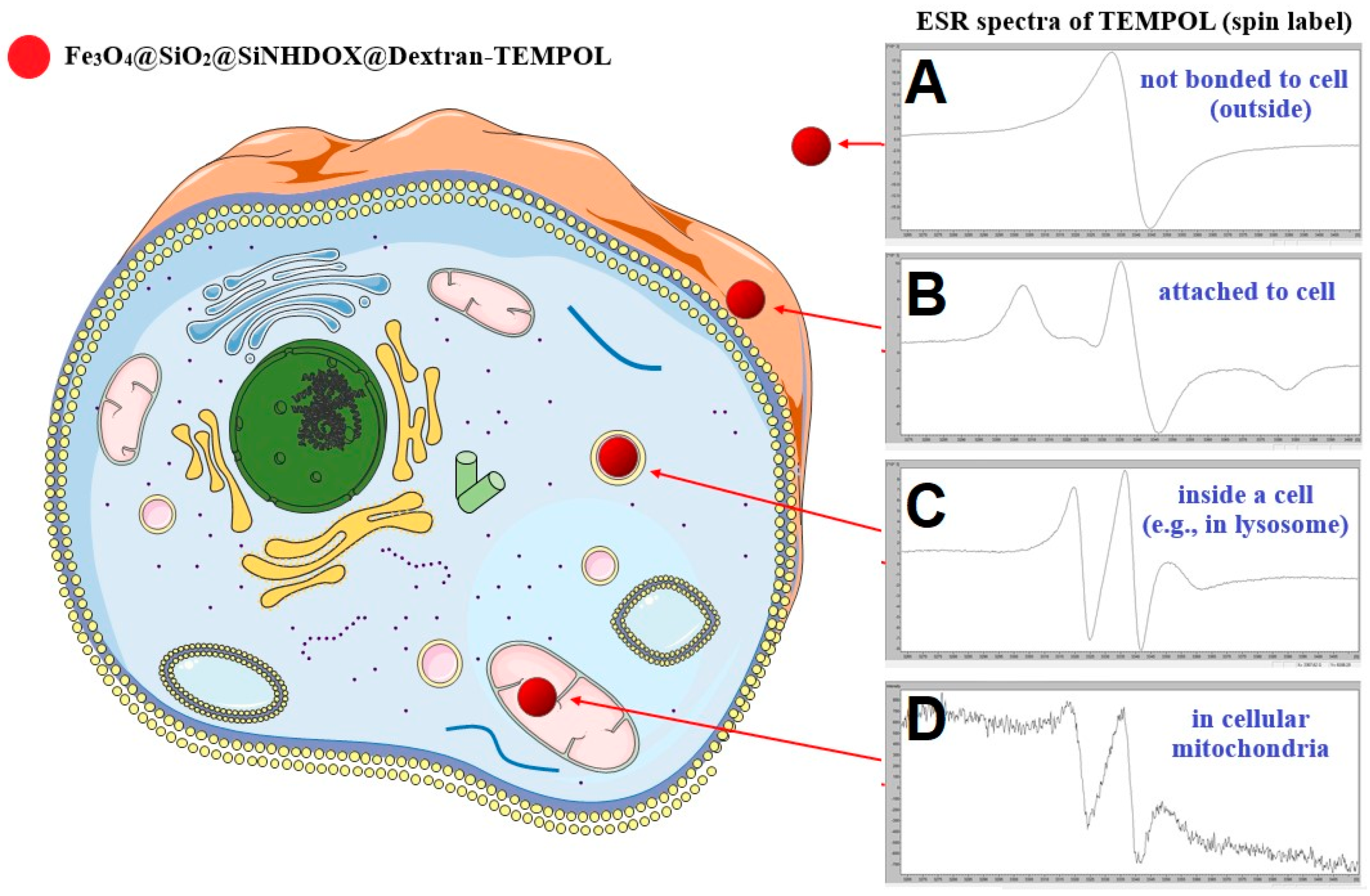
5. Monitoring of Magnetite-Based Nanocarriers Interacting with Cells in Biological Systems
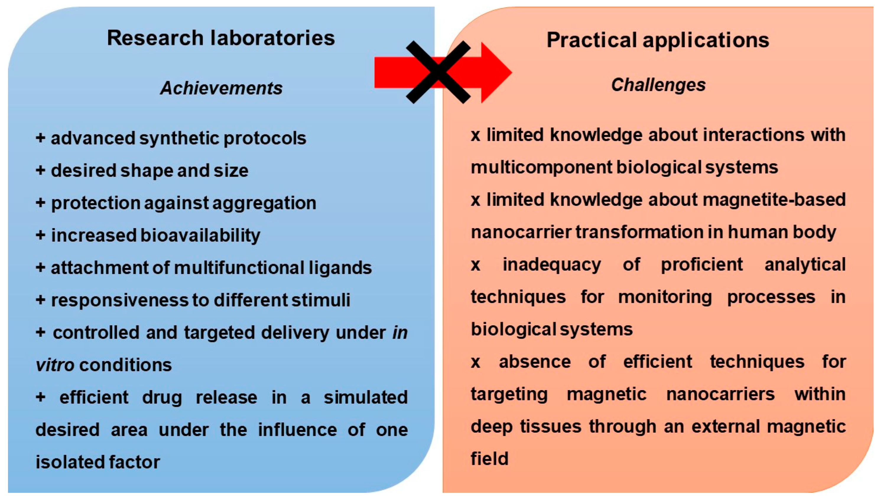
6. Perspectives for Future Studies
7. Conclusions
Author Contributions
Funding
Institutional Review Board Statement
Informed Consent Statement
Data Availability Statement
Conflicts of Interest
References
- Shabatina, T.I.; Vernaya, O.I.; Shabatin, V.P.; Melnikov, M.Y. Magnetic Nanoparticles for Biomedical Purposes: Modern Trends and Prospects. Magnetochemistry 2020, 6, 30. [Google Scholar] [CrossRef]
- Rukhsar, M.; Ahmad, Z.; Rauf, A.; Zeb, H.; Ur-Rehman, M.; Hemeg, H.A. An Overview of Iron Oxide (Fe3O4) Nanoparticles: From Synthetic Strategies, Characterization to Antibacterial and Anticancer Applications. Crystals 2022, 12, 1809. [Google Scholar] [CrossRef]
- Shabestari Khiabani, S.; Farshbaf, M.; Akbarzadeh, A.; Davaran, S. Magnetic Nanoparticles: Preparation Methods, Applications in Cancer Diagnosis and Cancer Therapy. Artif. Cells Nanomed. Biotechnol. 2017, 45, 6–17. [Google Scholar] [CrossRef] [PubMed]
- Fatmawati, T.; Shiddiq, M.; Armynah, B.; Tahir, D. Synthesis Methods of Fe3O4 Nanoparticles for Biomedical Applications. Chem. Eng. Technol. 2023, 46, 2356–2366. [Google Scholar] [CrossRef]
- Dudchenko, N.; Pawar, S.; Perelshtein, I.; Fixler, D. Magnetite Nanoparticles: Synthesis and Applications in Optics and Nanophotonics. Materials 2022, 15, 2601. [Google Scholar] [CrossRef]
- Rao, M.S.; Rao, C.S.; Kumari, A.S. Synthesis, Stability, and Emission Analysis of Magnetite Nanoparticle-Based Biofuels. J. Eng. Appl. Sci. 2022, 69, 79. [Google Scholar] [CrossRef]
- Kritika; Roy, I. Therapeutic Applications of Magnetic Nanoparticles: Recent Advances. Mater. Adv. 2022, 3, 7425–7444. [Google Scholar] [CrossRef]
- Al-Madhagi, H.; Yazbik, V.; Abdelwahed, W.; Alchab, L. Magnetite Nanoparticle Co-Precipitation Synthesis, Characterization, and Applications: Mini Review. BioNanoScience 2023, 13, 853–859. [Google Scholar] [CrossRef]
- Gallo-Cordova, A.; Almeida Streitwieser, D.; Del Puerto Morales, M.; Ovejero, J.G. Magnetic Iron Oxide Colloids for Environmental Applications. In Colloids—Types, Preparation and Applications; Nageeb Rashed, M., Ed.; IntechOpen: London, UK, 2021; ISBN 978-1-83962-969-3. [Google Scholar]
- Ajinkya, N.; Yu, X.; Kaithal, P.; Luo, H.; Somani, P.; Ramakrishna, S. Magnetic Iron Oxide Nanoparticle (IONP) Synthesis to Applications: Present and Future. Materials 2020, 13, 4644. [Google Scholar] [CrossRef]
- Montiel Schneider, M.G.; Martín, M.J.; Otarola, J.; Vakarelska, E.; Simeonov, V.; Lassalle, V.; Nedyalkova, M. Biomedical Applications of Iron Oxide Nanoparticles: Current Insights Progress and Perspectives. Pharmaceutics 2022, 14, 204. [Google Scholar] [CrossRef]
- Khizar, S.; Ahmad, N.M.; Zine, N.; Jaffrezic-Renault, N.; Errachid-El-Salhi, A.; Elaissari, A. Magnetic Nanoparticles: From Synthesis to Theranostic Applications. ACS Appl. Nano Mater. 2021, 4, 4284–4306. [Google Scholar] [CrossRef]
- Tran, H.-V.; Ngo, N.M.; Medhi, R.; Srinoi, P.; Liu, T.; Rittikulsittichai, S.; Lee, T.R. Multifunctional Iron Oxide Magnetic Nanoparticles for Biomedical Applications: A Review. Materials 2022, 15, 503. [Google Scholar] [CrossRef] [PubMed]
- Campana, L.G.; Falci, C.; Basso, M.; Sieni, E.; Dughiero, F. Clinical Electrochemotherapy for Chest Wall Recurrence from Breast Cancer. In Electroporation-Based Therapies for Cancer; Elsevier: Amsterdam, The Netherlands, 2014; pp. 3–33. ISBN 978-1-907568-15-2. [Google Scholar]
- Wagalgave, S.M.; Birajdar, S.S.; Malegaonkar, J.N.; Bhosale, S.V. Patented AIE Materials for Biomedical Applications. In Progress in Molecular Biology and Translational Science; Elsevier: Amsterdam, The Netherlands, 2021; Volume 185, pp. 199–223. ISBN 978-0-323-99604-4. [Google Scholar]
- Gao, P.; Wang, H.; Cheng, Y. Strategies for Efficient Photothermal Therapy at Mild Temperatures: Progresses and Challenges. Chin. Chem. Lett. 2022, 33, 575–586. [Google Scholar] [CrossRef]
- Chan, R.W.; Lau, J.Y.C.; Lam, W.W.; Lau, A.Z. Magnetic Resonance Imaging. In Encyclopedia of Biomedical Engineering; Elsevier: Amsterdam, The Netherlands, 2019; pp. 574–587. ISBN 978-0-12-805144-3. [Google Scholar]
- Avasthi, A.; Caro, C.; Pozo-Torres, E.; Leal, M.P.; García-Martín, M.L. Magnetic Nanoparticles as MRI Contrast Agents. Top. Curr. Chem. 2020, 378, 40. [Google Scholar] [CrossRef] [PubMed]
- Ezike, T.C.; Okpala, U.S.; Onoja, U.L.; Nwike, C.P.; Ezeako, E.C.; Okpara, O.J.; Okoroafor, C.C.; Eze, S.C.; Kalu, O.L.; Odoh, E.C.; et al. Advances in Drug Delivery Systems, Challenges and Future Directions. Heliyon 2023, 9, e17488. [Google Scholar] [CrossRef] [PubMed]
- Vargason, A.M.; Anselmo, A.C.; Mitragotri, S. The Evolution of Commercial Drug Delivery Technologies. Nat. Biomed. Eng. 2021, 5, 951–967. [Google Scholar] [CrossRef]
- Adepu, S.; Ramakrishna, S. Controlled Drug Delivery Systems: Current Status and Future Directions. Molecules 2021, 26, 5905. [Google Scholar] [CrossRef]
- Sahini, M.G.; Banyikwa, A.T. Superparamagnetic Iron Oxide Nanoparticles for Drug Delivery Applications. In Advanced and Modern Approaches for Drug Delivery; Elsevier: Amsterdam, The Netherlands, 2023; pp. 817–850. ISBN 978-0-323-91668-4. [Google Scholar]
- Anik, M.I.; Hossain, M.K.; Hossain, I.; Mahfuz, A.M.U.B.; Rahman, M.T.; Ahmed, I. Recent Progress of Magnetic Nanoparticles in Biomedical Applications: A Review. Nano Sel. 2021, 2, 1146–1186. [Google Scholar] [CrossRef]
- Malhotra, N.; Lee, J.-S.; Liman, R.A.D.; Ruallo, J.M.S.; Villaflores, O.B.; Ger, T.-R.; Hsiao, C.-D. Potential Toxicity of Iron Oxide Magnetic Nanoparticles: A Review. Molecules 2020, 25, 3159. [Google Scholar] [CrossRef]
- Frtús, A.; Smolková, B.; Uzhytchak, M.; Lunova, M.; Jirsa, M.; Kubinová, Š.; Dejneka, A.; Lunov, O. Analyzing the Mechanisms of Iron Oxide Nanoparticles Interactions with Cells: A Road from Failure to Success in Clinical Applications. J. Control. Release 2020, 328, 59–77. [Google Scholar] [CrossRef]
- Wang, F.; Geng, J.; Qi, X.; Zhang, P.; Zhang, H.; He, X.; Li, Z.; Yu, R.; Li, J.; Li, B.; et al. Facile Solvothermal Synthesis of Monodisperse Superparamagnetic Mesoporous Fe3O4 Nanospheres for pH-Responsive Controlled Drug Delivery. Colloids Surf. A 2021, 622, 126643. [Google Scholar] [CrossRef]
- Wang, F.; Qi, X.; Geng, J.; Liu, X.; Li, D.; Zhang, H.; Zhang, P.; He, X.; Li, B.; Li, Z.; et al. Template-Free Construction of Hollow Mesoporous Fe3O4 Nanospheres as Controlled Drug Delivery with Enhanced Drug Loading Capacity. J. Mol. Liq. 2022, 347, 118000. [Google Scholar] [CrossRef]
- Yoon, H.-M.; Kang, M.-S.; Choi, G.-E.; Kim, Y.-J.; Bae, C.-H.; Yu, Y.-B.; Jeong, Y.-I. Stimuli-Responsive Drug Delivery of Doxorubicin Using Magnetic Nanoparticle Conjugated Poly(Ethylene Glycol)-g-Chitosan Copolymer. Int. J. Mol. Sci. 2021, 22, 13169. [Google Scholar] [CrossRef]
- Wu, T.-C.; Lee, P.-Y.; Lai, C.-L.; Lai, C.-H. Synthesis of Multi-Functional Nano-Vectors for Target-Specific Drug Delivery. Polymers 2021, 13, 451. [Google Scholar] [CrossRef] [PubMed]
- Hosny, N.M.; Abbass, M.; Ismail, F.; El-Din, H.M.N. Radiation Synthesis and Anticancer Drug Delivery of Poly(Acrylic Acid/Acrylamide) Magnetite Hydrogel. Polym. Bull. 2023, 80, 4573–4588. [Google Scholar] [CrossRef]
- Javadian, S.; Najafi, K.; Sadrpoor, S.M.; Ektefa, F.; Dalir, N.; Nikkhah, M. Graphene Quantum Dots Based Magnetic Nanoparticles as a Promising Delivery System for Controlled Doxorubicin Release. J. Mol. Liq. 2021, 331, 115746. [Google Scholar] [CrossRef]
- Karimi, S.; Namazi, H. A Photoluminescent Folic Acid-Derived Carbon Dot Functionalized Magnetic Dendrimer as a pH-Responsive Carrier for Targeted Doxorubicin Delivery. New J. Chem. 2021, 45, 6397–6405. [Google Scholar] [CrossRef]
- Silva, A.S.; Diaz De Tuesta, J.L.; Sayuri Berberich, T.; Delezuk Inglez, S.; Bertão, A.R.; Çaha, I.; Deepak, F.L.; Bañobre-López, M.; Gomes, H.T. Doxorubicin Delivery Performance of Superparamagnetic Carbon Multi-Core Shell Nanoparticles: pH Dependence, Stability and Kinetic Insight. Nanoscale 2022, 14, 7220–7232. [Google Scholar] [CrossRef] [PubMed]
- Demin, A.M.; Vakhrushev, A.V.; Valova, M.S.; Korolyova, M.A.; Uimin, M.A.; Minin, A.S.; Pozdina, V.A.; Byzov, I.V.; Tumashov, A.A.; Chistyakov, K.A.; et al. Effect of the Silica–Magnetite Nanocomposite Coating Functionalization on the Doxorubicin Sorption/Desorption. Pharmaceutics 2022, 14, 2271. [Google Scholar] [CrossRef]
- Li, Y.; Dong, D.; Qu, Y.; Li, J.; Chen, S.; Zhao, H.; Zhang, Q.; Jiao, Y.; Fan, L.; Sun, D. A Multidrug Delivery Microrobot for the Synergistic Treatment of Cancer. Small 2023, 19, 2301889. [Google Scholar] [CrossRef]
- Lee, H.; Park, S. Magnetically Actuated Helical Microrobot with Magnetic Nanoparticle Retrieval and Sequential Dual-Drug Release Abilities. ACS Appl. Mater. Interfaces 2023, 15, 27471–27485. [Google Scholar] [CrossRef]
- Khasraw, M.; Bell, R.; Dang, C. Epirubicin: Is It like Doxorubicin in Breast Cancer? A Clinical Review. Breast 2012, 21, 142–149. [Google Scholar] [CrossRef]
- Taheri-Kafrani, A.; Shirzadfar, H.; Abbasi Kajani, A.; Kudhair, B.K.; Jasim Mohammed, L.; Mohammadi, S.; Lotfi, F. Functionalized Graphene Oxide/Fe3O4 Nanocomposite: A Biocompatible and Robust Nanocarrier for Targeted Delivery and Release of Anticancer Agents. J. Biotechnol. 2021, 331, 26–36. [Google Scholar] [CrossRef] [PubMed]
- Geyik, G.; Işıklan, N. Multi-Stimuli-Sensitive Superparamagnetic κ-Carrageenan Based Nanoparticles for Controlled 5-Fluorouracil Delivery. Colloids Surf. A 2022, 634, 127960. [Google Scholar] [CrossRef]
- Ali, Z.; Sajid, M.; Ahmed, M.M.; Hanif, M.; Manzoor, S. Synthesis of Green Fluorescent Cross-Linked Molecularly Imprinted Polymer Bound with Anti-Cancerous Drug (Docetaxel) for Targeted Drug Delivery. Polym. Bull. 2024, 81, 679–696. [Google Scholar] [CrossRef]
- Romdoni, Y.; Kadja, G.T.M.; Kitamoto, Y.; Khalil, M. Synthesis of Multifunctional Fe3O4@SiO2-Ag Nanocomposite for Antibacterial and Anticancer Drug Delivery. Appl. Surf. Sci. 2023, 610, 155610. [Google Scholar] [CrossRef]
- Bray, F.; Ferlay, J.; Soerjomataram, I.; Siegel, R.L.; Torre, L.A.; Jemal, A. Global Cancer Statistics 2018: GLOBOCAN Estimates of Incidence and Mortality Worldwide for 36 Cancers in 185 Countries. CA Cancer J. Clin. 2018, 68, 394–424. [Google Scholar] [CrossRef]
- Kreis, K.; Plöthner, M.; Schmidt, T.; Seufert, R.; Schreeb, K.; Jahndel, V.; Maas, S.; Kuhlmann, A.; Zeidler, J.; Schramm, A. Healthcare Costs Associated with Breast Cancer in Germany: A Claims Data Analysis. Eur. J. Health Econ. 2020, 21, 451–464. [Google Scholar] [CrossRef]
- Kurczewska, J. Chitosan-Based Nanoparticles with Optimized Parameters for Targeted Delivery of a Specific Anticancer Drug—A Comprehensive Review. Pharmaceutics 2023, 15, 503. [Google Scholar] [CrossRef]
- Ferreira, L.L.; Oliveira, P.J.; Cunha-Oliveira, T. Epigenetics in Doxorubicin Cardiotoxicity. In Pharmacoepigenetics; Elsevier: Amsterdam, The Netherlands, 2019; pp. 837–846. ISBN 978-0-12-813939-4. [Google Scholar]
- Ehsanimehr, S.; Moghadam, P.N.; Dehaen, W.; Irannejad, V.S. PEI Grafted Fe3O4@SiO2@SBA-15 Labeled FA as a pH-Sensitive Mesoporous Magnetic and Biocompatible Nanocarrier for Targeted Delivery of Doxorubicin to MCF-7 Cell Line. Colloids Surf. A 2021, 615, 126302. [Google Scholar] [CrossRef]
- Jalali, S.; Moghadam, P.N.; Shafiei-Irannejad, V. Synthesis of Magnetic Nanocarrier Conjugated by Folate Based on Tragacanth and In Vitro Investigation of Their Efficiency on Breast Cancer Cells. Starch 2023, 75, 2200092. [Google Scholar] [CrossRef]
- Parvaresh, A.; Izadi, Z.; Nemati, H.; Derakhshankhah, H.; Jaymand, M. Redox- and pH-Responsive Alginate-Based Magnetic Hydrogel: “Smart” Drug Delivery and Protein Corona Studies. J. Mol. Liq. 2023, 382, 121990. [Google Scholar] [CrossRef]
- Liu, Q.; Tan, Z.; Zheng, D.; Qiu, X. pH-Responsive Magnetic Fe3O4/Carboxymethyl Chitosan/Aminated Lignosulfonate Nanoparticles with Uniform Size for Targeted Drug Loading. Int. J. Biol. Macromol. 2023, 225, 1182–1192. [Google Scholar] [CrossRef] [PubMed]
- Naghaviyan, A.; Hashemi-Moghaddam, H.; Zavareh, S.; Ebrahimi Verkiani, M.; Meuller, A. Synergistic Effect Evaluation of Magnetotherapy and a Cationic–Magnetic Nanocomposite Loaded with Doxorubicin for Targeted Drug Delivery to Breast Adenocarcinoma. Mol. Pharm. 2023, 20, 101–117. [Google Scholar] [CrossRef]
- Radu, I.-C.; Mirica, A.-C.I.; Hudita, A.; Tanasa, E.; Iovu, H.; Zaharia, C.; Galateanu, B. Thermosensitive Behavior Defines the Features of Poly(N-Isopropylacrylamide)/Magnetite Nanoparticles for Cancer Management. Appl. Sci. 2023, 13, 4870. [Google Scholar] [CrossRef]
- Bekaroğlu, M.G.; Kiriş, A.; Başer, H.N.; İşçi, S. Stabilizer Effect of Tumor-Targeting Ligands on the Drug Delivering Fe3O4 Nanoparticles. Appl. Phys. A 2023, 129, 182. [Google Scholar] [CrossRef]
- Popova, V.; Poletaeva, Y.; Chubarov, A.; Dmitrienko, E. pH-Responsible Doxorubicin-Loaded Fe3O4@CaCO3 Nanocomposites for Cancer Treatment. Pharmaceutics 2023, 15, 771. [Google Scholar] [CrossRef]
- Markhulia, J.; Kekutia, S.; Mikelashvili, V.; Saneblidze, L.; Tsertsvadze, T.; Maisuradze, N.; Leladze, N.; Czigány, Z.; Almásy, L. Synthesis, Characterization, and In Vitro Cytotoxicity Evaluation of Doxorubicin-Loaded Magnetite Nanoparticles on Triple-Negative Breast Cancer Cell Lines. Pharmaceutics 2023, 15, 1758. [Google Scholar] [CrossRef] [PubMed]
- Li, M.; Wu, J.; Lin, D.; Yang, J.; Jiao, N.; Wang, Y.; Liu, L. A Diatom-Based Biohybrid Microrobot with a High Drug-Loading Capacity and pH-Sensitive Drug Release for Target Therapy. Acta Biomater. 2022, 154, 443–453. [Google Scholar] [CrossRef]
- Gong, D.; Celi, N.; Zhang, D.; Cai, J. Magnetic Biohybrid Microrobot Multimers Based on Chlorella Cells for Enhanced Targeted Drug Delivery. ACS Appl. Mater. Interfaces 2022, 14, 6320–6330. [Google Scholar] [CrossRef]
- Zhu, Y.; Jia, H.; Jiang, Y.; Guo, Y.; Duan, Q.; Xu, K.; Shan, B.; Liu, X.; Chen, X.; Wu, F. A Red Blood Cell-derived Bionic Microrobot Capable of Hierarchically Adapting to Five Critical Stages in Systemic Drug Delivery. Exploration 2023, 20230105. [Google Scholar] [CrossRef]
- Gong, D.; Sun, L.; Li, X.; Zhang, W.; Zhang, D.; Cai, J. Micro/Nanofabrication, Assembly, and Actuation Based on Microorganisms: Recent Advances and Perspectives. Small Struct. 2023, 4, 2200356. [Google Scholar] [CrossRef]
- Ramadan, I.; Nassar, M.Y.; Gomaa, A. In-Vitro Investigation of the Anticancer Efficacy of Carboplatin-Loaded Chitosan Nanocomposites Against Breast and Liver Cancer Cell Lines. J. Polym. Environ. 2023, 31, 1102–1115. [Google Scholar] [CrossRef]
- Nie, Z.; Vahdani, Y.; Cho, W.C.; Bloukh, S.H.; Edis, Z.; Haghighat, S.; Falahati, M.; Kheradmandi, R.; Jaragh-Alhadad, L.A.; Sharifi, M. 5-Fluorouracil-Containing Inorganic Iron Oxide/Platinum Nanozymes with Dual Drug Delivery and Enzyme-like Activity for the Treatment of Breast Cancer. Arab. J. Chem. 2022, 15, 103966. [Google Scholar] [CrossRef]
- Li, J.; Zhou, Y.; Yan, S.; Wu, W.; Sharifi, M. Core-Shell Iron Oxide-Platinium@metal Organic Framework/Epirubicin Nanospheres: Synthesis, Characterization and Anti-Breast Cancer Activity. Arab. J. Chem. 2023, 16, 105229. [Google Scholar] [CrossRef]
- Parsa, F.; Setoodehkhah, M.; Atyabi, S.M. Loading and Release Study of Ciprofloxacin from Silica-Coated Magnetite Modified by Iron-Based Metal-Organic Framework (MOF) as a Nonocarrier in Targeted Drug Delivery System. Inorg. Chem. Commun. 2023, 155, 111056. [Google Scholar] [CrossRef]
- Khodadadi, E.; Mahjoub, S.; Arabi, M.S.; Najafzadehvarzi, H.; Nasirian, V. Fabrication and Evaluation of Aptamer-Conjugated Paclitaxel-Loaded Magnetic Nanoparticles for Targeted Therapy on Breast Cancer Cells. Mol. Biol. Rep. 2021, 48, 2105–2116. [Google Scholar] [CrossRef] [PubMed]
- Cadena Castro, D.; Gatti, G.; Martín, S.E.; Uberman, P.M.; García, M.C. Promising Tamoxifen-Loaded Biocompatible Hybrid Magnetic Nanoplatforms against Breast Cancer Cells: Synthesis, Characterization and Biological Evaluation. New J. Chem. 2021, 45, 4032–4045. [Google Scholar] [CrossRef]
- Karimi, S.; Namazi, H. Synthesis of Folic Acid-Conjugated Glycodendrimer with Magnetic β-Cyclodextrin Core as a pH-Responsive System for Tumor-Targeted Co-Delivery of Doxorubicin and Curcumin. Colloids Surf. A 2021, 627, 127205. [Google Scholar] [CrossRef]
- Ahmadi, F.; Akbari, J.; Saeedi, M.; Seyedabadi, M.; Ebrahimnejad, P.; Ghasemi, S.; Nokhodchi, A. Efficient Synergistic Combination Effect of Curcumin with Piperine by Polymeric Magnetic Nanoparticles for Breast Cancer Treatment. J. Drug Deliv. Sci. Technol. 2023, 86, 104624. [Google Scholar] [CrossRef]
- Seyyedi Zadeh, E.; Ghanbari, N.; Salehi, Z.; Derakhti, S.; Amoabediny, G.; Akbari, M.; Asadi Tokmedash, M. Smart pH-Responsive Magnetic Graphene Quantum Dots Nanocarriers for Anticancer Drug Delivery of Curcumin. Mater. Chem. Phys. 2023, 297, 127336. [Google Scholar] [CrossRef]
- Matiyani, M.; Rana, A.; Pal, M.; Rana, S.; Melkani, A.B.; Sahoo, N.G. Polymer Grafted Magnetic Graphene Oxide as a Potential Nanocarrier for pH-Responsive Delivery of Sparingly Soluble Quercetin against Breast Cancer Cells. RSC Adv. 2022, 12, 2574–2588. [Google Scholar] [CrossRef] [PubMed]
- Yang, W.-S.; Zeng, X.-F.; Liu, Z.-N.; Zhao, Q.-H.; Tan, Y.-T.; Gao, J.; Li, H.-L.; Xiang, Y.-B. Diet and Liver Cancer Risk: A Narrative Review of Epidemiological Evidence. Br. J. Nutr. 2020, 124, 330–340. [Google Scholar] [CrossRef] [PubMed]
- Wang, L.; Liang, L.; Shi, S.; Wang, C. Study on the Application of Doxorubicin-Loaded Magnetic Nanodrugs in Targeted Therapy of Liver Cancer. Appl. Bionics Biomech. 2022, 2022, 2756459. [Google Scholar] [CrossRef]
- Mdlovu, N.V.; Lin, K.-S.; Weng, M.-T.; Lin, Y.-S. Design of Doxorubicin Encapsulated pH-/Thermo-Responsive and Cationic Shell-Crosslinked Magnetic Drug Delivery System. Colloids Surf. B 2022, 209, 112168. [Google Scholar] [CrossRef]
- Carrera Espinoza, M.J.; Lin, K.-S.; Weng, M.-T.; Kunene, S.C.; Lin, Y.-S.; Liu, S.-Y. Magnetic Boron Nitride Nanosheets-Based on pH-Responsive Smart Nanocarriers for the Delivery of Doxorubicin for Liver Cancer Treatment. Colloids Surf. B 2023, 222, 113129. [Google Scholar] [CrossRef]
- Wang, X.; Ma, Q.; Wen, C.; Gong, T.; Li, J.; Liang, W.; Li, M.; Wang, Y.; Guo, R. Folic Acid and Deoxycholic Acid Derivative Modified Fe3O4 Nanoparticles for Efficient pH-Dependent Drug Release and Multi-Targeting against Liver Cancer Cells. RSC Adv. 2021, 11, 39804–39812. [Google Scholar] [CrossRef]
- Chai, J.; Ma, Y.; Guo, T.; He, Y.; Wang, G.; Si, F.; Geng, J.; Qi, X.; Chang, G.; Ren, Z.; et al. Assembled Fe3O4 Nanoparticles on Zn Al LDH Nanosheets as a Biocompatible Drug Delivery Vehicle for pH-Responsive Drug Release and Enhanced Anticancer Activity. Appl. Clay Sci. 2022, 228, 106630. [Google Scholar] [CrossRef]
- Ebadi, M.; Rifqi Md Zain, A.; Tengku Abdul Aziz, T.H.; Mohammadi, H.; Tee, C.A.T.; Rahimi Yusop, M. Formulation and Characterization of Fe3O4@PEG Nanoparticles Loaded Sorafenib; Molecular Studies and Evaluation of Cytotoxicity in Liver Cancer Cell Lines. Polymers 2023, 15, 971. [Google Scholar] [CrossRef] [PubMed]
- Iacobazzi, R.M.; Vischio, F.; Arduino, I.; Canepa, F.; Laquintana, V.; Notarnicola, M.; Scavo, M.P.; Bianco, G.; Fanizza, E.; Lopedota, A.A.; et al. Magnetic Implants In Vivo Guiding Sorafenib Liver Delivery by Superparamagnetic Solid Lipid Nanoparticles. J. Colloid Interface Sci. 2022, 608, 239–254. [Google Scholar] [CrossRef]
- Ebadi, M.; Bullo, S.; Buskaran, K.; Hussein, M.Z.; Fakurazi, S.; Pastorin, G. Dual-Functional Iron Oxide Nanoparticles Coated with Polyvinyl Alcohol/5-Fluorouracil/Zinc-Aluminium-Layered Double Hydroxide for a Simultaneous Drug and Target Delivery System. Polymers 2021, 13, 855. [Google Scholar] [CrossRef]
- Ibrahim, S.; Baig, B.; Hisaindee, S.; Darwish, H.; Abdel-Ghany, A.; El-Maghraby, H.; Amin, A.; Greish, Y. Development and Evaluation of Crocetin-Functionalized Pegylated Magnetite Nanoparticles for Hepatocellular Carcinoma. Molecules 2023, 28, 2882. [Google Scholar] [CrossRef]
- Mansour, W.; El Fedawy, S.F.; Atta, S.A.; Zarie, R.M.; Fouad, N.T.A.; Maher, S.; Hussein, T.M.; Abdel Aziz, D.M.; Kamel, M. Targeted Therapy for HCC Using Dumbbell-like Nanoparticles Conjugated to Monoclonal Antibodies against VEGF and Cancer Stem Cell Receptors in Mice. Cancer Nano. 2023, 14, 14. [Google Scholar] [CrossRef]
- Yusefi, M.; Lee-Kiun, M.S.; Shameli, K.; Teow, S.-Y.; Ali, R.R.; Siew, K.-K.; Chan, H.-Y.; Wong, M.M.-T.; Lim, W.-L.; Kuča, K. 5-Fluorouracil Loaded Magnetic Cellulose Bionanocomposites for Potential Colorectal Cancer Treatment. Carbohydr. Polym. 2021, 273, 118523. [Google Scholar] [CrossRef] [PubMed]
- Mirzaghavami, P.S.; Khoei, S.; Khoee, S.; Shirvalilou, S. Folic Acid-Conjugated Magnetic Triblock Copolymer Nanoparticles for Dual Targeted Delivery of 5-Fluorouracil to Colon Cancer Cells. Cancer Nano. 2022, 13, 12. [Google Scholar] [CrossRef]
- Shirvalilou, S.; Khoee, S.; Khoei, S.; Karimi, M.R.; Sadri, E.; Shirvaliloo, M. Targeted Magnetochemotherapy Modified by 5-Fu-Loaded Thermally on/off Switching Nanoheaters for the Eradication of CT26 Murine Colon Cancer by Inducing Apoptotic and Autophagic Cell Death. Cancer Nano. 2023, 14, 11. [Google Scholar] [CrossRef]
- Farmanbar, N.; Mohseni, S.; Darroudi, M. Green Synthesis of Chitosan-Coated Magnetic Nanoparticles for Drug Delivery of Oxaliplatin and Irinotecan against Colorectal Cancer Cells. Polym. Bull. 2022, 79, 10595–10613. [Google Scholar] [CrossRef]
- Zhu, H.; Zhang, L.; Kou, F.; Zhao, J.; Lei, J.; He, J. Targeted Therapeutic Effects of Oral Magnetically Driven Pectin Nanoparticles Containing Chlorogenic Acid on Colon Cancer. Particuology 2024, 84, 53–59. [Google Scholar] [CrossRef]
- Siegel, R.L.; Miller, K.D.; Jemal, A. Cancer Statistics, 2019. CA Cancer J. Clin. 2019, 69, 7–34. [Google Scholar] [CrossRef]
- Darroudi, M.; Nazari, S.E.; Asgharzadeh, F.; Khalili-Tanha, N.; Khalili-Tanha, G.; Dehghani, T.; Karimzadeh, M.; Maftooh, M.; Fern, G.A.; Avan, A.; et al. Fabrication and Application of Cisplatin-Loaded Mesoporous Magnetic Nanobiocomposite: A Novel Approach to Smart Cervical Cancer Chemotherapy. Cancer Nano. 2022, 13, 36. [Google Scholar] [CrossRef]
- Ramezani Farani, M.; Azarian, M.; Heydari Sheikh Hossein, H.; Abdolvahabi, Z.; Mohammadi Abgarmi, Z.; Moradi, A.; Mousavi, S.M.; Ashrafizadeh, M.; Makvandi, P.; Saeb, M.R.; et al. Folic Acid-Adorned Curcumin-Loaded Iron Oxide Nanoparticles for Cervical Cancer. ACS Appl. Bio Mater. 2022, 5, 1305–1318. [Google Scholar] [CrossRef]
- Zhao, Q.; Xie, P.; Li, X.; Wang, Y.; Zhang, Y.; Wang, S. Magnetic Mesoporous Silica Nanoparticles Mediated Redox and pH Dual-Responsive Target Drug Delivery for Combined Magnetothermal Therapy and Chemotherapy. Colloids Surf. A 2022, 648, 129359. [Google Scholar] [CrossRef]
- Amiryaghoubi, N.; Abdolahinia, E.D.; Nakhlband, A.; Aslzad, S.; Fathi, M.; Barar, J.; Omidi, Y. Smart Chitosan–Folate Hybrid Magnetic Nanoparticles for Targeted Delivery of Doxorubicin to Osteosarcoma Cells. Colloids Surf. B 2022, 220, 112911. [Google Scholar] [CrossRef]
- Sabouri, Z.; Labbaf, S.; Karimzadeh, F.; Baharlou-Houreh, A.; McFarlane, T.V.; Esfahani, M.H.N. Fe3O4/Bioactive Glass Nanostructure: A Promising Therapeutic Platform for Osteosarcoma Treatment. Biomed. Mater. 2021, 16, 035016. [Google Scholar] [CrossRef] [PubMed]
- Niu, G.; Yousefi, B.; Qujeq, D.; Marjani, A.; Asadi, J.; Wang, Z.; Mir, S.M. Melatonin and Doxorubicin Co-Delivered via a Functionalized Graphene-Dendrimeric System Enhances Apoptosis of Osteosarcoma Cells. Mater. Sci. Eng. C 2021, 119, 111554. [Google Scholar] [CrossRef] [PubMed]
- Puiu, R.A.; Balaure, P.C.; Constantinescu, E.; Grumezescu, A.M.; Andronescu, E.; Oprea, O.-C.; Vasile, B.S.; Grumezescu, V.; Negut, I.; Nica, I.C.; et al. Anti-Cancer Nanopowders and MAPLE-Fabricated Thin Films Based on SPIONs Surface Modified with Paclitaxel Loaded β-Cyclodextrin. Pharmaceutics 2021, 13, 1356. [Google Scholar] [CrossRef] [PubMed]
- Li, D.; Fan, Y.; Liu, M.; Huang, S.; Wang, S. The Effect of Using Albumin-Perfluorohexane/Cisplatin-Magnetite Nanoparticles Produced by Hydrothermal Method against Gastric Cancer Cells through Combination Therapy. Arab. J. Chem. 2023, 16, 104758. [Google Scholar] [CrossRef]
- Al-Obaidy, R.; Haider, A.J.; Al-Musawi, S.; Arsad, N. Targeted Delivery of Paclitaxel Drug Using Polymer-Coated Magnetic Nanoparticles for Fibrosarcoma Therapy: In Vitro and In Vivo Studies. Sci. Rep. 2023, 13, 3180. [Google Scholar] [CrossRef] [PubMed]
- Khalil, M.; Haq, E.A.; Dwiranti, A.; Prasedya, E.S.; Kitamoto, Y. Bifunctional Folic-Conjugated Aspartic-Modified Fe3O4 Nanocarriers for Efficient Targeted Anticancer Drug Delivery. RSC Adv. 2022, 12, 4961–4971. [Google Scholar] [CrossRef]
- Dhavale, R.P.; Dhavale, R.P.; Sahoo, S.C.; Kollu, P.; Jadhav, S.U.; Patil, P.S.; Dongale, T.D.; Chougale, A.D.; Patil, P.B. Chitosan Coated Magnetic Nanoparticles as Carriers of Anticancer Drug Telmisartan: pH-Responsive Controlled Drug Release and Cytotoxicity Studies. J. Phys. Chem. Solids 2021, 148, 109749. [Google Scholar] [CrossRef]
- Takke, A.; Shende, P. Magnetic-Core-Based Silibinin Nanopolymeric Carriers for the Treatment of Renal Cell Cancer. Life Sci. 2021, 275, 119377. [Google Scholar] [CrossRef]
- Jahanban-Esfahlan, R.; Soleimani, K.; Derakhshankhah, H.; Haghshenas, B.; Rezaei, A.; Massoumi, B.; Farnudiyan-Habibi, A.; Samadian, H.; Jaymand, M. Multi-Stimuli-Responsive Magnetic Hydrogel Based on Tragacanth Gum as a De Novo Nanosystem for Targeted Chemo/Hyperthermia Treatment of Cancer. J. Mater. Res. 2021, 36, 858–869. [Google Scholar] [CrossRef]
- Soleimani, K.; Arkan, E.; Derakhshankhah, H.; Haghshenas, B.; Jahanban-Esfahlan, R.; Jaymand, M. A Novel Bioreducible and pH-Responsive Magnetic Nanohydrogel Based on β-Cyclodextrin for Chemo/Hyperthermia Therapy of Cancer. Carbohydr. Polym. 2021, 252, 117229. [Google Scholar] [CrossRef]
- Tsai, L.-H.; Young, T.-H.; Yen, C.-H.; Yao, W.-C.; Chang, C.-H. Intratumoral Thermo-Chemotherapeutic Alginate Hydrogel Containing Doxorubicin Loaded PLGA Nanoparticle and Heating Agent. Int. J. Biol. Macromol. 2023, 251, 126221. [Google Scholar] [CrossRef]
- Gong, T.; Wang, X.; Zhu, H.; Wen, C.; Ma, Q.; Li, X.; Li, M.; Guo, R.; Liang, W. Folic Acid–Maltodextrin Polymer Coated Magnetic Graphene Oxide as a NIR-Responsive Nano-Drug Delivery System for Chemo-Photothermal Synergistic Inhibition of Tumor Cells. RSC Adv. 2023, 13, 12609–12617. [Google Scholar] [CrossRef]
- Liu, X.; Wang, C.; Wang, X.; Tian, C.; Shen, Y.; Zhu, M. A Dual-Targeting Fe3O4@C/ZnO-DOX-FA Nanoplatform with pH-Responsive Drug Release and Synergetic Chemo-Photothermal Antitumor In Vitro and In Vivo. Mater. Sci. Eng. C 2021, 118, 111455. [Google Scholar] [CrossRef] [PubMed]
- Ma, H.; Yu, G.; Cheng, J.; Song, L.; Zhou, Z.; Zhao, Y.; Zhao, Q.; Liu, L.; Wei, X.; Yang, M. Design of an Injectable Magnetic Hydrogel Based on the Tumor Microenvironment for Multimodal Synergistic Cancer Therapy. Biomacromolecules 2023, 24, 868–885. [Google Scholar] [CrossRef] [PubMed]
- Wen, C.; Cheng, R.; Gong, T.; Huang, Y.; Li, D.; Zhao, X.; Yu, B.; Su, D.; Song, Z.; Liang, W. β-Cyclodextrin-Cholic Acid-Hyaluronic Acid Polymer Coated Fe3O4-Graphene Oxide Nanohybrids as Local Chemo-Photothermal Synergistic Agents for Enhanced Liver Tumor Therapy. Colloids Surf. B 2021, 199, 111510. [Google Scholar] [CrossRef] [PubMed]
- Işıklan, N.; Hussien, N.A.; Türk, M. Hydroxypropyl Cellulose Functionalized Magnetite Graphene Oxide Nanobiocomposite for Chemo/Photothermal Therapy. Colloids Surf. A 2023, 656, 130322. [Google Scholar] [CrossRef]
- Hu, W.; Qi, Q.; Hu, H.; Wang, C.; Zhang, Q.; Zhang, Z.; Zhao, Y.; Yu, X.; Guo, M.; Du, S.; et al. Fe3O4 Liposome for Photothermal/Chemo-Synergistic Inhibition of Metastatic Breast Tumor. Colloids Surf. A 2022, 634, 127921. [Google Scholar] [CrossRef]
- Xia, Y.; Xu, R.; Ye, S.; Yan, J.; Kumar, P.; Zhang, P.; Zhao, X. Microfluidic Formulation of Curcumin-Loaded Multiresponsive Gelatin Nanoparticles for Anticancer Therapy. ACS Biomater. Sci. Eng. 2023, 9, 3402–3413. [Google Scholar] [CrossRef] [PubMed]
- Liu, N.; Wu, L.; Zuo, W.; Lin, Q.; Liu, J.; Jin, Q.; Xiao, Z.; Chen, L.; Zhao, Y.; Zhou, J.; et al. pH/Thermal-Sensitive Nanoplatform Capable of On-Demand Specific Release to Potentiate Drug Delivery and Combinational Hyperthermia/Chemo/Chemodynamic Therapy. ACS Appl. Mater. Interfaces 2022, 14, 29668–29678. [Google Scholar] [CrossRef] [PubMed]
- Zhang, X.; Wei, P.; Wang, Z.; Zhao, Y.; Xiao, W.; Bian, Y.; Liang, D.; Lin, Q.; Song, W.; Jiang, W.; et al. Herceptin-Conjugated DOX-Fe3O4/P(NIPAM-AA-MAPEG) Nanogel System for HER2-Targeted Breast Cancer Treatment and Magnetic Resonance Imaging. ACS Appl. Mater. Interfaces 2022, 14, 15956–15969. [Google Scholar] [CrossRef] [PubMed]
- Fattahi Nafchi, R.; Ahmadi, R.; Heydari, M.; Rahimipour, M.R.; Molaei, M.J.; Unsworth, L. In Vitro Study: Synthesis and Evaluation of Fe3O4/CQD Magnetic/Fluorescent Nanocomposites for Targeted Drug Delivery, MRI, and Cancer Cell Labeling Applications. Langmuir 2022, 38, 3804–3816. [Google Scholar] [CrossRef] [PubMed]
- Zohreh, N.; Rastegaran, Z.; Hosseini, S.H.; Akhlaghi, M.; Istrate, C.; Busuioc, C. pH-Triggered Intracellular Release of Doxorubicin by a Poly(Glycidyl Methacrylate)-Based Double-Shell Magnetic Nanocarrier. Mater. Sci. Eng. C 2021, 118, 111498. [Google Scholar] [CrossRef]
- Hasani, M.; Jafari, S.; Akbari Javar, H.; Abdollahi, H.; Rashidzadeh, H. Cell-Penetrating Peptidic GRP78 Ligand-Conjugated Iron Oxide Magnetic Nanoparticles for Tumor-Targeted Doxorubicin Delivery and Imaging. ACS Appl. Bio Mater. 2023, 6, 1019–1031. [Google Scholar] [CrossRef]
- García-García, G.; Caro, C.; Fernández-Álvarez, F.; García-Martín, M.L.; Arias, J.L. Multi-Stimuli-Responsive Chitosan-Functionalized Magnetite/Poly(ε-Caprolactone) Nanoparticles as Theranostic Platforms for Combined Tumor Magnetic Resonance Imaging and Chemotherapy. Nanomed. Nanotechnol. Biol. Med. 2023, 52, 102695. [Google Scholar] [CrossRef]
- Wang, X.; Qi, Y.; Hu, Z.; Jiang, L.; Pan, F.; Xiang, Z.; Xiong, Z.; Jia, W.; Hu, J.; Lu, W. Fe3O4@PVP@DOX Magnetic Vortex Hybrid Nanostructures with Magnetic-Responsive Heating and Controlled Drug Delivery Functions for Precise Medicine of Cancers. Adv. Compos. Hybrid Mater. 2022, 5, 1786–1798. [Google Scholar] [CrossRef]
- Yusefi, M.; Shameli, K.; Hedayatnasab, Z.; Teow, S.-Y.; Ismail, U.N.; Azlan, C.A.; Rasit Ali, R. Green Synthesis of Fe3O4 Nanoparticles for Hyperthermia, Magnetic Resonance Imaging and 5-Fluorouracil Carrier in Potential Colorectal Cancer Treatment. Res. Chem. Intermed. 2021, 47, 1789–1808. [Google Scholar] [CrossRef]
- Zhang, C.; Wang, M.; Zhang, J.; Zou, B.; Wang, Y. Self-Template Synthesis of Mesoporous and Biodegradable Fe3O4 Nanospheres as Multifunctional Nanoplatform for Cancer Therapy. Colloids Surf. B 2023, 229, 113467. [Google Scholar] [CrossRef]
- Wang, H.; Xu, S.; Fan, D.; Geng, X.; Zhi, G.; Wu, D.; Shen, H.; Yang, F.; Zhou, X.; Wang, X. Multifunctional Microcapsules: A Theranostic Agent for US/MR/PAT Multi-Modality Imaging and Synergistic Chemo-Photothermal Osteosarcoma Therapy. Bioact. Mater. 2022, 7, 453–465. [Google Scholar] [CrossRef]
- Zablotskii, V.; Polyakova, T.; Dejneka, A. Effects of High Magnetic Fields on the Diffusion of Biologically Active Molecules. Cells 2021, 11, 81. [Google Scholar] [CrossRef]
- Yadegari Dehkordi, S.; Firoozabadi, S.M.; Forouzandeh Moghadam, M.; Shankayi, Z. Endocytosis Induction by High-Pulsed Magnetic Fields to Overcome Cell Membrane Barrier and Improve Chemotherapy Efficiency. Electromagn. Biol. Med. 2021, 40, 438–445. [Google Scholar] [CrossRef]
- Alromi, D.; Madani, S.; Seifalian, A. Emerging Application of Magnetic Nanoparticles for Diagnosis and Treatment of Cancer. Polymers 2021, 13, 4146. [Google Scholar] [CrossRef] [PubMed]
- Krzyminiewski, R.; Dobosz, B.; Schroeder, G.; Kurczewska, J. The Principles of a New Method, MNF-3D, for Concentration of Magnetic Particles in Three-Dimensional Space. Meas. J. Int. Meas. Confed. 2017, 112, 137–140. [Google Scholar] [CrossRef]
- Dobosz, B.; Schroeder, G.; Kurczewska, J. Comments on “The Principles of a New Method, MNF-3D, for Concentration of Magnetic Particles in Three-Dimensional Space”. Measurement 2023, 218, 113146. [Google Scholar] [CrossRef]
- Auría-Soro, C.; Nesma, T.; Juanes-Velasco, P.; Landeira-Viñuela, A.; Fidalgo-Gomez, H.; Acebes-Fernandez, V.; Gongora, R.; Almendral Parra, M.J.; Manzano-Roman, R.; Fuentes, M. Interactions of Nanoparticles and Biosystems: Microenvironment of Nanoparticles and Biomolecules in Nanomedicine. Nanomaterials 2019, 9, 1365. [Google Scholar] [CrossRef]
- Abarca-Cabrera, L.; Fraga-García, P.; Berensmeier, S. Bio-Nano Interactions: Binding Proteins, Polysaccharides, Lipids and Nucleic Acids onto Magnetic Nanoparticles. Biomater. Res. 2021, 25, 12. [Google Scholar] [CrossRef] [PubMed]
- Malatesta, M. Transmission Electron Microscopy as a Powerful Tool to Investigate the Interaction of Nanoparticles with Subcellular Structures. Int. J. Mol. Sci. 2021, 22, 12789. [Google Scholar] [CrossRef]
- Rennick, J.J.; Johnston, A.P.R.; Parton, R.G. Key Principles and Methods for Studying the Endocytosis of Biological and Nanoparticle Therapeutics. Nat. Nanotechnol. 2021, 16, 266–276. [Google Scholar] [CrossRef]
- Sousa De Almeida, M.; Susnik, E.; Drasler, B.; Taladriz-Blanco, P.; Petri-Fink, A.; Rothen-Rutishauser, B. Understanding Nanoparticle Endocytosis to Improve Targeting Strategies in Nanomedicine. Chem. Soc. Rev. 2021, 50, 5397–5434. [Google Scholar] [CrossRef] [PubMed]
- FitzGerald, L.I.; Johnston, A.P.R. It’s What’s on the Inside That Counts: Techniques for Investigating the Uptake and Recycling of Nanoparticles and Proteins in Cells. J. Colloid Interface Sci. 2021, 587, 64–78. [Google Scholar] [CrossRef]
- Nowak-Jary, J.; Machnicka, B. In Vivo Biodistribution and Clearance of Magnetic Iron Oxide Nanoparticles for Medical Applications. Int. J. Nanomed. 2023, 18, 4067–4100. [Google Scholar] [CrossRef]
- Spoială, A.; Ilie, C.-I.; Motelica, L.; Ficai, D.; Semenescu, A.; Oprea, O.-C.; Ficai, A. Smart Magnetic Drug Delivery Systems for the Treatment of Cancer. Nanomaterials 2023, 13, 876. [Google Scholar] [CrossRef] [PubMed]
- Huang, H.; Zhang, Z.; Li, G. A Review of Magnetic Nanoparticle-Based Surface-Enhanced Raman Scattering Substrates for Bioanalysis: Morphology, Function and Detection Application. Biosensors 2022, 13, 30. [Google Scholar] [CrossRef]
- Ovcherenko, S.S.; Chinak, O.A.; Chechushkov, A.V.; Dobrynin, S.A.; Kirilyuk, I.A.; Krumkacheva, O.A.; Richter, V.A.; Bagryanskaya, E.G. Uptake of Cell-Penetrating Peptide RL2 by Human Lung Cancer Cells: Monitoring by Electron Paramagnetic Resonance and Confocal Laser Scanning Microscopy. Molecules 2021, 26, 5442. [Google Scholar] [CrossRef] [PubMed]
- Krzyminiewski, R.; Dobosz, B.; Schroeder, G.; Kurczewska, J. ESR as a Monitoring Method of the Interactions between TEMPO-Functionalized Magnetic Nanoparticles and Yeast Cells. Sci. Rep. 2019, 9, 18733. [Google Scholar] [CrossRef]
- Krzyminiewski, R.; Dobosz, B.; Krist, B.; Schroeder, G.; Kurczewska, J.; Bluyssen, H.A.R. ESR Method in Monitoring of Nanoparticle Endocytosis in Cancer Cells. Int. J. Mol. Sci. 2020, 21, 4388. [Google Scholar] [CrossRef]
- Shashni, B.; Nagasaki, Y. Newly Developed Self-Assembling Antioxidants as Potential Therapeutics for the Cancers. J. Pers. Med. 2021, 11, 92. [Google Scholar] [CrossRef]
- Arfin, S.; Jha, N.K.; Jha, S.K.; Kesari, K.K.; Ruokolainen, J.; Roychoudhury, S.; Rathi, B.; Kumar, D. Oxidative Stress in Cancer Cell Metabolism. Antioxidants 2021, 10, 642. [Google Scholar] [CrossRef]
- Dobosz, B.; Krzyminiewski, R.; Kucińska, M.; Murias, M.; Schroeder, G.; Kurczewska, J. Spin Probes as Scavengers of Free Radicals in Cells. Appl. Sci. 2022, 12, 7999. [Google Scholar] [CrossRef]


| Fabrication | Coprecipitation Hydrothermal synthesis Thermal parsing Sol–gel synthesis Sonochemical method Laser ablation Ball milling |
| Characterization | FTIR, NMR SEM, TEM EDX XPS XRD N2 adsorption–desorption BET, BJH DLS TGA VSM, SQUID |
| Application | Biomedical Wastewater treatment Energy Electronics Agriculture Others |
Disclaimer/Publisher’s Note: The statements, opinions and data contained in all publications are solely those of the individual author(s) and contributor(s) and not of MDPI and/or the editor(s). MDPI and/or the editor(s) disclaim responsibility for any injury to people or property resulting from any ideas, methods, instructions or products referred to in the content. |
© 2024 by the authors. Licensee MDPI, Basel, Switzerland. This article is an open access article distributed under the terms and conditions of the Creative Commons Attribution (CC BY) license (https://creativecommons.org/licenses/by/4.0/).
Share and Cite
Kurczewska, J.; Dobosz, B. Recent Progress and Challenges Regarding Magnetite-Based Nanoparticles for Targeted Drug Delivery. Appl. Sci. 2024, 14, 1132. https://doi.org/10.3390/app14031132
Kurczewska J, Dobosz B. Recent Progress and Challenges Regarding Magnetite-Based Nanoparticles for Targeted Drug Delivery. Applied Sciences. 2024; 14(3):1132. https://doi.org/10.3390/app14031132
Chicago/Turabian StyleKurczewska, Joanna, and Bernadeta Dobosz. 2024. "Recent Progress and Challenges Regarding Magnetite-Based Nanoparticles for Targeted Drug Delivery" Applied Sciences 14, no. 3: 1132. https://doi.org/10.3390/app14031132
APA StyleKurczewska, J., & Dobosz, B. (2024). Recent Progress and Challenges Regarding Magnetite-Based Nanoparticles for Targeted Drug Delivery. Applied Sciences, 14(3), 1132. https://doi.org/10.3390/app14031132