
The Influence of Ionizing Radiation on Paclitaxel-Loaded Nanoparticles Based on PLGA

 ,
,  and
and 
Abstract
:1. Introduction
2. Materials and Methods
2.1. Materials
2.2. Synthesis of Homo- and Copolymers via Ring-Opening Polymerization
2.3. Preparation of Paclitaxel-Loaded Nanoparticles
2.4. Irradiation of Nanoparticles and Polymeric Matrices
2.5. Structural Analysis of Polymers
2.6. Gel Permeation Chromatography
2.7. Biological Assay
2.8. Thermal Properties
2.9. Dynamic Light Scattering
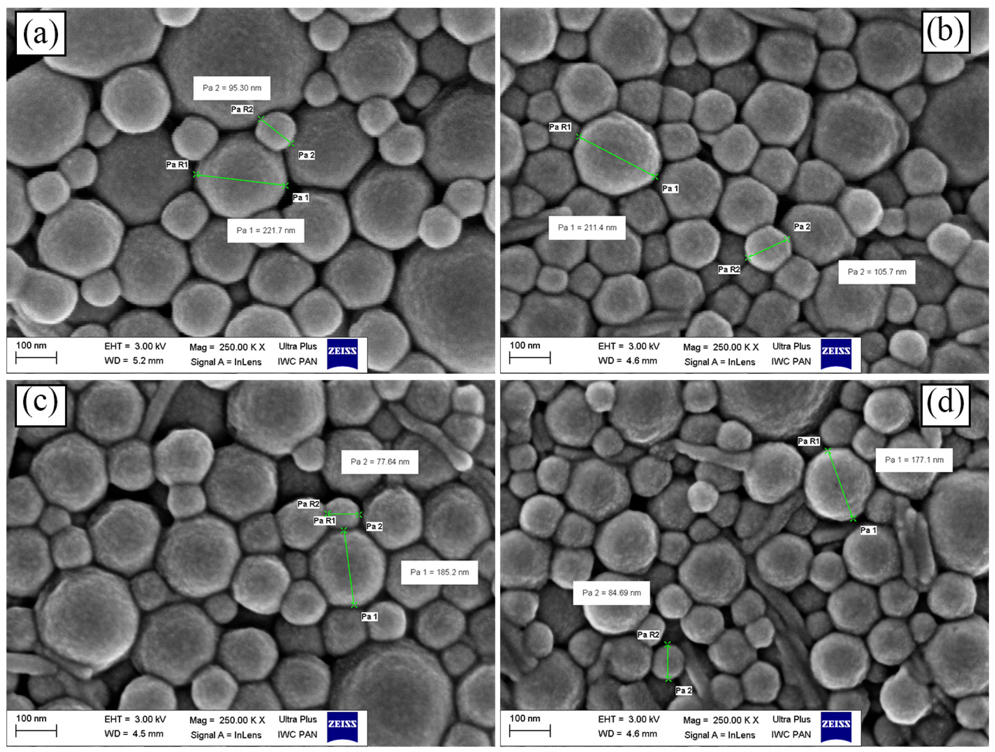
2.10. Surface Morphology
2.11. Drug Content
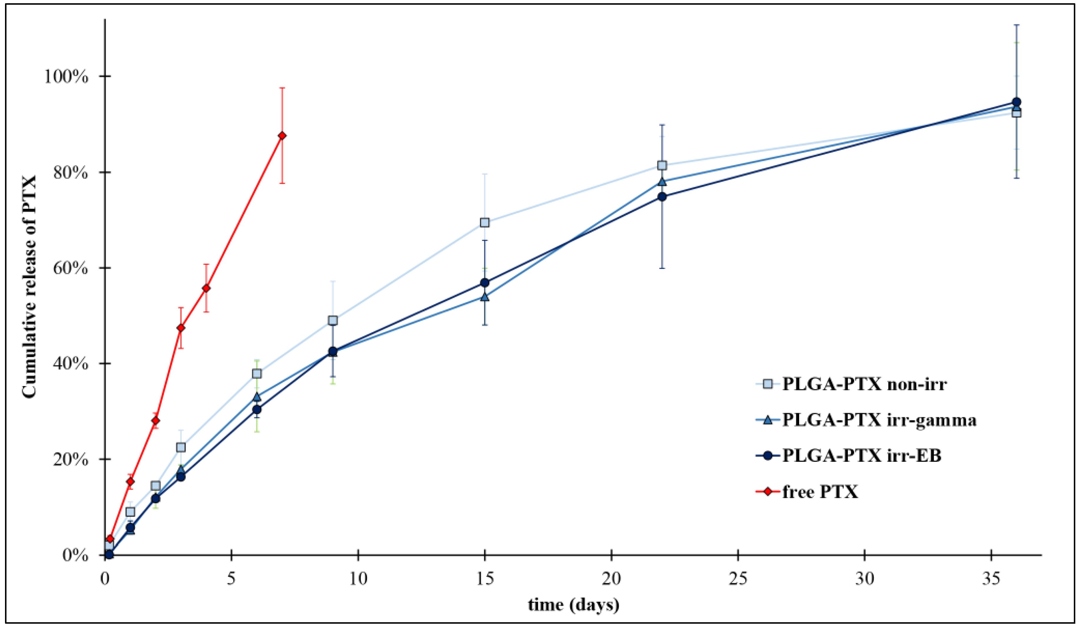
2.12. In Vitro Drug Release Study
3. Results and Discussion
3.1. Synthesis and Characterization of Polymers
3.2. The Influence of Ionizing Radiation on Thermal Properties of Polyesters
3.3. Evaluation of the Stability of PTX-Loaded Nanoparticles to Irradiation
3.4. In Vitro Drug Release
4. Conclusions
Author Contributions
Funding
Institutional Review Board Statement
Informed Consent Statement
Data Availability Statement
Acknowledgments
Conflicts of Interest
Sample Availability
Abbreviations
| ACN | acetonitrile |
| BiOct3 | bismuth 2-ethylhexanoate |
| Đ | dispersity index (GPC) |
| DDSs | drug delivery systems |
| EB | electron-beam |
| EE | encapsulation efficiency |
| GA | glycolide |
| G | glycolyl unit |
| GG | glycolidyl unit |
| ΔHc | enthalpy of cold crystallization |
| ΔHm | enthalpy of melting |
| IR | induction ratio |
| L | lactyl unit |
| LL | lactidyl unit |
| L-LA | l-lactide |
| lGG | average length of glycolidyl units |
| lLL | average length of lactidyl blocks |
| Mn | number average molecular mass |
| NRU | neutral red uptake |
| PBS | phosphate-buffered saline |
| PDI | dispersity index (DLS) |
| PEG | poly(ethylene glycol) |
| PLGA | copolymer of l-lactide and glycolide |
| PTX | paclitaxel |
| PVA | poly(vinyl alcohol) |
| ROP | ring-opening polymerization |
| Tc | temperature of cold crystallization |
| TFA | trifluoroacetic acid |
| Tg | glass transition temperature |
| TII | transesterification of the second mode |
| Tm | temperature of melting |
| Xc | crystalline phase content |
| ZP | zeta potential |
References
- WHO. Cancer Fact Sheet. 2022. Available online: https://www.who.int/news-room/fact-sheets/detail/cancer (accessed on 26 May 2022).
- Surapaneni, M.S.; Das, S.K.; Das, N.G. Designing Paclitaxel Drug Delivery Systems Aimed at Improved Patient Outcomes: Current Status and Challenges. ISRN Pharmacol. 2012, 2012, 623139. [Google Scholar] [CrossRef] [PubMed]
- Stefanowicz, Z.; Sobczak, M.; Piętniewicz, A.; Kołodziejski, W. Macromolecular conjugates of paclitaxel: Synthesis, characterization, and In Vitro paclitaxel release studies based on HPLC validated method. Acta Chromatogr. 2016, 28, 99–117. [Google Scholar] [CrossRef]
- Martins, K.F.; Messias, A.D.; Leite, F.L.; Duek, E.A.R. Preparation and characterization of paclitaxel-loaded PLDLA microspheres. Mat. Res. 2014, 17, 650–656. [Google Scholar] [CrossRef]
- Council of Europe. Suppl. 9.2. European Directorate for the Quality of Medicines & HealthCare; Council of Europe: London, UK, 2017; pp. 4333–4350. [Google Scholar]
- Tapia-Guerrero, Y.S.; Del Prado-Audelo, M.L.; Borbolla-Jiménez, F.V.; Gomez, D.M.G.; García-Aguirre, I.; Colín-Castro, C.A.; Morales-González, J.A.; Leyva-Gómez, G.; Magaña, J.J. Effect of UV and Gamma Irradiation Sterilization Processes in the Properties of Different Polymeric Nanoparticles for Biomedical Applications. Materials 2020, 13, 1090. [Google Scholar] [CrossRef]
- Raeiszadeh, M.; Adeli, B. A Critical Review on Ultraviolet Disinfection Systems against COVID-19 Outbreak: Applicability, Validation, and Safety Considerations. ACS Photonics 2020, 7, 2941–2951. [Google Scholar] [CrossRef] [PubMed]
- Igartua, M.; Hernández, R.M.; Rosas, J.E.; Patarroyo, M.E.; Pedraz, J.L. γ-Irradiation effects on biopharmaceutical properties of PLGA microspheres loaded with SPf66 synthetic vaccine. Eur. J. Pharm. Biopharm. 2008, 69, 519–526. [Google Scholar] [CrossRef] [PubMed]
- Domańska, I.M.; Oledzka, E.; Sobczak, M. Sterilization process of polyester based anticancer-drug delivery systems. Int. J. Pharm. 2020, 587, 119663. [Google Scholar] [CrossRef] [PubMed]
- Maksimenko, O.; Pavlov, E.; Toushov, E.; Molin, A.; Stukalov, Y.; Prudskova, T.; Feldman, V.; Kreuter, J.; Gelperina, S. Radiation sterilisation of doxorubicin bound to poly(butyl cyanoacrylate) nanoparticles. Int. J. Pharm. 2008, 356, 325–332. [Google Scholar] [CrossRef]
- Dorati, R.; Genta, I.; Montanari, L.; Cilurzo, F.; Buttafava, A.; Faucitano, A.; Conti, B. The effect of γ-irradiation on PLGA/PEG microspheres containing ovalbumin. J. Control. Release 2005, 107, 78–90. [Google Scholar] [CrossRef]
- Fernandezcarballido, A.; Puebla, P.; Herrerovanrell, R.; Pastoriza, P. Radiosterilisation of indomethacin PLGA/PEG-derivative microspheres: Protective effects of low temperature during gamma-irradiation. Int. J. Pharm. 2006, 313, 129–135. [Google Scholar] [CrossRef]
- EMA. Guideline on the Sterilisation of the Medicinal Product, Active Substance, Excipient and Primary Container. EMA/CHMP/CVMP/QWP/850374/2015. European Medicines Agency, 2019. Available online: https://www.ema.europa.eu/en/documents/scientific-guideline/guideline-sterilisation-medicinal-product-active-substance-excipient-primary-container_en.pdf (accessed on 18 May 2023).
- Sakar, F.; Özer, A.Y.; Erdogan, S.; Ekizoglu, M.; Kart, D.; Özalp, M.; Colak, S.; Zencir, Y. Nano drug delivery systems and gamma radiation sterilization. Pharm. Dev. Technol. 2017, 22, 775–784. [Google Scholar] [CrossRef] [PubMed]
- Athanasiou, K. Sterilization, toxicity, biocompatibility and clinical applications of polylactic acid/polyglycolic acid copolymers. Biomaterials 1996, 17, 93–102. [Google Scholar] [CrossRef] [PubMed]
- Wang, J.; Ng, C.W.; Win, K.Y.; Shoemakers, P.; Lee, T.K.Y.; Feng, S.S.; Wang, C.H. Release of paclitaxel from polylactide-co-glycolide (PLGA) microparticles and discs under irradiation. J. Microencapsul. 2003, 20, 317–327. [Google Scholar] [CrossRef] [PubMed]
- Song, T.-T.; Yuan, X.-B.; Sun, A.-P.; Wang, H.; Kang, C.-S.; Ren, Y.; He, B.; Sheng, J.; Pu, P.-Y. Preparation of injectable paclitaxel sustained release microspheres by spray drying for inhibition of glioma in vitro. J. Appl. Polym. Sci. 2010, 115, 1534–1539. [Google Scholar] [CrossRef]
- Dobrzynski, P.; Kasperczyk, J.; Janeczek, H.; Bero, M. Synthesis of Biodegradable Copolymers with the Use of Low Toxic Zirconium Compounds. 1. Copolymerization of Glycolide with l -Lactide Initiated by Zr(Acac)4. Macromolecules 2001, 34, 5090–5098. [Google Scholar] [CrossRef]
- Jing, Y.; Yang, M.; Dai, S.; Quan, C.; Liu, J.; Jiang, Q.; Zhang, C.; Liu, B. Microwaves promote transesterification in the rapid synthesis of methoxy-poly(ethylene glycol)-block-poly(l-lactide-random-glycolide). Polymer 2018, 136, 187–193. [Google Scholar] [CrossRef]
- EN ISO 10993-5:2009; Biological Evaluation of Medical Devices—Part 5: Tests for In Vitro Cytotoxicity (ISO 10993-5:2009). Annex A Neutral Red Uptake (NRU) Cytotoxicity Test. International Organization for Standardization: Geneva, Switzerland, 2009.
- ISO/FDIS 13829:2000; Water Quality-Determination of the Genotoxocity of Water and Waste Water Using the Umu-Test. International Organization for Standarization: Geneva, Switzerland, 2000.
- Sarasua, J.-R.; Prud’homme, R.E.; Wisniewski, M.; Le Borgne, A.; Spassky, N. Crystallization and Melting Behavior of Polylactides. Macromolecules 1998, 31, 3895–3905. [Google Scholar] [CrossRef]
- Magazzini, L.; Grilli, S.; Fenni, S.E.; Donetti, A.; Cavallo, D.; Monticelli, O. The Blending of Poly(glycolic acid) with Polycaprolactone and Poly(l-lactide): Promising Combinations. Polymers 2021, 13, 2780. [Google Scholar] [CrossRef]
- Kasiński, A.; Zielińska-Pisklak, M.; Kowalczyk, S.; Plichta, A.; Zgadzaj, A.; Oledzka, E.; Sobczak, M. Synthesis and Characterization of New Biodegradable Injectable Thermosensitive Smart Hydrogels for 5-Fluorouracil Delivery. Int. J. Mol. Sci. 2021, 22, 8330. [Google Scholar] [CrossRef]
- Dash, S.; Murthy, P.N.; Nath, L.; Chowdhury, P. Kinetic Modeling on Drug Release from Controlled Drug Delivery Systems. Acta Pol. Pharm. Drug Res. 2010, 67, 217–223. [Google Scholar]
- Domańska, I.; Zalewska, A.; Cieśla, K.; Plichta, A.; Głuszewski, W.; Łyczko, M.; Kowalczyk, S.; Oledzka, E.; Sobczak, M. The influence of electron beam and gamma irradiation on paclitaxel-loaded nanoparticles of fully randomized copolymers in relation to potential sterilization. Faculty of Chemistry, Warsaw University of Technology, 3 Noakowskiego Str., 00-664 Warsaw, Poland. 2023; manuscript submitted. [Google Scholar]
- Plikk, P.; Odelius, K.; Hakkarainen, M.; Albertsson, A.C. Finalizing the properties of porous scaffolds of aliphatic polyesters through radiation sterilization. Biomaterials 2006, 27, 5335–5347. [Google Scholar] [CrossRef] [PubMed]
- Loo, S.C.J.; Ooi, C.P.; Boey, Y.C.F. Radiation effects on poly(lactide-co-glycolide) (PLGA) and poly(l-lactide) (PLLA). Polym. Degrad. Stab. 2004, 83, 259–265. [Google Scholar] [CrossRef]
- Davison, L.; Themistou, E.; Buchanan, F.; Cunningham, E. Low temperature gamma sterilization of a bioresorbable polymer, PLGA. Radiat. Phys. Chem. 2018, 143, 27–32. [Google Scholar] [CrossRef]
- Loo, J.S.C.; Ooi, C.P.; Boey, F.Y.C. Degradation of poly(lactide-co-glycolide) (PLGA) and poly(l-lactide) (PLLA) by electron beam radiation. Biomaterials 2005, 26, 1359–1367. [Google Scholar] [CrossRef]
- Dorati, R.; Colonna, C.; Tomasi, C.; Bruni, G.; Genta, I.; Modena, T.; Conti, B. Long-Term Effect of Gamma Irradiation on the Functional Properties and Cytocompatibility of Multiblock Co-Polymer Films. J. Biomater. Sci. Polym. Ed. 2012, 23, 2223–2240. [Google Scholar] [CrossRef]
- Walo, M.; Rzepna, M. Recent Developments in Radiation Processing of Polymers; Institute of Nuclear Chemistry and Technology: Warszawa, Poland, 2020. [Google Scholar]
- Pandita, D.; Ahuja, A.; Velpandian, T.; Lather, V.; Dutta, T.; Khar, R.K. Characterization and in vitro assessment of paclitaxel loaded lipid nanoparticles formulated using modified solvent injection technique. Pharmazie 2009, 64, 301–310. [Google Scholar] [CrossRef]
- Nandhakumar, S.; Dhanaraju, M.D.; Sundar, V.D.; Heera, B. Influence of surface charge on the in vitro protein adsorption and cell cytotoxicity of paclitaxel loaded poly(ε-caprolactone) nanoparticles. Bull. Fac. Pharm. Cairo Univ. 2017, 55, 249–258. [Google Scholar] [CrossRef]
- Musumeci, T.; Ventura, C.; Giannone, I.; Ruozi, B.; Montenegro, L.; Pignatello, R.; Puglisi, G. PLA/PLGA nanoparticles for sustained release of docetaxel. Int. J. Pharm. 2006, 325, 172–179. [Google Scholar] [CrossRef]
- Jusu, S.M.; Obayemi, J.D.; Salifu, A.A.; Nwazojie, C.C.; Uzonwanne, V.; Odusanya, O.S.; Soboyejo, W.O. Drug encapsulated blend of PLGA PEG microspheres: In vitro and in vivo study of the efects of localized/targeted drug delivery on the treatment of triple negative breast cancer. Sci. Rep. 2020, 10, 14188. [Google Scholar] [CrossRef]
- Singhvi, G.; Singh, M. Review: In-Vitro Drug Release Characterization Models. Int. J. Pharm. Stud. Res. 2011, 2, 77–84. [Google Scholar]
- Głuszewski, W.; Zagórski, Z.P. Radiation effects in polypropylene/polystyrene blends as the model of aromatic protection effects. Nukleonika 2008, 53, S21–S24. [Google Scholar]
- Ferry, M.; Ngono, Y. Energy transfer in polymers submitted to ionizing radiation: A review. Radiat. Phys. Chem. 2021, 180, 109320. [Google Scholar] [CrossRef]


| Sample | Genotoxicity Assay | Cytotoxicity Assay | |||
|---|---|---|---|---|---|
| −S9 a | +S9 b | ||||
| G | IR a | G | IR b | Cells Viability (%) | |
| PLGA | 0.97 ± 0.07 | 0.80 ± 0.07 | 0.97 ± 0.07 | 1.31 ± 0.40 | 106 ± 6 |
| Positive Control | 1.04 ± 0.01 | 3.30 ± 0.28 | 0.87 ± 0.04 | 2.53 ± 0.41 | 1 ± 1 |
| Negative Control | 1.00 ± 0.01 | 1.00 ± 0.05 | 1.00 ± 0.04 | 1.00 ± 0.08 | 111 ± 1 |
| Irradiation | Mn (kDa) a | ΔMn (%) ab | Đ a | FGG | lLLe | lGGe | TII (LGL) | TII (GLG) |
|---|---|---|---|---|---|---|---|---|
| non-irr | 13.79 ± 0.10 | n.a. | 1.83 ± 0.01 | 0.15 | 4.33 | 1.23 | 0.15 | 1.14 |
| irr-γ | 11.89 ± 0.64 | −13.82 | 1.95 ± 0.09 | 0.16 | 3.35 | 1.28 | 0.13 | 1.38 |
| irr-e− | 11.85 ± 0.15 | −14.07 | 1.96 ± 0.03 | 0.16 | 4.61 | 1.22 | 0.15 | 0.79 |
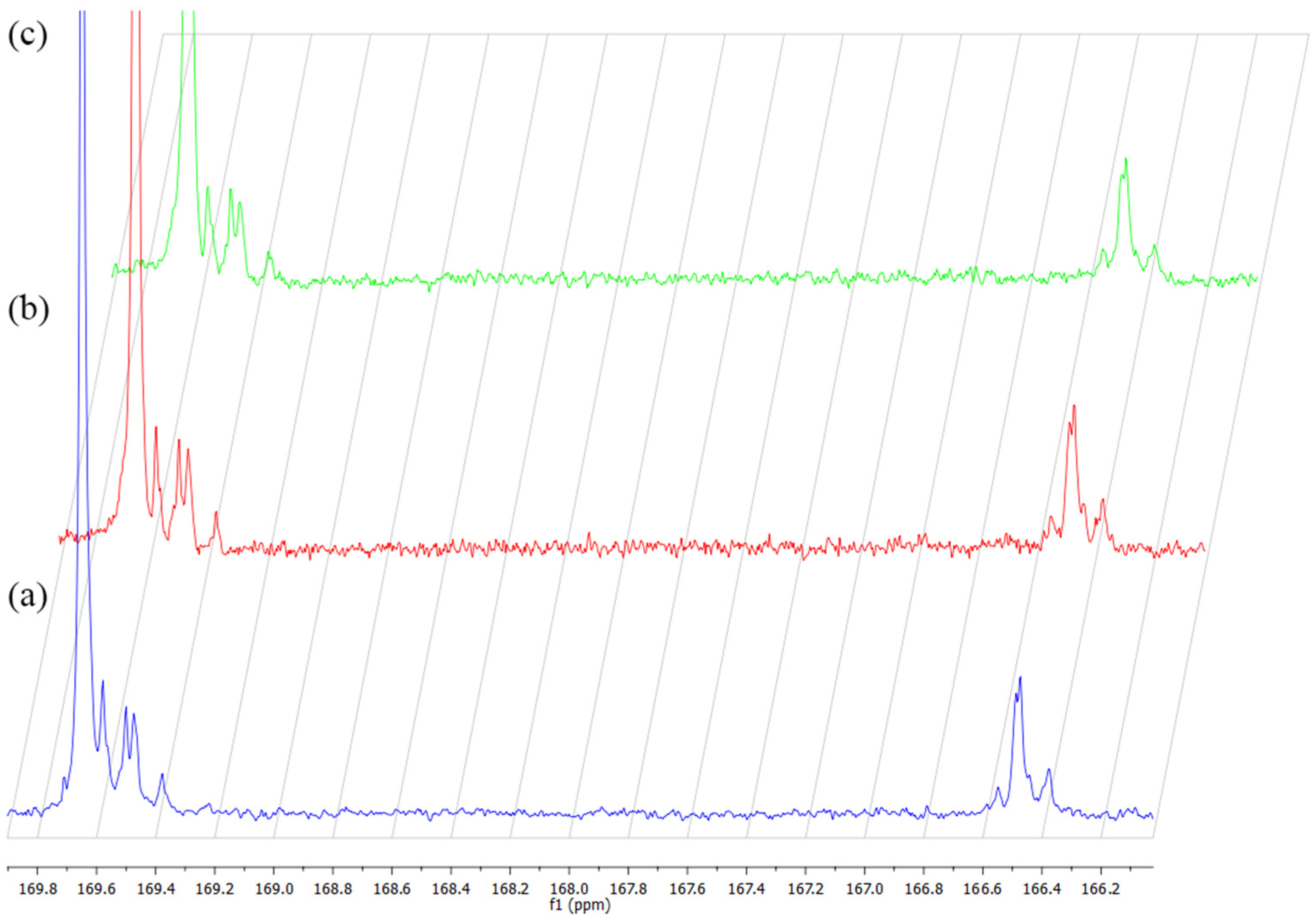
| Chemical Shift (ppm) | Sequence |
|---|---|
| 169.65 | LLLL |
| 169.58 | LLGG |
| 169.50 | LGLL |
| 169.47 | GGLL |
| 169.38 | GLG |
| 166.55 | GGLG |
| 166.49 | GGLL |
| 166.47 | GGGG |
| 166.44 | LGL |
| 166.38 | LLGG |
| Irradiation | Tg b (°C) | Tmin a (°C) | Tc a (°C) | Tm a (°C) | ∆Hc a (J g−1) | ∆Hm a (J g−1) | Xc a (%) |
|---|---|---|---|---|---|---|---|
| non-irr | 48.1 | 77.3 | 103.7 | 138.0 (Tm1) | 8.52 | 50.16 | 35.1 |
| 147.1 (Tm2) | |||||||
| irr-γ | 47.6 | 76.8 | 102.6 | 137.4 (Tm1) | 15.00 | 41.92 | 22.7 |
| 146.2 (Tm2) | |||||||
| irr-e− | 48.2 | 76.7 | 103.3 | 137.3 (Tm1) | 4.76 | 42.06 | 31.4 |
| 146.5 (Tm2) |
| Irradiation | Size (nm) | PDI | ZP (mV) |
|---|---|---|---|
| PLGA | 253.6 ± 4.1 | 0.19 ± 0.03 | −22.48 ± 0.41 |
| PLGA-PTX non-irr | 253.5 ± 3.9 | 0.17 ± 0.03 | −20.83 ± 1.17 |
| PLGA-PTX irr-γ | 256.4 ± 3.1 | 0.20 ± 0.02 | −36.73 ± 1.26 |
| PLGA-PTX irr-e− | 253.6 ± 2.1 | 0.17 ± 0.02 | −33.75 ± 0.57 |
| Irradiation | Zero-Order | First-Order | Higuchi | Korsmeyer–Peppas | ||
|---|---|---|---|---|---|---|
| R2 | R2 | R2 | KH a | R2 | n b | |
| non-irr | 0.881 | 0.998 | 0.981 | 17.805 | 0.995 | 0.793 |
| irr-γ | 0.949 | 0.963 | 0.991 | 17.906 | 0.977 | 0.857 |
| irr-e | 0.946 | 0.974 | 0.995 | 17.820 | 0.993 | 0.857 |
Disclaimer/Publisher’s Note: The statements, opinions and data contained in all publications are solely those of the individual author(s) and contributor(s) and not of MDPI and/or the editor(s). MDPI and/or the editor(s) disclaim responsibility for any injury to people or property resulting from any ideas, methods, instructions or products referred to in the content. |
© 2023 by the authors. Licensee MDPI, Basel, Switzerland. This article is an open access article distributed under the terms and conditions of the Creative Commons Attribution (CC BY) license (https://creativecommons.org/licenses/by/4.0/).
Share and Cite
Domańska, I.M.; Figat, R.; Zalewska, A.; Cieśla, K.; Kowalczyk, S.; Kędra, K.; Sobczak, M. The Influence of Ionizing Radiation on Paclitaxel-Loaded Nanoparticles Based on PLGA. Appl. Sci. 2023, 13, 11052. https://doi.org/10.3390/app131911052
Domańska IM, Figat R, Zalewska A, Cieśla K, Kowalczyk S, Kędra K, Sobczak M. The Influence of Ionizing Radiation on Paclitaxel-Loaded Nanoparticles Based on PLGA. Applied Sciences. 2023; 13(19):11052. https://doi.org/10.3390/app131911052
Chicago/Turabian StyleDomańska, Izabela M., Ramona Figat, Aldona Zalewska, Krystyna Cieśla, Sebastian Kowalczyk, Karolina Kędra, and Marcin Sobczak. 2023. "The Influence of Ionizing Radiation on Paclitaxel-Loaded Nanoparticles Based on PLGA" Applied Sciences 13, no. 19: 11052. https://doi.org/10.3390/app131911052
APA StyleDomańska, I. M., Figat, R., Zalewska, A., Cieśla, K., Kowalczyk, S., Kędra, K., & Sobczak, M. (2023). The Influence of Ionizing Radiation on Paclitaxel-Loaded Nanoparticles Based on PLGA. Applied Sciences, 13(19), 11052. https://doi.org/10.3390/app131911052