Article

Application of a Validated HPLC Method for the Determination of Resveratrol, Ferulic Acid, Quercetin, Retinol, and α-Tocopherol in a Cold Cream—Permeability Study

by
Athanasia Karavalasi
1,
Sofia Almpani
1,
Panagiota Tserkezou
1,
Konstantina Chachlioutaki
2,
Georgios Kamaris
1 and
Catherine K. Markopoulou
1,*
1
Laboratory of Pharmaceutical Analysis, Department of Pharmacy, Aristotle University of Thessaloniki, 54124 Thessaloniki, Greece
2
Laboratory of Pharmaceutical Technology, Department of Pharmacy, Aristotle University of Thessaloniki, 54124 Thessaloniki, Greece
*
Author to whom correspondence should be addressed.
Appl. Sci. 2024, 14(24), 11843; https://doi.org/10.3390/app142411843
Submission received: 20 November 2024 / Revised: 12 December 2024 / Accepted: 15 December 2024 / Published: 18 December 2024
(This article belongs to the Special Issue Research on Organic and Medicinal Chemistry)

Abstract

:
Due to the rapid increase in the use of anti-aging cosmetic products, there is a need to develop valid analytical methods to control their quality. The present work deals with the development and validation of a new chromatographic method for the quantitative determination of five lipophilic components (resveratrol, ferulic acid, quercetin, retinol, and α-tocopherol), with anti-aging properties, in a cold cream (w/o). For the HPLC-UV separation of the active ingredients, an HS, Discovery® Supelco (Supelco Inc., Bellefonte, PA, USA), C18 column (25 cm × 4.6 mm), 5 μm (at 40 °C) was used as a stationary phase while a binary system of A: Acetonitrile with formic acid 0.2% and B: H2O with formic acid 0.2%, in gradient elution (flow 1.5 mL·min−1), was used as mobile. The analytical method was validated according to ICH guidelines Q2(R2), where linearity (r2 ≥ 0.998), selectivity, precision (% recovery 97.1–101.9), and accuracy (%RSD < 2) were evaluated. The processing of the samples for the recovery of the five analytes from the cream was investigated by experimental design methodology and the cross D-optimal technique (% recovery 98.5–102.9, %RSD < 2%, n = 5). Finally, the same analysis was applied to study the transdermal penetration of the active ingredients incorporated in cold cream (over a period of 8 h). Their behavior was compared with the corresponding one in suspension using Franz cells in a vertical arrangement. The new method is considered reliable for the analysis of the anti-aging product.

1. Introduction

The skin is considered the largest organ of the human body since it occupies a total surface area of 2 m2 [1]. Its main role is to protect the underlying organs from the external environment and the penetration of pathogenic microorganisms. It consists of the epidermis, dermis, and subcutaneous fat tissue.
The natural process of aging in humans is characterized by skin deterioration and the appearance of health problems. Skin aging is a process that occurs naturally and can mark a person’s self-esteem, socialization, and quality of life [2]. It is due to exogenous and endogenous environmental factors. Extrinsic aging is characterized by laxity, wrinkles, discoloration, and dilation of blood vessels [3]. The accumulation of amorphous elastin material is another characteristic of extrinsically aged skin that can lead to a decrease in elasticity. In addition, collagen fibers are damaged and fragmented [4]. Intrinsic or chronological aging is difficult to reverse and is related to genetic factors such as hormone levels [5], genotype, and endocrine metabolism [6]. From a medical perspective, aging is associated with the accumulation of cellular damage and the inability of stem cells to promote both regeneration and restoration of their normal integrity [7]. Aged skin is biologically characterized by the decomposition of the dermal extracellular matrix, where collagen, elastin fibers [2], glycosaminoglycans, and hyaluronic acid decrease.
Nowadays, many people, especially women, spend a lot of money on cosmetics and drugs for the treatment and prevention of aging. Thus, in recent decades, this problem has been a field of interest for scientific groups for the development of new cosmetic products. Creams have a wide application in cosmetology and are the most frequent forms of cosmetics for daily use. By the term “cream”, we mean a solid or semi-solid emulsion (o/w or w/o), which is intended for application to the skin or mucosa [8].
Resveratrol (RSV), ferulic acid (FERA), quercetin (QR), retinol, RTN (vitamin A), and α-tocopherol, α-TOC (vitamin E) are five active pharmaceutical ingredients (APIs) that, according to the existing literature, contribute to the reduction in facial aging. Their use in cosmetic products is mainly due to their antioxidant and anti-inflammatory effects. The substances are presented as regulators in cellular functions, in the expression of genes that reduce oxidative stress, and in the control of free radical formation [9]. Therefore, their use contributes to the hydration and vascularization of the skin, with the result that wrinkles are removed, and the skin, due to an increase in collagen, acquires elasticity and hardness [10,11,12,13]. Sunlight is another reason that leads to premature aging. Park and Lee utilized resveratrol (5–100 µM) in UV/B-irradiated HaCaT keratinocytes and observed that it increased keratinocyte survival [14,15]. Accordingly, vitamin E can reduce hyperpigmentation caused by UV radiation because it exhibits photoprotection [16]. Resveratrol may also slow down the skin aging process due to its role as a sirtuin activator (regeneration and cellular enzyme) [17] while exhibiting antibacterial, antiviral, antifungal, and anti-inflammatory properties [18]. Likewise, retinol is essential for the growth, reproduction, and maintenance of epithelial tissues as well as for wound healing [19]. Taking into account that the five active ingredients work synergistically, meaning that their therapeutic, healing, and regenerative properties are either complementary or additive, it is recommended to combine them in cosmetic formulations in order to achieve the optimal result. Their suggested percentage composition in cosmetics is for ferulic acid 0.5–1% [20], for resveratrol up to 5%, for quercetin 0.1–3, up to 0.3% for retinol [21], and up to 5% [22] for vitamin E. Therefore, in the context of the present work, a new formulation was prepared and then evaluated, incorporating the five active ingredients. The formulation belongs to the cold cream category, i.e., it is a water-in-oil (w/o) emulsion with a greasy texture that ensures prolonged contact at the application surface, compared to other semi-solid forms. Cold cream helps restore moisture to dry skin by creating a feeling of coolness due to the evaporation of water. In addition, it is non-irritating, and when applied to the skin, it provides softness.
From an analytical point of view, chromatography is considered the method of choice for the reliable determination and suitability of such formulations. Most analyses reporting individually to one of these active ingredients (ferulic acid, resveratrol, quercetin, retinol, and tocopherol) are performed with HPLC or high-performance thin-layer chromatography (HPTLC) or even gas chromatography [23,24,25,26,27,28,29]. Their separation conditions may vary depending on the study and the substrate. In HPLC, usually RP-C18 [30,31] or C8 [32] columns are used as stationary phases, and water with organic solvents, such as acetonitrile and/or methanol and ethanol, is preferred as mobile phases [33,34].
It is noteworthy that although the five active ingredients (APIs) are widely used in cosmetics, to the best of our knowledge, there is no literature report for their simultaneous determination, which makes the present study innovative. The only related mention is an HPLC-UV method suitable for the determination of QR, RTN, and α-TOC in nanoemulsion [35]. Relevant analyses have also been described for their binary mixtures, such as that of quercetin with resveratrol [32], retinol with tocopherol [36], or tocopherol with quercetin [37].
Thus, in the present work, five active ingredients were determined in cold cream (w/o) using a new validated HPLC-DAD method. Due to the complex substrate, the processing of the samples was investigated by experimental design methodology (D-Optimal cross) [38,39]. API’s stability (at 45 °C and sonication) was also studied over a period of 80 min. Finally, the same analytical method was applied for the in vitro permeation study of the five drugs using Franz cells. Their behavior was compared after being incorporated into a simple suspension and a cream. Samples after lyophilization were reconstituted and analyzed.
A new formulation with anti-aging properties is proposed to contribute to the scientific community’s efforts to combat aging. For the analysis of its five active ingredients, an innovative, reliable, and flexible analytical method suitable for routine analysis is developed.

2. Materials and Methods

2.1. Instrumentation

The analytical method was implemented on an HPLC–20AD liquid chromatography setup (Shimadzu, Kyoto, Japan) consisting of a SIL-20A HT autosampler (Shimadzu), a CTO-20A column oven (Shimadzu), and an SPD-M20A photon array detector. For the separation of the samples, a Supelco Discovery (Supelco Inc., Bellefonte, PA, USA) C18 (25 cm × 4.6 mm) 5 μm column and a binary mobile phase consisting of (A): H2O with formic acid 0.2% and (B): acetonitrile with formic acid 0.2%, in gradient elution, were used. All samples were injected in duplicate, and results were processed through LC Solution software 2020 (Shimadzu).

2.2. Reagents, Solvents and Materials

Resveratrol 99.0% (3,4′,5-trihydroxystilbene, C14H12O3, RSV) from TCI Chemicals (Tokyo, Japan), quercetin dihydrate 99.0% (3,3′,4′,5′7-pentahydroxyflavone, C15H14O9, QR) from BioChemika-Fluka (Buchs, Switzerland), trans-ferulic acid 99.0% (4-hydroxy-3-methoxycinnamic acid, C10H10O4, FERA) from Sigma-Aldrich (Shanghai, China), retinol 95% (vitamin A, C20H30O, Retinol, RTN) from Sigma-Aldrich (Buchs, Switzerland), and α-Tocopherol 95.5% (vitamin E, C29H50O2, α-Tocopherol, α-TOC) from Sigma-Aldrich (Darmstadt, Germany) were used as active ingredients (Figure 1). Coconut oil, cetyl alcohol, borax, and cera alba (excipients) were purchased from Chemco (Athens, Greece). For the preparation of the new formulation (cold cream), one of the raw materials used was resveratrol, which originated from the extract of grape (Vitis vinifera) and Japanese acacia (Polygpnum cuspidatum). The extract is sold under the brand name Regener Life Resveratrol® (Company: Natural Factors, Coquitlam, BC, Canada, purchased from a local pharmacy) in 500 mg capsules with 250 mg of pure and natural trans-resveratrol.
The solvents for the mobile phase or for processing the samples were of high purity and suitable for use (HPLC grade). More specifically, methanol (CH3OH, MeOH) (Chem-Lab, Zedelgem, Belgium), acetonitrile (ACN) (Honeywell, Charlotte, NC, USA), and ethanol (CH3CH2OH, EtOH) (Honeywell) were used. Water was ultrapure for HPLC (18.2 MΩ cm resistivity) and produced through a purification system (B30, Adrona SIA, Riga, Latvia).
Formic acid (CH2O2, FA) Sigma-Aldrich (Darmstadt, Germany) and trifluoroacetic acid (C2HF3O2, TFA), Sigma-Aldrich (Darmstadt, Germany), were utilized to adjust the pH, while the final diluent of the samples consisted of MeOH-ACN-F.A. 50:50:0.2 v/v/v.
Phosphate-buffered saline (PBS) pH 7.4 was prepared by dissolving 8.0 g NaCl, 1.44 g Na2HPO4, 0.24 g KH2PO4 (Merck, Darmstadt, Germany), and 0.20 g KCl (Chem-Lab nv, Zedelgem, Belgium) in 1 L of distilled water. For the in vitro permeability experiments, a dialysis tubing cellulose membrane (flat width 43 mm) was obtained from Sigma-Aldrich (Darmstadt, Germany).

2.3. Standard Solutions

To prepare the stock solutions of the five substances, 7.8 mg RSV, 5.2 mg FERA, 9.4 mg QR, 10.5 mg RTN, and 13.5 α-TOC were accurately weighed into a 10 mL volumetric flask. The flask was filled up to the mark with MeOH-ACN-F.A. solvent, 50:50:0.2 v/v/v, and placed in an ultrasonic bath for approximately 2 min (stock solution). This was followed by an intermediate dilution (100 μL to 25 mL) used for the six standard solutions of the calibration curve

2.4. Cream Preparation

The ingredients of the classic cold cream (w/o) were used as excipients of the formulation [40]. More specifically, the fatty phase contains white wax, cetyl alcohol, and paraffin oil, while the aqueous carrier contains borax (emulsifier) and deionized water. The active ingredients, resveratrol 1% w/w, ferulic acid 0.5% w/w, quercetin 1% w/w, retinol 0.3% w/w, and α-tocopherol 2% w/w, were incorporated into the cream after an appropriate process. Therefore, further research was conducted to determine the optimal amount of each excipient to achieve a satisfactory formulation texture. Table 1 presents five different cream bases that were prepared and compared with each other.
Cream type 1 had the consistency of the classic cold cream recipe. Its texture was quite greasy; it spread very easily on the skin, but it did not leave a nice feeling after its application. When cream 2 was applied to the skin, the oiliness was quite noticeable and the smell was quite strong, probably due to the almond oil. In creams 3, 4, 5, the amount of coconut oil was gradually reduced until the desired oiliness and texture were reached. Creams 3 and 4 had the best characteristics, as they did not leave a greasy feeling on the skin, spread very easily, had the desired shine, and did not clump. In order to choose the optimum composition, the two creams were left in a cool place, for about 3 weeks, and re-examined. Of the two cases, type 3 cream showed the best result and was prepared according to the following procedure: 2 g of white wax was weighed and placed in a water bath at 70 °C to melt. At the same time, the two lipophilic vitamins A and E, resveratrol, ferulic acid, and quercetin were quantitatively added to a plastic mortar containing 7 g of coconut oil, and the whole mixture was mixed with a pestle. After homogenization, cetyl alcohol and white wax were added gradually with simultaneous mixing while the mixture was placed in a water bath (45 °C) to prevent its solidification. With a similar procedure, the excipients of the aqueous phase were weighed and placed in a water bath at 45 °C. This was followed by the mixing of the two phases, where the aqueous was slowly incorporated into the fatty phase. After the cream base cooled, it was stored.

2.5. In Vitro Permeability Studies—Franz Cells Apparatus

The in vitro permeability profile of the five APIs incorporated into the cold cream or suspension was studied using a vertical Franz cell diffusion system. The cells consist of two compartments, a donor (upper chamber) and an acceptor (lower chamber), with a cellulose membrane (effective diffusion surface 4.9 cm2, receptor volume 20 mL) located between them. The donor compartment was either uniformly coated with 250 mg of cream or with 250 µL of suspension, while the receiver was filled with 20 mL of degassed PBS pH 7.4. During the experiments, sink conditions were maintained under continuous agitation (90 rpm) at 37 °C. Experiments were repeated in triplicate for both cold creams and suspensions, while samples containing only the substrate (blank) were also performed. At regular time intervals (2, 3, 4, 6, 8 h), and within 8 h, a sample volume of 0.5 mL was taken from the receiver compartment and replaced with fresh pre-warmed PBS.
After the samples were collected, they were lyophilized to completely remove the aqueous medium. Then, 400 µL of methanol was added and the samples were sonicated (15 min), placed in the freezer for 10 min and centrifuged. Two hundred microliters of supernatant was quantitatively transferred to a vial to which 200 μL of acetonitrile with 0.2% formic acid was added, mixed (vortexed for 2 min) and analyzed by HPLC. Quantification of the membrane residue after completion of the experiment was performed according to the formulation analysis procedure.
The cumulative amount of permeated compound per unit area (µg/cm2) was plotted against time (h), and the steady-state flux (Jss) was calculated from the slope of the linear section of the plot. The apparent permeability coefficient (Papp) was derived by Equation (1).
P app = J ss C d , ( cm · h 1 )
where:
Cd is the initial concentration of the drug in the donor compartment and
Jss is the steady state flux.

3. Results and Discussion

3.1. Investigation of the Chromatographic Method

In the present analytical method, special attention had to be paid to the chromatographic separations and column cleanup due to the wide range of lipophilicity of the analytes (LogP values) as well as the complex fatty substrate. More specifically, FERA has an intermediate polarity (LogP = 1.5), in contrast to α-TOC, which is extremely lipophilic (LogP = 10.5). On the other hand, FERA (LogP = 1.5), QR (LogP = 2.2), and RSV (LogP = 3.4) are likely to present separation problems due to their similar polarity. Τo determine the appropriate chromatographic conditions, a series of experiments were conducted regarding the choice of the stationary phase and its temperature, the composition of the mobile phase and its flow rate, as well as the sample diluent. Given the circumstances, and with the aim of creating a non-time-consuming analytical method that could be used in routine analyses, it was obvious that the mobile phase had to be with gradient elution. Therefore, a simplified mobile phase with gradient elution (and buffers) was initially used, which was subsequently modified.
Regarding the stationary phase, an ACE® phenyl (Avantor®, Radnor, PA, USA; 150 mm × 4.6 mm, 5 µm) and C18 columns of different dimensions and particles size (a) Merck (Merck KGaA, Darmstadt, Germany; 125 mm × 4.6 mm, 5 µm) (b) MZ® (MZ-Analysentechnik, Mainz, Germany; 150 × 4.6 mm), 3 µm (c) Aqua Evosphere Fortis® (Fortis Technologies, Neston, UK; 250 mm × 4.6 mm, 5 μm) and (d) HS, Discovery® Supelco (Supelco, Bellefonte, PA, USA; 250 mm × 4.6 mm, 5 μm), were tested under the same analytical conditions. After investigation, phenyl was discarded because the peaks of RSV and QR were not sufficiently separated (Supplementary Figure S1A). Among the C18 columns, only the two (Discovery and Aqua Evosphere) with the largest dimensions (25 cm × 4.6 mm) exhibited the best behavior. Between them, the C18 Discovery was chosen, and the mobile phase was further studied using acetonitrile as the organic solvent (A: acetonitrile with 0.2% formic acid and B: H2O with 0.2% formic acid). The case of replacing acetonitrile with methanol was rejected from the beginning mainly because the total run time would increase significantly. Another issue that had to be addressed was that the gradient elution program should be as smooth as possible in order to minimize baseline noise. The one chosen, for sufficient peak separation in a short period of time, included the following steps: Initially and up to the first four minutes, the % content of phase B was 20%. Then from 4 to 9 min it went linearly to 100%, where it was maintained until 17 min. Then in the next 3 min the system returned to 20% of phase B, where it remained until 25 min, where the injection was completed (total run).
Given the presence of acetonitrile in the mobile phase in high concentration, an inorganic salt could not be used as a buffer solution because it would create precipitation problems. Thus, the performance of the proposed mobile phase was tested after the addition of three different acids (at their optimal concentrations): 0.2% formic acid (FA), 0.1% trifluoroacetic acid (TFA), and 0.2% acetic acid (ACA). Of these, 0.2% formic acid showed the best separation (Supplementary Figure S2).
Last but not least, the flow rate of the mobile phase and the column temperature should be determined to ensure short elution times of the analytes at relatively low pressures. Thus, three flow rates (0.8, 1.0, and 1.5 mL·min−1) were investigated at temperatures from 25–40 °C. A flow rate of 1.5 mL·min−1 and a column temperature of 40 °C were finally selected.
To ensure the sufficient dissolution of the five drugs and the good quality of the chromatographic peaks, the choice of the appropriate diluent was deemed necessary. More specifically, due to the presence of the two very lipophilic vitamins, A and E, the presence of methanol was crucial to ensure their dissolution. On the other hand, methanol alone did not give isometric chromatographic peaks but required the addition of acetonitrile and 0.2% formic acid. To determine their % participation, mixtures ranging from 100:0% to 40:60% MeOH-ACN with 0.2% F.A. were tested. The combination of 50% methanol with 0.2% FA and 50% acetonitrile with 0.2% FA gave narrower and more isometric peaks.
Finally, to improve the sensitivity of the method, the appropriate maximum UV wavelengths (λmax) were defined for each substance. Based on their full spectra, resveratrol and ferulic acid gave a maximum at 320 nm, retinol at 326 nm, quercetin at 370 nm, and α-tocopherol at 290 nm. (Supplementary Figures S3 and S4).

3.2. Method Validation

According to ICH Q2(R2) guidelines, an assay is suitable for its intended use when it reliably ensures the purity and quality of the active substances in the pharmaceutical formulation [41]. The following assessments were carried out as part of the evaluation of the proposed analytical method.

3.2.1. System Suitability

Suitability control parameters depend on the procedure being validated and are intended to ensure that system performance is adequate for the proposed analysis. The main parameters tested, based on the USP pharmacopoeia, are listed in Table 2.
According to the APIs retention times (tr), ferulic acid is the first eluted tr = 4.4, followed by resveratrol and quercetin and then by retinol and α-tocopherol. Considering the logP values of the analytes, this order was expected, as ferulic acid is the most polar compound, while α-tocopherol is the most lipophilic. Their tailing coefficient values (Tf) were generally acceptable and within the range of 0.8–1.8.

3.2.2. Specificity

Specificity is defined as the ability to qualitatively determine the analyte in the presence of other components (degradation products, related substances, or matrix). In practice, the elution times of the analyte and that of possible impurities in the sample should not be identical.
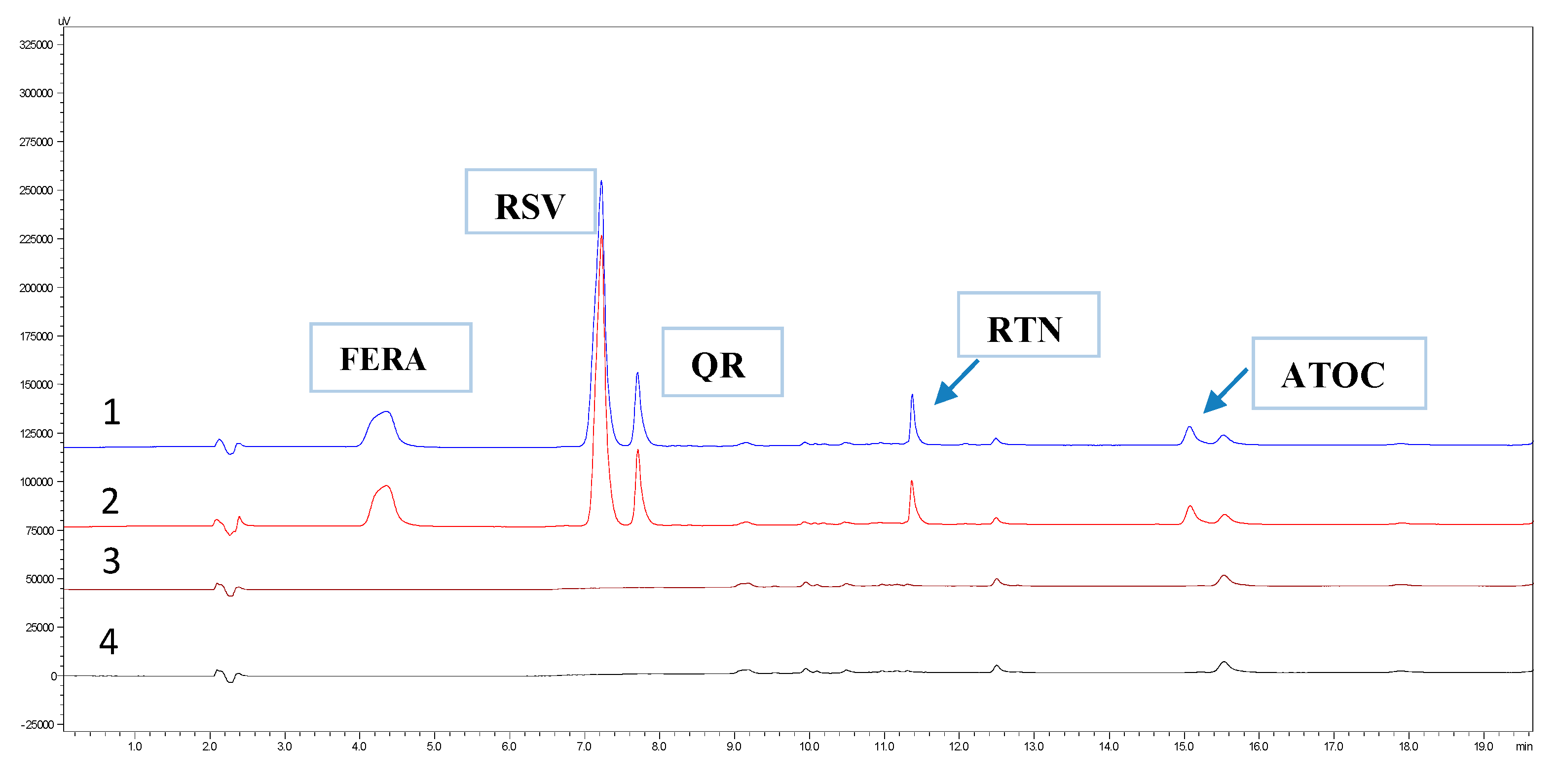
Under the present conditions, the chromatograms of a blank (MeOH/ACN/FA 0.2%) and a standard solution were compared (Figure 2). Comparing the results, it was found that in the elution time of the five substances, there was no interfering peak (co-elution).

3.2.3. Linearity

The linearity is evaluated based on the correlation diagram of concentrations (standard solutions) versus UV absorbance values (Table 3). Standard calibration curves were calculated by the least-squares method. Τhe good linearity of the method was also estimated by the % y-intercept values (intercept value × 100/100% response) for each analyte, which should be <2%.

3.2.4. Precision

Precision was tested within the same day (intra-day precision) and between three consecutive days (intermediate precision) at three concentration levels (low, intermediate, and high). In all cases, three repetitions were carried out for each sample (Table 4). Intra-day precision was additionally calculated after six repetitions at 100% of the labeled amount for each drug. The results indicate that the analytical method is reproducible, as the RSD % values were <2%.

3.2.5. Accuracy

To determine the precision, the % recovery values of the test substances at three corresponding concentration levels were calculated. The results are listed in Table 4 and range from 97.1–102.0%.

3.2.6. Limit of Detection (LOD) and Limit of Quantification (LOQ)

The detection and quantification limits were determined (Table 2) based on Equations (1) and (2):
LOD = 3.3   ×   S . D . S l o p e
LOQ = 10   ×   S . D . S l o p e
where S.D. is the standard deviation of the intercept and slope is the slope variable of the calibration curve.

3.2.7. Robustness

For the robustness of the system, the effect of small changes (±1) of some chromatographic parameters (λmax, flow rate of the mobile phase, and column temperature) on the tailing factor (Tf) and the AUC value (area under the curve) for each substance was investigated separately. According to the results (% RSD), none of these small changes affected the efficiency of the method (Table 5).

3.3. Quantitative Determination of Resveratrol at the Plant Extract

In the final product (cold cream), instead of the reference standard of resveratrol, RSV derived from the Regener Life Resveratrol® formulation was used. Therefore, the % recovery value of the substance had to be determined according to the following procedure. The content of one capsule was quantitatively transferred into a 25 mL volumetric flask and dissolved with MeOH-ACN-FA, 50:50:0.2 v/v/v. The sample was sonicated to facilitate dissolution of RSV, diluted (C = 25 μg·mL−1), centrifuged, and analyzed by HPLC (Supplementary Figure S5). The % recovery value of RSV was calculated (% recovery = 102.0%) relative to a reference standard solution (STD) with the same concentration.

3.4. Stability Study

Since the five active ingredients were subjected to heating (during the preparation of the cream and their analysis), it was deemed necessary to check their stability at a temperature of 45 °C and using an ultrasonic bath. For this reason, a mixed standard solution of RSV 0.780 μg·mL−1, FERA 0.515 μg·mL−1, QR 2.35 μg·mL−1, RTN 1.05 μg·mL−1, and ATOC 13.5 μg·mL−1 (diluent MeOH/ACN/FA 0.2%) was prepared. The solution was placed in conditions under examination, and at regular time intervals of 0, 10, 30, 60, and 80 min, samples were taken and analyzed by HPLC. The results show (Figure 3) that at 45 °C the substances are not destroyed, as they always give similar AUC values.

4. Extraction Procedure via Experimental Design

Finding the appropriate conditions for the best recovery of the APIs from the cream substrate was investigated using experimental design methodology and the “Design Expert 11” software. The study was carried out in order to determine the extraction solvents (methanol or acetonitrile), their participation ratios, as well as the time the samples should be subjected to ultrasonication and freezing. Thus, a D-optimal model was utilized, where the combination data of a mixture (A: acetonitrile B: methanol) was crossed with two factors, which were C: sonication and D: freezing time (Table 6).
Based on the experimental design methodology, 18 proposed combinations were applied, aiming at the optimal % recovery of the 5 APIs from the substrate (10 mg cream). For their quantification, a standard (STD) solution with the same concentration (FERA = 5 μg·mL−1, RSV = 10 μg·mL−1, QR = 10 μg·mL−1, RTN = 3 μg·mL−1, a-TOC = 20 μg·mL−1), a blank (cream substrate), and a blank (diluent) were also analyzed (Figure 2).
According to the results, only in three of the five responses were the understudy experimental factors significant (p-value < 0.05) and affecting the recoveries (% RTN, % RSV, and % FERA). The order of the combined (mixture × 2 factors) model was cubic × mean (p < 0.0001) for RTN, quadratic × mean (p = 0.0001) for RSV, and mean × linear (p = 0.0001) for FERA.
Subsequently, the significance of the examined factors (A, B, C, D) was assessed by the analysis of variance (ANOVA). Based on the results, it was found that factor D was not significant in any of these cases. Indeed, freezing is used to ensure the purity of the samples (precipitation of fatty impurities due to very low temperature) and is likely not affecting the recoveries of the substances. The overall evaluation of the models is determined by the values of specific statistical parameters presented in Table 7. The F value expresses the ratio of the systematic dispersion to the random unsystematic. When F is >1, it means that the factor has a significant effect, and the modeling is satisfactory. It is desirable that “R-squared” and “adjusted R-squared” have similar values, as this ideal case means that 100% of the observed variance can be explained by the model. Finally, the term “adequate precision” expresses the signal-to-noise ratio, and values > 4 are preferred.
The good predictive ability of the proposed models can also be revealed by the plots of the residuals against the predicted values, where there are no outliers (Supplementary Figure S6).
In order to find the best solution of the model, two approaches were applied:
(A) Graphical: It is based on the superimposition of the isometric curves in a single diagram (overlay plot) and then graphical localization of the region of optimal solutions (Supplementary Figure S7). Based on the diagram, it was found that the shaded region, characterized as the region of optimal solutions, satisfies the objectives set.
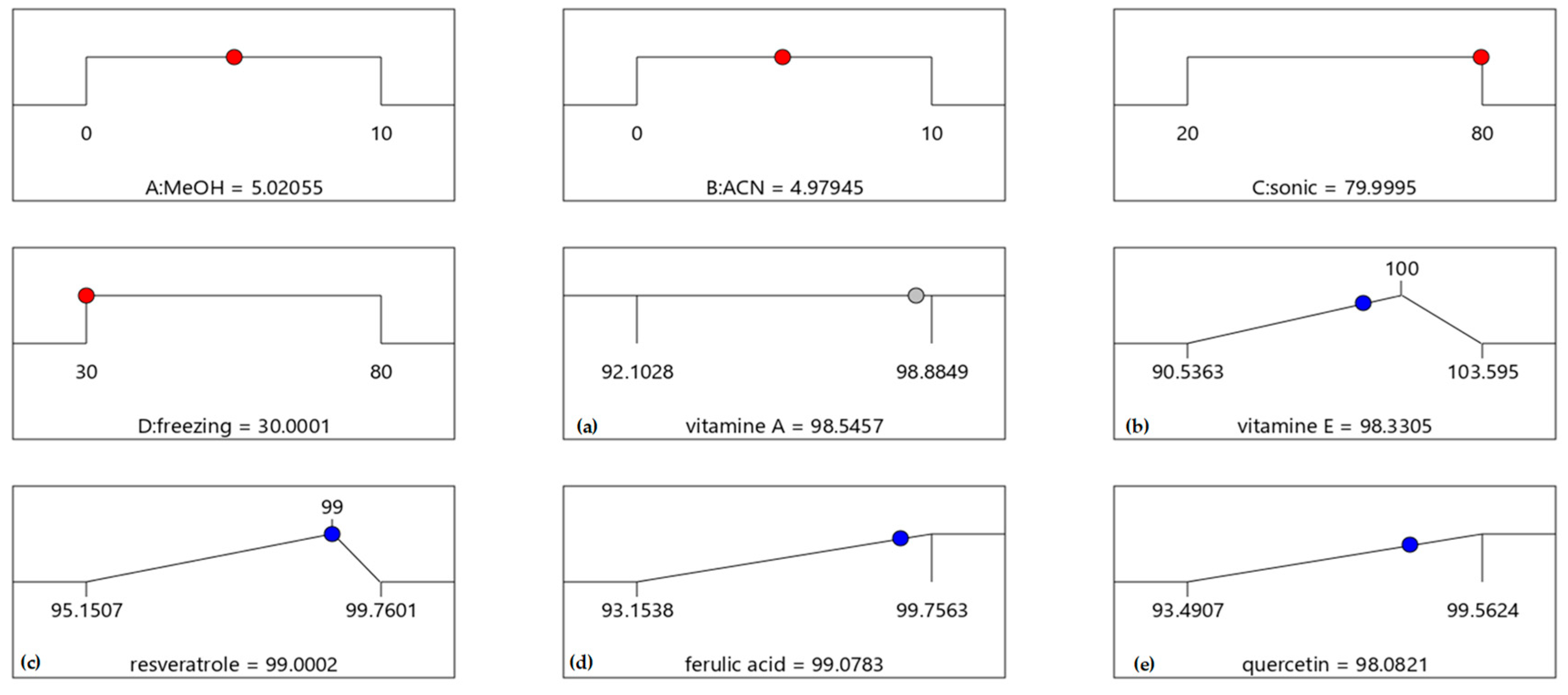
(B) Mathematical: It is based on the construction of mathematical optimization relations such as the desirability function whose value must tend to 1.0 (Figure 4).
The model suggested solvent composition was 5 mL MeOH and 5 mL ACN; the ideal sonication time was 80 min, and the freezing time was 30 min (desirability = 0.865). Commenting on the results, we could say that methanol and ultrasonication contribute to the effective recovery of the five APIs from the cream, while acetonitrile and freezing help to sample purification. To check the reliability of the method, five samples were analyzed, and their % recovery values were calculated (Table 8).
The resulting recoveries demonstrate the effectiveness of the method since they range from 98.5–102.8%.

5. Permeability Study with Franz Cells

After formulating the cold cream and developing a reliable analytical method, an in vitro penetration study of APIs was performed using vertical Franz cells and a cellulose membrane setup. Although cellulose is generally characterized as a polar membrane, on its surface contains OH and carbonyl groups as functional groups and a carbon skeleton (CH2) that give it a partially amphiphilic character. We could say that the hydrophobic nature of the glycopyranose plane allows the cellulose chains to stack through hydrophobic interactions, creating a polymer-like structure. Thus, its structural similarity to biological membranes (phospholipid bilayer) allows cellulose to be used as a marker of drug permeation in in vitro studies [42]. Zhu et al. reveal that a hydrophilic artificial membrane simulates more closely drug permeation into tissue at levels comparable to Caco-2 permeation and is significantly superior to in silico techniques [43]. It should, of course, be noted that the epidermis, due to the existence of keratinocytes on its surface, is a special category of tissue and needs additional ex vivo and in vivo experiments in order to be studied thoroughly.
In the present permeation study, comparative experiments were performed using two different carriers, cold cream and an aqueous suspension. Both formulations, since the five actives were incorporated, were studied in Franz cells in triplicate. After sampling, and to remove PBS salts, samples were lyophilized and reconstituted with methanol.
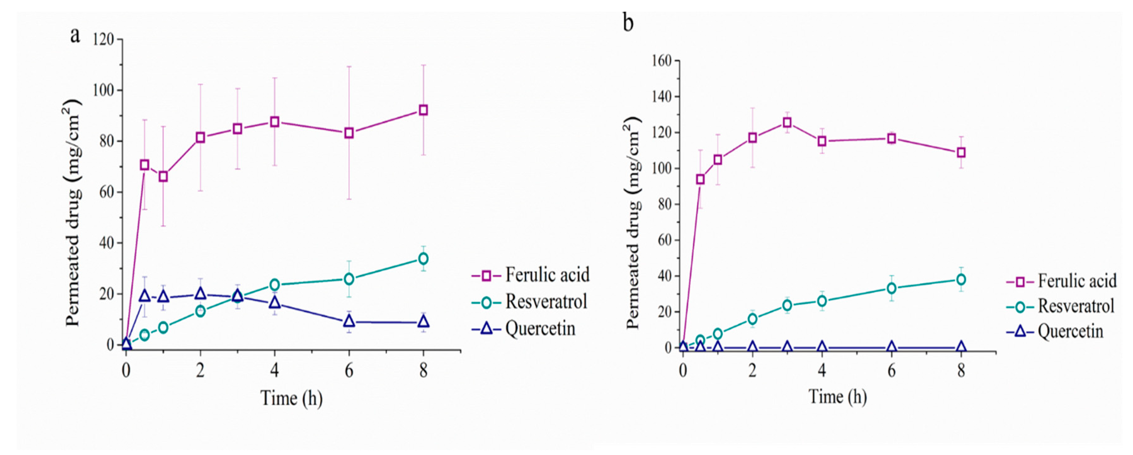
Supplementary Table S1 provides an overall presentation of the most important physicochemical properties of the five drugs under study, which determine the permeability (paracellular, intercellular, and intercellular) of a molecule through a tissue or membrane. The profile of the cumulative amount of each compound that permeated per unit area (mg·cm−2), at the sampling times, is illustrated in Figure 5, while their steady-state flux Jss and Papp coefficient values are recorded in Table 9.
According to the results, the substance with the greatest tendency to penetrate the membrane is ferulic acid, followed by resveratrol and quercetin, while the two vitamins show no activity. This behavior was partly expected since ferulic acid is the most polar substance and has the lowest molecular weight. In fact, its high solubility in water indicates that the aqueous carrier is the most effective. RSV, although more lipophilic than QR, is more membrane permeable due to its smaller size (MW). Furthermore, it appears that quercetin, when incorporated into the suspension, barely penetrates the membrane, while when present in the cream, its penetration is more significant. This is probably due to the fact that it is more soluble in the cold cream (due to coconut and almond oils) than in the aqueous suspension [44]. Regarding the two vitamins, it was found that due to their high lipophilicity and molecular weight, they do not penetrate the membrane in either of the two carriers. The different penetration abilities of the five substances are desirable since they must act both on the layers of the epidermis and on the inner layers of the skin. Given that they have a similar and/or complementary action, they can treat, in this way, the problem of aging.
Table 10 illustrates the amounts of APIs added to the donor (Franz cells) and the corresponding amounts trapped in the membrane or transferred to the acceptor.
Comparing the results, it appears that the carrier that ensures the best sample homogeneity, especially for the most lipophilic substances, is the cream (w/o), while the suspension presents integration problems. An additional reason why the aqueous suspensions are generally avoided is that they usually hydrolyze and destroy the API’s [45].

6. Conclusions

The present research study aims to develop a reliable and sensitive analytical method for the quantitative determination of five antioxidant drugs in a cold cream prepared in the laboratory. Purification conditions for the quantitative recovery of the analytes from the cream substrate were decided based on an experimental design methodology. The method was efficient, easy to operate, and short enough to be applied in routine analyses. A corresponding method was successfully used to study the transdermal penetration of the five drugs (in cream and suspension) with vertical Franz cells. Based on the results, the APIs act synergistically since their degree of penetration into the skin is different, ensuring an overall better antioxidant regenerative effect on the skin.
The overall effort is expected to yield new insights into the field of anti-aging cosmetics.

Supplementary Materials

The following supporting information can be downloaded at https://www.mdpi.com/article/10.3390/app142411843/s1, Figure S1: Chromatographic separation of the five substances in (A) phenyl and (B) C18 columns at 290, 370, 326 and 320 nm; Table S1: Physicochemical properties of the studied compounds; Figure S2: Chromatograms (at 290 nm) of three different mobile phases with acetonitrile/water 50:50 v/v and (1) formic acid 0.2% (FA), (2) acetic acid 0.2% (ACA) and (3) trifluoroacetic acid 0.1% (TFA); Figure S3: Ultraviolet (UV) spectra of the analytes; Figure S4: Chromatogram of the five substances at different wavelengths and Blank sample chromatogram; Figure S5: Chromatogram of resveratrol in the formulation (tR = 8.6 min); Figure S6: Residue plots of the five substances; Figure S7: Diagram of the superposition of the isometric curves.

Author Contributions

Conceptualization, C.K.M. and A.K.; methodology, C.K.M.; software, C.K.M.; formal analysis, all authors; investigation, all authors; writing—original draft preparation, C.K.M.; writing—review and editing, C.K.M., S.A. and A.K. All authors have read and agreed to the published version of the manuscript.

Funding

This research received no external funding.

Institutional Review Board Statement

Not applicable.

Informed Consent Statement

Not applicable.

Data Availability Statement

The raw data supporting the conclusions of this article can be made available by the authors upon reasonable request.

Conflicts of Interest

The authors declare no conflicts of interest.

References

  1. Gallo, R.L. Human Skin Is the Largest Epithelial Surface for Interaction with Microbes. J. Investig. Dermatol. 2017, 137, 1213–1214. [Google Scholar] [CrossRef] [PubMed]
  2. Bonté, F.; Girard, D.; Archambault, J.C.; Desmoulière, A. Skin Changes During Ageing. In Biochemistry and Cell Biology of Ageing: Part II Clinical Science; Springer: Singapore, 2019; pp. 249–280. [Google Scholar] [CrossRef]
  3. Kim, J.; Kim, J.; Lee, Y.I.; Almurayshid, A.; Jung, J.Y.; Lee, J.H. Effect of a topical antioxidant serum containing vitamin C, vitamin E, and ferulic acid after Q-switched 1064-nm Nd:YAG laser for treatment of environment-induced skin pigmentation. J. Cosmet. Dermatol. 2020, 19, 2576–2582. [Google Scholar] [CrossRef]
  4. Sunder, S. Relevant Topical Skin Care Products for Prevention and Treatment of Aging Skin. Facial Plast. Surg. Clin. N. Am. 2019, 27, 413–418. [Google Scholar] [CrossRef] [PubMed]
  5. Puizina-Ivic, N. Skin aging. Acta Dermatovenerol. Alp. Panon. Adriat. 2008, 17, 47–54. [Google Scholar]
  6. Sjerobabski-Masnec, I.; Situm, M. Skin aging. Acta Clin. Croat. 2010, 49, 515–518. [Google Scholar] [PubMed]
  7. Cao, C.; Xiao, Z.; Wu, Y.; Ge, C. Diet and Skin Aging—From the Perspective of Food Nutrition. Nutrients 2020, 12, 870. [Google Scholar] [CrossRef]
  8. Prajakta, S.; Shahu, K. Formulation and Evaluation of Vanishing Herbal Cream of Crude Drugs. Asian J. Pharm. Res. Dev. 2020, 8, 66–69. [Google Scholar] [CrossRef]
  9. Niki, E.; Abe, K. Vitamin E: Structure, Properties and Functions. In Vitamin E: Chemistry and Nutritional Benefits; Food Chemistry, Function and Analysis Series; The Royal Society of Chemistry: London, UK, 2019; Chapter 1; pp. 1–11. [Google Scholar] [CrossRef]
  10. Janssens-Böcker, C.; Kerscher, M. Skin Anti-Aging Benefits of a 2% Resveratrol Emulsion. J. Cosmet. Dermatol. Sci. Appl. 2021, 11, 155–168. [Google Scholar] [CrossRef]
  11. Zduńska, K.; Dana, A.; Kolodziejczak, A.; Rotsztejn, H. Antioxidant Properties of Ferulic Acid and Its Possible Application. Ski. Pharmacol. Physiol. 2018, 31, 332–336. [Google Scholar] [CrossRef]
  12. Zasada, M.; Budzisz, E.; Kolodziejska, J.; Kalinowska-Lis, U. An evaluation of the physicochemical parameters and the content of the active ingredients in original formulas containing retinol. J. Cosmet. Dermatol. 2020, 19, 2374–2383. [Google Scholar] [CrossRef]
  13. Quan, T. Human Skin Aging and the Anti-Aging Properties of Retinol. Biomolecules 2023, 13, 1614. [Google Scholar] [CrossRef] [PubMed]
  14. Park, K.; Lee, J.H. Protective effects of resveratrol on UVB-irradiated HaCaT cells through attenuation of the caspase pathway. Oncol. Rep. 2008, 19, 413–417. [Google Scholar] [CrossRef] [PubMed]
  15. Shin, E.J.; Lee, J.S.; Hong, S.; Lim, T.G.; Byun, S. Quercetin Directly Targets JAK2 and PKCδ and Prevents UV-Induced Photoaging in Human Skin. Int. J. Mol. Sci. 2019, 20, 5262. [Google Scholar] [CrossRef]
  16. Rattanawiwatpong, P.; Wanitphakdeedecha, R.; Bumrungpert, A.; Maiprasert, M. Anti-aging and brightening effects of a topical treatment containing vitamin C, vitamin E, and raspberry leaf cell culture extract: A split-face, randomized controlled trial. J. Cosmet. Dermatol. 2020, 19, 671–676. [Google Scholar] [CrossRef] [PubMed]
  17. Truong, V.L.; Jun, M.; Jeong, W.S. Role of resveratrol in regulation of cellular defense systems against oxidative stress. BioFactors 2018, 44, 36–49. [Google Scholar] [CrossRef]
  18. Wolter, F.; Stein, J. Biological activities of resveratrol and its analogs. Drugs Future 2002, 27, 949–959. [Google Scholar] [CrossRef]
  19. Zinder, R.; Cooley, R.; Vlad, L.G.; Molnar, J.A. Vitamin A and Wound Healing. Nutr. Clin. Pract. 2019, 34, 839–849. [Google Scholar] [CrossRef]
  20. Lin, F.-H.; Lin, J.-Y.; Gupta, R.D.; Tournas, J.A.; Burch, J.A.; Selim, M.A.; Monteiro-Riviere, N.A.; Grichnik, J.M.; Zielinski, J.; Pinnell, S.R. Ferulic Acid Stabilizes a Solution of Vitamins C and E and Doubles its Photoprotection of Skin. J. Investig. Dermatol. 2005, 125, 826–832. [Google Scholar] [CrossRef] [PubMed]
  21. Zouboulis, C.C.; Ganceviciene, R.; Liakou, A.I.; Theodoridis, A.; Elewa, R.; Makrantonaki, E. Aesthetic aspects of skin aging, prevention, and local treatment. Clin. Dermatol. 2019, 37, 365–372. [Google Scholar] [CrossRef] [PubMed]
  22. Thiele, J.J.; Hsieh, S.N.; Ekanayake-Mudiyanselage, S. Vitamin E: Critical review of its current use in cosmetic and clinical dermatology. Dermatol. Surg. 2005, 31, 805–813. [Google Scholar] [CrossRef] [PubMed]
  23. Casagrande, R.; Baracat, M.M.; Georgetti, S.R.; Verri, W.A., Jr.; Vicentini, F.T.; Rafael, J.A.; Jabor, J.R.; Fonseca, M.J. Method validation and stability study of quercetin in topical emulsions. Química Nova 2009, 32, 1939–1942. [Google Scholar] [CrossRef]
  24. Fasolo, D.; Schwingel, L.; Holzschuh, M.; Bassani, V.; Teixeira, H. Validation of an isocratic LC method for determination of quercetin and methylquercetin in topical nanoemulsions. J. Pharm. Biomed. Anal. 2007, 44, 1174–1177. [Google Scholar] [CrossRef]
  25. Lai, J.F.; Franke, A.A. Analysis of circulating lipid-phase micronutrients in humans by HPLC: Review and overview of new developments. J. Chromatogr. B 2013, 931, 23–41. [Google Scholar] [CrossRef] [PubMed]
  26. Gundersen, T.E.; Blomhoff, R. Qualitative and quantitative liquid chromatographic determination of natural retinoids in biological samples. J. Chromatogr. A 2001, 935, 13–43. [Google Scholar] [CrossRef] [PubMed]
  27. Tee-ngam, P.; Nunant, N.; Rattanarat, P.; Siangproh, W.; Chailapakul, O. Simple and Rapid Determination of Ferulic Acid Levels in Food and Cosmetic Samples Using Paper-Based Platforms. Sensors 2013, 13, 13039–13053. [Google Scholar] [CrossRef] [PubMed]
  28. Barberousse, H.; Roiseux, O.; Robert, C.; Paquot, M.; Deroanne, C.; Blecker, C. Analytical methodologies for quantification of ferulic acid and its oligomers. J. Sci. Food Agric. 2008, 88, 1494–1511. [Google Scholar] [CrossRef]
  29. Marcato, D.C.; Spagnol, C.M.; Salgado, H.R.N.; Isaac, V.L.B.; Corrêa, M.A. New and potential properties, characteristics, and analytical methods of ferulic acid: A review. Braz. J. Pharm. Sci. 2022, 58, e18747. [Google Scholar] [CrossRef]
  30. Wang, J.; Shi, A.; Agyei, D.; Wang, Q. Formulation of water-in-oil-in-water (W/O/W) emulsions containing trans-resveratrol. RSC Adv. 2017, 7, 35917–35927. [Google Scholar] [CrossRef]
  31. Scalia, S.; Trotta, V.; Iannuccelli, V.; Bianchi, A. Bianchi Enhancement of in vivo human skin penetration of resveratrol by chitosan-coated lipid microparticles. Colloids Surf. B Biointerfaces 2015, 135, 42–49. [Google Scholar] [CrossRef] [PubMed]
  32. Silva, L.F.D.; Guerra, C.C.; Czermainski, A.B.C.; Ferrari, L.; Bergold, A.M. Validation of a chromatographic method to routine analysis of trans-resveratrol and quercetin in red wines. Pesqui. Agropecuária Bras. 2017, 52, 335–343. [Google Scholar] [CrossRef]
  33. Mansour, F.R.; Abdallah, I.A.; Bedair, A.; Hamed, M. Analytical Methods for the Determination of Quercetin and Quercetin Glycosides in Pharmaceuticals and Biological Samples. Crit. Rev. Anal. Chem. 2023, 54, 1–26. [Google Scholar] [CrossRef] [PubMed]
  34. Fiod Riccio, B.V.; Fonseca-Santos, B.; Colerato Ferrari, P.; Chorilli, M. Characteristics, Biological Properties and Analytical Methods of Trans-Resveratrol: A Review. Crit. Rev. Anal. Chem. 2020, 50, 339–358. [Google Scholar] [CrossRef] [PubMed]
  35. Kamaris, G.; Dalavitsou, A.; Markopoulou, C.K. Development and Validation of an HPLC-DAD Method for the Determination of Seven Antioxidants in a Nano-Emulsion: Formulation and Stability Study. Separations 2024, 11, 43. [Google Scholar] [CrossRef]
  36. Thongchai, W.; Liawruangrath, B. Development of HPLC analysis for the determination of retinol and alpha tocopherol in corn oil nanoemulsion lotion. Int. Food Res. J. 2016, 23, 1367. [Google Scholar]
  37. Ahmed, O.A.; El-Bassossy, H.M.; El-Sayed, H.M.; El-Hay, S.S.A. Rp-HPLC Determination of Quercetin in a Novel D-α-Tocopherol Polyethylene Glycol 1000 Succinate Based SNEDDS Formulation: Pharmacokinetics in Rat Plasma. Molecules 2021, 26, 1435. [Google Scholar] [CrossRef]
  38. Benedetti, B.; Caponigro, V.; Ardini, F. Experimental Design Step by Step: A Practical Guide for Beginners. Crit. Rev. Anal. Chem. 2022, 52, 1015–1028. [Google Scholar] [CrossRef]
  39. Politis, S.N.; Colombo, P.; Colombo, G.; Rekkas, D.M. Design of experiments (DoE) in pharmaceutical development. Drug Dev. Ind. Pharm. 2017, 43, 889–901. [Google Scholar] [CrossRef]
  40. Yadav, R.; Thakur, S.; Parihar, R.; Chauhan, U.; Chanana, A.; Chawra, H.S. Pharmaceutical Preparation and Evaluation of Cold Cream. Int. J. Innov. Sci. Res. Technol. 2023, 8, 1069–1075. [Google Scholar]
  41. ICH. ICH Q2(R2) Guideline on Validation of Analytical Procedures. 2023. Available online: https://www.ema.europa.eu/en/ich-q2r2-validation-analytical-procedures-scientific-guideline (accessed on 5 November 2023).
  42. Tsanaktsidou, E.; Chatzitaki, A.T.; Chatzichristou, A.; Fatouros, D.G.; Markopoulou, C.K. A Comparative Study and Prediction of the Ex Vivo Permeation of Six Vaginally Administered Drugs across Five Artificial Membranes and Vaginal Tissue. Molecules 2024, 29, 2334. [Google Scholar] [CrossRef]
  43. Berben, P.; Bauer-Brandl, A.; Brandl, M.; Faller, B.; Flaten, G.E.; Jacobsen, A.C.; Brouwers, J.; Augustijns, P. Drug permeability profiling using cell-free permeation tools: Overview and applications. Eur. J. Pharm. Sci. 2018, 119, 219–233. [Google Scholar] [CrossRef] [PubMed]
  44. Moghimipour, E.; Farsimadan, N.; Salimi, A. Ocular Delivery of Quercetin Using Microemulsion System: Design, Characterization, and Ex-vivo Transcorneal Permeation. Iran. J. Pharm.Res. IJPR 2022, 21, e127486. [Google Scholar] [CrossRef]
  45. Francioso, A.; Mastromarino, P.; Restignoli, R.; Boffi, A.; d’Erme, M.; Mosca, L. Improved stability of trans-resveratrol in aqueous solutions by carboxymethylated (1, 3/1, 6)-β-D-glucan. J. Agric. Food Chem. 2014, 62, 1520–1525. [Google Scholar] [CrossRef] [PubMed]
Figure 1. Chemical structures of RSV, FERA, QR, RTN, and α-ΤΟC.
Applsci 14 11843 g001
Figure 2. Chromatogram at 290 nm of (1) standard solution, (2) sample, (3) blank of diluent, and (4) substrate blank.
Applsci 14 11843 g002
Figure 3. Stability study of substances at the temperature of 45 °C.
Applsci 14 11843 g003
Figure 4. Suggested optimal values of factors (A) MeOH (mL), (B) ACN (mL), (C) sonication time (min) and (D) freezing time (min) and predicted % recovery values of (a): vitamin A, (b): vitamin E, (c): resveratrol, (d): ferulic acid, (e): quercetin.
Applsci 14 11843 g004
Figure 5. Permeability study of ferulic acid, resveratrol, and quercetin (a) cream, (b) suspension.
Applsci 14 11843 g005
Table 1. Cream bases with different proportions of ingredients.
IngredientsCream 1Cream 2Cream 3Cream 4Cream 5
White wax2.0 g2.0 g2.0 g2.0 g2.0 g
Cetyl alcohol0.3 g0.3 g0.3 g0.3 g0.3 g
Coconut oil9.0 g3.0 g7.0 g4.5 g3.0 g
Almond oil-3.0 g---
Borax0.16 g0.16 g0.16 g0.16 g0.16 g
Deionized water5.16 g5.16 g5.16 g5.16 g5.16 g
Total16.62 g16.62 g14.62 g12.12 g10.62 g
Table 2. System suitability parameters.
SubstancesRetention Time Tailing Factor Capacity FactorResolutionTheoretical Plates HΕΤP
mm × 10−3
(min)(Tf)(k′)(Rs)(Ν)
FERA4.40.91.1 1109225.4
RSV7.20.92.67.513,90218.0
QR7.21.62.92.543,3345.8
RTN11.41.84.725.099,7902.5
α-TOC15.01.76.520.071,3173.5
Table 3. Statistical results for linearity.
AnalytesConcentration Range (μg·mL−1) Calibration Curves%y-Intercept ValuesCorrelation Coefficient (r2)LOD (μg·mL−1)LOQ (μg·mL−1)
RSV0.4–3.2y = 186,124x − 1035.50.750.9980.130.39
FERA0.2–2.1 y = 128,958x − 199.10.30.9990.040.12
QR1.2–9.4y = 98,150x − 1391.00.60.9980.481.45
RTN0.6–4.2 y = 87,131x − 705.80.80.9990.220.66
ATOC6.4–27.0 y = 4171.8x − 1260.82.00.9982.286.91
Table 4. Results of the reliability tests.
APIsIntraday PrecisionInter-Day Precision
Concentrations%RSD (n = 3) 1st Day% Recovery%RSD (n = 6)%RSD (n = 3)
(μg·mL−1)2nd Day3rd DayTotal
FERA0.12.0102.0 2.01.22.0
0.31.498.70.11.20.11.0
2.10.199.6 0.31.50.2
RSV0.20.1101.0 0.81.42.0
0.470.1100.10.12.01.71.5
3.11.6100.5 0.30.41.0
QR0.60.1100.2 1.60.11.2
1.41.397.10.11.41.62.0
9.41.3100.8 0.20.21.9
RTN0.31.0100.8 1.40.21.6
0.60.5100.70.20.30.51.2
4.21.597.6 0.20.40.4
α-TOC3.20.799.7 2.00.31.7
8.11.6100.00.60.00.41.3
271.7100.2 0.40.20.4
Table 5. Robustness tests for the five analytes.
% RSD
ParametersAUC RSVTf RSVAUC QRTf QRRs RSV-QRAUC FERATf FERAAUC RTNTf RTNAUC α-TOCTf α-TOC
According to USP
Flow rate mL/min5.74.95.92.95.53.75.05.91.04.76.5
(1.4, 1.5, 1.6)
Column T (°C)1.04.50.85.73.50.84.95.12.63.84.5
(39°,40°, 41°)
λmax0.41.10.11.22.50.51.22.34.92.01.4
Table 6. D-optimal experimental design considered limits and selected responses.
Mixture UnitsLimits
AMeOHmL0–10
BACNmL0–10
Process
Csonicmin20–80 min
Dfreezingmin30–80 min
Responses
R1RTN% Recovery
R2A-TOC% Recovery
R3RSV% Recovery
R4FERA% Recovery
R5QR% Recovery
Table 7. Best fitting models for selected responses.
ANOVARTNRSVFERA
R20.79310.83840.7279
Adjusted R20.74880.80380.6916
Predicted R20.59590.73670.6082
Adeq. Precision11.369.8110.03
F17.8924.2120.06
C.V. %1.030.631.63
Table 8. % recovery values (ν = 5) applying the proposed purification procedure.
% Recovery
SampleRSVFERAQRRTNα-TOC
198.999.998.6100.999.3
299.499.499.5102.9100.8
399.3100.098.9102.5100.0
499.199.798.5101.399.7
598.599.197.7101.199.0
%RSD0.40.40.60.90.7
Table 9. Steady-state flux Jss and Papp coefficient values for ferulic acid, resveratrol, and quercetin.
CellJ (μg/cm2/h)Papp (h/cm2) × 10−3
FERARSVQRFERARSVQR
Cream2.344 ± 0.1313.276 ± 0.6741.890 ± 0.3390.455 ± 0.0260.323 ± 0.0660.188 ± 0.034
Solution3.303 ± 0.8014.466 ± 0.566-0.578 ± 0.1350.315 ± 0.065-
Table 10. Amounts of APIs loaded and found in the reference sample (cold cream and suspension), in the donor, and acceptor chambers and in the membrane tissue.
Amount of Drug in Suspension Amount of Drug in Cream
DrugReference SampleDonorAcceptorMembraneReference SampleDonorAcceptorMembrane
Loaded
(μg)
Found
(μg)
Loaded Sample
(μg)
Final Found
(μg)
Final Found
(μg)
Loaded
(μg)
Found
(μg)
Loaded Sample
(μg)
Final Found
(μg)
Final Found
(μg)
RTN750.0337.5750.0-315.22040.02092.52040.0-1875.8
α-TOC5000.01887.55000.0-1988.6639.16590.0639.1-5765.7
RSV2600.0545.02600.0169.8349.43235.33163.03235.3155.52693.8
FERA1300.0565.01300.0432.431.51641.61426.31641.6397.7726.2
QR2500.02232.52500.0-2132.33417.03652.53417.022.72871.6
Disclaimer/Publisher’s Note: The statements, opinions and data contained in all publications are solely those of the individual author(s) and contributor(s) and not of MDPI and/or the editor(s). MDPI and/or the editor(s) disclaim responsibility for any injury to people or property resulting from any ideas, methods, instructions or products referred to in the content.