Article

Sjögren’s Syndrome: The Role of Serological Profiles Versus Minor Salivary Gland Histopathology

1
Institute of Clinical Dentistry, Faculty of Dentistry, University of Oslo, 0317 Oslo, Norway
2
Department of Clinical Medicine, Faculty of Medicine, University of Bergen, 5020 Bergen, Norway
*
Author to whom correspondence should be addressed.
Appl. Sci. 2024, 14(24), 11482; https://doi.org/10.3390/app142411482
Submission received: 8 September 2024 / Revised: 21 November 2024 / Accepted: 3 December 2024 / Published: 10 December 2024

Abstract

:
Between the years 2008 and 2023, 306 patients were referred to the Department of Oral Surgery and Oral Medicine, Faculty of Dentistry, University of Oslo, for salivary gland biopsy after clinical suspicion of Sjögren’s syndrome. This study aimed to investigate possible associations between serology and minor salivary gland histopathology by stratifying patients according to serology features (negative, single, or double seropositivity according to anti-SSA and anti-SSB), focus score (FS, negative, or actual score), and germinal centers (GCs). Information was collected from referrals and visits. Collected information included oral and ocular dryness symptoms, sialometry, Schirmer I test, histopathological analysis, and serological findings. Patients were classified using the 2002 American European Consensus Group classification criteria and were stratified into seven subgroups according to their serological profiles and FS, which were compared. The majority of patients were females, and the most frequently referred age group was 50–59 years of age. Most patients had both oral and ocular symptoms. Seropositive patients had higher median FS compared to seronegative patients. Double-seropositive patients had significantly higher median FS compared to single-seropositive patients. The results indicated that anti-SSB together with anti-SSA plays a role in disease severity in the salivary glands. In addition to anti-SSA, anti-SSB should be routinely evaluated as a possible predictor of the severity of inflammatory destruction in the salivary gland tissue.

1. Introduction

Sjögren’s syndrome (SS) is an autoimmune disease characterized by chronic inflammation that manifests with sicca syndrome—an array of dryness symptoms arising from impaired salivary gland and/or lacrimal gland functioning [1,2]. The chronic inflammation and autoantibody production lead to destruction of exocrine glands through mononuclear cell infiltration. On a microscopic level, chronic inflammatory cells are recognized in the salivary gland biopsy [3,4] and/or the lacrimal gland biopsy [4]. The etiology of SS remains unknown, though current discussion suggests an interaction between genetics, hormones, and environmental factors. Prior infection is also speculated to be a contributing part [2,5].
There is no curative treatment for SS, and available treatment options are aimed principally at managing symptoms. Xerostomia (the subjective sensation of dry mouth) is a key complaint in oral manifestations of SS, and other symptoms—such as difficulty with eating, swallowing, speaking, and gustatory perception—are well discussed [6]. For the management of xerostomia and its associated symptoms, it is recommended that patients drink small amounts of water regularly, use chewing gum or lozenges to stimulate salivary secretion, use oral lubricants to substitute saliva, apply oral gels, and use specialized mouthwash and toothpaste, as well as oral rinses [7]. Xeropthalmia (dryness of the eyes due to inadequate tear production) is SS’s most common ocular manifestation. Related ocular symptoms include a burning sensation, redness of the eyes, itching, and sensitivity to light [8]. Current therapeutic options for ocular manifestations of SS include artificial tear drops, immunosuppressant medications, and tear-stimulating drugs [7]. Other recommendations are humidity-preserving glasses and surgery to reduce fluid drainage from the eyes (punctal occlusion) [7,9]. Overall, SS has strong implications for patient health outcomes, where studies show that having the disease exposes the patient to a higher risk for dental caries, oral and ocular infections [6,8], lower quality of life [10], and an increased risk of lymphoma [2,11].
Currently, there are no diagnostic criteria for SS. Instead, classification criteria are used to assist in the diagnostic work-up of patients [12], where the final diagnosis is made by considering the full clinical picture of each patient, involving a comprehensive evaluation of symptoms, clinical findings, and laboratory results. Newer studies also make the distinction of non-Sjögren’s sicca syndrome patients (non-SS)—those who share the symptoms of dryness common to (primary Sjögren’s syndrome (pSS) patients but do not fulfil the specific classification criteria [13]. This distinction is important as recent findings suggest that this group has a poorer general and oral health-related quality of life than SS patients, where individually tailored treatment would be desirable to achieve the best patient outcomes [13].
Commonly used criteria to classify patients with pSS include the 2002 American–European Consensus Group (AECG) classification criteria [14] and the 2016 American College of Rheumatology/European League Against Rheumatism (ACR-EULAR) classification criteria [15].
The complexity of the varying clinical presentations of SS suggests that a pSS diagnosis may be an umbrella term representing more than one pathology [16]. Yet, the possible number of pathologies and their pathogenesis remain unclear. Overall, there is a clear need to stratify SS patients in order to develop streamline treatment.
There is a growing body of evidence that a positive focus score (FS)—defined as the accumulation of 50 or more inflammatory cells in a 4 mm2 area of glandular tissue—correlates with disease severity, with research showing an association with the development of salivary gland dysfunction, extra-glandular manifestations such as arthritis, and the development of lymphoma [11,17]. Likewise, the presence of germinal centers (GCs)—ectopic lymphoid-like structures found in salivary gland biopsies—has been shown to be associated with worse patient outcomes [11,18]. Even amongst non-SS patients, a histopathological abnormality is not uncommon, with studies showing significant replacement of glandular tissue with fatty infiltrations [19].
Further, evidence is growing that there is an association between seropositivity and disease severity [20]. Yet, only a few studies use comprehensive datasets that differentiate serologic phenotypes or use a sufficiently large dataset to make meaningful conclusions. To be more precise, the current classification alternatives do not report the heterogeneity in SS-associated patterns for antinuclear antibodies (ANA)—antibodies that target cellular nuclear components—specifically anti-SSA and anti-SSB antibodies. More specifically, the AECG classification criteria do not differentiate between single-seropositive patients (anti-SSA or anti-SSB antibodies) or double-seropositive patients (anti-SSA and anti-SSB antibodies) in its classification, where either yields a positive finding; and the ACR-EULAR classification only utilizes the presence of anti-SSA antibodies as a positive score. This is significant when one considers the health implications of different combinations of antibodies, where studies hint at an association between the different serologic phenotypes and disease activity [20].
These findings reinforce the need to stratify SS patients with routinely collected information, such as serology and histopathology to differentiate disease subtypes. This may help improve diagnosis and provide targeted treatment and adequate follow-up of affected patients.

Aims and Goals

To investigate associations between serology and histopathology by stratifying the patients in the Faculty of Dentistry’s dataset according to serology (negative, single, or double seropositivity), FS, and GCs. Further, the AECG and ACR-EULAR classification criteria are compared for the present dataset.

2. Materials and Methods
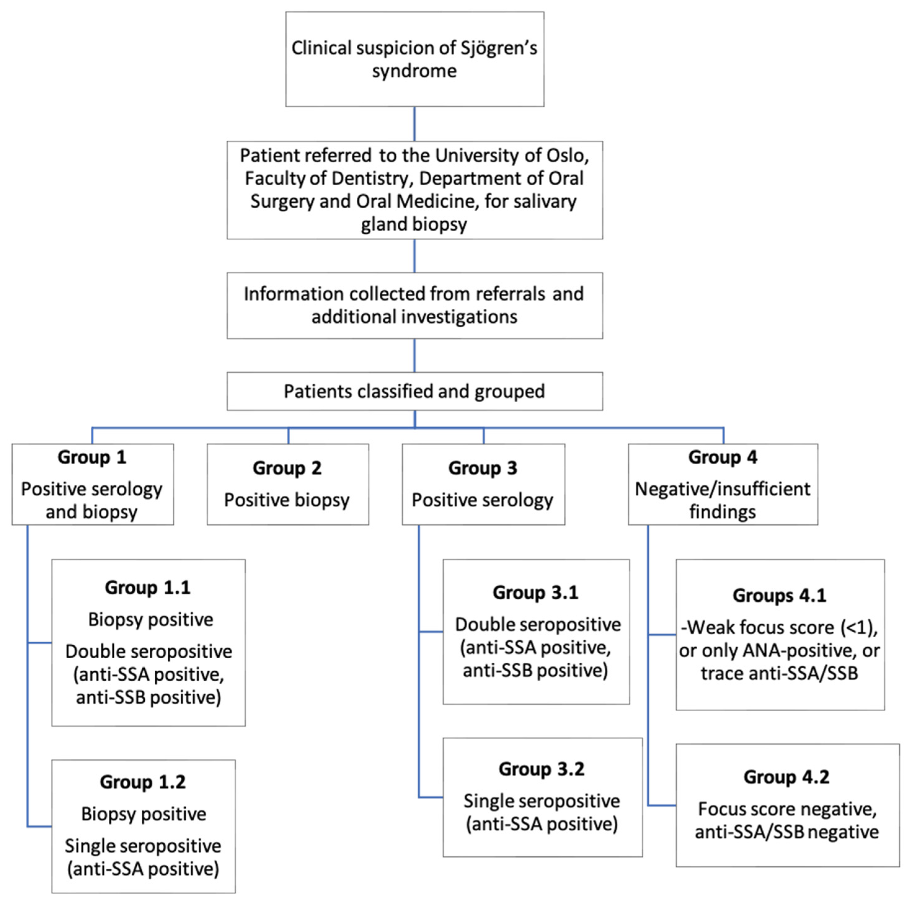
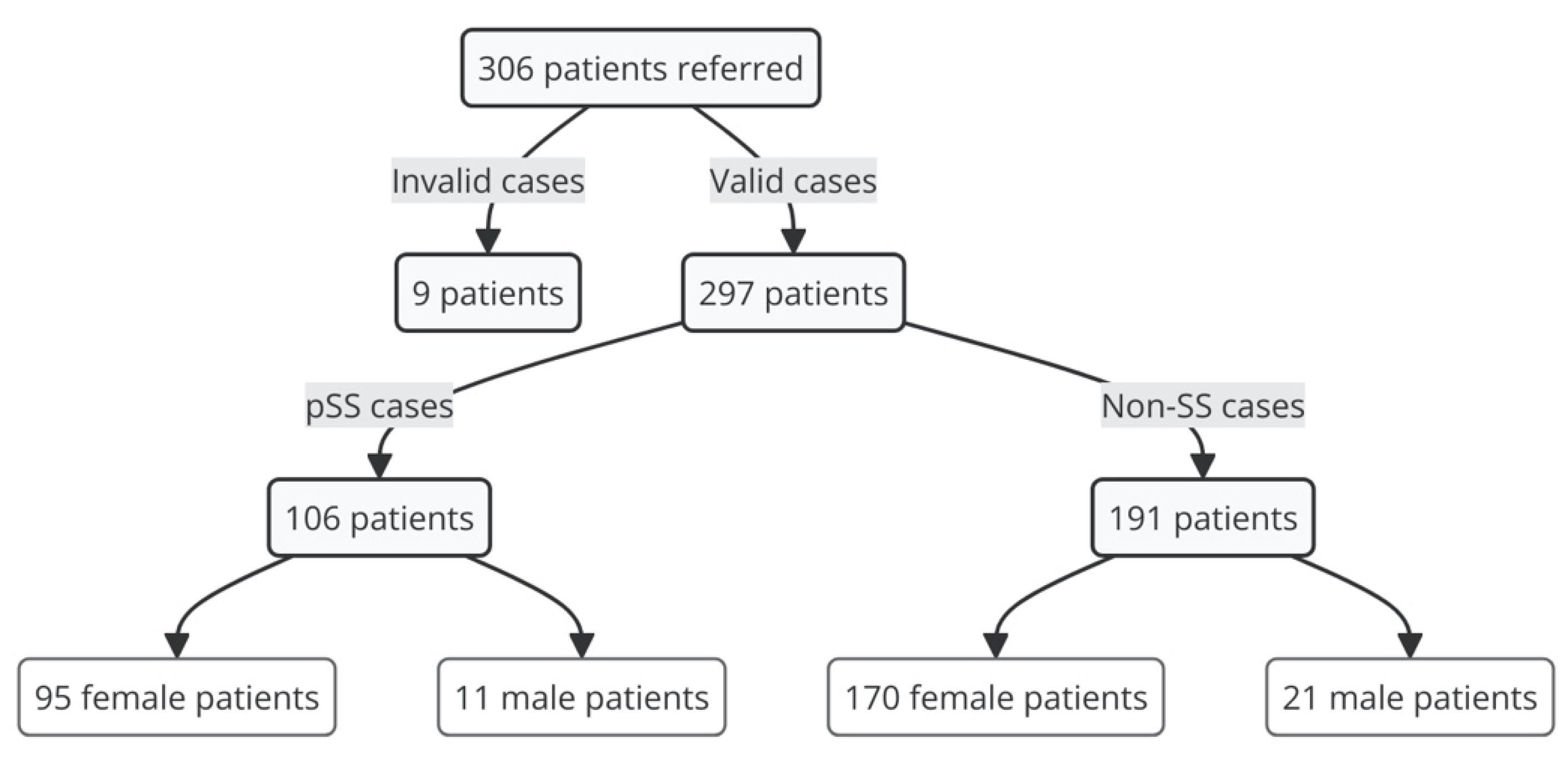
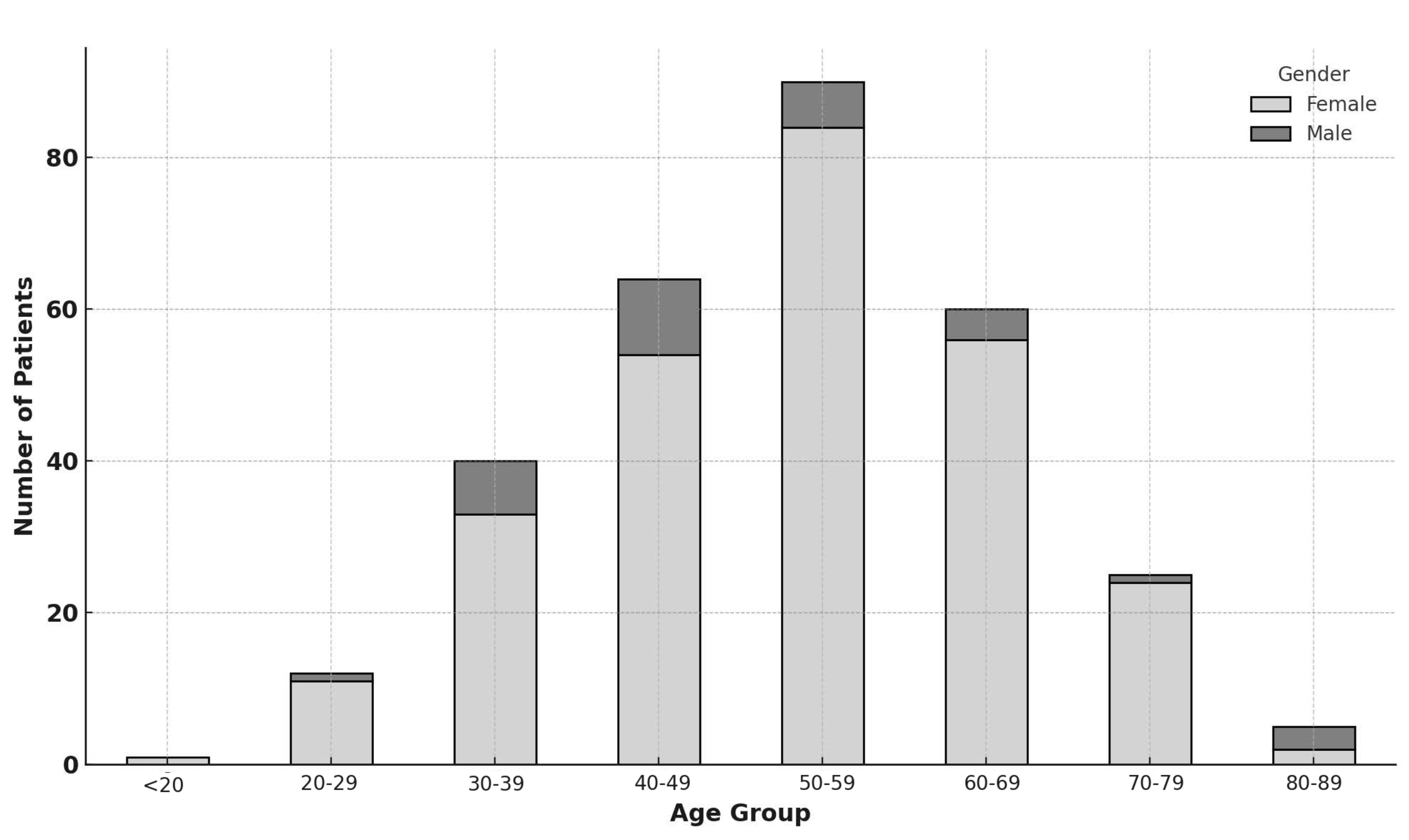
2.1. Population

This study uses data collected from patients referred to the University of Oslo, Faculty of Dentistry, Department of Oral Surgery and Oral Medicine, for salivary gland biopsy between 2008 and 2023 after clinical suspicion of pSS; an 8-year-longer time-frame than an earlier study utilizing this dataset [21]. Rheumatologists comprised the largest referring group.

2.2. Clinical Investigations

Subjective and objective ocular and oral signs and symptoms were collected from referral forms and during patient visits to the Department of Oral Surgery and Oral Medicine for patients whose information was unavailable from referrals. Information collected included ocular and oral dryness symptoms, sialometry, Schirmer I test, histological findings from biopsy, and serological findings.
The Schirmer I test is used to assess tear production via the wetting of a standardized paper strip inserted into the lower eyelid over a period of 5 min without anesthesia. The wetting of the paper was measured in millimeters, and recordings of ≤5 mm per 5 min were considered pathological [22]. Sialometry, or salivary flow, was recorded by collecting unstimulated whole saliva for 15 min to assess the degree of salivary hypofunction. A result of ≤1.5 mL was considered pathological [14]. Results of serological tests measuring anti-SSA antibody titer were collected, with results routinely including anti-SSB.
Biopsies were collected by one oral surgeon at the University of Oslo, Faculty of Dentistry (J.L.J.). The preferred biopsy site was on the left inside of the lower lip. The surgical technique and site for biopsy extraction were similar for all patients, and 5–10 salivary glands were routinely collected. Biopsies were transported to Haukeland University Hospital, Department of Pathology, in formalin and then paraffin-embedded, followed by hematoxylin and eosin staining. The tissue sections were then examined by one oral pathologist (K.S.), and an FS of ≥1 was considered pathological [3]. Adipose tissue was not included in the total surface area for calculating the FS. Reports from the histological evaluations included data regarding replacement of glandular tissue by adipose tissue, the atrophy of glandular tissue, and the presence of GCs. The use of the same surgeon/pathologist ensured the quality, consistency, and reliability of the results.
The 2002 AECG classification [14] was used to classify patients in this study as it is comprehensive, utilizing both clinical and serologic features. Furthermore, criteria necessary for the AECG classification were available from the patient data; however, ocular staining—1 of the 5 measured criteria in the ACR-EULAR [15]—was available only for a limited number of patients. Therefore, to avoid missing data bias, we used the AECG classification criteria.

2.3. Data Analysis and Presentation

Patients’ data were incorporated into the database utilized by Samuelsen et al. [21]. Patients were then classified and stratified into subgroups according to serological and histopathological phenotype. An overview of the subgroups is listed below (Figure 1).
Statistical analysis was conducted using statistical software packages SPSS (version 29.0.0.0) and GraphPad Prism (version 10.2.0). FS medians were compared using the Mann–Whitney U-test, as FS values were not normally distributed (confirmed through normality checks). To address the influence of potential confounders (age, gender, and GC status), a generalized linear model (GLM) with a gamma distribution and log link function was used.

3. Results

In total, 306 patients were referred for biopsy between 2008 and 2023. Nine participants were excluded from the study due to missing data, being repeat patients, or after conferring with the referring doctor. Figure 2 illustrates the division of these patients according to classification and, further, according to gender.
The distribution of patients according to age group is illustrated below (Figure 3). The majority of patients were found in the age group 50–59 years, representing 30% of referrals. The least-referred patient age group was patients < 20 years (patients aged between 18 and 19 years).

Patient Groups

Referred patients were divided into four groups, with seven subgroups in total (Figure 1). The subgroups were determined by which objective findings were fulfilled. pSS patients were found in groups 1.1, 1.2, 2, 3.1, and 3.2. Our data showed that 106 patients met the criteria for pSS classification.
Table 1, Table 2 and Table 3 show the number of patients in each group, their age-related descriptives (minimum age, maximum age, mean age), and the FS descriptives for groups 1.1 (biopsy-positive and double-seropositive), 1.2 (biopsy-positive and double-seropositive), and 2 (biopsy-positive) (minimum FS, maximum FS, mean FS, median FS). This information was shown according to gender for all referred patients, pSS patients, and non-SS patients.
Women formed the majority of all referrals (89.2%) and were the largest referred gender among both pSS (89.6%) and non-SS patients (89.0%).
There were 106 pSS patients in groups 1.1, 1.2, 2, 3.1, and 3.2. Most patients were classified based on positive findings from biopsy and serology (41 patients, representing 38.7%), followed by serology only (36 patients, representing 34.0%) and histology only (29 patients, representing 27.4%).
Most patients reported having both oral and ocular symptoms, irrespective of being classified as pSS or non-SS. Few patients had only one symptom of dryness. Patients with no symptoms of dryness formed the smallest referred group among all patient groups.
Differences in average FS between seropositive and seronegative patients, as well as between single- and double seropositive patients, are shown in Figure 4a,b. The median FS was significantly higher in the double-seropositive group compared to the single-seropositive group (difference between medians = 0.9, p < 0.05) (Figure 4a). The median FS was also higher in the seropositive group compared to the seronegative group; however, this finding was not statistically significant (difference between medians = 0.5, p > 0.05) (Figure 4b). Using a generalized linear model with a gamma distribution and log link function, possible confounding factors such as age, gender, and the presence of GC were investigated. After adjustment, similar to the previous results, we found that double-seropositive patients had a significantly higher FS than single-seropositive patients, and when comparing seropositive patients to seronegative patients, there was no significant difference in FS.
Histological analysis of salivary gland biopsies showed that 26 pSS patients were GC-positive. The median FS of GC-positive patients was significantly higher than GC-negative patients (difference between medians = 1.9, p < 0.05 *), as shown in Figure 5a. However, there were no significant differences when the number of GC-positive patients in group 1.1 (biopsy-positive and anti-SSA- and anti-SSB-positive patients), group 1.2 (biopsy-positive and anti-SSA-positive patients), and group 2 (only biopsy-positive patients) were compared. The group division of GC-positive patients is shown below in Figure 5b. GC-positive patients accounted for 24.5% of all patients classified as pSS.
We compared the diagnostic performance of the ACR-EULAR classification criteria against the AECG classification criteria by analyzing the number of true positives, true negatives, false positives, and false negatives, which were 101, 181, 8, and 7, respectively, in order to see if our classification of pSS patients would be similar regardless of the criteria used. The analysis showed sensitivity = 93.5%, specificity = 95.8%, positive predictive value = 92.7%, accuracy = 95.0%, and kappa = 89.1%.
When using FS as the ground truth for determining the presence of glandular involvement in patients (FS ≥ 1), we found for the AECG criteria (n = 297) true positives: 70, false positives: 36, false negatives: 1, and true negatives: 190. For the ACR-EULAR criteria (n = 291 due to 6 patients lacking subjective symptoms), we found true positives: 66, false positives: 43, false negatives; 1, and true negatives: 181.

4. Discussion

This paper describes patients referred to the University of Oslo’s Department of Oral Surgery and Oral Medicine for minor salivary gland biopsy between 2008 and 2023 after clinical suspicion of SS. In total, 306 patients were referred, of whom 106 met the AECG classification criteria for pSS and 191 did not (Figure 2). Thus, pSS patients represented 35.6% of all referrals, and non-SS patients represented 64.3%.
The importance of both serology and biopsy in the investigation of SS are in line with other studies [12], and this is seen in the large number of pSS classifications that they account for, individually and together (Figure 4). More specifically, the importance of serological tests in the classification of SS is highlighted since 45 patients could not have been classified without positive serological findings, representing 42.5% of cases. Likewise, the importance of biopsy is highlighted, since 38 patients could not be classified without positive histological findings, representing 35.8% of cases.
Our study showed significant differences between the median FS of single- and double-seropositive pSS patients (p < 0.05); more specifically, double-seropositive patients had a higher median FS than single-seropositive patients. The findings in the present study are in line with the results from Baldini et al., suggesting that “patients with two or more laboratory abnormalities should be more closely monitored” [23]. Furthermore, these results suggest that serologic phenotypes are likely to be a significant factor in the variability of FS, implying that SS may not be a distinct or uniform disease; rather, it may be composed of unique subgroups of patients with varying disease mechanisms and severity. Liao et al. concluded that salivary gland biopsy has a role in this stratification [24]. Moreover, our findings question the removal of anti-SSB from the ACR-EULAR classification criteria where only anti-SSA is evaluated, whereas in the AECG criteria, anti-SSA and/or anti-SSB are included. Our results may indicate that both anti-SSA and anti-SSB should be routinely evaluated. Further, among the 70 biopsy-positive pSS patients, there were 41 seropositive and 29 seronegative pSS cases, corresponding to 58.6% and 41.4% of these cases, respectively. The results showed a difference between their median FS, where seropositive patients had a higher FS than seronegative patients (Figure 4b). However, the results did not reveal a significant difference (p > 0.05). This is in contrast to other studies [25,26]; thus, our results should be interpreted cautiously since the relatively small sample size may have given rise to a type 2 error where we fail to see significant differences which may, in fact, exist.
Of the 297 patients, 26 patients were GC-positive, accounting for 8.8% of all referrals. The difference in median FS between GC-positive and GC-negative patients was significantly different (p < 0.05) (Figure 5a), with GC-positive patients having a larger median FS (median FS = 3) than GC-negative patients (median FS = 1.1). Thus, routinely screening patients presenting with higher FS for GCs may be beneficial since positive histological findings may represent a higher risk for complications such as lymphoma [25,27]. Thus, a salivary gland biopsy may reveal important information about the state of the disease in the target organ. Accordingly, studies recommend routine biopsy for patients diagnosed with pSS that have not taken a biopsy, as the presence of inflammatory foci and GCs may otherwise be missed [21,28]. In the present paper, 11 GC-positive patients could have been classified with pSS without a biopsy, corresponding to 10.4% of all pSS-classified patients. Accordingly, GC-positive patients represent a significant proportion of pSS diagnoses, suggesting that mandatory biopsy and screening may benefit all pSS patients. Unfortunately, many rheumatologists are unwilling to refer seropositive patients as they already fulfil the classification criteria combined with the fear of complications associated with the biopsy [11].
Comparing the ACR-EULAR classification criteria to the AECG classification criteria revealed a high sensitivity (93.5%) and specificity (95.8%), representing a strong performance in correctly identifying positive and negative cases, respectively. The positive predictive value (92.7%) and overall accuracy (95.0%) further highlighted the reliability of the ACR-EULAR criteria. Also, the high kappa value (89.1%) indicated strong agreement between the two sets of criteria in their classification of SS. These values were found even though one of the components utilized in the ACR-EULAR classification criteria—ocular staining—was missing for most patients, highlighting the robustness of the ACR-EULAR classification criteria. This underlines that the ACR-EULAR classification criteria represent adequate tools for classifying pSS patients, as observed by others [29,30,31]. Overall, these findings suggest that the results from our study are likely to be equally valid regardless of the classification criteria used.
Similar to a previous study [21], a key strength of this study is the use of one oral surgeon and one pathologist. This ensures that the variability in salivary gland biopsy technique and evaluation is limited. Additionally, our patient sample size is larger than a previous study utilizing the same dataset [21], allowing us to make more meaningful reasonings from statistical analyses. Weaknesses in our study may arise from a sampling bias due to the majority of referrals coming from one national region; therefore, inferences may not represent the whole population [21]. At the same time, the stratification of larger groups of patients into more homogeneous, but smaller, subgroups could have increased the risk of type II errors, where true effects may not have been observed as statistically significant. Nonetheless, the trends that we did observe between the subgroups showed the presence of significant differences, affirming the need for further investigation to validate these findings. Improvements in future studies could be made by taking more biopsies and utilizing each patient’s highest FS value or assigning an average FS based on several samples, since recent studies [32] show significant variations in FS values of each biopsy despite coming from the same patient.

5. Conclusions

A significant benefit of stratifying patients according to serologic phenotypes was identified; more specifically, there was a significant difference between the median FS of single- and double-seropositive patients, where double-seropositive patients had a higher median FS than single-seropositive patients. Our study, in particular, distinguishes between single- and double-seropositive patients, offering new insights into the seropositive patient subgroup. Double-seropositive patients displayed a significantly higher median FS compared to single-seropositive patients, suggesting more severe glandular involvement. Our results may indicate that both anti-SSA alone and anti-SSA and anti-SSB combined should be evaluated in the future.

Author Contributions

Conceptualization, J.L.J. and H.H. (Håvard Hynne); methodology, J.L.J. and K.S.; formal analysis, S.U., H.H. (Håkon Holm), A.R. and H.H. (Håvard Hynne); investigation, J.L.J. and K.S.; writing—original draft preparation, S.U., H.H. (Håkon Holm) and A.R.; writing—review and editing, J.L.J., K.S. and H.H. (Håvard Hynne); visualization, S.U., H.H. (Håkon Holm) and A.R.; supervision, J.L.J. and H.H. (Håvard Hynne); project administration, J.L.J. All authors have read and agreed to the published version of the manuscript.

Funding

This research received no external funding.

Institutional Review Board Statement

This study was conducted in accordance with the Declaration of Helsinki, and approval was obtained from the Regional Committees for Medical and Health Research Ethics, region southeast (REK 2010/1292 and REK 2015/363).

Informed Consent Statement

Informed consent was obtained from all subjects involved in this study.

Data Availability Statement

The original contributions presented in the study are included in the article, further inquiries can be directed to the corresponding author.

Conflicts of Interest

The authors declare no conflicts of interest.

References

  1. Nocturne, G.; Mariette, X. Advances in Understanding the Pathogenesis of Primary Sjögren’s Syndrome. Nat. Rev. Rheumatol. 2013, 9, 544–556. [Google Scholar] [CrossRef] [PubMed]
  2. Brito-Zerón, P.; Baldini, C.; Bootsma, H.; Bowman, S.J.; Jonsson, R.; Mariette, X.; Sivils, K.; Theander, E.; Tzioufas, A.; Ramos-Casals, M. Sjögren Syndrome. Nat. Rev. Dis. Primers 2016, 2, 16047. [Google Scholar] [CrossRef] [PubMed]
  3. Fisher, B.A.; Jonsson, R.; Daniels, T.; Bombardieri, M.; Brown, R.M.; Morgan, P.; Bombardieri, S.; Ng, W.F.; Tzioufas, A.G.; Vitali, C.; et al. Standardisation of Labial Salivary Gland Histopathology in Clinical Trials in Primary Sjögren’s Syndrome. Ann. Rheum Dis. 2017, 76, 1161–1168. [Google Scholar] [CrossRef]
  4. Vitali, C.; Moutsopoulos, H.M.; Bombardieri, S. The European Community Study Group on Diagnostic Criteria for Sjögren’s Syndrome. Sensitivity and Specificity of Tests for Ocular and Oral Involvement in Sjögren’s Syndrome. Ann. Rheum Dis. 1994, 53, 637–647. [Google Scholar] [CrossRef] [PubMed]
  5. Nakamura, H.; Kawakami, A.; Eguchi, K. Mechanisms of Autoantibody Production and the Relationship between Autoantibodies and the Clinical Manifestations in Sjögren’s Syndrome. Transl. Res. J. Lab. Clin. Med. 2006, 148, 281–288. [Google Scholar] [CrossRef]
  6. van der Reijden, W.A.; Vissink, A.; Veerman, E.C.; Amerongen, A.V. Treatment of Oral Dryness Related Complaints (Xerostomia) in Sjögren’s Syndrome. Ann. Rheum Dis. 1999, 58, 465–474. [Google Scholar] [CrossRef]
  7. Ramos-Casals, M.; Brito-Zerón, P.; Bombardieri, S.; Bootsma, H.; De Vita, S.; Dörner, T.; Fisher, B.A.; Gottenberg, J.E.; Hernandez-Molina, G.; Kocher, A.; et al. Eular Recommendations for the Management of Sjögren’s Syndrome with Topical and Systemic Therapies. Ann. Rheum Dis. 2020, 79, 3–18. [Google Scholar] [CrossRef] [PubMed]
  8. Tsifetaki, N.; Kitsos, G.; Paschides, C.A.; Alamanos, Y.; Eftaxias, V.; Voulgari, P.V.; Psilas, K.; Drosos, A.A. Oral Pilocarpine for the Treatment of Ocular Symptoms in Patients with Sjögren’s Syndrome: A Randomised 12 Week Controlled Study. Ann. Rheum Dis. 2003, 62, 1204–1207. [Google Scholar] [CrossRef]
  9. Craig, J.P.; Nelson, J.D.; Azar, D.T.; Belmonte, C.; Bron, A.J.; Chauhan, S.K.; de Paiva, C.S.; Gomes, J.A.P.; Hammitt, K.M.; Jones, L.; et al. Tfos Dews Ii Report Executive Summary. Ocul. Surf. 2017, 15, 802–812. [Google Scholar] [CrossRef]
  10. Ramia, T.; Daada, S.; Chaabene, I.; Klii, R.; Hammami, S.; Ines, K.; Kechida, M. Ab0539 Depression, Self-Esteem and Quality of Life in Sjögren’s Syndrome. Ann. Rheum. Dis. 2022, 81 (Suppl. S1), 1397. [Google Scholar] [CrossRef]
  11. Carubbi, F.; Alunno, A.; Cipriani, P.; Bartoloni, E.; Baldini, C.; Quartuccio, L.; Priori, R.; Valesini, G.; De Vita, S.; Bombardieri, S.; et al. A Retrospective, Multicenter Study Evaluating the Prognostic Value of Minor Salivary Gland Histology in a Large Cohort of Patients with Primary Sjögren’s Syndrome. Lupus 2015, 24, 315–320. [Google Scholar] [CrossRef] [PubMed]
  12. Dal Pozzolo, R.; Cafaro, G.; Perricone, C.; Calvacchi, S.; Bruno, L.; Colangelo, A.; Tromby, F.; Gerli, R.; Bartoloni, E. Salivary Gland Biopsy as a Prognostic Tool in Sjögren’s Syndrome. Expert Rev. Clin. Immunol. 2024, 20, 1139–1147. [Google Scholar] [CrossRef] [PubMed]
  13. Tashbayev, B.; Garen, T.; Palm, O.; Chen, X.; Herlofson, B.B.; Young, A.; Hove, L.H.; Rykke, M.; Singh, P.B.; Aqrawi, L.A.; et al. Patients with Non-Sjogren’s Sicca Report Poorer General and Oral Health-Related Quality of Life Than Patients with Sjogren’s Syndrome: A Cross-Sectional Study. Sci. Rep. 2020, 10, 2063. [Google Scholar] [CrossRef] [PubMed]
  14. Vitali, C.; Bombardieri, S.; Jonsson, R.; Moutsopoulos, H.M.; Alexander, E.L.; Carsons, S.E.; Daniels, T.E.; Fox, P.C.; Fox, R.I.; Kassan, S.S.; et al. Classification Criteria for Sjögren’s Syndrome: A Revised Version of the European Criteria Proposed by the American-European Consensus Group. Ann. Rheum Dis. 2002, 61, 554–558. [Google Scholar] [CrossRef]
  15. Shiboski, C.H.; Shiboski, S.C.; Seror, R.; Criswell, L.A.; Labetoulle, M.; Lietman, T.M.; Rasmussen, A.; Scofield, H.; Vitali, C.; Bowman, S.J.; et al. 2016 American College of Rheumatology/European League against Rheumatism Classification Criteria for Primary Sjögren’s Syndrome: A Consensus and Data-Driven Methodology Involving Three International Patient Cohorts. Arthritis Rheumatol. 2017, 69, 35–45. [Google Scholar] [CrossRef]
  16. Berry, J.S.; Tarn, J.; Casement, J.; Duret, P.M.; Scott, L.; Wood, K.; Johnsen, S.J.; Nordmark, G.; Devauchelle-Pensec, V.; Seror, R.; et al. Examining the Biological Pathways Underlying Clinical Heterogeneity in Sjogren’s Syndrome: Proteomic and Network Analysis. Ann. Rheum Dis. 2024, 83, 88–95. [Google Scholar] [CrossRef]
  17. Daniels, T.E.; Cox, D.; Shiboski, C.H.; Schiødt, M.; Wu, A.; Lanfranchi, H.; Umehara, H.; Zhao, Y.; Challacombe, S.; Lam, M.Y.; et al. Associations between Salivary Gland Histopathologic Diagnoses and Phenotypic Features of Sjögren’s Syndrome among 1726 Registry Participants. Arthritis Rheum. 2011, 63, 2021–2030. [Google Scholar] [CrossRef]
  18. Risselada, A.P.; Looije, M.F.; Kruize, A.A.; Bijlsma, J.W.; van Roon, J.A. The Role of Ectopic Germinal Centers in the Immunopathology of Primary Sjögren’s Syndrome: A Systematic Review. Semin Arthritis Rheum 2013, 42, 368–376. [Google Scholar] [CrossRef]
  19. Skarstein, K.; Aqrawi, L.A.; Oijordsbakken, G.; Jonsson, R.; Jensen, J.L. Adipose Tissue Is Prominent in Salivary Glands of Sjogren’s Syndrome Patients and Appears to Influence the Microenvironment in These Organs. Autoimmunity 2016, 49, 338–346. [Google Scholar] [CrossRef]
  20. Fonzetti, S.; Fulvio, G.; La Rocca, G.; García, I.C.N.; Governato, G.; Izzetti, R.; Donati, V.; Ferro, F.; Mosca, M.; Baldini, C. Pos1459 the Weight of Anti-La/Ssb Antibodies in Ro/Ssa-Positive Patients with a Newly Diagnosed Sjögren’s Syndrome: A Phenotype Characterized by a More Severe Major and Minor Glandular Involvement and a Higher Biological Activity. Ann. Rheum. Dis. 2023, 82 (Suppl. S1), 1083–1084. [Google Scholar]
  21. Samuelsen, T.; Aqrawi, L.; Skarstein, K.; Jensen, J. Evaluering Av Spyttkjertelbiopsier Ved Utredning Av Sjögrens Syndrom. Nor. Tann. Tid. 2021, 131, 988–994. [Google Scholar] [CrossRef] [PubMed]
  22. Wolffsohn, J.S.; Arita, R.; Chalmers, R.; Djalilian, A.; Dogru, M.; Dumbleton, K.; Gupta, P.K.; Karpecki, P.; Lazreg, S.; Pult, H.; et al. Tfos Dews Ii Diagnostic Methodology Report. Ocul. Surf. 2017, 15, 539–574. [Google Scholar] [CrossRef] [PubMed]
  23. Baldini, C.; Pepe, P.; Quartuccio, L.; Priori, R.; Bartoloni, E.; Alunno, A.; Gattamelata, A.; Maset, M.; Modesti, M.; Tavoni, A.; et al. Primary Sjogren’s Syndrome as a Multi-Organ Disease: Impact of the Serological Profile on the Clinical Presentation of the Disease in a Large Cohort of Italian Patients. Rheumatology 2014, 53, 839–844. [Google Scholar] [CrossRef]
  24. Liao, R.; Yang, H.T.; Li, H.; Liu, L.X.; Li, K.; Li, J.J.; Liang, J.; Hong, X.P.; Chen, Y.L.; Liu, D.Z. Recent Advances of Salivary Gland Biopsy in Sjögren’s Syndrome. Front. Med. 2021, 8, 792593. [Google Scholar] [CrossRef]
  25. Baer, A.N.; DeMarco, M.M.; Shiboski, S.C.; Lam, M.Y.; Challacombe, S.; Daniels, T.E.; Dong, Y.; Greenspan, J.S.; Kirkham, B.W.; Lanfranchi, H.E.; et al. The Ssb-Positive/Ssa-Negative Antibody Profile Is Not Associated with Key Phenotypic Features of Sjögren’s Syndrome. Ann. Rheum Dis. 2015, 74, 1557–1561. [Google Scholar] [CrossRef] [PubMed]
  26. Conticini, E.; Bardelli, M.; Vitale, A.; De Stefano, R.; Falsetti, P.; Selvi, E.; Bacarelli, M.R.; D’Alessandro, R.; Cantarini, L.; Frediani, B.; et al. Diagnostic Role of Minor Salivary Glands Biopsy in Sjögren’s Syndrome: Correlations between Histology and Autoimmunity in a Large, Monocentric Cohort. Reumatologia 2023, 61, 109–115. [Google Scholar] [CrossRef]
  27. Chatzis, L.; Vlachoyiannopoulos, P.G.; Tzioufas, A.G.; Goules, A.V. New Frontiers in Precision Medicine for Sjogren’s Syndrome. Expert Rev. Clin. Immunol. 2021, 17, 127–141. [Google Scholar] [CrossRef]
  28. Risselada, A.P.; Kruize, A.A.; Goldschmeding, R.; Lafeber, F.P.J.G.; Bijlsma, J.W.J.; van Roon, J.A.G. The Prognostic Value of Routinely Performed Minor Salivary Gland Assessments in Primary Sjögren’s Syndrome. Ann. Rheum. Dis. 2014, 73, 1537–1540. [Google Scholar] [CrossRef] [PubMed]
  29. Rasmussen, A.; Ice, J.A.; Li, H.; Grundahl, K.; Kelly, J.A.; Radfar, L.; Stone, D.U.; Hefner, K.S.; Anaya, J.M.; Rohrer, M.; et al. Comparison of the American-European Consensus Group Sjogren’s Syndrome Classification Criteria to Newly Proposed American College of Rheumatology Criteria in a Large, Carefully Characterised Sicca Cohort. Ann. Rheum Dis. 2014, 73, 31–38. [Google Scholar] [CrossRef]
  30. Tsuboi, H.; Hagiwara, S.; Asashima, H.; Takahashi, H.; Hirota, T.; Noma, H.; Umehara, H.; Kawakami, A.; Nakamura, H.; Sano, H.; et al. Comparison of Performance of the 2016 Acr-Eular Classification Criteria for Primary Sjögren’s Syndrome with Other Sets of Criteria in Japanese Patients. Ann. Rheum Dis. 2017, 76, 1980–1985. [Google Scholar] [CrossRef]
  31. Le Goff, M.; Cornec, D.; Jousse-Joulin, S.; Guellec, D.; Costa, S.; Marhadour, T.; Le Berre, R.; Genestet, S.; Cochener, B.; Boisrame-Gastrin, S.; et al. Comparison of 2002 Aecg and 2016 Acr/Eular Classification Criteria and Added Value of Salivary Gland Ultrasonography in a Patient Cohort with Suspected Primary Sjögren’s Syndrome. Arthritis Res. Ther. 2017, 19, 269. [Google Scholar] [CrossRef] [PubMed]
  32. Ono, J.; Toya, S.; Ogura, I.; Okada, Y. Study of Clinical Factors, Focus Score, Lymphocyte Type and Nf-Κb Pathway in Sjögren’s Syndrome. Odontology 2023, 111, 207–216. [Google Scholar] [CrossRef] [PubMed]
Figure 1. Overview of the stratification criteria for patients referred for biopsy.
Applsci 14 11482 g001
Figure 2. Schematic illustration of the distribution of referred patients according to classification and gender. Patients classified as having primary Sjögren’s syndrome (pSS) or non-Sjögren’s sicca syndrome (non-SS) are shown. The 2002 American–European Consensus Group classification criteria were used to classify the patients.
Applsci 14 11482 g002
Figure 3. Age and gender distribution of patients referred for salivary gland biopsy.
Applsci 14 11482 g003
Figure 4. (a) Median focus scores of double-seropositive patients (anti-SSA- and anti-SSB-positive) and single-seropositive patients (anti-SSA patients/anti-SSB-positive). (b) Median focus scores of seropositive patients (anti-SSA/SSB-positive) and seronegative patients (anti-SSA/SSB-negative). Error bars represent 95% CI. * Significant difference between the groups’ median focus scores (p < 0.05).
Applsci 14 11482 g004
Figure 5. (a) The median focus score for germinal-center-positive and germinal-center-negative patients. Error bars represent 95% CI. * Significant difference between focus scores (p < 0.05). (b) Distribution of germinal-center-positive patients in the biopsy-positive groups. The groups represented are as follows: group 1.1: biopsy-positive and anti-SSA- and anti-SSB-positive patients; group 1.2: biopsy-positive and anti-SSA-positive patients; group 2: only biopsy-positive patients.
Applsci 14 11482 g005
Table 1. Distribution of all referred patients by group, gender, and age. Patients were classified with primary Sjögren’s syndrome (pSS) according to the 2002 American–European Consensus Group classification criteria.
Group 1.1
(Biopsy-Positive and Double-Seropositive)
Group 1.2
(Biopsy-Positive and Single-Seropositive)
Group 2
(Biopsy-Positive)
Group 3.1
(Double-Seropositive)
Group 3.2
(Single-Seropositive)
Group 4.1
(FS < 1 and/or Trace ANA and/or Trace SSA/SSB)
Group 4.2
(Biopsy-Negative and Seronegative)
Total
Number of patients2318305458393297
Percentage of referred patients7.7%6.1%10.1%1.7%15.2%27.9%31.3%100.0%
Gender
Male
Frequency313187932
Min. age30474382332836
Max. age80476182697985
Mean age48.747.052.782.045.950.753.1
Female
Frequency2017274377684265
Min. age26183921243225
Max. age84747668777681
Mean age48.548.455.851.050.752.854.8
Table 2. Distribution of primary Sjögren’s syndrome (pSS) patients by group, age, and focus score (FS). Patients were classified with primary Sjögren’s syndrome (pSS) according to the 2002 American–European Consensus Group classification criteria.
Group 1.1
(Biopsy-Positive and Double-Seropositive)
Group 1.2
(Biopsy-Positive and Single-Seropositive)
Group 2
(Biopsy-Positive)
Group 3.1
(Double-Seropositive)
Group 3.2
(Single-Seropositive)
Group 4.1
(FS < 1 and/or Trace ANA and/or Trace SSA/SSB)
Group 4.2
(Biopsy-Negative and Seronegative)
Total
Number of patients23182933300106
Percentage of total pSS classifications21.7%17.0%27.4%2.8%31.1%0.0%0.0%100%
Gender
Male
Frequency313040011
Min. age304743 33
Max. age804761 52
Mean age48.747.052.7 41.3
Female
Frequency2017263290095
Min. age2618395424
Max. age8474766877
Mean age48.548.455.761.050.5
Focus Score
Lowest FS1.01.01.00.00.0
Highest FS10.03.33.70.00.9
Mean FS3.31.61.70.00.04
Median FS2.01.11.50.00.0
Table 3. Distribution of non-SS patients by group, gender, and age. Patients were classified with primary Sjögren’s syndrome (pSS) according to the 2002 American–European Consensus Group classification criteria.
Group 1.1
(Biopsy-Positive and Double-Seropositive)
Group 1.2
(Biopsy-Positive and Single-Seropositive)
Group 2
(Biopsy-Positive)
Group 3.1
(Double-Seropositive)
Group 3.2
(Single-Seropositive)
Group 4.1
(FS < 1 and/or Trace ANA and/or Trace SSA/SSB)
Group 4.2
(Biopsy-Negative and Seronegative)
Total
Number of patients0012128393191
Percentage of all referrals 0.5%1.0%6.3%43.5%48.7%100.0%
Gender
Male
Frequency000147921
Min. age 82422836
Max. age 82697985
Mean age 8250.550.753.1
Female
Frequency001187684170
Min. age 6021403225
Max. age 6021677681
Mean age 602151.452.854.8
Disclaimer/Publisher’s Note: The statements, opinions and data contained in all publications are solely those of the individual author(s) and contributor(s) and not of MDPI and/or the editor(s). MDPI and/or the editor(s) disclaim responsibility for any injury to people or property resulting from any ideas, methods, instructions or products referred to in the content.

