Article

Exploration of the Anti-Photoaging Mechanisms of Lactiplantibacillus plantarum TWK10 in a UVB-Induced Mouse Model

1
Department of Food Science, Fu Jen Catholic University, New Taipei City 242062, Taiwan
2
Department of Food Science, Nutrition, and Nutraceutical Biotechnology, Shih Chien University, Taipei 104336, Taiwan
3
Culture Collection & Research Institute, SYNBIO TECH INC., Kaohsiung City 821011, Taiwan
*
Author to whom correspondence should be addressed.
Appl. Sci. 2024, 14(20), 9497; https://doi.org/10.3390/app14209497
Submission received: 13 September 2024 / Revised: 13 October 2024 / Accepted: 14 October 2024 / Published: 17 October 2024
(This article belongs to the Section Food Science and Technology)

Abstract

:
Functional foods have shown promise in mitigating skin aging. This study aimed to evaluate the effects of Lactiplantibacillus plantarum TWK10 (LPTWK10) and its spray-dried supernatant powder on ultraviolet B (UVB)-induced skin photoaging in female BALB/c nude mice. Over a 13-week period of UVB exposure and concurrent administration of high doses of LPTWK10 or its spray-dried fermentation supernatant, significant improvements were observed, skin wrinkles were notably reduced, transepidermal water loss rate decreased by 68.94–70.77%, and stratum corneum hydration increased by 76.97–112.24%. Furthermore, LPTWK10 was effective in reducing erythema and inflammation while enhancing skin lightness. Histological assessments revealed substantial reductions in epidermal hyperplasia and collagen degradation. Additionally, LPTWK10 was found to influence critical mechanisms associated with collagen metabolism and proinflammatory cytokine production. In summary, LPTWK10 attenuates photoaging through modulation of collagen metabolism and reduction in inflammatory responses, suggesting its potential as a functional ingredient for delaying photoaging.

1. Introduction

The skin, which is the largest organ of the body, is divided into the epidermis, dermis, and hypodermis. The outermost layer of the epidermis is composed mainly of keratinocytes, which serve as a barrier against external damage [1]. The dermis is composed of fibrous connective tissue, extracellular matrix, and cells. Its primary components include collagen, elastin fibers, and glycoproteins, which maintain the structural durability and elasticity of the skin and serve as a major source of moisture [2]. Fibroblasts synthesize and secrete collagen and elastin. Subcutaneous tissue is located in the deepest layer of the skin and contains many fat cells that store energy and provide physical insulation [3]. Skin aging can be classified as endogenous and exogenous aging. Endogenous aging, also known as natural aging, is the gradual atrophy of the skin and other organs with age. Atrophy of the collagen and elastin fibers in the extracellular matrix of the skin results in a loss of skin elasticity, resulting in thinning of the epidermis and the formation of fine lines with age [4]. Exogenous aging is primarily due to environmental factors such as sun exposure, environmental pollution, smoking, alcohol consumption, and poor nutrition. Ultraviolet (UV) radiation, which is the principal cause of skin photoaging, alters the connective tissue of the dermis, leading to the degradation and loss of collagen as well as the abnormal accumulation of elastin fibers [2,5,6]. This ultimately increases epidermal thickness and decreases skin elasticity, leading to skin aging and damage [7]. Photoaging is characterized by deep wrinkle formation, loss of lightness and elasticity, uneven pigmentation, laxity, roughness, and capillary dilation [8,9].
UV waves, which are electromagnetic waves with wavelengths of 10–400 nm, are classified based on wavelength into UVA (320–400 nm), UVB (280–320 nm), and UVC (100–280 nm) [10]. Of the UV radiation reaching the surface of the Earth, 5–10% is UVB, and 90–95% is UVA. However, UVB has higher energy levels than those of UVA and acts on the epidermis [11], making it more damaging to the skin than UVA and a major contributor to epidermal cell hypertrophy, keratosis, and dermatitis. Prolonged skin exposure to UVB promotes the production of large amounts of reactive oxygen species (ROS), resulting in imbalanced oxidative stress in the body, which directly damages cellular DNA and cell membrane lipids and proteins, increases prostaglandin E2 levels and generates nitric oxide [12]. Furthermore, these effects result in inflammatory reactions such as erythema, sunburn, sensation of heat, and eventually skin cancer [13].
UV irradiation of the skin causes excessive production of ROS and pro-inflammatory hormones such as tumor necrosis factor-α (TNF-α), interleukin (IL)-6, and IL-1β and activates the mitogen-activated protein kinase (MAPK) signaling pathway, causing NF-κB and activator protein-1 (AP-1) to enter the nucleus, thereby regulating downstream biological responses. This upregulates matrix metalloprotease (MMP) expression in the skin, leading to collagen breakdown [14]. In particular, IL-1α, IL-1β, and TNF-α enhance IL-6 production and release in keratinocytes [15]; IL-6 can also induce MMPs through an autocrine mechanism [2]. In addition, UV modulates the transforming growth factor-β signaling pathway and inhibits collagen regeneration, leading to photoaging-related phenomena such as skin dryness and wrinkles [16].
Previous experimental results from our laboratory have demonstrated that the indigenous lactic acid bacterial strain Lactiplantibacillus plantarum TWK10 (LPTWK10), derived from Taiwanese pickles, not only has the capacity to modulate blood pressure in spontaneously hypertensive rats [17] but also alleviates cognitive impairments associated with hypertension-induced vascular dementia [18]. Continuous administration of TWK10-fermented soy milk to hypertensive rats with vascular dementia for 13 weeks led to significant reductions in escape latency times in the Morris water maze test, including in reference memory, probe trials, and working memory assessments [18]. Furthermore, both animal models and human studies have indicated that TWK10 supplementation significantly enhances muscle mass and improves exercise performance [19]. Additionally, Chang et al. (2015) demonstrated that the ethanol extract of TWK10-fermented soy milk, when co-cultured with B16 melanoma cells, effectively inhibits melanin synthesis by modulating the expression of melanin biosynthesis-related proteins [20]. Further investigations by our team have explored the potential dermatological benefits of TWK10, revealing that the ethanol extract of TWK10-fermented soy milk not only attenuates the rate of blood glucose elevation in diabetic rats but also promotes wound healing [21]. These findings suggest that TWK10 and its fermentation products exhibit a range of bioactivities, warranting further investigation into their potential to mitigate UVB-induced skin photoaging.
Female HR-1 mice fed with 1 × 10⁹ CFU/100 μL of Bifidobacterium breve strain Yakult for 9 days exhibited significantly reduced transepidermal water loss (TEWL) and stratum corneum (SC) hydration caused by UVB irradiation, as well as significant inhibition of UVB-induced increases in hydrogen peroxide content, protein oxidation, lipid oxidation, and xanthine oxidase activity in the skin [22]. HR-1 mice tube-fed continuously for 7 weeks with 2 × 10⁹ CFU/day of B. breve B-3 exhibit significant alleviation of changes in TEWL, skin hydration, and epidermal thickening, reduced tight junction and basement membrane damage caused by prolonged UVB irradiation, and substantially inhibit IL-1β production in the skin [23]. Human dermal fibroblasts treated with 1 × 10⁹ CFU/mL of L. plantarum HY7714 exhibit markedly inhibited protein expression of UVB-induced MMP-1 and procollagen and also inhibit the expression of the JUK and phosphorylated c-Jun proteins [24].
The purpose of this study was to investigate the effects of the viable form of LPTWK10 and its spray-dried supernatant powder—containing metabolites and secreted compounds—on UVB-induced skin photoaging in induced in female immunodeficient mice. We aimed to evaluate the potential of both samples in preventing or ameliorating skin photoaging, as well as to explore any differences between them. By examining both forms, we sought to determine whether the bioactive compounds in the supernatant could provide similar or enhanced skin protection compared with live bacteria, potentially leading to alternative formulations for skincare applications.

2. Materials and Methods

2.1. Sample Preparation

The LPTWK10 or LPTWK10 spray-dried supernatant powders were prepared by in-house methods at the laboratory of SYNBIO TECH INC.(Kaohsiung, Taiwan) as experimental samples. The live bacteria LPTWK10 powder was prepared by inoculating 3% LPTWK10 into a medium containing 60 g/L glucose, 20 g/L yeast extract, 1 g/L dipotassium phosphate, and 0.1 g/L magnesium sulfate. The mixture was incubated at 37 °C for 16 h. Following incubation, the bacteria were collected by centrifugation and mixed with an equal amount of cryoprotectant (100 g/L maltodextrin and 10 g/L glycerol) before being freeze-dried to obtain the powder. In contrast, the LPTWK10 supernatant powder, which contains no bacterial cells, was prepared using a similar process. After incubation, the LPTWK10 cells were removed, and the supernatant was spray-dried with maltodextrin as a carrier. The spray-drying process utilized a liquid-to-solid ratio of 3.5:1 and a drying temperature of 120–130 °C.

2.2. Animals

This animal trial was approved by the Institutional Animal Care and Use Committee (IACUC) of Fu Jen Catholic University (IACUC No. A10830 and A10831). Animal experimental models were designed based on those described by Satoh et al. [23] and Kim et al. [24], with some modifications. Seventy-two female five-week-old BALB/c nude mice were housed following the Rules for the Establishment of an Animal Experimentation Management Unit issued by the Council of Agriculture of the Executive Yuan of the Republic of China (Executive Yuan, 2018). Experimental animals were housed in an animal room maintained at a constant temperature (22 ± 2 °C) and 60% relative humidity with a light cycle of 12 h. Food and water were replenished every three days and maintained at a sufficient level for the animals to feed ad libitum. In addition, the animal weight was measured weekly and used for both health monitoring and as baseline values for calculating the sample doses needed for each group of animals in subsequent weeks [23,24].
At 7 weeks of age and assuming no significant difference in body weight among the groups (p > 0.05), the mice were divided into the following nine groups (Table 1): normal control (NC), ultraviolet B (UVB), positive control treated with hyaluronic acid following UV irradiation [25], three groups treated with different doses of LPTWK10 following UV irradiation (LPLD, LPMD, and LPHD), and three groups treated with different doses of spray-dried LPTWK10 supernatant powder following UV irradiation (SPLD, SPMD, and SPHD). Samples were converted to weekly doses based on the differences in body weight in each group of animals. Sample suspensions were freshly prepared in 0.1 mL sterile distilled water before tube feeding and used immediately.

2.3. UVB Induction

At 7 weeks of age, the dorsal skin of nude mice was irradiated with UVB to induce skin photoaging [23,24]. Mice were anesthetized using isoflurane inhalation and then irradiated with a UVB irradiator (BIO-LINK BLX-312, Vilber Lourmat, France). To avoid severe skin lesions caused by repeated UVB irradiation, a minimum erythema dose (MED) of 36 mJ/cm2 was selected as the initial UVB irradiation dose. Irradiation was performed three times per week. Nude mice were analyzed using the DermaLab Combo system (Cortex Technology, Hadsund, Denmark) before and after irradiation to determine the dorsal skin parameters, including TEWL, SC hydration, lightness (L* value), and erythema (a* value). The dose of UVB irradiation increased gradually with prolonged induction, namely 54 mJ/cm2 at weeks 2–4, 72 mJ/cm2 at weeks 5–7, 108 mJ/cm2 at weeks 8–10, and 144 mJ/cm2 at weeks 11–13.

2.4. Preparation of Serum and Skin Tissue Extracts

Nude mice were euthanized using CO2 asphyxiation, and 1 mL of blood was slowly withdrawn from the vena cava using a collection needle, placed in a collection tube containing a coagulant, and allowed to stand for 15 min for the blood to coagulate. The sample was then centrifuged at 3000× g for 10 min at 4 °C. The upper serum layer was collected and stored at −80 °C. The collected dorsal skin was partially fixed with 10% neutral formalin for tissue sectioning. A total of 0.06 g of skin was weighed and added to 0.6 mL of phosphate-buffered saline with an appropriate quantity of zirconium oxide beads for tissue homogenization using a tissue homogenizer (Fastprep-24 Classic, MP Biomedicals, Irvine, CA, USA) at 6 m/s three times at 30 s each time. The homogenate was centrifuged at 5000× g for 20 min at 4 °C, and the supernatant was collected and stored at −80 °C for subsequent analysis.

2.5. Measurement of Inflammatory Cytokine Levels in Skin Tissue

A commercially available analytical kit (Biolegend Inc., San Diego, CA, USA) was used to measure levels of the inflammation-associated factors TNF-α (No. 430905), IL-6 (No. 431305), and IL-1β (No. 432605) in skin tissue. The experimental procedure was performed by following the kit manufacturer’s instructions. In brief, using IL-1β as an example, 100 μL of capture antibody was added to a 96-well-dish and placed in a 4 °C refrigerator overnight for coating. The following day, the liquid was removed from the wells, and the wells were washed four times with 300 μL of wash buffer, ensuring maximum removal of the wash buffer on the last wash. Next, 200 μL of assay dilution A buffer was added to the well and allowed to react for 1 h at room temperature on a shaker at 500 rpm. The wells were washed with a wash buffer, and the washing procedure was repeated four times. Serum from each group of nude mice, as well as 100 μL of standard, were added and allowed to react at room temperature for 2 h, followed by four wash procedures. Next, 100 μL of avidin-horseradish peroxidase (HRP) solution, TMB substrate solution, and stop solution were added to the wells following the manufacturer’s instructions. Finally, the absorbance of each well was measured at a wavelength of 450 nm and aligned with the standard curve to determine the serum IL-1β levels. Similar procedures were followed for TNF-α and IL-6 with their specific capture antibody and antibody reagents.

2.6. Measurement of Collagen-Related Indicators

Commercially available analytical kits (Elabscience Bionovation Inc., Houston, TX, USA) were used to determine the levels of the inflammation-associated factors MMP-1 (E-EL-M0779), MMP-9 (E-EL-M3052), procollagen I C-terminal propeptide (PICP, E-EL-M0231), and procollagen III C-terminal propeptide (PIIICP, E-EL-M0367). The experimental procedure was performed following the manufacturer’s instructions. In brief, using MMP-1 as an example, 100 μL of standard or skin tissue extracts were separately added to a 96-well dish and allowed to react at 37 °C for 90 min. Immediately after separating the liquid, 100 μL of a biotinylated detection antibody working solution was added to each well and allowed to react at 37 °C. After 1 h, the wells were washed thrice with wash buffer, 100 μL of an HRP conjugate working solution was added, and the 96-well dish was incubated at 37 °C for 30 min. The wells were washed five times with wash buffer, and 90 μL of the substrate reagent was added and allowed to react at 37 °C for 15 min, followed by the addition of 50 μL stop solution. Finally, the absorbance of each well was measured at a wavelength of 450 nm and aligned with the standard curve to calculate MMP-1 protein content in the skin tissue fluid. The MMP-9, PICP, and PIIICP assay procedures were similar, except for differences in biotinylated detection antibody working solution reagents.

2.7. Histopathology and Immunohistochemical Staining of Skin Sections

Histopathologic examination was performed with assistance from the Animal Disease Diagnostic Center of National Chung Hsing University Veterinary Medical Teaching Hospital for tissue sectioning and histopathologic interpretation. Briefly, dorsal skin tissues were fixed in 10% neutral formalin solution, dehydrated, embedded in paraffin, processed into 5–7 µm sections, stained with hematoxylin and eosin (H&E) and Masson’s trichrome, and microscopically analyzed to determine the thickness of the epidermis and the collagen fiber content in the dermis. Finally, quantification was performed using the Image-Pro PLUS 7.0 software (Media Cybernetics Inc., Rockville, MD, USA).
Immunohistochemical staining, including claudin-1, laminin, and collagen IV assays, was performed by Rapid Science Co., Ltd. (Taichung, Taiwan). The dorsal skin tissue was fixed in 10% neutral formalin, processed into slides, and imaged and recorded microscopically. Immunohistochemical staining was quantified using Image-Pro PLUS 7.0 software (Media Cybernetics Inc.). For quantification, three areas were randomly selected and averaged.

2.8. Statistical Analyses

The experiments in this study were repeated at least thrice, and the results are expressed as the mean ± standard deviation. Statistical analyses were performed using the IBM SPSS Statistics 18 (IBM, Endicott, NY, USA). One-way analysis of variance was used for statistical analysis, and the differences between groups were compared using Duncan’s multiple range test. Differences with p < 0.05 were considered statistically significant.

3. Results and Discussion

3.1. Wrinkles on the Dorsal Skin of Nude Mice

BALB/c nude mice were selected for this study because of their unique characteristics that make them particularly suitable for investigating UVB-induced skin aging. These mice are athymic, resulting in a lack of functional T lymphocytes and an impaired immune response [26]. This immunodeficiency minimizes variability in immune-mediated responses, allowing for a more accurate assessment of the direct effects of UVB exposure on skin aging. Furthermore, BALB/c nude mice possess skin properties that closely resemble those of human skin, including similar epidermal layers and keratinization processes. This similarity enhances the translational relevance of the findings to human conditions. Studies have demonstrated that these mice develop UVB-induced skin damage and aging features that parallel those observed in humans, including alterations in skin elasticity and collagen degradation [25]. Additionally, the consistent and reproducible response of BALB/c nude mice to UVB exposure makes them an effective model for evaluating the impact of potential therapeutic interventions [27]. Thus, the use of BALB/c nude mice provides a valuable and relevant model for studying UVB-induced skin aging and assessing the efficacy of anti-aging treatments.
Gilchrest (2013) demonstrated that skin aging is characterized by coarse wrinkles, abnormal keratinocyte proliferation, dryness, laxity, and hyperpigmentation. In particular, the degree of wrinkle formation can be used as an indicator of skin damage [28,29]. Figure 1 shows the appearance of wrinkles on the back of nude mice in each group. These wrinkles increased in number and became drier after 13 weeks of UVB irradiation. At week 13 of administering high doses of LPTWK10 or the spray-dried LPTWK10 supernatant, the dorsal skin of the nude mice was devoid of wrinkles and smoother than that of mice in the UVB-induced group. These results show that high doses of LPTWK10 or the spray-dried LPTWK10 supernatant effectively minimize the formation of wrinkles on the skin.

3.2. Dorsal Skin Water Loss in Nude Mice

TEWL (g/m2/h) is a marker of skin barrier function [30,31]. The initial TEWL ranged from 14.89 ± 4.25 g/m2/h to 19.96 ± 5.24 g/m2/h, and no significant difference (p > 0.05) was found among the groups, indicating that all the mice were at the same baseline before induction (Figure 2A). At week 7 of UVB irradiation, the TEWL of mice in the UVB-induced group tended to be significantly higher than that of the NC group by approximately 2.37 g/m2/h (p < 0.05) and was significantly higher than that of the NC group at week 13 by approximately 14.87 g/m2/h. In contrast, the administration of LPTWK10 or spray-dried LPTWK10 supernatant at low, medium, and high doses over a 13-week period resulted in significant reductions in transepidermal water loss (TEWL) in the dorsal skin, with decreases of approximately 12.34–16.08 g/m²/h and 5.39–15.91 g/m²/h, respectively, when compared with the UVB group (p < 0.05). These results suggest a dose-dependent effect, indicating that the administration of low, medium, and high doses of LPTWK10 or the spray-dried LPTWK10 supernatant is not associated with skin damage or toxicity and is effective in enhancing the barrier function of the skin.

3.3. SC Hydration of the Dorsal Skin of Nude Mice

The SC is the outermost layer of the skin and functions as a barrier to prevent moisture loss. It is composed primarily of lipids and keratinocytes and contains ceramides, cholesterol, and free fatty acids [32]. In particular, ceramides maintain SC hydration, which indicates the moisture content of the skin and also protects the skin from external damage and pathogens [32]. When the skin barrier is compromised, an increase in TEWL is accompanied by a decrease in the SC hydration level [33]. At week 13 of UVB irradiation, hydration in the UVB group was significantly (p < 0.05) lower than that in the NC group by 47.37% (Figure 2B). At week 13, compared with the UVB group, the mice administered low, medium, and high doses of LPTWK10 exhibited significant increases of approximately 63.32, 90.31, and 112.24% (p < 0.05), respectively, in the dorsal skin hydration level, whereas those administered low, medium, and high doses of the spray-dried LPTWK10 supernatant exhibited significant increases of approximately 47.45, 79.89, and 76.97% (p < 0.05), respectively. These results indicate that low, medium, and high doses of LPTWK10 and the spray-dried LPTWK10 supernatant increase skin moisture content, and LPTWK10 exhibits dose-dependent effects.

3.4. Erythema, Melanin, and Lightness of the Dorsal Skin of Nude Mice

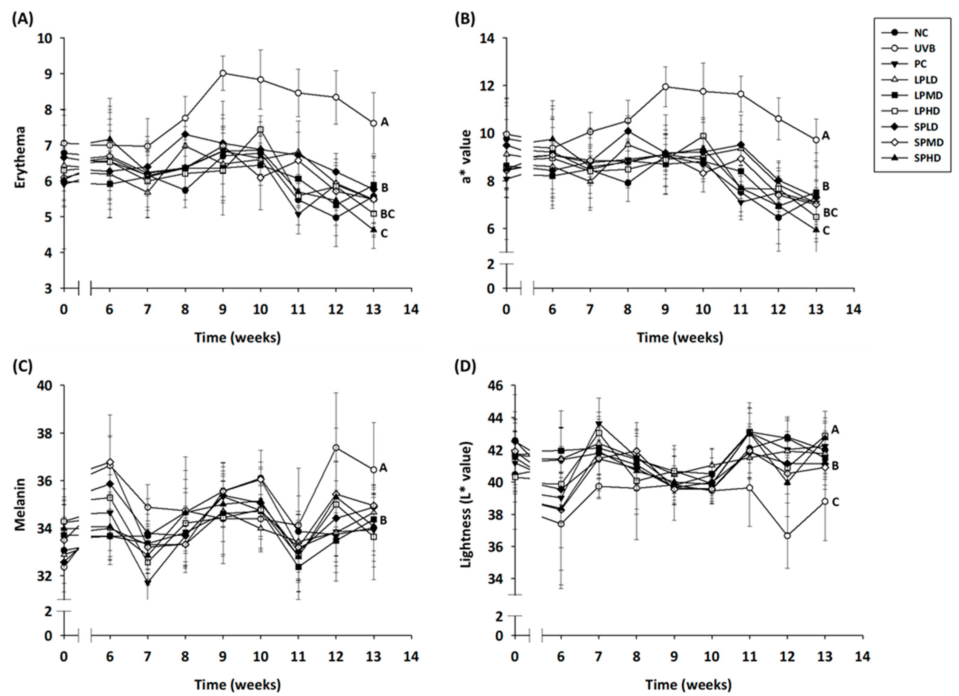
Erythema and a* values are both indicators of the degree of skin inflammation [31]. Figure 3A,B show the erythema and a* values in the dorsal skin of nude mice in each group. The initial erythema values of the dorsal skin ranged from 5.90 ± 0.46 to 7.05 ± 0.93, and the initial a* values ranged from 8.07 ± 0.46 to 9.95 ± 1.20. The two groups did not differ significantly (p > 0.05), suggesting that all animals were at the same baseline before induction. At week 8 of UVB irradiation, the erythema and a* values in the UVB group were significantly higher than those in the NC group by approximately 2.01 and 1.60, respectively, and at 13 weeks of irradiation, they were significantly higher than those in the NC group by 2.04 and 2.41, respectively. At week 13 of administering low, medium, and high doses of LPTWK10 or the spray-dried LPTWK10 supernatant, the mice exhibited significant reductions (p < 0.05) of approximately 1.73–2.53 and 1.85–2.99, respectively, in erythema values and approximately 2.20–3.23 and 2.41–3.79, respectively, in a* values relative to those in the UVB group mice.
Melanin is produced by melanocytes in the basal layer of the epidermis. Melanocytes can protect the skin by absorbing UV radiation. Prolonged exposure to UV radiation may lead to abnormal melanin accumulation, contributing to pigmentation and even the potential for other skin lesions [34,35]. The initial melanin values ranged from 32.36 ± 1.40 to 34.29 ± 0.85, and the initial L* values ranged from 40.30 ± 1.52 to 42.58 ± 1.10 (Figure 3C,D). No significant difference (p > 0.05) was noted between the groups, suggesting that all animals were at the same baseline before induction. At week 12 of UVB irradiation, the melanin and L* values significantly increased by approximately 3.65 and decreased by approximately 2.45, respectively, in the UVB group compared with those in the NC group. At week 13 of irradiation, compared with the NC group, the UVB group exhibited significantly higher melanin value in the dorsal skin by approximately 2.44, and the L* value was significantly lower by approximately 3.20. In contrast, at week 13 of treatment with low, medium, and high doses of LPTWK10 or the spray-dried LPTWK10 supernatant, the mice exhibited significant decreases in melanin values of approximately 1.79–2.82 and 1.50–2.37, respectively, and significant increases in L* values of approximately 2.70–4.08 and 2.12–3.94, respectively, relative to those in the UVB group mice (p < 0.05). Studies by Liu et al. (2022) and Chang et al. (2015) indicate that LPTWK10 possesses antioxidant activity and inhibits melanin biosynthesis in melanocytes. Therefore, it is hypothesized that LPTWK10 can enhance the skin’s antioxidant defenses against oxidative stress caused by UVB exposure. By reducing melanin production and improving overall skin tone, it may help prevent cellular damage and promote skin health [20,36]. These results show that LPTWK10 or the spray-dried LPTWK10 supernatant can reduce skin erythema and inflammation, decrease melanin values, and increase skin lightness, suggesting a promising avenue for the development of anti-photoaging interventions.

3.5. Pro-Inflammatory Cytokines in the Dorsal Skin of Nude Mice

UVB-induced skin damage is associated with the production of pro-inflammatory cytokines such as TNF-ɑ, IL-6, and IL-1β by keratinocytes in the epidermis [37]. UV light induces the release of IL-1 by keratinocytes, which further triggers inflammatory responses in the deep layers of the skin and contributes to the synthesis and release of other pro-inflammatory cytokines [38]. The release of pro-inflammatory cytokines is essential for regulating skin damage. In particular, IL-6 is highly correlated with inflammatory skin diseases [39].
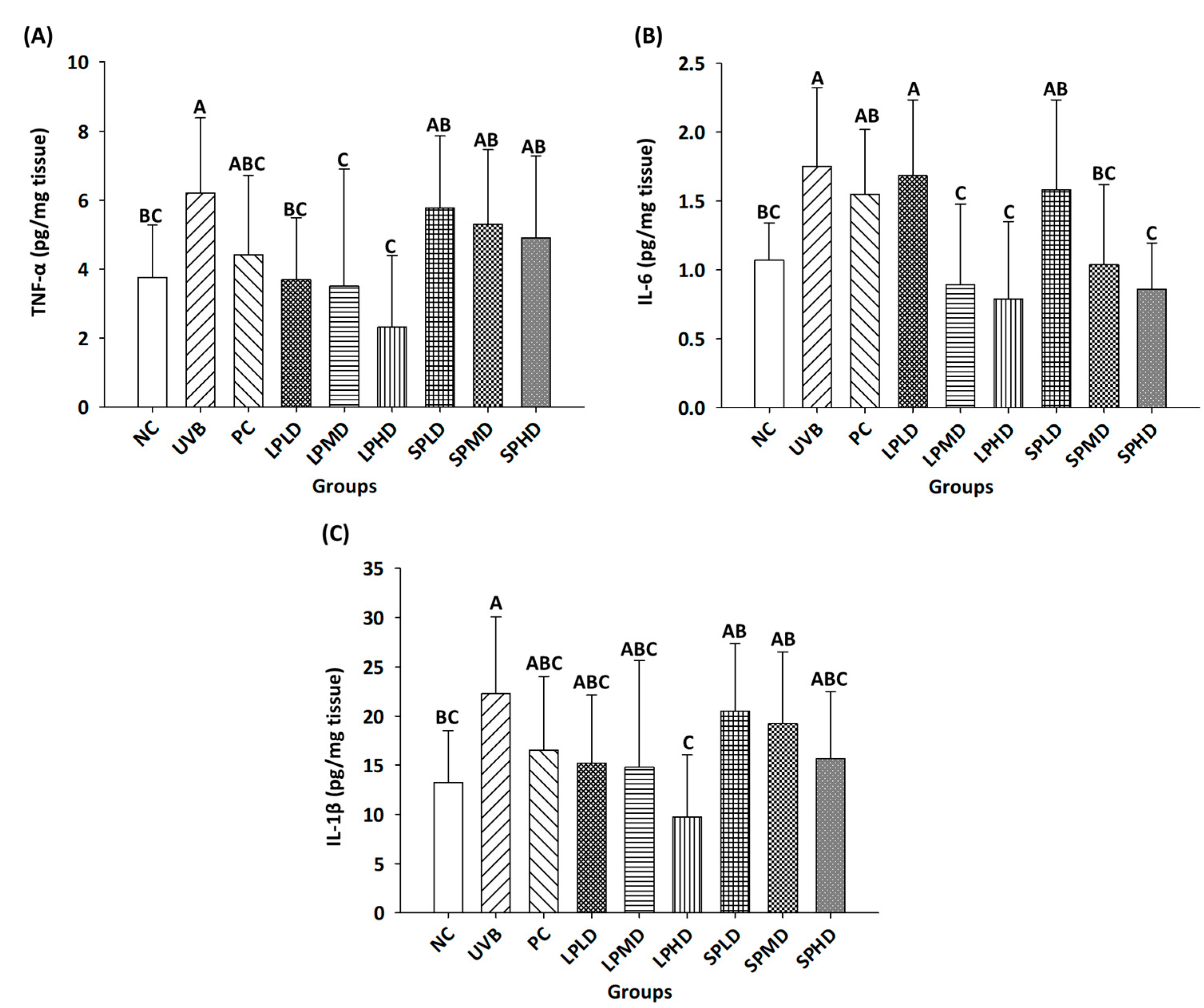
At week 13 of UVB irradiation, the TNF-α, IL-6, and IL-1β contents in the UVB group were significantly higher than those in the NC group (Figure 4A–C). Relative to this observation, the following findings were observed at week 13 in the respective treatment groups. LPTWK10 treatment significantly decreased the TNF-α content by 40.60–62.77% (p < 0.05). Treatment with medium and high doses of LPTWK10 and the spray-dried LPTWK10 supernatant significantly decreased the IL-6 content by 48.97–55.04% and 40.66–50.98%, respectively (p < 0.05). High-dose LPTWK10 administration significantly decreased the IL-1β content by approximately 56.19% (p < 0.05). Both LPTWK10 and its spray-dried supernatant effectively downregulate pro-inflammatory cytokine levels in the skin, with high-dose LPTWK10 demonstrating the most significant reduction in TNF-α, IL-6, and IL-1β levels in the dorsal skin of nude mice subjected to UVB irradiation-induced photoaging. This reduction in cytokine levels may alleviate inflammation-induced skin damage, thereby contributing to improved skin hydration and reduced erythema (Figure 2 and Figure 3A). These findings highlight the potential of LPTWK10 as a therapeutic agent for mitigating the effects of photoaging.

3.6. Determination of Collagen-Related Proteins in the Dorsal Skin of Nude Mice

The dermis includes collagen types I, II, III, and IV, with type I accounting for approximately 90% of the connective tissue protein, followed by type III, both of which are associated with elasticity. During skin aging, the amount of type I collagen decreases while the amount of type III collagen increases. Furthermore, the ratio of type III to type I collagen in the skin increases with UVB irradiation [40]. In addition, increased type III collagen levels may be a response to skin damage caused by UV irradiation, similar to the increased levels observed in scar tissue [7]. Therefore, controlling the ratio of type III to type I collagen can effectively promote anti-aging effects [14]. Collagen, of which type I collagen is the predominant type, is synthesized in the form of procollagen precursors secreted outside the dermis cells to form regularly arranged fibrous structures [32,41].
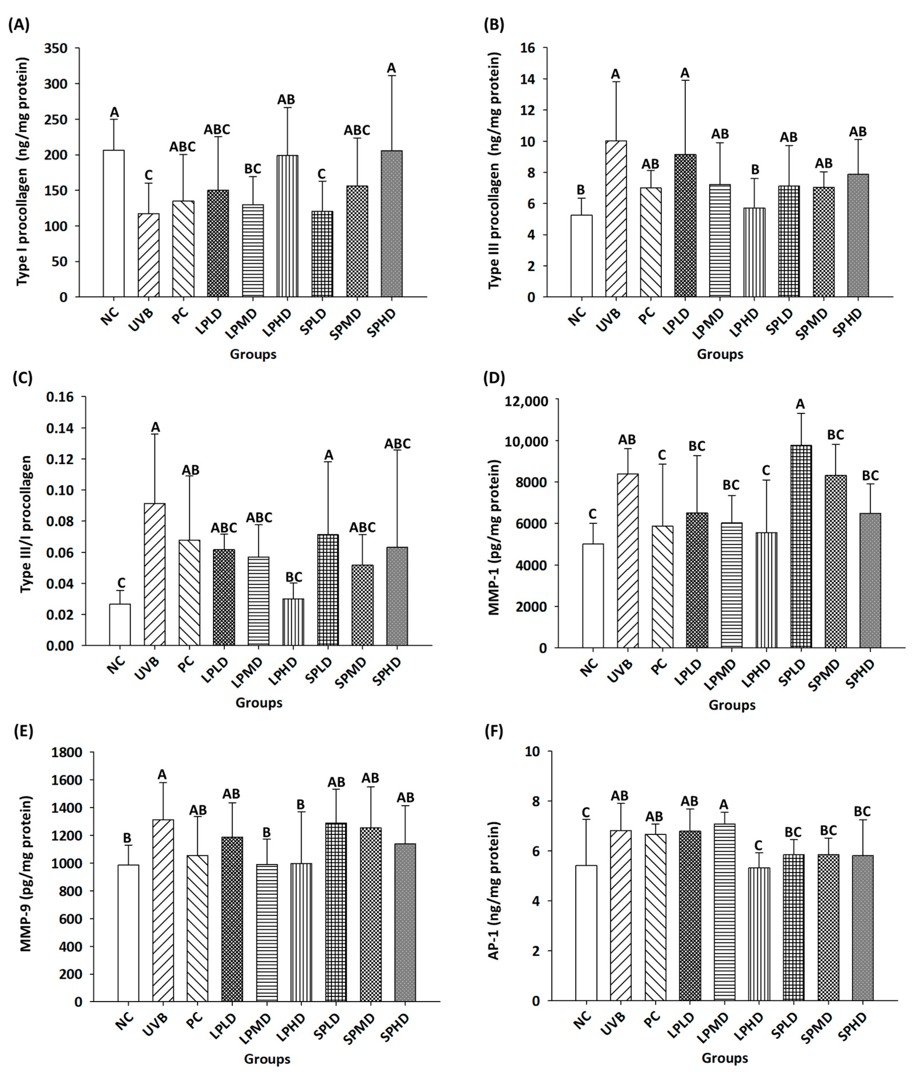
The following findings related to procollagen content were observed at week 13 of UVB and LPTWK10 treatments. The type I procollagen content in the UVB group was significantly lower than that in the NC group by approximately 43.22% (Figure 5A), which significantly increased on treatment with a high dose of LPTWK10 or the spray-dried LPTWK10 supernatant by approximately 69.88 and 75.68%, respectively (p < 0.05). The type III procollagen content in the UVB group was significantly higher than that in the NC group (90.81%), which significantly decreased by 43.07% on treatment with a high dose of LPTWK10 (p < 0.05) (Figure 5B). The ratio of type III to type I procollagen in the skin was further compared, with a lower ratio indicating protective effects for the skin (Figure 5C). The type III/type I pro-collagen ratio was significantly higher in the UVB group than in the NC group, at approximately 0.06, which significantly decreased by approximately 0.06 on treatment with a high dose of LPTWK10 (p < 0.05).
MMPs are zinc ion-dependent endopeptidases involved in the degradation of various extracellular matrix (ECM) components and function in wound repair [8]. MMPs can be subdivided into collagenases, gelatinases, stromelysins, and membrane types based on receptor specificity, domain structure, and cell surface binding properties [42]. MMP-1 is a major collagenase that breaks down collagen and also degrades other ECM components such as elastin, laminin, and fibronectin [43]; hence, it is considered an indicator of photoaging. Increased MMP-1 levels accelerate skin aging, leading to wrinkle formation and compromised skin integrity [41]. MMP-9 is a gelatinase that degrades type IV collagen, a major component of the basement membrane of the skin, during photoaging [44]. Prolonged exposure of the skin to UV radiation upregulates ROS production, leading to the peroxidation of cell membranes, which in turn damages keratinocytes and fibroblasts. For UV-irradiated collagen, only MMP-1 can cleave type I and type III procollagen and denature collagen, which is further catabolized by MMP-3 and MMP-9 [44,45].
The following findings related to MMP levels were observed at week 13 of UVB and LPTWK10 treatments. Compared with the NC group, the UVB-treated group showed 67.86% higher MMP-1 levels, which significantly decreased by 33.85% on treatment with a high dose of LPTWK10 (p < 0.05) (Figure 5D). MMP-9 levels were significantly higher in the UVB group than those in the NC group by 33.20%, which significantly decreased by approximately 24.41 and 23.99%, respectively, on treatment with a medium or high dose of LPTWK10 (p < 0.05) (Figure 5E). These results show that high doses of LPTWK10 effectively lowered the levels of MMP-1 and MMP-9, thereby inhibiting collagen cleavage.
AP-1, which is composed of phosphorylated c-Jun and c-Fos as its subunits, plays an essential role in photoaging by upregulating the gene expression levels of MMP-1, MMP-3, and MMP-9, which in turn leads to collagen cleavage in the dermis [46]. The UVB-induced increase in ROS levels stimulates MAPK phosphorylation, which activates the transcription factors AP-1 and NF-κB and regulates MMP-1, MMP-3, and MMP-9, leading to collagen degradation [44]. At week 13, AP-1 levels were significantly higher in the UVB group than those in the NC group by 26.00% (Figure 5F), which significantly decreased by 21.98% on treatment with a high dose of LPTWK10 (p < 0.05). After 8 weeks treated with mixed probiotics, the expressions of MMPs were significantly decreased [47]. Moreover, HR-1 hairless male mice, which induced photoaging by UVB irradiation, were treated with L. acidophilus and exhibited significant downregulation of MMP-1 and MMP-9 expressions [48]. This shows that high doses of LPTWK10 are effective in inhibiting AP-1 levels, which in turn mitigates the increases in MMP-1 and MMP-9 levels, thereby downregulating collagen degradation. By regulating MMPs and enhancing procollagen production, LPTWK10 may help maintain skin elasticity and reduce wrinkle formation, which aligns with the previously reported results on wrinkle count (Figure 1). This mechanism is crucial for understanding how LPTWK10 contributes to skin health and mitigates the effects of photoaging.
The studies indicated that B. breve improved UVB-induced photoaging by increasing the activities of anti-oxidation enzymes and decreasing the production of pro-inflammation cytokines [22,23]. L. plantarum HY7714 has been confirmed through animal studies to improve skin wrinkles by reducing the expressions and activities of MMPs associated with collagen degradation [24]. It is worth mentioning that LPTWK10 and LPTWK10 spray-dried supernatant powders mitigate the photoaging process not only through antioxidant and anti-inflammatory activities but also by reducing collagen degradation via the regulation of MMP expressions. This indicates that LPTWK10 and its spray-dried supernatant powder can improve photoaging through multiple mechanisms.

3.7. Dorsal Skin Tissue Sections of Nude Mice

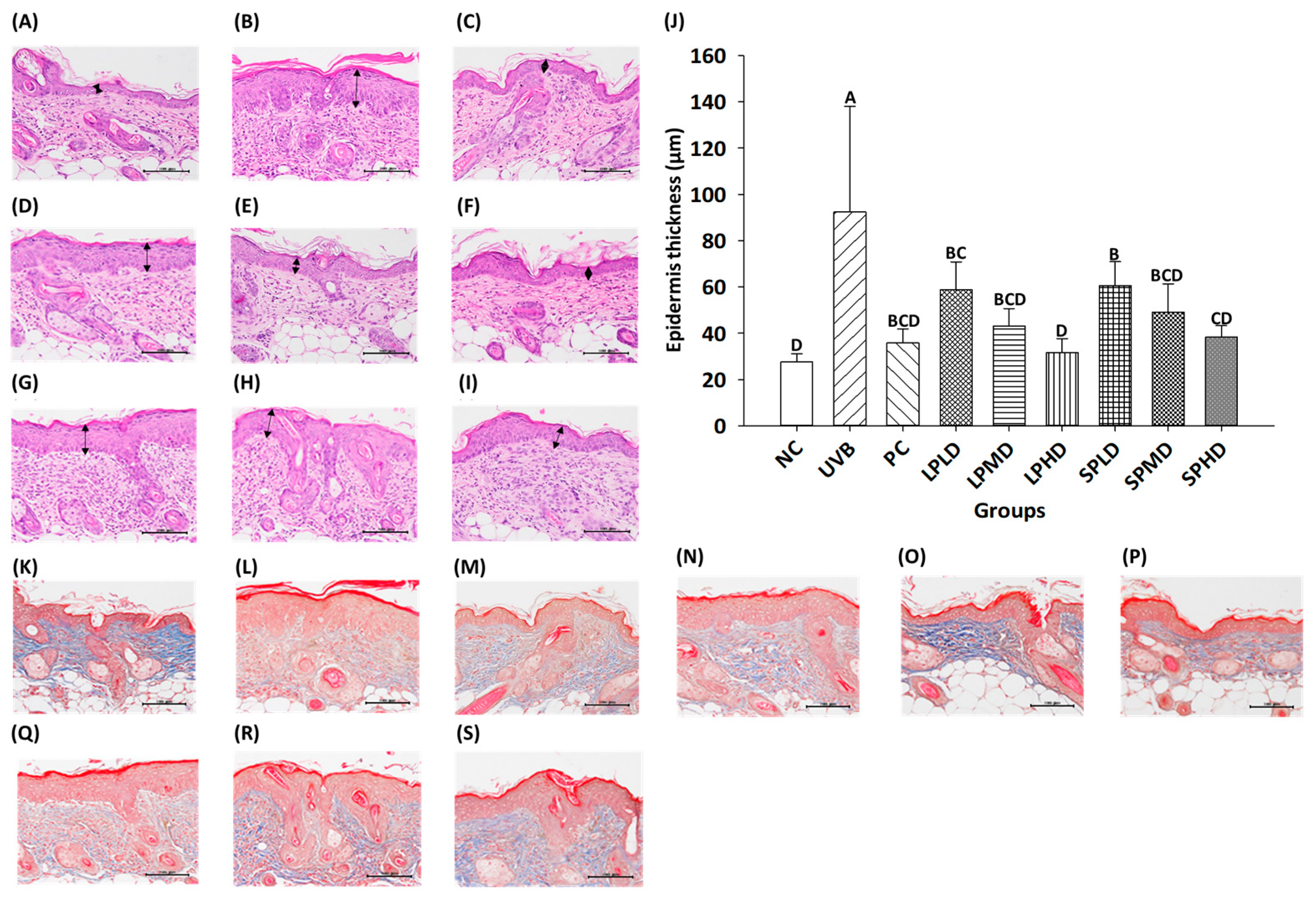
Prolonged UV exposure causes damage to collagen fibers and excessive and abnormal deposition of elastin fibers [6,40]. Ultimately, chronic exposure to sunlight induces keratinocyte proliferation and epidermal hyperplasia, which in turn enhances epidermal thickness and collagen degradation and downregulates collagen production, thereby reducing skin elasticity and leading to skin aging and damage [49,50]. In addition, epidermal thickness increases with increasing UVB exposure to prevent further damage to skin tissue and reduce the potential for malignant transformation [31]. Figure 6 shows the H&E staining of dorsal skin tissue sections for each group. The black arrows in the figure indicate epidermal thickness. The following findings were observed at week 13 of UVB and LPTWK10 treatments. The epidermal thickness in UVB-induced nude mice was significantly higher than that in the NC group by 2.34-fold, which decreased significantly on treatment with LPTWK10 or spray-dried LPTWK10 supernatant by 36.35–65.68% and 34.48–58.59%, respectively (p < 0.05); this effect was dose-dependent. MT staining of dorsal skin tissue sections revealed that the collagen content in the dermis was lower than that in the NC group, which increased in all groups treated with low, medium, and high doses of LPTWK10 or spray-dried LPTWK10 supernatant (Figure 6K–S). Thus, LPTWK10 and spray-dried LPTWK10 supernatants are effective in improving epidermal hyperplasia and inhibiting collagen degradation.

3.8. Immunohistochemical Staining of Dorsal Skin Tissue in Nude Mice

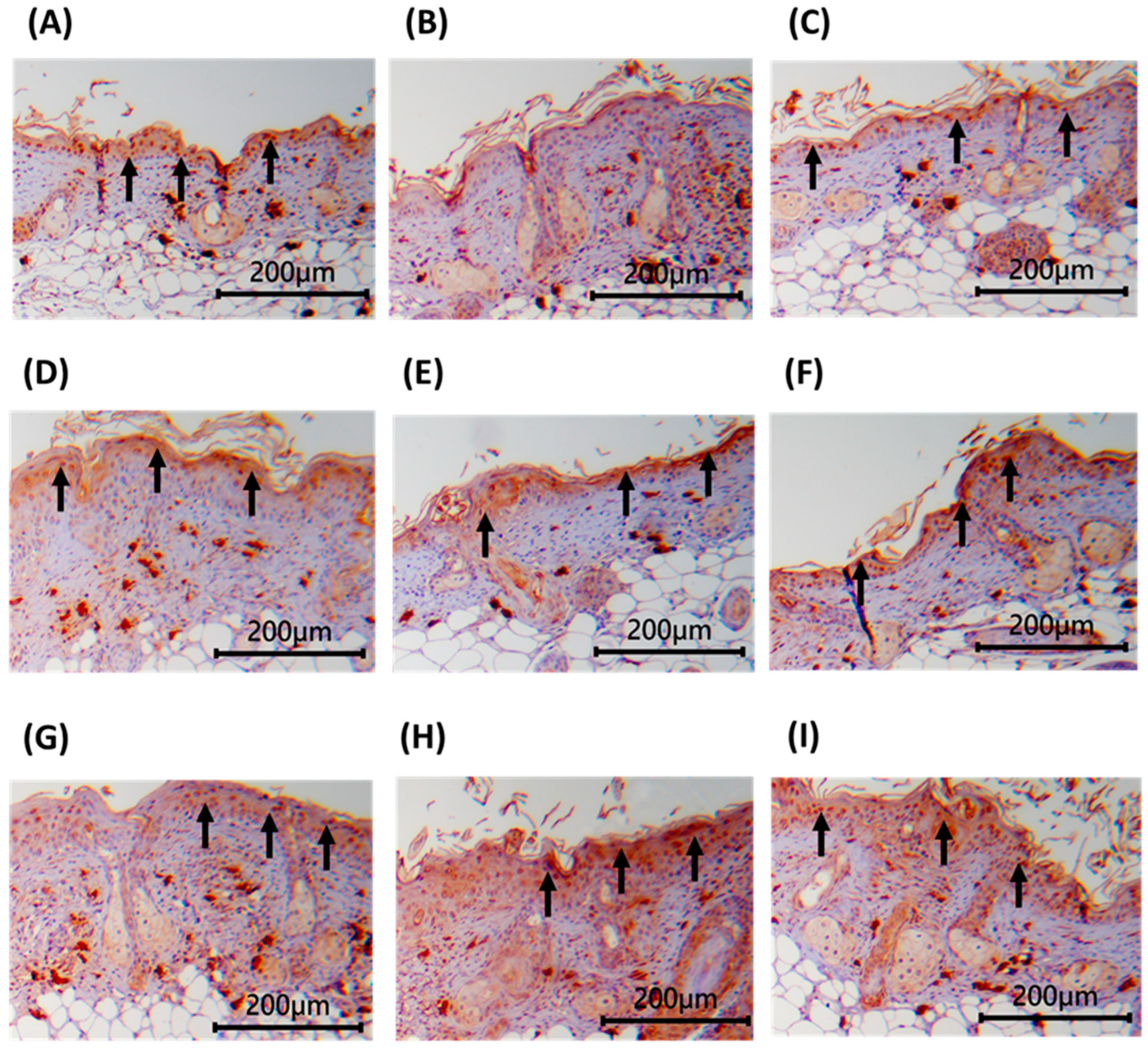
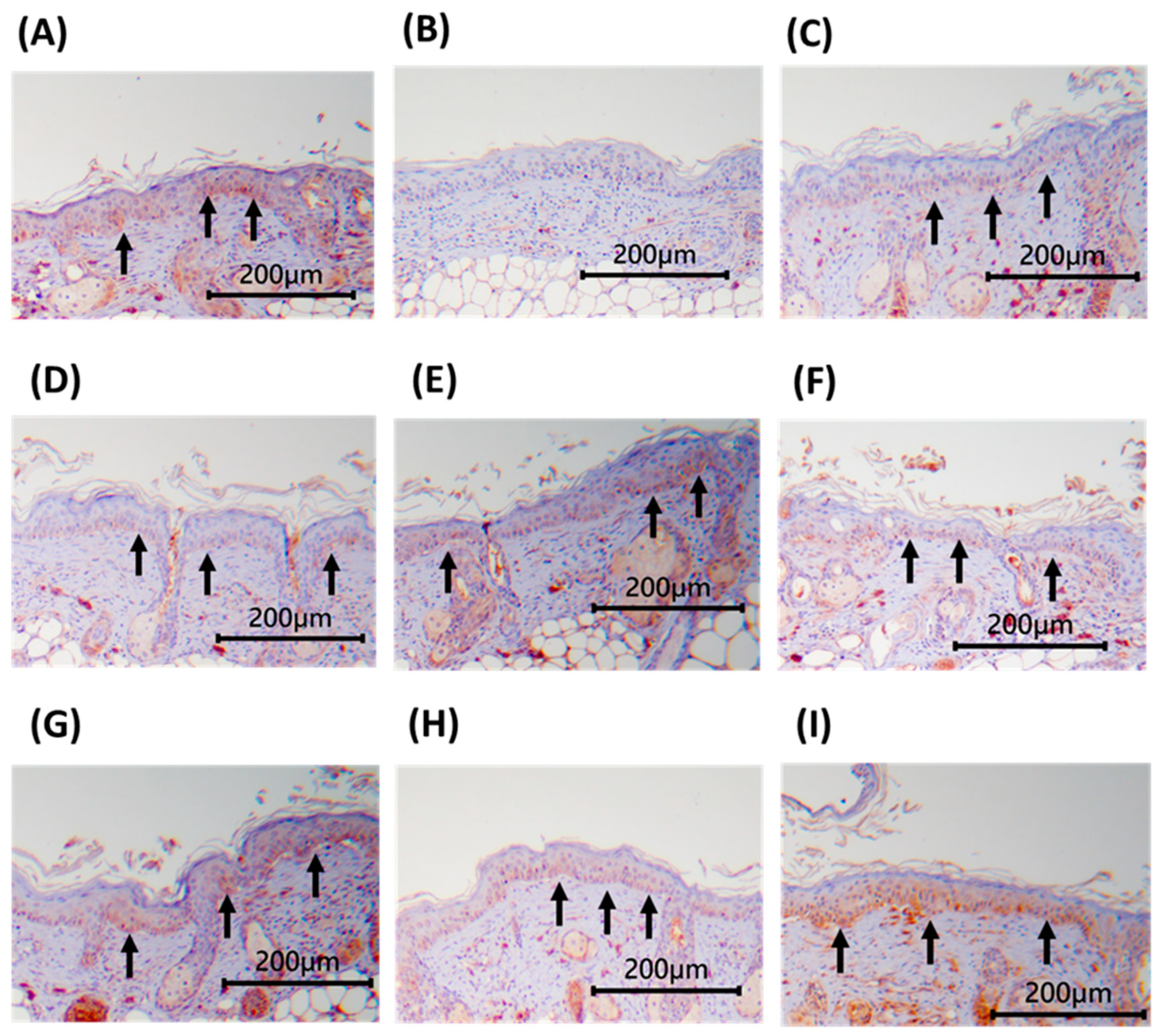
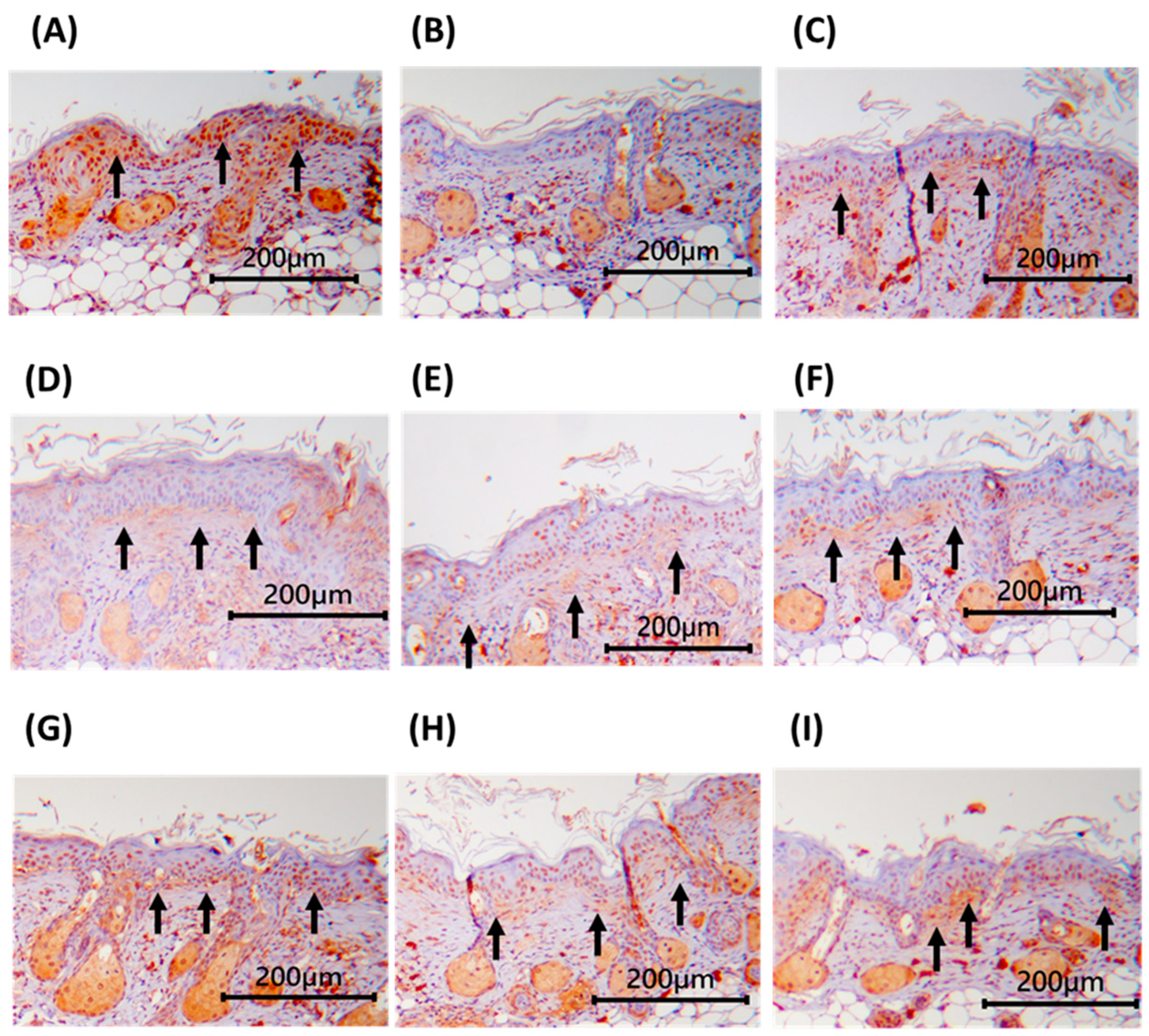
The epidermis consists of the stratum basale, stratum spinosum, stratum granulosum, and SC. The physical barrier of the skin is composed primarily of the SC and tight junctions (TJs) [51]. The SC forms a continuous layer of keratinocytes, which serve as the first line of defense of the skin, whereas TJs control the permeability of substances such as small molecules, electrolytes, and water while preventing pathogen invasion [33]. TJs are composed primarily of two multiprotein complexes. The transmembrane protein claudin-1 is expressed abundantly in keratinocytes and directly interacts with cytoplasmic plaques [52]. Claudin-1-deficient mice exhibit abnormalities in the stratum granulosum and, in severe cases, die within one day of birth [53]. The basement membrane (BM) is a multimolecular structure composed primarily of laminin and type IV collagen. It is firmly anchored between the epidermis and dermis and can regulate epidermal differentiation, proliferation, and epidermal-dermal interactions. UV-induced damage to the BM results in the formation of a multilayered structure that contributes to the aging of the dermis and epidermis [54,55]. Increased ROS levels are a key factor in skin photoaging. Following UV irradiation, ROS are generated in the epidermis and dermis, resulting in the degradation of the BM and ECM, disruption of cell-cell adhesion, and apoptosis, ultimately leading to the disruption of barrier function and reduction in the water content of the skin [23,55].
Figure 7, Figure 8 and Figure 9 show the immunohistochemical staining results of dorsal skin tissue sections, wherein target proteins appear brown. At week 13, claudin-1, laminin, and type IV collagen levels in the dorsal skin tissues of the UVB-treated group were lower than those in the NC group. These levels increased in all three groups on treatment with LPTWK10 or the spray-dried LPTWK10 supernatant at low, medium, and high doses. The results indicate that LPTWK10 or the spray-dried LPTWK10 supernatant decelerates the degradation of claudin-1, laminin, and type IV collagen proteins in the skin, thereby ameliorating the impairment of skin barrier function. In addition, the increase in stratum corneum hydration observed in our results may be linked to the ability of LPTWK10 to enhance the skin barrier function, thereby reducing moisture loss and improving overall skin hydration levels.
The above results suggest that both LPTWK10 and its spray-dried supernatant powders can mitigate UVB-induced skin photoaging in female immunodeficient mice through different pathways. Studies have shown that gut microbiota dysbiosis can increase intestinal permeability, allowing gut microorganisms or their metabolites to translocate into the circulatory system and accumulate in the skin, leading to systemic inflammation and compromising skin health. Probiotics may modulate the gut-skin axis by improving gut microbiota composition [56] In addition to reducing UVB-induced ROS levels and enhancing antioxidant enzyme activities in skin tissue [22,57], Lactobacillus can regulate the AP-1 pathway, thereby reducing the increase in MMP activities during photoaging. This regulation helps prevent collagen loss, contributing to overall skin health. Research has demonstrated that lysates from Lactobacillus effectively inhibit MMP-1 expression, further supporting the role of probiotics in protecting against UVB-induced skin damage. Additionally, metabolites of probiotics have shown protective effects against UVB-induced oxidative stress and collagen degradation through modulation of signaling pathways such as p38 MAPK [29,58]. The primary active substances in LPTWK10 spray-dried supernatant powders are likely derived from the metabolites of lactic acid bacteria. For example, short-chain fatty acids (SCFAs) have been documented to exhibit strong anti-inflammatory activity. SCFAs can regulate the gut-skin axis by improving gut microbiota composition and reducing intestinal permeability, thereby decreasing systemic inflammation that may negatively impact skin health. Additionally, SCFAs enhance skin barrier function by preventing TEWL, which improves skin hydration and elasticity, thus reducing the effects of photoaging. Furthermore, some probiotics produce antioxidant enzymes, such as superoxide dismutase (SOD) and catalase, during their metabolic processes. These enzymes effectively neutralize the excessive ROS generated by UVB exposure, thereby mitigating skin cell damage [59].
Our experimental results indicate that the effects of LPTWK10 are superior to those of the LPTWK10 spray-dried supernatant powders, with the highest dose of LPTWK10 demonstrating the greatest ability to mitigate UVB-induced photoaging. This difference in efficacy may be attributed to the distinct bioactive components present in each form. LPTWK10 consists of live bacteria, which not only enhance skin health through their own activity but also have the potential to generate beneficial metabolites in the gut when interacting with prebiotics. Consequently, in addition to the direct effects of the bacteria, there may be additive benefits from these gut-derived metabolites. In contrast, LPTWK10 spray-dried supernatant powders primarily contain bacterial metabolites and secreted compounds. While these metabolites, such as SCFAs and lactic acid, exhibit anti-inflammatory and antioxidant properties, they may not fully replicate the comprehensive range of benefits provided by live probiotics. Additionally, the stability and bioavailability of active compounds in the spray-dried supernatant could influence its efficacy. Some metabolites may degrade or lose potency during the spray-drying process or subsequent storage, potentially diminishing their effectiveness compared with live LPTWK10.

4. Conclusions

In this study, 13 weeks of UVB irradiation led to significant skin damage, evidenced by prominent wrinkle formation, a marked decrease in dorsal skin moisture (p < 0.05), and observable erythema, inflammation, melanin accumulation, and reduced skin lightness, confirming the successful induction of photoaging. Oral administration of LPTWK10 or its spray-dried supernatant to UVB-exposed nude mice resulted in a significant reduction in wrinkle count, moisture loss, erythema, and melanin accumulation (p < 0.05). The observed anti-photoaging effects were attributed to the inhibition of pro-inflammatory factor production and modulation of protein expression related to collagen synthesis. Notably, the most effective dose of LPTWK10 in mitigating UVB-induced skin damage was found to be 2 × 10¹⁰ CFU/kg bw/day. In conclusion, our study offers compelling evidence for the efficacy of LPTWK10 and its spray-dried supernatant powder in mitigating UVB-induced skin photoaging. By elucidating their distinct mechanisms of action and exploring their combined potential in skincare applications, we aim to provide valuable insights into the field of dermatology. In the future, further research can focus not only on leveraging the strengths of both sample types to enhance skin protection but also on conducting human trials to investigate the mechanisms of action of LPTWK10 and its spray-dried supernatant powder under a broader range of skin conditions.

Author Contributions

Conceptualization, T.-H.L., C.-C.L., J.-S.L. and T.-Y.T.; Data curation, T.-H.L., W.-J.L. and M.-C.C.; Formal analysis, T.-H.L., W.-J.L. and M.-C.C.; Methodology, T.-H.L., M.-C.C., J.-S.L. and T.-Y.T.; Project administration, T.-H.L. and Y.-C.C.; Visualization, T.-H.L.; Validation, W.-J.L.; Investigation, W.-J.L.; Writing – original draft, T.-H.L. and W.-J.L.; Writing – review & editing, T.-H.L., M.-C.C., Y.-C.C., C.-C.L., J.-S.L. and T.-Y.T.; Resources, Y.-C.C., J.-S.L. and T.-Y.T.; Funding acquisition, T.-Y.T.; Supervision, T.-Y.T. All authors have read and agreed to the published version of the manuscript.

Funding

This research received no external funding.

Institutional Review Board Statement

This animal study protocol was approved by the Institutional Animal Care and Use Committee (IACUC) of Fu Jen Catholic University (IACUC No. A10830 and A10831, approved on 16 June 2020).

Informed Consent Statement

Not applicable.

Data Availability Statement

The original contributions presented in the study are included in the article, further inquiries can be directed to the corresponding author.

Conflicts of Interest

Authors Yi-Chen Cheng, Chia-Chia Lee and Jin-Seng Lin were employed by the company SYNBIO TECH INC. The remaining authors declare that the research was conducted in the absence of any commercial or financial relationships that could be construed as a potential conflict of interest.

References

  1. Montagna, W.; Parakkal, P.F. 5—Blood Supply. In The Structure & Function of Skin, 3rd ed.; Montagna, W., Parakkal, P.F., Eds.; Academic Press: Cambridge, MA, USA, 1974; pp. 142–156. [Google Scholar] [CrossRef]
  2. Yi, R.; Zhang, J.; Sun, P.; Qian, Y.; Zhao, X. Protective Effects of Kuding Tea (Ilex kudingcha C. J. Tseng) Polyphenols on UVB-Induced Skin Aging in SKH1 Hairless Mice. Molecules 2019, 24, 1016. [Google Scholar] [CrossRef]
  3. Lawton, S. Skin 1: The Structure and Functions of the Skin. Nurs. Times 2019, 115, 30–33. [Google Scholar] [CrossRef]
  4. Miyamae, Y.; Yamakawa, Y.; Kawabata, M.; Ozaki, Y. A Combined Near-Infrared Diffuse Reflectance Spectroscopy and Principal Component Analysis Method of Assessment for the Degree of Photoaging and Physiological Aging of Human Skin. Anal. Sci. 2012, 28, 1159–1164. [Google Scholar] [CrossRef] [PubMed]
  5. Rijken, F.; Bruijnzeel-Koomen, C.A.F.M. Photoaged Skin: The Role of Neutrophils, Preventive Measures, and Potential Pharmacological Targets. Clin. Pharmacoloy Ther. 2011, 89, 120–124. [Google Scholar] [CrossRef]
  6. Zhang, J.A.; Yin, Z.; Ma, L.W.; Yin, Z.Q.; Hu, Y.Y.; Xu, Y.; Wu, D.; Permatasari, F.; Luo, D.; Zhou, B.R. The Protective Effect of Baicalin against UVB Irradiation Induced Photoaging: An In Vitro and In Vivo Study. PLoS ONE 2014, 9, e99703. [Google Scholar] [CrossRef]
  7. Plastow, S.R.; Harrison, J.A.; Young, A.R. Early Changes in Dermal Collagen of Mice Exposed to Chronic UVB Irradiation and the Effects of a UVB Sunscreen. J. Investig. Dermatol. 1988, 91, 590–592. [Google Scholar] [CrossRef]
  8. Lee, D.H.; Oh, J.H.; Chung, J.H. Glycosaminoglycan and Proteoglycan in Skin Aging. J. Dermatol. Sci. 2016, 83, 174–181. [Google Scholar] [CrossRef]
  9. Wulf, H.C.; Sandby-Møller, J.; Kobayasi, T.; Gniadecki, R. Skin Aging and Natural Photoprotection. Micron 2004, 35, 185–191. [Google Scholar] [CrossRef]
  10. Avila Acevedo, J.G.; Espinosa González, A.M.; De Maria y Campos, D.M.; Benitez Flores Jdel, C.; Hernández Delgado, T.; Flores Maya, S.; Campos Contreras, J.; Muñoz López, J.L.; García Bores, A.M. Photoprotection of Buddleja cordata Extract against UVB-Induced Skin Damage in SKH-1 Hairless Mice. BMC Complement. Altern. Med. 2014, 14, 281. [Google Scholar] [CrossRef]
  11. García-Bores, A.M.; Espinosa-González, A.M.; Reyna-Campos, A.; Cruz-Toscano, S.; Benítez-Flores, J.C.; Hernández-Delgado, C.T.; Flores-Maya, S.; Urzúa-Meza, M.; Peñalosa-Castro, I.; Céspedes-Acuña, C.L.; et al. Lippia graveolens Photochemopreventive Effect against UVB Radiation-Induced Skin Carcinogenesis. J. Photochem. Photobiol. B Biol. 2017, 167, 72–81. [Google Scholar] [CrossRef]
  12. Halliday, G.M. Inflammation, Gene Mutation and Photoimmunosuppression in Response to UV-Induced Oxidative Damage Contributes to Photocarcinogenesis. Mutat. Res. 2005, 571, 107–120. [Google Scholar] [CrossRef]
  13. Katiyar, S.K.; Matsui, M.S.; Mukhtar, H. Kinetics of UV Light-Induced Cyclobutane Pyrimidine Dimers in Human Skin in Vivo: An Immunohistochemical Analysis of Both Epidermis and Dermis. Photochem. Photobiol. 2000, 72, 788–793. [Google Scholar] [CrossRef]
  14. Li, G.; Fang, T.; Zhang, Q.; Tan, A.; Cheng, Y.; Zhou, Q.; Liu, M.; Ta, X.; Huang, L.H.; Rouseff, R.; et al. Protective Effects of Polymethoxyflavone-Rich Cold-Pressed Orange Peel Oil against Ultraviolet B-Induced Photoaging on Mouse Skin. J. Funct. Foods. 2020, 67, 103834. [Google Scholar] [CrossRef]
  15. Robinson, E.S.; Werth, V.P. The Role of Cytokines in the Pathogenesis of Cutaneous Lupus Erythematosus. Cytokine 2015, 73, 326–334. [Google Scholar] [CrossRef]
  16. Park, B.; Hwang, E.; Seo, S.A.; Cho, J.G.; Yang, J.E.; Yi, T.H. Eucalyptus Globulus Extract Protects against UVB-Induced Photoaging by Enhancing Collagen Synthesis via Regulation of TGF-β/Smad Signals and Attenuation of AP-1. Arch. Biochem. Biophys. 2018, 637, 31–39. [Google Scholar] [CrossRef]
  17. Liu, Y.Y.; Zeng, S.Y.; Leu, Y.L.; Tsai, T.Y. Antihypertensive Effect of a Combination of Uracil and Glycerol Derived from Lactobacillus plantarum Strain TWK10-Fermented Soy Milk. J. Agric. Food Chem. 2015, 63, 7333–7342. [Google Scholar] [CrossRef]
  18. Liu, T.H.; Chiou, J.; Tsai, T.Y. Effects of Lactobacillus plantarum TWK10-Fermented Soymilk on Deoxycorticosterone Acetate-Salt-Induced Hypertension and Associated Dementia in Rats. Nutrients 2016, 8, 260. [Google Scholar] [CrossRef]
  19. Chen, Y.M.; Wei, L.; Chiu, Y.S.; Hsu, Y.J.; Tsai, T.Y.; Wang, M.F.; Huang, C.C. Lactobacillus plantarum TWK10 Supplementation Improves Exercise Performance and Increases Muscle Mass in Mice. Nutrients 2016, 8, 205. [Google Scholar] [CrossRef]
  20. Chang, C.J.; Dai, R.Y.; Leu, Y.L.; Tsai, T.Y. Effects of the Melanogenic Inhibitor, Uracil, Derived from Lactobacillus plantarum TWK10-Fermented Soy Milk on Anti-Melanogenesis in B16f0 Mouse Melanoma Cells. J. Funct. Foods 2015, 17, 314–327. [Google Scholar] [CrossRef]
  21. Chuang, Y.C.; Cheng, M.C.; Lee, C.C.; Chiou, T.Y.; Tsai, T.Y. Effect of Ethanol Extract from Lactobacillus plantarum TWK10-Fermented Soymilk on Wound Healing in Streptozotocin-Induced Diabetic Rat. AMB Express 2019, 9, 163. [Google Scholar] [CrossRef]
  22. Ishii, Y.; Sugimoto, S.; Izawa, N.; Sone, T.; Chiba, K.; Miyazaki, K. Oral Administration of Bifidobacterium breve Attenuates UV-Induced Barrier Perturbation and Oxidative Stress in Hairless Mice Skin. Arch. Dermatol. Res. 2014, 306, 467–473. [Google Scholar] [CrossRef]
  23. Satoh, T.; Murata, M.; Iwabuchi, N.; Odamaki, T.; Wakabayashi, H.; Yamauchi, K.; Abe, F.; Xiao, J.Z. Effect of Bifidobacterium breve B-3 on Skin Photoaging Induced by Chronic UV Irradiation in Mice. Benef. Microbes 2015, 6, 497–504. [Google Scholar] [CrossRef]
  24. Kim, H.M.; Lee, D.E.; Park, S.D.; Kim, Y.T.; Kim, Y.J.; Jeong, J.W.; Jang, S.S.; Ahn, Y.T.; Sim, J.H.; Huh, C.S.; et al. Oral Administration of Lactobacillus plantarum HY7714 Protects Hairless Mouse against Ultraviolet B-Induced Photoaging. J. Microbiol. Biotechnol. 2014, 24, 1583–1591. [Google Scholar] [CrossRef]
  25. Zhou, Y.; Bai, R.; Huang, Y.; Li, W.; Chen, J.; Cheng, Z.; Wu, X.; Diao, Y. The Anti-Photoaging Effect of C-phycocyanin on Ultraviolet B-Irradiated Balb/c-nu Mouse Skin. Front. Bioeng. Biotechnol. 2023, 11, 1229387. [Google Scholar] [CrossRef]
  26. Gilhar, A.; Etzioni, A. The Nude Mouse Model for the Study of Human Skin Disorders. Dermatology 1994, 189, 5–8. [Google Scholar] [CrossRef]
  27. Fan, Y.; Jeong, J.H.; You, G.Y.; Park, J.U.; Choi, T.H.; Kim, S. An Experimental Model Design for Photoaging. J. Craniofac. Surg. 2015, 26, e467–e471. [Google Scholar] [CrossRef]
  28. Gilchrest, B.A. Photoaging. J. Investig. Dermatol. 2013, 133, E2–E6. [Google Scholar] [CrossRef]
  29. Hara, Y.; Hirao, T.; Iwai, I. Facial Expression under Stiff Stratum Corneum Leads to Strain Concentrations, Followed by Residual Wrinkle Formation. Int. J. Cosmet. Sci. 2017, 39, 66–71. [Google Scholar] [CrossRef]
  30. Kong, Y.H.; Xu, S.P. Juglanin Administration Protects Skin against UVB-Induced Injury by Reducing Nrf2-Dependent ROS Generation. Int. J. Mol. Med. 2020, 46, 67–82. [Google Scholar] [CrossRef]
  31. Kuo, Y.H.; Lin, T.Y.; You, Y.J.; Wen, K.C.; Sung, P.J.; Chiang, H.M. Antiinflammatory and Antiphotodamaging Effects of Ergostatrien-3β-ol, Isolated from Antrodia camphorata, on Hairless Mouse Skin. Molecules 2016, 21, 1213. [Google Scholar] [CrossRef]
  32. Son, D.J.; Jung, J.C.; Choi, Y.M.; Ryu, H.Y.; Lee, S.; Davis, B.A. Wheat Extract Oil (Weo) Attenuates UVB-Induced Photoaging Via Collagen Synthesis in Human Keratinocytes and Hairless Mice. Nutrients 2020, 12, 300. [Google Scholar] [CrossRef]
  33. Li, Z.; Jiang, R.; Wang, M.; Zhai, L.; Liu, J.; Xu, X.; Sun, L.; Zhao, D. Ginsenosides Repair UVB-Induced Skin Barrier Damage in Balb/c Hairless Mice and HaCat Keratinocytes. J. Ginseng. Res. 2022, 46, 115–125. [Google Scholar] [CrossRef]
  34. Costin, G.E.; Hearing, V.J. Human Skin Pigmentation: Melanocytes Modulate Skin Color in Response to Stress. FASEB J. 2007, 21, 976–994. [Google Scholar] [CrossRef]
  35. Solano, F. Melanins: Skin Pigments and Much More—Types, Structural Models, Biological Functions, and Formation Routes. New J. Sci. 2014, 2014, 498276. [Google Scholar] [CrossRef]
  36. Liu, T.H.; Chiang, W.T.; Cheng, M.C.; Tsai, T.Y. Effects of Germination Black Soy Milk Fermented with Lactobacillus plantarum TWK10 on Anti-Oxidative and Anti-Melanogenesis. Appl. Sci. 2022, 12, 277. [Google Scholar] [CrossRef]
  37. Aoki, R.; Aoki-Yoshida, A.; Suzuki, C.; Takayama, Y. Protective Effect of Indole-3-Pyruvate against Ultraviolet B-Induced Damage to Cultured HaCat Keratinocytes and the Skin of Hairless Mice. PLoS ONE 2014, 9, e96804. [Google Scholar] [CrossRef]
  38. Pillai, S.; Oresajo, C.; Hayward, J. Ultraviolet Radiation and Skin Aging: Roles of Reactive Oxygen Species, Inflammation and Protease Activation, and Strategies for Prevention of Inflammation-Induced Matrix Degradation—A Review. Int. J. Cosmet. Sci. 2005, 27, 17–34. [Google Scholar] [CrossRef]
  39. Wang, X.P.; Schunck, M.; Kallen, K.J.; Neumann, C.; Trautwein, C.; Rose-John, S.; Proksch, E. The Interleukin-6 Cytokine System Regulates Epidermal Permeability Barrier Homeostasis. J. Investig. Dermatol. 2004, 123, 124–131. [Google Scholar] [CrossRef]
  40. Fan, J.; Zhuang, Y.; Li, B. Effects of Collagen and Collagen Hydrolysate from Jellyfish Umbrella on Histological and Immunity Changes of Mice Photoaging. Nutrients 2013, 5, 223–233. [Google Scholar] [CrossRef]
  41. Kim, H.R.; Jeong, D.H.; Kim, S.; Lee, S.W.; Sin, H.S.; Yu, K.Y.; Jeong, S.I.; Kim, S.Y. Fermentation of Blackberry with L. plantarum JBMI F5 Enhance the Protection Effect on UVB-Mediated Photoaging in Human Foreskin Fibroblast and Hairless Mice through Regulation of MAPK/NF-κB Signaling. Nutrients 2019, 11, 2429. [Google Scholar] [CrossRef]
  42. Jabłońska-Trypuć, A.; Matejczyk, M.; Rosochacki, S. Matrix Metalloproteinases (MMPs), the Main Extracellular Matrix (ECM) Enzymes in Collagen Degradation, as a Target for Anticancer Drugs. J. Enzyme Inhib. Med. Chem. 2016, 31, 177–183. [Google Scholar] [CrossRef]
  43. Gupta, A.; Kaur, C.D.; Jangdey, M.; Saraf, S. Matrix Metalloproteinase Enzymes and Their Naturally Derived Inhibitors: Novel Targets in Photocarcinoma Therapy. Ageing Res. Rev. 2014, 13, 65–74. [Google Scholar] [CrossRef]
  44. Zhang, G.; Wang, P.; Wang, X. Skin Ageing and Cancer. In The Role of Matrix Metalloproteinase in Human Body Pathologies; Francesco, T., Ed.; IntechOpen: Rijeka, Croatia, 2017; Chapter 6. [Google Scholar] [CrossRef]
  45. Feng, X.X.; Yu, X.T.; Li, W.J.; Kong, S.Z.; Liu, Y.H.; Zhang, X.; Xian, Y.F.; Zhang, X.J.; Su, Z.R.; Lin, Z.X. Effects of Topical Application of Patchouli Alcohol on the UV-Induced Skin Photoaging in Mice. Eur. J. Pharm. Sci. 2014, 63, 113–123. [Google Scholar] [CrossRef]
  46. Freitas-Rodríguez, S.; Folgueras, A.R.; López-Otín, C. The Role of Matrix Metalloproteinases in Aging: Tissue Remodeling and Beyond. Biochim. Biophys. Acta Mol. Cell Res. 2017, 1864, 2015–2025. [Google Scholar] [CrossRef]
  47. Seo, E.; Song, H.H.; Kim, H.; Kim, B.-Y.; Park, S.; Suh, H.J.; Ahn, Y. Oral Administration of Mixed Probiotics Improves Photoaging by Modulating the Cecal Microbiome and MAPK Pathway in UVB-Irradiated Hairless Mice. Mol. Nutr. Food Res. 2023, 67, 2200841. [Google Scholar] [CrossRef]
  48. Im, A.R.; Kim, H.S.; Hyun, J.W.; Chae, S. Potential for Tyndalized Lactobacillus acidophilus as an Effective Component in Moisturizing Skin and Anti-Wrinkle Products. Exp. Ther. Med. 2016, 12, 759–764. [Google Scholar] [CrossRef]
  49. Kim, H.; Oh, I.; Park, K.H.; Kim, N.M.; Do, J.H.; Cho, Y. Stimulatory Effect of Dietary Red Ginseng on Epidermal Hydration and Ceramide Levels in Ultraviolet-Irradiated Hairless Mice. J. Med. Food 2009, 12, 746–754. [Google Scholar] [CrossRef]
  50. Yaar, M.; Gilchrest, B.A. Photoageing: Mechanism, Prevention and Therapy. Br. J. Dermatol. 2007, 157, 874–887. [Google Scholar] [CrossRef]
  51. Kobayashi, M.; Shu, S.; Marunaka, K.; Matsunaga, T.; Ikari, A. Weak Ultraviolet B Enhances the Mislocalization of Claudin-1 Mediated by Nitric Oxide and Peroxynitrite Production in Human Keratinocyte-Derived HaCat Cells. Int. J. Mol. Sci. 2020, 21, 7138. [Google Scholar] [CrossRef]
  52. Yokouchi, M.; Kubo, A. Maintenance of Tight Junction Barrier Integrity in Cell Turnover and Skin Diseases. Exp. Dermatol. 2018, 27, 876–883. [Google Scholar] [CrossRef]
  53. Sugawara, T.; Iwamoto, N.; Akashi, M.; Kojima, T.; Hisatsune, J.; Sugai, M.; Furuse, M. Tight Junction Dysfunction in the Stratum Granulosum Leads to Aberrant Stratum Corneum Barrier Function in Claudin-1-Deficient Mice. J. Dermatol. Sci. 2013, 70, 12–18. [Google Scholar] [CrossRef]
  54. Park, E.K.; Lee, H.J.; Lee, H.; Kim, J.H.; Hwang, J.; Koo, J.I.; Kim, S.H. The Anti-Wrinkle Mechanism of Melatonin in UVB Treated HaCat Keratinocytes and Hairless Mice via Inhibition of ROS and Sonic Hedgehog Mediated Inflammatory Proteins. Int. J. Mol. Sci. 2018, 19, 1995. [Google Scholar] [CrossRef]
  55. Tamaru, E.; Watanabe, M.; Nomura, Y. Dietary Immature Citrus unshiu Alleviates UVB- Induced Photoaging by Suppressing Degradation of Basement Membrane in Hairless Mice. Heliyon 2020, 6, e04218. [Google Scholar] [CrossRef]
  56. Sinha, S.; Lin, G.; Ferenczi, K. The Skin Microbiome and the Gut-Skin Axis. Clin. Dermatol. 2021, 39, 829–839. [Google Scholar] [CrossRef]
  57. Teng, Y.; Huang, Y.; Danfeng, X.; Tao, X.; Fan, Y. The Role of Probiotics in Skin Photoaging and Related Mechanisms: A Review. Clin. Cosmet. Investig. Dermatol. 2022, 15, 2455–2464. [Google Scholar] [CrossRef]
  58. Im, A.R.; Lee, B.; Kang, D.J.; Chae, S. Protective Effects of Tyndallized Lactobacillus acidophilus IDCC 3302 Against UVB-induced Photodamage to Epidermal Keratinocytes Cells. Int. J. Mol. Med. 2019, 43, 2499–2506. [Google Scholar] [CrossRef]
  59. Gao, T.; Wang, X.; Li, Y.; Ren, F. The Role of Probiotics in Skin Health and Related Gut-Skin Axis: A Review. Nutrients 2023, 15, 3123. [Google Scholar] [CrossRef]
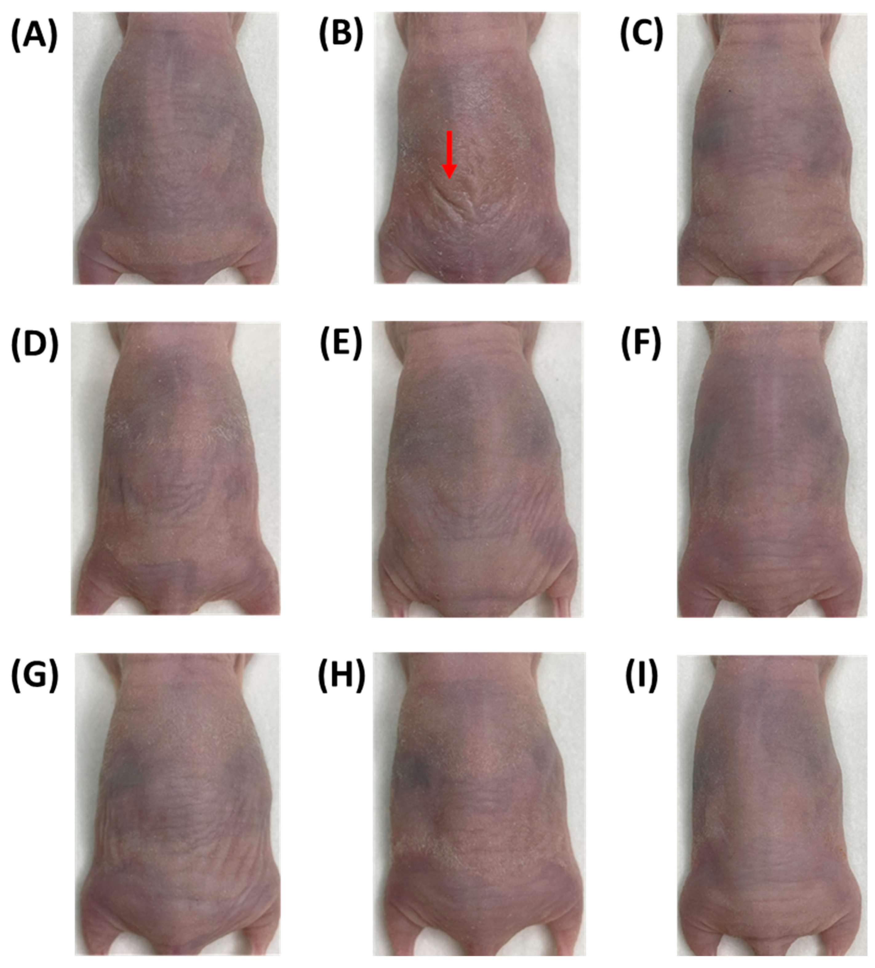
Figure 1. Effects of the administration of LPTWK10 or LPTWK10 spray-dried supernatant powders on dorsal wrinkles in UVB-induced nude mice. (A) NC (B) UVB (C) PC (D) LPLD (E) LPMD (F) LPHD (G) SPLD (H) SPMD (I) SPHD. Data are presented as mean ± SD (n = 7 or 8). UVB: ultraviolet B; NC: normal control; PC: positive control; LPLD, LPMD, and LPHD: administration of LPTWK10 at 2 × 10⁸ CFU/kg bw/day, 2 × 10⁹ CFU/kg bw/day and 2 × 10¹⁰ CFU/kg bw/day, respectively; SPLD, SPMD and SPHD: administration of TWK10 spray-dried supernatant powders at 0.30 g/kg bw/day, 0.60 g/kg bw/day, and 1.20 g/kg bw/day, respectively. Red arrow: wrinkles.
Applsci 14 09497 g001
Figure 2. Effects of the administration of LPTWK10 or LPTWK10 spray-dried supernatant powders on (A) dorsal TEWL and (B) dorsal hydration of the stratum corneum in UVB-induced nude mice. Data are presented as mean ± SD (n = 7 or 8). Values with different uppercase letters were significantly different by Duncan’s multiple range test (p < 0.05). TEWL: transepidermal water loss; UVB: ultraviolet B; NC: normal control; PC: positive control; LPLD, LPMD and LPHD: administration of LPTWK10 viable cells at 2 × 10⁸ CFU/kg bw/day, 2 × 10⁹ CFU/kg bw/day and 2 × 10¹⁰ CFU/kg bw/day, respectively; SPLD, SPMD and SPHD: administration of TWK10 spray-dried supernatant powders at 0.30 g/kg bw/day, 0.60 g/kg bw/day, and 1.20 g/kg bw/day, respectively.
Applsci 14 09497 g002
Figure 3. Effects of the administration of LPTWK10 or LPTWK10 spray-dried supernatant powders on dorsal (A) erythema (B) a* value (C) melanin and (D) lightness in UVB-induced nude mice. Data are presented as mean ± SD (n = 7−8). Values with different uppercase letters were significantly different in the same weeks by Duncan’s multiple range test (p < 0.05). UVB: ultraviolet B; NC: normal control; PC: positive control; LPLD, LPMD and LPHD: administration of LPTWK10 viable cells at 2 × 10⁸ CFU/kg bw/day, 2 × 10⁹ CFU/kg bw/day and 2 × 10¹⁰ CFU/kg bw/day, respectively; SPLD, SPMD and SPHD: administration of LPTWK10 spray-dried supernatant powders at 0.30 g/kg bw/day, 0.60 g/kg bw/day, and 1.20 g/kg bw/day, respectively.
Applsci 14 09497 g003
Figure 4. Effects of the administration of LPTWK10 or LPTWK10 spray-dried supernatant powders on dorsal (A) TNF-α, (B) IL-6, and (C) IL-1β levels in UVB-induced nude mice. Data are presented as mean ± SD (n = 7 or 8). Values with different uppercase letters were significantly different by Duncan’s multiple range test (p < 0.05). TNF-α: tumor necrosis factor-α; IL-6: interleukin-6; IL-1β: interleukin-1β; UVB: ultraviolet B; NC: normal control; PC: positive control; LPLD, LPMD and LPHD: administration of LPTWK10 viable cells at 2 × 10⁸ CFU/kg bw/day, 2 × 10⁹ CFU/kg bw/day and 2 × 10¹⁰ CFU/kg bw/day, respectively; SPLD, SPMD and SPHD: administration of LPTWK10 spray-dried supernatant powders at 0.30 g/kg bw/day, 0.60 g/kg bw/day, and 1.20 g/kg bw/day, respectively.
Applsci 14 09497 g004
Figure 5. Effects of the administration of LPTWK10 or LPTWK10 spray-dried supernatant powders on dorsal (A) Type I procollagen, (B) Type III procollagen, (C) Type III procollagen/Type I procollagen, (D) MMP-1, (E) MMP-9 and (F) AP-1 levels in UVB-induced nude mice. Data are presented as mean ± SD (n = 7 or 8). Values with different uppercase letters were significantly different by Duncan’s multiple range test (p < 0.05). UVB: ultraviolet B; NC: normal control; PC: positive control; LPLD, LPMD and LPHD: administration of LPTWK10 viable cells at 2 × 10⁸ CFU/kg bw/day, 2 × 10⁹ CFU/kg bw/day and 2 × 10¹⁰ CFU/kg bw/day, respectively; SPLD, SPMD and SPHD: administration of LPTWK10 spray-dried supernatant powders at 0.30 g/kg bw/day, 0.60 g/kg bw/day, and 1.20 g/kg bw/day, respectively.
Applsci 14 09497 g005
Figure 6. Effects of the administration of LPTWK10 viable cells or LPTWK10 spray-dried supernatant powders on dorsal histopathological findings (AI) H&E stain, (J) epidermis thickness level and (KS) MT stain in UVB-induced nude mice. (A,K) NC, (B,L) UVB, (C,M) PC, (D,N) LPLD, (E,O) LPMD, (F,P) LPHD, (G,Q) SPLD, (H,R) SPMD, and (I,S) SPHD. Original magnification, 400×; scale bar = 100 μm. Data are presented as mean ± SD (n = 7 or 8). H&E: hematoxylin and eosin; UVB: ultraviolet B; NC: normal control; PC: positive control; LPLD, LPMD and LPHD: administration of LPTWK10 viable cells at 2 × 10⁸ CFU/kg bw/day, 2 × 10⁹ CFU/kg bw/day and 2 × 10¹⁰ CFU/kg bw/day, respectively; SPLD, SPMD and SPHD: administration of LPTWK10 spray-dried supernatant powders at 0.30 g/kg bw/day, 0.60 g/kg bw/day, and 1.20 g/kg bw/day, respectively. Black arrow: epidermis thickness. Values with different uppercase letters were significantly different by Duncan’s multiple range test (p < 0.05).
Applsci 14 09497 g006
Figure 7. Effects of the administration of LPTWK10 or LPTWK10 spray-dried supernatant powders on dorsal immunohistochemistry stain (claudin-1) in UVB-induced nude mice. (A) NC, (B) UVB, (C) PC, (D) LPLD, (E) LPMD, (F) LPHD, (G) SPLD, (H) SPMD, and (I) SPHD. Original magnification, 100×; scale bar = 200 μm. Data are presented as mean ± SD (n = 7 or 8). MT: Masson’s trichrome; UVB: ultraviolet B; NC: normal control; PC: positive control; LPLD, LPMD, and LPHD: administration of LPTWK10 viable cells at 2 × 10⁸ CFU/kg bw/day, 2 × 10⁹ CFU/kg bw/day and 2 × 10¹⁰ CFU/kg bw/day, respectively; SPLD, SPMD and SPHD: administration of LPTWK10 spray-dried supernatant powders at 0.30 g/kg bw/day, 0.60 g/kg bw/day, and 1.20 g/kg bw/day, respectively. Black arrow: claudin-1 protein in the epidermis layer.
Applsci 14 09497 g007
Figure 8. Effects of the administration of LPTWK10 or LPTWK10 spray-dried supernatant powders on dorsal immunohistochemistry stain (laminin) in UVB-induced nude mice. (A) NC, (B) UVB, (C) PC, (D) LPLD, (E) LPMD, (F) LPHD, (G) SPLD, (H) SPMD, and (I) SPHD. Original magnification, 100×; scale bar = 200 μm. Data are presented as mean ± SD (n = 7 or 8). MT: Masson’s trichrome; UVB: ultraviolet B; NC: normal control; PC: positive control; LPLD, LPMD, and LPHD: administration of LPTWK10 viable cells at 2 × 10⁸ CFU/kg bw/day, 2 × 10⁹ CFU/kg bw/day and 2 × 10¹⁰ CFU/kg bw/day, respectively; SPLD, SPMD and SPHD: administration of LPTWK10 spray-dried supernatant powders at 0.30 g/kg bw/day, 0.60 g/kg bw/day, and 1.20 g/kg bw/day, respectively. Black arrow: laminin protein in the basement membrane.
Applsci 14 09497 g008
Figure 9. Effects of the administration of LPTWK10 or LPTWK10 spray-dried supernatant powders on dorsal immunohistochemistry stain (type IV collagen) in UVB-induced nude mice. (A) NC, (B) UVB, (C) PC, (D) LPLD, (E) LPMD, (F) LPHD, (G) SPLD, (H) SPMD, and (I) SPHD. Original magnification, 100×; scale bar = 200 μm. Data are presented as mean ± SD (n = 7 or 8). MT: Masson’s trichrome; UVB: ultraviolet B; NC: normal control; PC: positive control; LPLD, LPMD and LPHD: administration of LPTWK10 viable cells at 2 × 10⁸, 2 × 10⁹ and 2 × 10¹⁰ CFU/kg bw/day, respectively; SPLD, SPMD and SPHD: administration of LPTWK10 spray-dried supernatant powders at 0.30, 0.60 and 1.20 g/kg bw/day, respectively. Black arrow: type IV collagen protein in the basement membrane.
Applsci 14 09497 g009
Table 1. Animal experimental groups.
GroupsNumber *UVB
Irradiation
SamplesDosage
(/kg bw/Day)
NC7-sterile water-
UVB8+sterile water-
PC7+hyaluronic acid60 mg
LPLD8+LPTWK10 (0.1×)2 × 10⁸ CFU
LPMD8+LPTWK10 (1.0×)2 × 10⁹ CFU
LPHD8+LPTWK10 (10.0×)2 × 10¹⁰ CFU
SPLD8+LPTWK10 spray-dried supernatant powders (0.5×)0.30 g
SPMD7+LPTWK10 spray-dried supernatant powders (1.0×)0.60 g
SPHD8+LPTWK10 spray-dried supernatant powders (2.0×)1.20 g
* Three experimental animals died during the acclimatization period and in the second week of the experiment. After a necropsy with the assistance of a veterinary surgeon, it was determined that the deaths were not due to the experimental design or human factors. Thus, in the end, the NC, PC, and SPMD groups consisted of seven experimental animals each, and the UVB, LPLD, LPMD, LPHD, SPLD, and SPHD groups consisted of eight experimental animals each.
Disclaimer/Publisher’s Note: The statements, opinions and data contained in all publications are solely those of the individual author(s) and contributor(s) and not of MDPI and/or the editor(s). MDPI and/or the editor(s) disclaim responsibility for any injury to people or property resulting from any ideas, methods, instructions or products referred to in the content.