Article

Recovery of Bioactive Components from Strawberry Seeds Residues Post Oil Extraction and Their Cosmetic Potential

1
Department of Analytical Chemistry, Medical University of Lublin, Chodźki 4a, 20-093 Lublin, Poland
2
Analytical Department, Łukasiewicz Research Network—New Chemical Syntheses Institute, Aleja Tysiąclecia Państwa Polskiego 13a, 24-110 Puławy, Poland
*
Author to whom correspondence should be addressed.
Appl. Sci. 2024, 14(2), 783; https://doi.org/10.3390/app14020783
Submission received: 28 December 2023 / Revised: 13 January 2024 / Accepted: 15 January 2024 / Published: 17 January 2024

Abstract

:

Featured Application

The residues from the fruit sector represent a rich reservoir of nutrients and biologically active compounds with health-promoting properties. Therefore, they should be valorized. The processing of strawberries generates up to several percent of waste and, until now, various methods of reusing them have been proposed that align with sustainable development policies. This study proposes the valorization of defatted strawberry seeds as cosmetic additives. It has been demonstrated that they can serve as a source of phenolic compounds with antioxidant and cytoprotective activity, desirable features in skincare products.

Abstract

Recently, there has been an increased interest in the valorization of byproducts generated during fruit processing. An example of this is the waste produced during the processing of strawberries. For instance, it has been evidenced that strawberries seeds can be a valuable source of oil. The goal of this paper was to investigate the potential of strawberry seed residues after oil extraction (defatted seeds) as a source of phenolics with possible cosmetic applications. The components were recovered using water and ethanol mixture, assisted by heat, ultrasound, and microwave. The extracts were characterized through ultra-high performance liquid chromatography with spectrophotometric and mass detectors (UPLC-DAD-MS), and the biological properties of the phenolic-rich fraction were assessed using antioxidant tests and a cell viability assay on human skin fibroblasts. The study revealed that defatted strawberry seeds are rich in low molecular weight phenolics, specifically in tiliroside, kaempferol 3-glucoside, and ellagic acid. Furthermore, the phenolic-rich fraction was effective in scavenging free radicals in human skin fibroblasts and showed cytoprotective activity against oxidative stress. This evidence suggests that defatted strawberry seeds are a valuable material for further processing to obtain a beneficial additive for skincare products.

1. Introduction

Recently, there has been an increased interest in the valorization of agri-food byproducts generated during fruit processing. This approach aligns with the global zero-waste trend and has economic and environmental benefits, reducing environmental pollution and supporting the development of innovative options across different branches of industry [1,2]. Utilizing materials that are considered as waste enables the effective utilization of available resources, and can result in the creation of new products or food/cosmetic additives with elevated pro-health values [3].
Strawberries (Fragaria × ananassa Duch.) are seasonal berries known for their taste and nutritional values which are commonly cultivated all over the world, with a global annual production reaching over 13 million tons [4,5]. Strawberries contain vitamin C, phenolics including anthocyanin, flavonoid, and ellagitannin [6] and the health-promoting properties of the fruits have been extensively studied [7]. Apart from direct consumption, they are intensively used for the manufacture of beverages, juices, and jams. However, the processing of the fruits generates up to even several percent of pomace consisting of seeds, stalks, and pulp, which are considered as by-product [8]. The examination of the chemical composition of pomace showed that it contains plenty of bioactive components from both the group of primary metabolites, including sugars, proteins, and fatty acids, and secondary ones such as phenolics, phytosterols, and tocols [9].
Certain recent articles have concentrated on the potential for valorization of strawberry pomace, waste left during the production of distillate, and seeds [8,10]. For example, it has been demonstrated that seeds contain a substantial amount of oil, characterized by a high level of α-linolenic acid, indicating its significant potential for cosmetic and pharmaceutical applications [11,12]. The seeds and strawberry waste are also characterized by a high content of hydrolyzable ellagitannins, demonstrating beneficial properties in gastrointestinal tract function, as evidenced by both in vitro [8,13] and in vivo studies [14,15]. Grzelak-Błaszczyk et al. demonstrated that defatted seeds contain significant amounts of protein, dietary fiber, phenolics, vitamins, and minerals [16] and that therefore they are a valuable additive to bread [17].
In this study, the objective was to establish the phytochemical profile of phenolic compounds from defatted seed residues and develop an effective isolation procedure for recovering low molecular weight phenolics from the residues. To assess the biological potential of the phenolic fraction, the effects on the viability of human skin fibroblasts and cellular metabolism were investigated, along with antioxidant potential and cytoprotective activity in cells with induced oxidative stress. All of these features are essential for the valorization of such material for cosmetic applications.

2. Materials and Methods
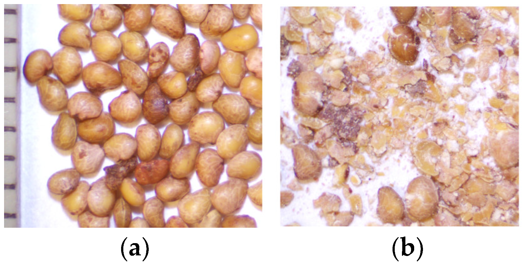
2.1. Plant Material

The research material consisted of seed remains after extraction of oil and it was provided from Łukasiewicz Research Network—New Chemical Syntheses Institute in Puławy (Poland). The extraction of oil was carried out using an in-house-built quarter-technical installation for supercritical CO2 extraction on scale [18].
The process was conducted at 40 °C in continuous mode, using 230 and 330 bar pressure, and a CO2 flow rate of 125 kg/h (a total extraction time was 120 min). The weight of seeds in the extractor was 4350 g. The amount of oils obtained as a result of extraction were 15.6% and 16.5%, respectively. The residues (Figure 1) were preserved at 4 °C until subjected to analysis.

2.2. Extraction

To assess the total content of low molecular phenolics in the defatted strawberry seeds, three step extraction was carried out using ethanol-water mixture with increased concentration of alcohol (60, 80 and 100%). Each step with fresh portion of solvent lasted 15 min and the extraction was assisted by ultrasound. Supernatants were collected, combined and adjusted to final volume of 5 mL.
To assess the effectiveness of different extraction techniques 1 g of the tested material was extracted using an ethanol-water mixture with increased alcohol concentration (80, 90, and 100%) for 15 min. Heat reflux extraction (HRE) was carried out using a water bath at 60 °C and a reflux condenser as referenced from the literature [19]. Ultrasound-assisted extraction was conducted using the ultrasonic bath RU102H (Sonorex, Bandelin, Berlin, Germany) [19]. Extraction assisted by microwave was performed using a Plasmotronika UniClever system (Wroclaw, Poland) in open system and 60% of generator power [20]. Maceration with stirring (ME) was carried out at water bath at 40 °C with the assistance of a magnetic stirrer.

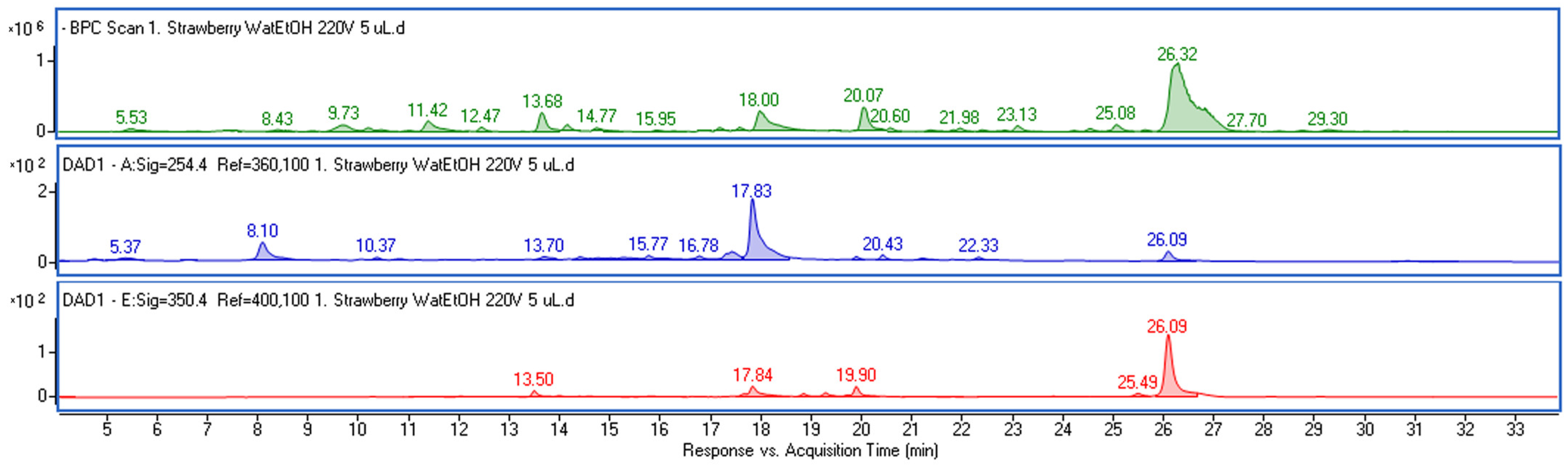
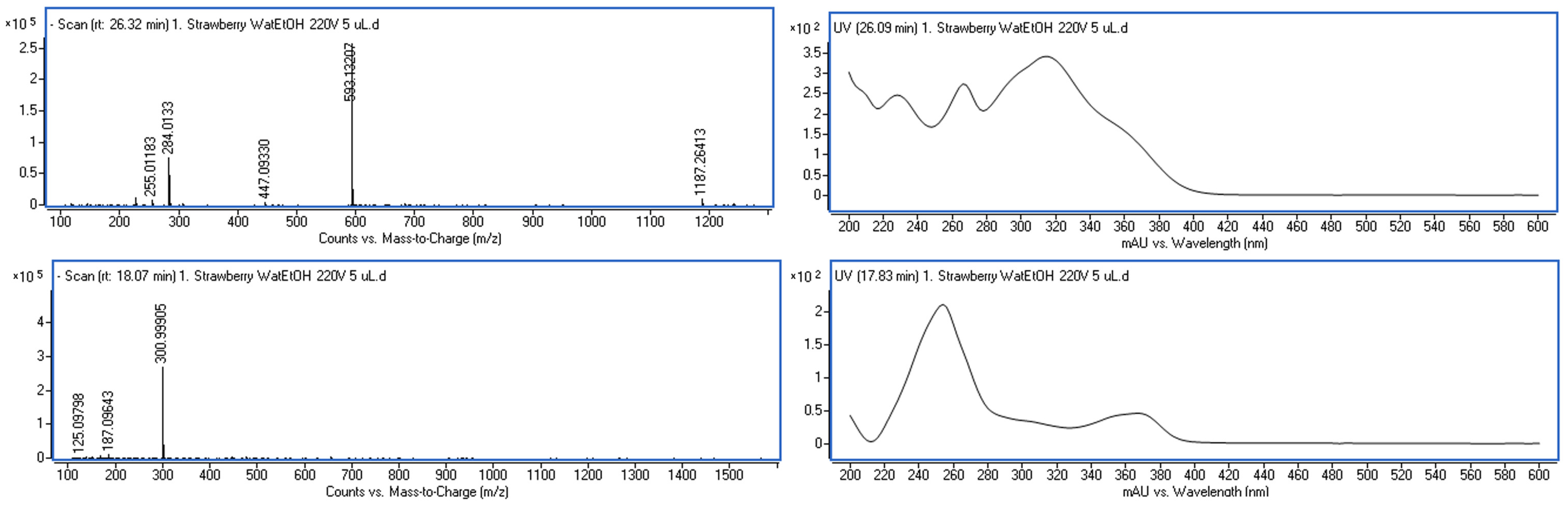
2.3. Chromatographic Analysis

Acetonitrile (ACN) and formic acid (FA) were MS grade (Sigma-Aldrich, St. Louis, MO, USA). An ultra-high performance liquid chromatograph (UHPLC) Infinity Series II combined with MS with electrospray ionization (ESI) and DAD detector (Agilent Technologies, Santa Clara, CA, USA) was used for phytochemical study. Chromatographic and MS conditions were detailed described in our previous work [21]. Briefly, the extract separation was carried out on Titan column (10 cm × 2.1 mm, 1.9 µm) (Supelco, Sigma-Aldrich). Elution was performed using mixture of water (A) and ACN (B), both acidified with 0.05% FA (flow rate was 0.2 mL/min. Gradient was as follows: 0–20 min from 98% A to 75% A; 20–32 min 75% A; 32–40 min from 75% A to 1% A. All standard compounds were from Sigma-Aldrich.

2.4. Cell Line Assay

Human skin fibroblast cell lines (ATCC® CRL-2522™, Manassas, VA, USA) were cultured in conditions given in literature [18]. The cells at density of 1 × 10⁵ cells/well were treated with varying concentration of the extracts for 24, 48 and 72 h to assess cytotoxicity. The dried extract was dissolved in DMSO and diluted appropriately in the medium. The concentration of DMSO in investigated solutions was below 0.5% and DMSO in concentration of 0.5% was used as control. For antioxidant assay the cells were pretreated with the extract (30 min) and then H2O2 at concentration of 250 µM was added as a inducer of oxidative stress [22]. After 6 h of incubation, cell viability and reactive oxygen species were investigated.

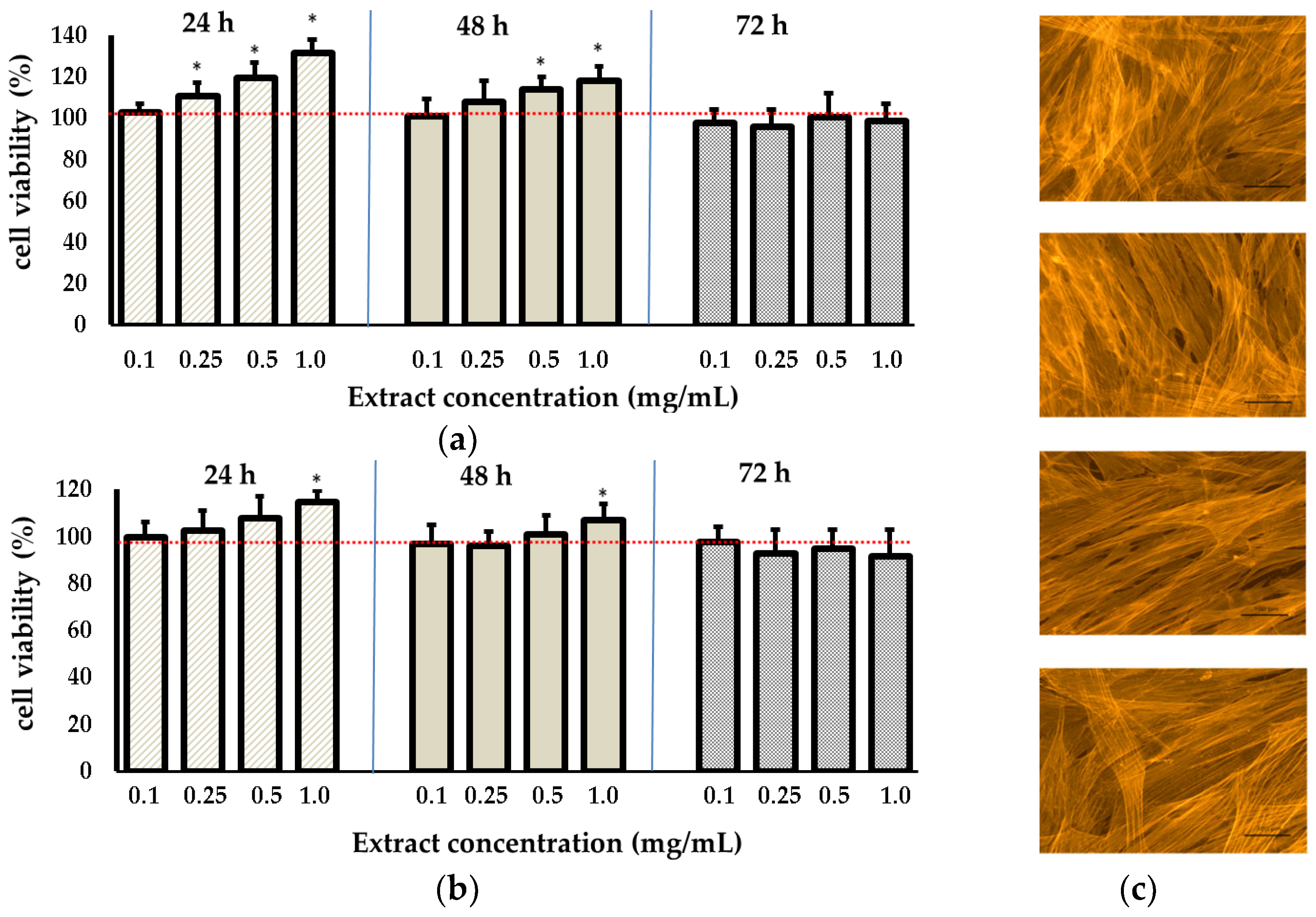
2.4.1. Cell Viability

Spectrophotometric tests were carried out using an E-max Microplate Reader (Molecular Devices Corporation, Menlo Park, CA, USA). Neutral Red (NR) and 3-(4,5-dimethylthiazole-2-yl)-2,5-diphenyltetrazolium bromide (MTT) dyes (Sigma-Aldrich) were used to assess the cytotoxicity of the extract according the procedure described previously [21].

2.4.2. Cytoskeleton Organization

Labeling of the cytoskeleton with F-actin was performed using tetra methyl rhodamine-isothiocyanate (TRITC) -phalloidin (1 μg mL−1) in bovine serum albumin (PBS) following the procedure described in the literature [23]. Cell observation was conducted using a fluorescence microscope (Olympus, BX51, Hamburg, Germany).

2.4.3. Intracellular Levels of Reactive Oxygen Species (ROS)

The procedure was described in detail in previous work [21]. Briefly, a solution of 10 μM H2DCFDA (Sigma Aldrich, St. Louis, MO, USA) was added to the cells and then the cells were incubated in the dark for 60 min. The fluorescence was measured using an excitation wavelength of λ = 485 nm and an emission wavelength of λ = 530 nm.

2.5. Statistic

The extractions were repeated two times. The values presented are mean ± standard deviation (SD) from three measurements. The calculation was performed using Statistica (ver. 13.3). A one-way ANOVA and Dunnett’s test were carried out. The graphs were prepared using Excel program.

3. Results and Discussion

3.1. Phenolic Composition of the Plant Material

Plant material was characterized in terms of low molecular weight phenolics, which are considered most suitable for dermatological purposes [24,25]. A three-step extraction process was employed to exhaustively isolate the components (Figure S1) and assess the total content in defatted seeds. Chromatographic analysis (Figure 2) revealed the presence of two predominant phenolic constituents in the extract with retention times of approximately 18 and 26.3 min. Both components exhibited strong absorbance in the region of 200–400 nm. Based on mass spectrometry data (Figure 3) and comparison with standards, the identified components were determined to be ellagic acid and kaempferol 3-O-coumaroyl-glucoside (tiliroside).
Additionally procyanidin, kaempferol glucoside, quercetin glucoside, pelargonidin-3-(malonyl)-glucoside, catechin, and catechin hexoside, along with derivatives of ellagic acid, including rhamnoside and methylellagic acid hexoside, were identified in the extracts. Mass spectrometry (MS) data and the quantification results of the identified metabolites in the extracts are presented in Table 1.
The determined content of the main identified compounds in defatted strawberry seeds were in accordance with the values documented in existing literature. For instance, Grzelak-Błaszczyk et al., who investigated material derived from three seasons of cultivation, reported free ellagic acid content ranging from 43.8 to 58.2 mg/100 g dry matter, anthocyanins from 9.8 to 15.2 mg/100 g, and kaempferol glycosides from 24.4 to 28 mg/100 g. The predominant flavonoid, tiliroside, was found to be in the range of 139.7 to 184.0 mg/100 g [16]. Tiliroside is a valuable plant metabolite with diverse biological activity commonly found in the plant kingdom [26]. Literature data have evidenced that this polyphenol exhibits anti-inflammatory, hepatoprotective, anti-hyperglycemic, and antidiabetic properties [27]. The richest source of tiliroside is the flowers of Tilia cordata, or the aerial parts of Potentilla spp. where its amount reaches above 1 mg/g [20,28]. Although the amount of tiliroside in defatted seeds is lower, it is worthy to note that it is relatively high compared to other plant [29].

3.2. Optimization of Extraction Procedure to Obtain Phenolic Rich Fraction

To determine the optimal extraction conditions for obtaining a fraction rich in low molecular phenolics the percentage content of specific compounds in the total yield of the obtained extract were considered. Water is the most environmentally friendly solvent with good swelling properties. However, the extraction efficacy of the main strawberry phenolic components using pure water was poor. Phenolic compounds content was very low or they were absent (Figure S2). Despite the ultrasound, or microwave treatment facilitating the release of various metabolites from the plant matrix, the low solubility of tiliroside and ellagic acid proved to be the main cause of low efficiency in isolation using water.
In further steps, increasing concentrations of ethanol in water were tested. Ethanol was chosen as the extraction solvent since this solvent is allowed for cosmetic purposes, thus eventual residues in the dried extract are permitted. Furthermore, ethanol is easy to evaporate and can be reused in the extraction process. As expected, the amount of phenolics increased with the rising concentration of ethanol, and only at a concentration above 70% the isolation of the considered metabolites was effective. The effectiveness of isolating a specific component using ultrasonic-assisted extraction was shown in Figure S3. Table 2 displays the results regarding quantitative determination of the main low molecular weight phenolics using maceration with stirring and extraction assisted by heating, ultrasound and microwaves.
The highest total extraction yield was achieved through heat reflux extraction, possibly due to the enhanced dissolution of carbohydrates at higher temperatures. On the other hand, the extraction at temperature of 60 °C yielded a lower amount of tiliroside and kaempferol 3-glucoside comparing to the maceration with stirring what may indicated on partially degradation of these components. Similar observation was made by Oniszczuk et al. who extracted tiliroside from flowers of T. cordata [20]. The assisted techniques were the most effective and the results for ellagic and tiliroside expressed per gram of plant material were not significantly different. However, in the case of kaempferol 3-glucoside, higher amount was detected when ultrasonic extraction was applied. The high effectiveness of ultrasonic extraction in isolating tiliroside and kaempferol 3-glucoside was also confirmed by literature data [20,30]. On the other hand, the content of compounds relative to the total extraction yield was lower for MAE compared to UAE. Furthermore, the lower values for kaempferol 3-glucoside and tiliroside may indicate the destructive effects of this technique. Similar phenomena were observed for some flavonoids treated by microwave [31]. However, it is worth mentioning that, in our study, we opted for relatively mild MAE conditions.
To increase the content of phenolics in total yield, the material was treated with water (stirring for 5 min) before final extraction. The process was carried in one and in two steps with fresh portion of water. It allows to partially elute water soluble components such as sugars or proteins. Furthermore, two variants of extraction were tested including two steps extraction with fresh potion of solvent (15 min for each step) and one step extraction lasted 30 min. The results are summarized in Table 3.
Pretreating the samples with water allowed for an increase of the concentration of compounds in the total yield to above 30%. Furthermore, as expected, extraction carried out in two steps, each lasting 15 min, was more effective than a single, longer extraction. Interestingly, the content of the compounds in the total yield obtained using the two-step process differed only slightly compared to the one-step 15-min process. While, in general, the amount of isolated compounds increased with the repetition of the extraction, the total yield also increased. However, the two-step extraction allowed for a more complete utilization of the raw material.

3.3. Biological Assays

To assess the cosmetic potential of the phenolic-rich fraction obtained from defatted seeds, biological tests were performed using human skin fibroblast cells. As a cosmetic additive should be safe and display no adverse effects on the cells, cytotoxicity of the extract was verified based on 24-h, 48-h, and 72-h assays of cell viability and cell metabolism. Two complementary assays were applied, including the neutral red test indicating the capacity to bind the dye within the lysosomes of viable cells, and MTT, showing the action of mitochondrial dehydrogenases, important enzymes responsible for metabolic activity [32,33]. The results are shown in Figure 4.
In general, at the tested concentrations, the extract was non-toxic throughout the entire evaluation period. Furthermore, after 24- and 48-h exposures to the highest tested concentration, both cell proliferation and cell metabolism were stimulated, increasing up to 132% in MTT and 115% in NR tests after 24 h. After 48 h, the values rose to 118% and 109%, respectively, compared to the untreated control. No negative impact on cellular cytoskeleton was also observed (Figure 4c).
ROS-scavenging activity is another highly appreciated feature of cosmetic products, as oxidative stress and excessive exposure to reactive oxygen species (ROS) promote skin aging and the development of skin inflammatory states [34]. Oxidative stress may lead to the disruption of the organization of the collagen network, resulting in structural changes in the skin and the occurrence of wrinkles. Furthermore, the damage to skin cells resulting from ROS action provokes increased production of cytokines and favors inflammation. Thus, supporting the endogenous antioxidative systems with effective exogenous antioxidants is essential for protection against the adverse effects of free radicals.
Figure 5 displays the effects of the extract from defatted strawberry seeds on skin fibroblasts with induced oxidative stress using H2O2 treatment as a model. The 2’,7’-dichlorodihydrofluorescein diacetate (H2DCFDA) reagent was used to investigate the impact of the extract on the redox imbalance caused by H2O2 stimulation. Nonfluorescent H2DCFDA is deacetylated by intracellular esterases, next oxidized by ROS and converted to fluorescent 2′,7′-dichlorofluorescein (DCF). As shown in Figure 5a, H2O2 stimulation increased ROS levels by approximately 33% compared to the control. The cells, when pre-treated with the extract, hindered the generation of excessive ROS, and at the highest tested concentration, ROS levels reduced to those observed in the untreated control. The extract also provided protection against H2O2 induced cytotoxicity (Figure 5b). In the experimental setting, a significant decline in cell viability was observed in the cells following treatment with H2O2, with the number of visible cells reduced to 59% compared to the control. In cells pretreated with the extract before the addition of H2O2, cell viability was restored, and at higher concentrations, it reached the level of the untreated control. This suggests that incorporating the extract mitigates the detrimental cellular impacts of H2O2 and acts as a protective measure under conditions of oxidative stress.
The beneficial effect of the extract on skin fibroblasts is likely attributed to the presence of tiliroside, 3-glucoside of kaempferol, and ellagic acid, as indicated by numerous scientific papers detailing their biological activities. The strong antioxidant activity of tiliroside, assessed using various colorimetric tests, including FRAP, DPPH, ABTS•+, and Fe2+ chelating assays, has been previously reported in the literature [35,36]. Moreover, Li et al. described its cytoprotective effect against the OH-induced damage in mesenchymal stem cell and they found that the activity is linked with p-coumaroyl moiety in the molecular structure [36]. Also, ellagic acid belongs to a potent antioxidant agent. Baek et al. reported its protective effects against oxidative stress induced by UV-B in skin cells, associated with the restoration of glutathione (GSH) and superoxide dismutase (SOD) levels which were reduced in fibroblasts subjected to UV-B irradiation [37]. Significant ROS scavenging activity was also reported for kaempferol 3-glucoside [38,39]. Furthermore, it has been evidenced that this flavonoid protect for cellular membrane against ROS [38]. The presence of these specific phenolics in the tested extract has also been beneficial, considering the other desirable properties of cosmetic formulation. In vivo studies have demonstrated that tiliroside exhibits a protective effect against inflammation in human skin provoked by UV radiation [40,41] and in a mouse ear inflammation model [35]. It also acts as a tyrosinase inhibitor, which may be useful in skin-whitening preparations [42]. Furthermore, Takeda et al. discovered that tiliroside stimulates the production of ceramides, the components of the outermost layer of the epidermis, essential for maintaining skin hydration. Consequently, it supports moisture retention and improves skin barrier function [27]. In turn, ellagic acid has shown significant activity towards the inhibition of some metalloproteinases, including hyaluronidase, elastase, collagenase, being accountable for the breakdown of the extracellular matrix and loss of skin firmness [43] and exerts a photoprotective effect [43,44].

4. Conclusions

Our study showed the defatted strawberry seeds are a rich reservoir of phenolic compounds that can be effectively extracted using a water and ethanol mixture. The investigation revealed that tiliroside, kaempferol glucoside, and ellagic acid were the most abundant components of the material. Isolated phenolic-rich fraction demonstrated significant antioxidant properties, as evidenced by its ability to scavenge free radicals in human skin fibroblasts. Additionally, the fraction exhibited cytoprotective activity in oxidative stress condition. Taking into account all of the above data, it can be concluded that defatted strawberry seeds may serve as a valuable material for subsequent processing to obtain a beneficial additive for skin care products.

Supplementary Materials

The following supporting information can be downloaded at: https://www.mdpi.com/article/10.3390/app14020783/s1, Figure S1: BPC chromatograms of extracts obtained during subsequent extraction steps. I—extraction with 60% ethanol, II—extraction with 80% ethanol, III—extraction with 100% ethanol, IV—repeated extraction with 100% ethanol; Figure S2: BPC chromatograms of extracts obtained during ultrasound assisted extraction using water (blue), 20% ethanol in water (green) and 50% ethanol in water (red); Figure S3: Effectiveness of isolating a specific component using ultrasonic-assisted extraction with increased ethanol concentration.

Author Contributions

Conceptualization, W.W., M.Ż., I.S. and M.W.; methodology, M.W., I.S., B.M. and K.T.; software, M.W. and I.S.; validation, M.W. and I.S.; formal analysis, W.W., M.Ż., M.W., B.M. and K.T.; investigation, W.W., M.Ż., I.S. and M.W.; resources, B.M. and K.T.; data curation, M.W.; writing—original draft preparation, W.W., M.Ż. and M.W.; writing—review and editing, I.S. and M.W.; visualization, W.W. and M.W.; supervision, I.S. and M.W.; project administration, I.S. and M.W. funding acquisition, I.S. and M.W. All authors have read and agreed to the published version of the manuscript.

Funding

This research received no external funding.

Institutional Review Board Statement

Not applicable.

Informed Consent Statement

Not applicable.

Data Availability Statement

The raw data supporting the conclusions of this article will be made available by the authors on request.

Conflicts of Interest

The authors declare no conflicts of interest.

References

  1. Kaim, U. Methods for the Valorisation of Agri-Food Waste According to the Circular Bioeconomy Concept. Eng. Sci. Technol. 2021, 2021, 94–112. [Google Scholar] [CrossRef]
  2. Facchini, F.; Silvestri, B.; Digiesi, S.; Lucchese, A. Agri-Food Loss and Waste Management: Win-Win Strategies for Edible Discarded Fruits and Vegetables Sustainable Reuse. Innov. Food Sci. Emerg. Technol. 2023, 83, 103235. [Google Scholar] [CrossRef]
  3. Sławińska, N.; Prochoń, K.; Olas, B. A Review on Berry Seeds—A Special Emphasis on Their Chemical Content and Health-Promoting Properties. Nutrients 2023, 15, 1422. [Google Scholar] [CrossRef]
  4. Qaderi, R.; Mezzetti, B.; Capocasa, F.; Mazzoni, L. Stability of Strawberry Fruit (Fragaria × Ananassa Duch.) Nutritional Quality at Different Storage Conditions. Appl. Sci. 2022, 13, 313. [Google Scholar] [CrossRef]
  5. Salas-Arias, K.; Irías-Mata, A.; Sánchez-Kopper, A.; Hernández-Moncada, R.; Salas-Morgan, B.; Villalta-Romero, F.; Calvo-Castro, L.A. Strawberry Fragaria × Ananassa Cv. Festival: A Polyphenol-Based Phytochemical Characterization in Fruit and Leaf Extracts. Molecules 2023, 28, 1865. [Google Scholar] [CrossRef] [PubMed]
  6. Newerli-Guz, J.; Śmiechowska, M.; Drzewiecka, A.; Tylingo, R. Bioactive Ingredients with Health-Promoting Properties of Strawberry Fruit (Fragaria × Ananassa Duchesne). Molecules 2023, 28, 2711. [Google Scholar] [CrossRef] [PubMed]
  7. Ribeiro, M.D.S.; Sebastià, N.; Montoro, A.; García-Martínez, E. Strawberry (Fragaria × Ananassa) and Kiwifruit (Actinidia Deliciosa) Extracts as Potential Radioprotective Agents: Relation to Their Phytochemical Composition and Antioxidant Capacity. Appl. Sci. 2023, 13, 8996. [Google Scholar] [CrossRef]
  8. Pukalskienė, M.; Pukalskas, A.; Dienaitė, L.; Revinytė, S.; Pereira, C.V.; Matias, A.A.; Venskutonis, P.R. Recovery of Bioactive Compounds from Strawberry (Fragaria × Ananassa) Pomace by Conventional and Pressurized Liquid Extraction and Assessment Their Bioactivity in Human Cell Cultures. Foods 2021, 10, 1780. [Google Scholar] [CrossRef]
  9. Davidson, M.; Louvet, F.; Meudec, E.; Landolt, C.; Grenier, K.; Périno, S.; Ouk, T.-S.; Saad, N. Optimized Single-Step Recovery of Lipophilic and Hydrophilic Compounds from Raspberry, Strawberry and Blackberry Pomaces Using a Simultaneous Ultrasound-Enzyme-Assisted Extraction (UEAE). Antioxidants 2023, 12, 1793. [Google Scholar] [CrossRef]
  10. Spagnuolo, C.; Moccia, F.; Tedesco, I.; Adabbo, E.; Panzella, L.; Russo, G.L.; Napolitano, A. Stillage Waste from Strawberry Spirit Production as a Source of Bioactive Compounds with Antioxidant and Antiproliferative Potential. Antioxidants 2023, 12, 421. [Google Scholar] [CrossRef]
  11. Raczyk, M.; Bryś, J.; Brzezińska, R.; Ostrowska-Ligęza, E.; Wirkowska-Wojdyła, M.; Górska, A. Quality Assessment of Cold-Pressed Strawberry, Raspberry and Blackberry Seed Oils Intended for Cosmetic Purposes. Acta Sci. Pol. Technol. Aliment. 2021, 20, 127–133. [Google Scholar] [CrossRef] [PubMed]
  12. Milala, J.; Grzelak-Błaszczyk, K.; Sójka, M.; Kosmala, M.; Dobrzyńska-Inger, A.; Rój, E. Changes of Bioactive Components in Berry Seed Oils during Supercritical CO 2 Extraction. J. Food Process. Preserv. 2018, 42, e13368. [Google Scholar] [CrossRef]
  13. Villamil-Galindo, E.; Gastélum-Estrada, A.; Chuck-Hernandez, C.; Antunes-Ricardo, M.; Reza-Zaldivar, E.E.; Piagentini, A.; Jacobo-Velázquez, D.A. Kinetic Ultrasound-Assisted Extraction as a Sustainable Approach for the Recovery of Phenolics Accumulated through UVA Treatment in Strawberry By-Products. Foods 2023, 12, 2989. [Google Scholar] [CrossRef] [PubMed]
  14. Kosmala, M.; Zduńczyk, Z.; Juśkiewicz, J.; Jurgoński, A.; Karlińska, E.; Macierzyński, J.; Jańczak, R.; Rój, E. Chemical Composition of Defatted Strawberry and Raspberry Seeds and the Effect of These Dietary Ingredients on Polyphenol Metabolites, Intestinal Function, and Selected Serum Parameters in Rats. J. Agric. Food Chem. 2015, 63, 2989–2996. [Google Scholar] [CrossRef]
  15. Juśkiewicz, J.; Król, B.; Kosmala, M.; Milala, J.; Zduńczyk, Z.; Żary-Sikorska, E. Physiological Properties of Dietary Ellagitannin-Rich Preparations Obtained from Strawberry Pomace Using Different Extraction Methods. Pol. J. Food Nutr. Sci. 2015, 65, 199–209. [Google Scholar] [CrossRef]
  16. Grzelak-Błaszczyk, K.; Karlińska, E.; Grzęda, K.; Rój, E.; Kołodziejczyk, K. Defatted Strawberry Seeds as a Source of Phenolics, Dietary Fiber and Minerals. LWT 2017, 84, 18–22. [Google Scholar] [CrossRef]
  17. Kowalczewski, P.Ł.; Walkowiak, K.; Masewicz, Ł.; Duda, A.; Poliszko, N.; Różańska, M.B.; Jeżowski, P.; Tomkowiak, A.; Mildner-Szkudlarz, S.; Baranowska, H.M. Wheat Bread Enriched with Raspberry and Strawberry Oilcakes: Effects on Proximate Composition, Texture and Water Properties. Eur. Food Res. Technol. 2019, 245, 2591–2600. [Google Scholar] [CrossRef]
  18. Wójciak, M.; Mazurek, B.; Tyśkiewicz, K.; Kondracka, M.; Wójcicka, G.; Blicharski, T.; Sowa, I. Blackcurrant (Ribes nigrum L.) Seeds—A Valuable Byproduct for Further Processing. Molecules 2022, 27, 8679. [Google Scholar] [CrossRef]
  19. Wójciak-Kosior, M.; Sowa, I.; Kocjan, R.; Nowak, R. Effect of Different Extraction Techniques on Quantification of Oleanolic and Ursolic Acid in Lamii Albi Flos. Ind. Crops Prod. 2013, 44, 373–377. [Google Scholar] [CrossRef]
  20. Oniszczuk, A.; Podgórski, R. Influence of Different Extraction Methods on the Quantification of Selected Flavonoids and Phenolic Acids from Tilia Cordata Inflorescence. Ind. Crops Prod. 2015, 76, 509–514. [Google Scholar] [CrossRef]
  21. Skalska-Kamińska, A.; Wójciak, W.; Żuk, M.; Paduch, R.; Wójciak, M. Protective Effect of Urtica Dioica Extract against Oxidative Stress in Human Skin Fibroblasts. Life 2023, 13, 2182. [Google Scholar] [CrossRef] [PubMed]
  22. Wójciak, M.; Feldo, M.; Borowski, G.; Kubrak, T.; Płachno, B.J.; Sowa, I. Antioxidant Potential of Diosmin and Diosmetin against Oxidative Stress in Endothelial Cells. Molecules 2022, 27, 8232. [Google Scholar] [CrossRef]
  23. Sowa, I.; Paduch, R.; Strzemski, M.; Zielińska, S.; Rydzik-Strzemska, E.; Sawicki, J.; Kocjan, R.; Polkowski, J.; Matkowski, A.; Latalski, M.; et al. Proliferative and Antioxidant Activity of Symphytum officinale Root Extract. Nat. Prod. Res. 2018, 32, 605–609. [Google Scholar] [CrossRef] [PubMed]
  24. Sun, M.; Deng, Y.; Cao, X.; Xiao, L.; Ding, Q.; Luo, F.; Huang, P.; Gao, Y.; Liu, M.; Zhao, H. Effects of Natural Polyphenols on Skin and Hair Health: A Review. Molecules 2022, 27, 7832. [Google Scholar] [CrossRef]
  25. Ratz-Łyko, A.; Arct, J.; Majewski, S.; Pytkowska, K. Influence of Polyphenols on the Physiological Processes in the Skin. Phytother. Res. 2015, 29, 509–517. [Google Scholar] [CrossRef]
  26. Grochowski, D.M.; Locatelli, M.; Granica, S.; Cacciagrano, F.; Tomczyk, M. A Review on the Dietary Flavonoid Tiliroside. Compr. Rev. Food Sci. Food Saf. 2018, 17, 1395–1421. [Google Scholar] [CrossRef] [PubMed]
  27. Takeda, S.; Shimoda, H.; Takarada, T.; Imokawa, G. Strawberry Seed Extract and Its Major Component, Tiliroside, Promote Ceramide Synthesis in the Stratum Corneum of Human Epidermal Equivalents. PLoS ONE 2018, 13, e0205061. [Google Scholar] [CrossRef]
  28. Tomczyk, M.; Bazylko, A.; Staszewska, A. Determination of Polyphenolics in Extracts of Potentilla Species by High-performance Thin-layer Chromatography Photodensitometry Method. Phytochem. Anal. 2010, 21, 174–179. [Google Scholar] [CrossRef]
  29. Nowak, R. Separation and Quantification of Tiliroside from Plant Extracts by SPE/RP-HPLC. Pharm. Biol. 2003, 41, 627–630. [Google Scholar] [CrossRef]
  30. Pieczykolan, A.; Pietrzak, W.; Nowak, R.; Pielczyk, J.; Łamacz, K. Optimization of Extraction Conditions for Determination of Tiliroside in Tilia L. Flowers Using an LC-ESI-MS/MS Method. J. Anal. Methods Chem. 2019, 2019, 9052425. [Google Scholar] [CrossRef]
  31. Jurinjak Tušek, A.; Šamec, D.; Šalić, A. Modern Techniques for Flavonoid Extraction—To Optimize or Not to Optimize? Appl. Sci. 2022, 12, 11865. [Google Scholar] [CrossRef]
  32. Babich, H.; Borenfreund, E. Applications of the Neutral Red Cytotoxicity Assay to In Vitro Toxicology. Altern. Lab. Anim. 1990, 18, 129–144. [Google Scholar] [CrossRef]
  33. Ghasemi, M.; Turnbull, T.; Sebastian, S.; Kempson, I. The MTT Assay: Utility, Limitations, Pitfalls, and Interpretation in Bulk and Single-Cell Analysis. Int. J. Mol. Sci. 2021, 22, 12827. [Google Scholar] [CrossRef] [PubMed]
  34. He, X.; Wan, F.; Su, W.; Xie, W. Research Progress on Skin Aging and Active Ingredients. Molecules 2023, 28, 5556. [Google Scholar] [CrossRef] [PubMed]
  35. Sala, A.; Recio, M.C.; Schinella, G.R.; Máñez, S.; Giner, R.M.; Cerdá-Nicolás, M.; Ríos, J.-L. Assessment of the Anti-Inflammatory Activity and Free Radical Scavenger Activity of Tiliroside. Eur. J. Pharmacol. 2003, 461, 53–61. [Google Scholar] [CrossRef]
  36. Li, X.; Tian, Y.; Wang, T.; Lin, Q.; Feng, X.; Jiang, Q.; Liu, Y.; Chen, D. Role of the P-Coumaroyl Moiety in the Antioxidant and Cytoprotective Effects of Flavonoid Glycosides: Comparison of Astragalin and Tiliroside. Molecules 2017, 22, 1165. [Google Scholar] [CrossRef]
  37. Baek, B.; Lee, S.H.; Kim, K.; Lim, H.-W.; Lim, C.-J. Ellagic Acid Plays a Protective Role against UV-B-Induced Oxidative Stress by up-Regulating Antioxidant Components in Human Dermal Fibroblasts. Korean J. Physiol. Pharmacol. 2016, 20, 269. [Google Scholar] [CrossRef]
  38. Park, S.N.; Kim, S.Y.; Lim, G.N.; Jo, N.R.; Lee, M.H. In Vitro Skin Permeation and Cellular Protective Effects of Flavonoids Isolated from Suaeda Asparagoides Extracts. J. Ind. Eng. Chem. 2012, 18, 680–683. [Google Scholar] [CrossRef]
  39. Taiwo, F.O.; Oyedeji, O.; Osundahunsi, M.T. Antimicrobial and Antioxidant Properties of Kaempferol-3-O-Glucoside and 1-(4-Hydroxyphenyl)-3-Phenylpropan-1-One Isolated from the Leaves of Annona Muricata (Linn.). J. Pharm. Res. Int. 2019, 26, 1–13. [Google Scholar] [CrossRef]
  40. Carola, C.; Pfluecker, F.; Graf, R.; Epstein, H.; Wirth, C. Tiliroside and Dihydroxy Methylchromone: From Nature to Cosmetic Applications. J. Appl. Cosmetol. 2010, 28, 109–123. [Google Scholar]
  41. Bae, J.; Choi, J.; Kang, S.; Lee, Y.; Park, J.; Kang, Y. Dietary Compound Ellagic Acid Alleviates Skin Wrinkle and Inflammation Induced by UV-B Irradiation. Exp. Dermatol. 2010, 19, e182–e190. [Google Scholar] [CrossRef] [PubMed]
  42. Lu, Y.; Chen, J.; Wei, D.; Wang, Z.; Tao, X. Tyrosinase Inhibitory Effect and Inhibitory Mechanism of Tiliroside from Raspberry. J. Enzym. Inhib. Med. Chem. 2009, 24, 1154–1160. [Google Scholar] [CrossRef] [PubMed]
  43. Tavares, W.D.S.; Martin Pastor, M.; Pérez, L.; Morán, M.D.C.; Sousa, F.F.O.D. Skin Repairing Potential of Ellagic Acid-Loaded Zein Nanoparticles: Chemical and Biopharmaceutical Characterization, Enzymatic Inhibition and Cytotoxicity over Keratinocytes. J. Mol. Liq. 2023, 384, 122198. [Google Scholar] [CrossRef]
  44. Shimogaki; Tanaka; Tamai; Masuda. In Vitro and in Vivo Evaluation of Ellagic Acid on Melanogenesis Inhibition. Int. J. Cosmet. Sci. 2000, 22, 291–303. [Google Scholar] [CrossRef] [PubMed]
Figure 1. Strawberry seeds (a) and strawberry seeds residues after oil extraction (b).
Applsci 14 00783 g001
Figure 2. Chromatograms of the extract from defatted seeds including base ion chromatogram (green), and chromatograms at wavelength of 254 nm (blue) and 350 nm (red).
Applsci 14 00783 g002
Figure 3. Mass and UV-Vis spectra of tiliroside (upper panels) and ellagic acid (lower panels).
Applsci 14 00783 g003
Figure 4. Impact of the different concentration of the extracts from Defatted strawberry seed on: (a) cell metabolism (MTT), (b) cell viability (NR) expressed as a % of control (dashed red line) after 24, 48 and 72 h of exposure. The data are means (n = 3) ± SD. * Indicates statistically significant difference between extract samples and control at p < 0.05. (c) F-actin cytoskeleton organization-staining with TRITC-phalloidine.
Applsci 14 00783 g004
Figure 5. Effect of extract in human skin fibroblast cells on (a) 2′,7′-dichlorodihydrofluorescein fluorescence and (b) cell viability assessed using NR test. The cells were pretreated with different extract concentration and exposured on H2O2. The data are means (n = 3) ± SD. * Indicates statistically significant difference vs. H2O2-stimulated cells (p < 0.05).
Applsci 14 00783 g005
Table 1. Data used for identification and the results of quantification of low molecular weight phenolics (mg/100 g of fresh plant material ± SD) extracted from defatted seed residues using 230 and 330 PSI.
RT (min)[M − H]FormulaNameSeeds Defatted at 230 PSI (mg/100 g)Seeds Defatted at
330 PSI (mg/100 g)
7.73451.12448C21H24O11catechin hexoside 28.7 ± 1.1 a9.1 ± 1.2 a
10.07577.13615C30H26O12Procyanidin 2++
10.41289.07211C15H14O6Catechin 15.6 ± 0.6 a6.1 ± 0.7 a
13.68517.09892C24H22O13pelargonidin-3-(malonyl)-glucoside 24.4 ± 0.5 a4.1 ± 0.5 a
17.70447.05881C20H16O12ellagic acid rhamnoside 13.6 ± 0.3 a4.4 ± 0.4 b
18.00300.99905C14H6O8ellagic acid 165.8 ± 5.9 a77.2 ± 6.8 b
18.44463.08843C21H20O12quercetin glucoside 18.1 ± 0.8 a10.3 ± 0.6 a
20.07447.09445 C21H20O11kaempferol glucoside 123.5 ± 2.2 a24.9 ± 2.4 a
20.55461.07357C21H18O12methylellagic acid hexoside 23.8 ± 0.4 a4.9 ± 0.5 b
26.32593.13207C30H26O13tiliroside 1131.7 ± 11.6 a136.4 ± 12.1 a
1 Components were confirmed by comparison with standard; 2 components were identified based on parent ion mass and data found in the literature; the different letter symbols indicate statistically significant differences between the values in the same line.
Table 2. The amount of main low molecular phenolics (µg/g of fresh plant material ±SD) extracted from defatted seeds using different techniques and increased ethanol concentration. The process was conducted in one run and lasted 15 min.
ConditionKaempferol-GEllagic AcidTilirosideYield (%)
HRE (60 °C) 80%90.8 ± 8.7 d355.5 ± 34.9 b,c874.9 ± 84.5 d6.91 ± 0.7 a
HRE (60 °C) 90%88.1 ± 8.4 d360.1 ± 35.8 a,b900.8 ± 90.1 c,d6.34 ± 0.6 a
HRE (60 °C) 100%87.4 ± 8.8 d365.1 ± 36.8 a,b898.7 ± 90.7 c,d5.71 ± 0.6 b
ME (40 °C) 80%111.8 ± 10.4 a,b350.3 ± 34.4 b,c898.7 ± 87.6 c,d4.51 ± 0.5 c
ME (40 °C) 90%101.5 ± 10.1 b,c357.9 ± 35.8 a–c910.4 ± 90.4 b4.66 ± 0.4 c
ME (40 °C) 100%99.1 ± 9.8 b,c 366.1 ± 36.4 a,b905.5 ± 88.7 b,c4.84 ± 0.5 c
UAE 80%122.3 ± 11.1 a345.4 ± 23.5 c984.1 ± 90.7 a4.49 ± 0.5 c
UAE 90%119.3 ± 10.4 a371.3 ± 33.3 a973.2 ± 91.4 a4.61 ± 0.5 c
UAE 100%102.3 ± 9.9 b,c370.3 ± 34.6 a986.5 ± 98.3 a4.75 ± 0.5 c
MAE 80%97.9 ± 9.8 c357.2 ± 33.9 a–c940.5 ± 88.8 a,b5.98 ± 0.6 b
MAE 90%96.7 ± 8.9 c,d370.5 ± 35.4 a911.8 ± 90.9 b5.81 ± 0.5 b
MAE 100%91.9 ± 9.4 d376.3 ± 36.7 a898.7 ± 86.5 c,d5.65 ± 0.5 b
HRE—heat reflux extraction, ME—maceration with stirring, UAE—ultrasound assisted extraction, MAE—microwave assisted extraction; the different letter symbols indicate statistically significant differences between the values in the same column.
Table 3. The content of main low molecular phenolics (mg/g of dried extract ± SD). The process was conducted before and after maceration with water in one or two extraction runs.
ComponentExtraction without PretreatmentPretreatment with Water
1 Step UAE 1 × 15 min2 Steps UAE
2 × 15 min
1 Step UAE
30 min
Kaempferol glucoside2.59 ± 0.21 a3.44 ± 0.31 b4.05 ± 0.44 b3.84 ± 0.31 b
Ellagic acid8.05 ± 0.84 a10.71 ± 1.02 b13.5 ± 1.11 c11.4 ± 0.91 b
Tiliroside21.11 ± 1.82 a28.08 ± 2.28 b36.5 ± 3.24 c31.8 ± 1.99 b
UAE—ultrasound assisted extraction; the different letter symbols indicate statistically significant differences between the values in the same line.
Disclaimer/Publisher’s Note: The statements, opinions and data contained in all publications are solely those of the individual author(s) and contributor(s) and not of MDPI and/or the editor(s). MDPI and/or the editor(s) disclaim responsibility for any injury to people or property resulting from any ideas, methods, instructions or products referred to in the content.

