Article

Coniferous Honeydew Honey: Antibacterial Activity and Anti-Migration Properties against Breast Cancer Cell Line (MCF-7)

by
Małgorzata Dżugan
1,*,
Ewa Ciszkowicz
2,
Monika Tomczyk
1,
Michał Miłek
1 and
Katarzyna Lecka-Szlachta
2
1
Department of Chemistry and Food Toxicology, Institute of Food Technology and Nutrition, University of Rzeszów, Ćwiklińskiej 1a, 35-601 Rzeszów, Poland
2
Department of Biotechnology and Bioinformatics, Faculty of Chemistry, Rzeszow University of Technology, Al. Powstańców Warszawy 6, 35-959 Rzeszów, Poland
*
Author to whom correspondence should be addressed.
Appl. Sci. 2024, 14(2), 710; https://doi.org/10.3390/app14020710
Submission received: 21 December 2023 / Revised: 11 January 2024 / Accepted: 12 January 2024 / Published: 14 January 2024
(This article belongs to the Special Issue Honey as a Therapeutic Agent)

Abstract

:
Four samples of fir honeydew honey from Podkarpackie labeled with a Protected Designation of Origin symbol were tested in terms of their physicochemical parameters, antioxidant, and antibacterial effects, as well as their anti-migration properties against a breast cancer cell line (MCF-7) and fibroblasts. The results confirmed the high quality of tested samples regarding the obligatory parameters, as well as the additional indicators used (antioxidant and enzymatic activity), compared to representative rapeseed honey. Among the tested bacterial strains, the greatest effectiveness was demonstrated against Klebsiella pneumoniae and Streptococcus spp. Moreover, the results obtained in the urease inhibition in vitro test suggested the potential use of honeydew honey in the treatment of urease-positive bacterial infections. For the first time, using a scratch test it was found that the Podkarpackie honeydew honey efficiently affected the migration of cancer breast cells, whereas it only slightly inhibited the movement of normal fibroblasts. It can be suggested that the quality of honey guaranteed by the Protected Designation of Origin label could be the key factor of honeydew honey’s bioactivity and its potential medicinal use.

1. Introduction

Honeydew honey is a type of honey which bees (Apis mellifera) produce from excretions of plant-sucking insects or secretions of plants. Thus, the composition of honeydew is dependent on both plant and aphid species, and may also be influenced by environmental factors including water stress and temperature [1,2]. Two varieties of honeydew honey are distinguished: deciduous honeydew honey and coniferous honeydew honey, which in Poland is mainly produced from Abies alba. This species occurs mainly in southern and south-eastern Poland. Podkarpackie, an ecologically clean region in south-eastern Poland, is a natural basin of coniferous forests with a predominance of fir trees; therefore, it has a huge potential for the production of honeydew honey. Podkarpackie honeydew honey was registered by the European Commission as a Protected Designation of Origin (PDO) in 2010 [3]. This label was established by the EC to protect the designations of origin of traditional regional food products.
Podkarpackie honeydew honey is characterized by a high content of macro- and microelements such as potassium, calcium, magnesium, manganese, copper, iron, and others [4,5]. This characteristic also results from its geographical origin because Podkarpackie’s soil is rich in mineral salts [6]. The characteristic dark color of Podkarpackie honeydew honey results from the species of fir trees (Abies alba) found in the described area [5]. Moreover, this Polish honeydew honey is characterized by a higher than standard content of simple sugars, which, combined with its moderate acidity, improves the flavor bouquet of the honey and reduces the sweet taste [7].
The increased interest in honeydew honey is due to its nutritional, sensory, and, therefore, medicinal properties, which are different from flower honey. Considering the organoleptic properties honeydew honey is distinguished by its dark color, which in pure fir honey becomes greenish, and specific sensory characteristics such as a delicate resin aroma [7]. Compared to nectar honey, honeydew honey is characterized by higher pH values, electrical conductivity, net absorbance, and ash content; a higher content of disaccharides and trisaccharides; and lower levels of monosaccharides [5,8]. Actually, according to the applicable standards, a special parameter that distinguishes honeydew honey is its electrical conductivity: for this variety of honey the minimum required is 0.8 mS/cm, while for all nectar honeys it is below the mentioned value [9]. However, researchers are still working on chemical markers of honeydew honey. The presence of melezitose was considered a characteristic feature of honeydew honey, and usually the level of this sugar may indicate what part of a given honey sample was produced from honeydew (also with regard to nectar-honeydew honey) [5,8]. It is known that a specific feature of honeydew honey is its positive specific rotation assessed polarimetrically, in contrast to nectar honeys [10]. Another marker of honeydew honey often analyzed by scientists is the polyphenol profile; however, the results have not been consistent. Among the proposed polyphenolic markers, protocatechuic acid (3,4-dihydroxybenzoic acid), specific for honeydew honey, is commonly determined [11,12]. Other authors state that the most frequently recurrent polyphenols determined in honeydew honeys are protocatechuic, p-hydroxybenzoic acid, caffeic acid, p-coumaric acid, ferulic acid, and vanillic acid, as well as the flavonoids chrysin and pinocembrin, which were also proposed to be honeydew honey markers [13,14]. In addition to polyphenols, which are considered to be the main bioactive ingredients, honeydew honey is characterized by a higher content of minerals, proteins, organic acids, enzymes, amino acids, and vitamins, which are also responsible for the antibacterial and antioxidant activity [5,7,8,15,16,17,18].
Many reports indicate that honeydew honey has multiple stronger antioxidant properties than most nectar honeys, excluding buckwheat honey, which is a comparable or slightly stronger antioxidant than honeydew honey [14,16,18]. The high antioxidant activity of honey directly translates into antiproliferative effects, and cytotoxic, genotoxic, and apoptotic effects for high concentrations of honey have been reported. Due to this, honeydew honey is considered a potential therapeutic agent for patients suffering from several cancers such as breast [19,20], liver [21], colorectal [22], and prostate cancer [19], among others [23,24]. However, most of these studies concern nectar honey, not honeydew honey. In addition to its antioxidant properties, the second-best-studied property of honey is its antibacterial properties. The antibacterial properties of honey are mainly associated with a naturally low pH, high osmolarity, hydrogen peroxide (H2O2), and the occurrence of numerous flavonoids, aromatic and volatile substances, defensin-1, methylglyoxal, and lysozyme. The mentioned factors act synergistically or individually [25,26,27]. Honey has been found to be effective against over 60 species of Gram-positive (Gram+) and negative (Gram−) bacteria [28]. The beneficial antibacterial properties of honey, especially for wound healing through increasing the epithelial proliferation and accelerating the absorption of edema around the wound, have been known since ancient times [29]. A diabetic foot ulcer (DFU) is a problem that affects almost 15% of diabetic patients. Of those who develop a foot ulcer, 6% will be hospitalized for infection or other complications related to the ulcer [29]. To overcome a DFU, wound cleansing, blood glucose control, and infection prevention are usually used; however, research is increasingly conducted on the effectiveness of using honey dressings. As is well known, the strength of antibacterial properties is significantly dependent on the variety. Unfortunately, there is a lack of research on the effectiveness of particular honey types in the treatment of DFU; therefore, it still remains a challenge for scientists [29].
Therefore, the aim of the study was to assess the therapeutic potential, including the antioxidant, antibacterial, and wound healing effects, of Polish fir honeydew honey of guaranteed quality, which may contribute to increasing the importance of this type of honey in medicinal applications. A novelty of the study was the first use of the scratch test to assess the biological activity of honey, which is standardly used to test the effectiveness of various substances in treating wounds and inhibiting the migration of cancer cells.

2. Materials and Methods

2.1. Honey Samples

Four coniferous (fir, Abies alba) honeydew honey samples and one rapeseed honey sample were selected for the study from the collection of the Department of Chemistry and Food Toxicology. All samples were collected during beekeeping seasons 2021–2022 in apiaries located in Podkarpackie Province (Poland). A detailed description of honey samples is included in Table 1.

2.2. Physicochemical Evaluation

2.2.1. Water Content and Water Activity

The water content was determined in liquefied honey samples using an electronic refractometer dedicated to honey (HI96800; Hanna Instruments, Woonsocket, RI, USA). The water activity was determined using Rotronic HC2-AW device, working with HW5 software (v1.0.0) (Rotronic AG, Bassersdorf, Switzerland) at a temperature of 25 ± 2 °C.

2.2.2. Conductivity

The conductivity of 20% aqueous solutions of each honey was measured using a CP-401 conductometer (Elmetron, Zabrze, Poland). The results were presented in mS/cm.

2.2.3. pH and Free Acidity

The pH value of 20% aqueous honey solutions were measured using SevenCompact™ S210 pH-meter (Mettler Toledo, Columbus, OH, USA). The free acidity was determined using the standard alkali titration method (0.1 M NaOH) up to pH = 8.3, controlled using pH-meter. The results were calculated as mval/kg of honey.

2.2.4. Color Analysis

The HI 96,785 colorimeter (Hanna Instruments, Woonsocket, RI, USA) was used for color determination in the Pfund scale. Since the results for honeydew honey exceeded 150 mm Pfund, an additional color assessment was performed by measuring the absorbance at 450 nm for the 1 g/mL solution of each honey in distilled water using Biosens UV5100 UV/Vis spectrophotometer (Biosens, Warszawa, Poland).

2.2.5. 5-Hydroxymethylfurfural Analysis

5-hydroxymethylfurfural (HMF) was determined in 20% solutions of each honey using the reflectometric method (RQflex20, Merck, Darmstadt, Germany). The measurement range of the method was from 1 to 60 mg/L.

2.2.6. Total Phenolic Content and Antioxidant Capacity

Total phenolic content using Folic–Ciocalteu method, as well as DPPH (2,2-diphenyl-1-picrylhydrazyl), FRAP (ferric ion reducing antioxidant power), and CUPRAC (cupric ion reducing antioxidant capacity) assays of antioxidant capacity, were tested for 20% honey solutions as described earlier for goldenrod honey [30]. Ferrous ions’ chelating ability was tested according to Mansour et al. [31]: 80 μL of distilled water and 40 μL of 0.2 mM FeSO4 were mixed with a honey solution, and then 40 μL of 2 mM ferrozine solution was added and the absorbance was measured after 10 min of incubation at room temperature at 562 nm (EPOCH2, BioTek, Winooski, VT, USA). The results were expressed as mg EDTA/kg of honey using a calibration curve (0.1–0.5 mg/mL, y = 140.5x, R2 = 0.8910). Copper(II) ion chelating ability was tested as described by Santos et al. [32]. Briefly, 30 μL of honey solution was mixed with 200 μL of acetate buffer (50 mM, pH 6.0), and 30 μL of CuSO4 solution (100 mg/L) was then added. After 2 min, 8.5 μL of 2 mM pyrocatechol violet was added to initiate the reaction. The microplate was shaken for 10 min and left for an additional 10 min. After the incubation, the absorbance was measured at 632 nm (EPOCH2, BioTek, Winooski, VT, USA). The results were expressed as mg EDTA/kg of honey using a calibration curve (0.025–0.5 mg/mL, y = 366.05x, R2 = 0.9997).

2.2.7. Protein Content

Soluble protein fraction was determined in honey extracts (20% w/v) using the Bradford method according to Dżugan et al. [33]. The calibration curve in the range of 6.25–200 µg/per 20 µL was prepared using bovine albumin as standard and was applied to calculate the protein content in mg of protein per kg.

2.2.8. Enzyme Analysis

The diastase activity in honey was determined using Phadebas Honey Diastase Test (Magle AB, Malmö, Sweden) spectrophotometrically according to the manufacturer’s instructions. The measured absorbance at 620 nm was expressed as a diastase number (DN). The activities of three glycosidases were evaluated using the p-nitrophenyl glycoside as substrate: N-acetyl-βglucosaminidase (NAG), β-galactosidase (β-GAL), and α-glucosidase (α-GLU). The analytic protocol has been described earlier [33]. Raw absorbance values were recalculated into enzymatic units mU (nmol/min/g).

2.2.9. Jack Bean Urease Inhibition Assay

Jack bean (Canavalia ensiformis) urease (Merck, Darmstadt, Germany) was used for testing the inhibition potential of tested honey samples according to the modified protocol of Tanaka et al. [34]. Twenty-five microliters of urease solution (4 U/mL) was mixed with an equal volume of 20% honey solution and preincubated for 30 min at 25 °C. Then, the reaction was initiated by adding of 200 μL of 100 mM phosphate buffer (pH 6.8) containing 500 mM urea and 0.002% phenol red. After 1 h of incubation (25 °C), the absorbance was measured at 570 nm using EPOCH2 (BioTek, Winooski, VT, USA). The urease inhibition (%) was calculated according to Equation (1):
I % = 1 A A C × 100 %
where A is the absorbance of the tested sample and AC is the absorbance of control sample (without inhibitor). Acetohydroxamic acid (Sigma Aldrich, St. Louis, MO, USA) was used as a positive control.

2.3. Bacterial Strains and Growth Conditions

The certified Gram-negative (Escherichia coli ATCC 10536, Klebsiella pneumoniae ATCC 13883) and Gram-positive (Listeria monocytogenes SLR 2249, Staphylococcus aureus ATCC 6538, Staphylococcus epidermidis ATCC 35984, Streptococcus agalactiae DSM 2134, Streptococcus pyogenes DSM20565) strains were used. All strains were stored at −80 °C in a 20% glycerol solution (Sigma-Aldrich, MO, USA). Second or third culture cultivated in Mueller–Hinton Broth (MHB) at 37 °C was used and the density was adjusted to 108 colony-forming units (CFU/mL), which corresponds to OD600 between 0.4 and 0.45 [35]. Immediately before starting the experiment, the given culture was diluted in MHB to a density of 105 CFU/mL.

2.4. Determination of Antibacterial Activity

The antibacterial activity of tested honey samples in MHB was determined via the two-fold microdilution method [36,37]. Sterile 96-well microtiter plates were used to prepare a series of two-fold dilutions of 1 g/mL of each sample in MHB, positive control (MHB without honey), solvent control (MHB with sterile H2O and sterile DMSO, ratio 3:1) and negative control (sterile MHB). Final concentrations were 50%, 25%, 12.5%, 6.25%, 3.13%, 1.56%, 0.78%, and 0.39%. An equal volume of bacterial working culture (105 CFU/mL) was added to each well containing 100 µL of prepared tested and control samples. After 24 h incubation at 37 °C, the MIC (minimum inhibitory concentration) values were determined as the lowest concentration of sample at which no bacterial growth was observed. In questionable cases, spectrophotometric measurement was performed at 600 nm to indicate the MIC value. The determination of minimum bactericidal concentration (MBC) was performed based on the MIC results by plating 100 µL of contents of a given well on the plate (MIC, 2 × MIC, 4 × MIC) on Mueller–Hinton Agar (MHA) plates [35,38]. After 24 h incubation at 37 °C, the plates were counted for a number of colonies that corresponded to a single bacterial cell in the mixture.

2.5. Cell Lines

A human breast cancer cell line (MCF-7, HTB-22) and human normal fibroblast cell line (BJ, CRL-2522) were employed for this study. The cells were cultured in Eagle’s Minimum Essential Medium (EMEM) (ATCC 30-2003) supplemented with 10% fetal bovine serum (FBS, ATCC 30-2020) and penicillin-streptomycin solution (ATCC 30-2300) at 37 °C in a humidified atmosphere containing 5% CO2.

2.6. Determination of Cytotoxicity

Normal (BJ) and cancer cell line (MCF-7) cells were treated for 48 h (37 °C and 5% CO2) with 0.1%, 1%, and 10% of tested honeys. For each independent experiment, untreated (growth control) and solvent (H2O) controls were included. Cytotoxicity was determined using an MTT assay, which is based on the measurement of cell viability using 2,5-diphenyl-3-(4,5-dimethyl-2-thiazolyl) tetrazolium bromide (MTT, Abcam, Inc., Cambridge, UK) [38]. After 48 h incubation with honey samples, 100 µL of MTT in PBS solution (0.5 mg/mL) was added to each well for 3 h in cell culture incubation conditions. After that, dimethyl sulfoxide (DMSO, Sigma-Aldrich, MO, USA) was used to dissolve the formed formazan crystals. Absorbance was measured at 570 nm with the use of a Varioskan™ LUX multimode microplate reader (Thermo Scientific, Waltham, MA, USA), and % of cell viability in comparison to growth control was calculated.

2.7. Measurement of Cell Migration Using Scratch Assay

To evaluate the migration of BJ and MCF-7 cells exposed to 0.1%, 1%, and 10% honey samples, a scratch assay, also known as wound healing assay, was performed. Due to the lack of significant differences between the concentrations of 1% and 10%, only the effects caused by the two lowest concentrations will be discussed in the following sections. Cells at a density of 5 × 10⁴ cells/mL were incubated overnight until 95% of confluency. Two scratch lines were made in three replications for each sample through the confluent monolayer using a sterile 200 µL pipette tip. After washing cells twice with Dulbecco’s Phosphate Buffered Saline (D-PBS, ATCC 4-X, Manassas, VA, USA), fresh EMEM containing 5% FBS with or without tested honey samples was added to cells at indicated concentrations. Photographs of each wound were captured using an Olympus IX83 inverted microscope (Olympus, Shinjuku, Japan) at indicated times. The different duration of the experiment resulted from the observation of cell migration in untreated control wells. The analysis was carried out until the cells of a specific cell line without the tested honeys reached 100% confluence and was completed after 64 h and 24 h for BJ and MCF-7 cells, respectively. The experiment was conducted in triplicate and two independent series (total n = 6). Obtained images were analyzed using ImageJ public domain software, and cell migration into the wound (%) was calculated according to Equation (2) [38]:
C e l l   M i g r a t i o n   % = A t = 0 A t = Δ t A t = 0
where At=0 is the initial scratch area and At=Δt is the scratch area after n hours of the initial scratch, both in µm2.

2.8. Statistical Analysis

All experiments were performed in three independent procedures. The data were statistically analyzed using GraphPad Prism (OriginLab, Northampton, MA, USA). The normality of the data was checked using Shapiro–Wilks tests. The differences between groups were assessed using one-way analysis of variance (ANOVA). The results were statistically significant at p < 0.05 in the 95% confidence interval.

3. Results and Discussion

3.1. Physicochemical Evaluation

For the tested honey samples, a basic physicochemical evaluation has been performed and the results are summarized in Table 2. The obtained quality parameters for all honey samples were within the range corresponding to applicable standards [9]: the water content was not exceeding 20% and the free acidity was below 40 mval/kg. The HMF level was much below the level of 30 mg/kg. For honeydew honey samples, the conductivity value was above 0.8 mS/cm, which confirms that the type of honey was declared correctly. In the case of rapeseed honey, the conductivity was 0.144 mS/cm, typical for nectar honeys (below 0.8 mS/cm). The values of the tested parameters obtained for honeydew honey were mostly within the ranges previously determined for a larger pool of samples from the same region [5], as well as other honeydew honey samples tested by other teams [8,39,40].
Water activity is an additional parameter not included in legal regulations, but it reflects well the microbiological stability of the product. It is assumed that the value of 0.6 is the limit preventing the growth of microorganisms [41]. The tested honeys showed lower values, except for the H4 sample, which had a water activity value slightly exceeding 0.6, which proves its microbiological safety. Generally, water activity for various honey varieties ranges between 0.410 and 0.691 [42]. Another parameter that clearly distinguishes honeydew honey is its dark color. The color of the tested samples could not be expressed using the Pfund scale as all the values exceeded the measuring range of the device (>150 mm Pfund). Among the samples previously examined in the work by Tomczyk et al. [5], some samples also showed such a dark color, and the average color value for honeydew honey from Podkarpacie was 140 mm Pfund. The simple method used to measure absorbance at 450 nm allowed for the differentiation of the four tested samples. For sample H1, the lowest absorbance value was obtained, and the remaining three showed similar values. It is common knowledge that dark-colored honeys are characterized by a higher content of bioactive substances and have stronger health-promoting effects than light honeys [43,44].

3.2. Total Phenolic Content and Antioxidant Capacity

The samples of honeydew honey were subjected to further analysis in terms of total polyphenols as well as antioxidant activity using various colorimetric methods (Table 3). Among the tested honeydew honey samples, the highest content of total polyphenols was in samples H3 and H4, while a significantly lower content was recorded in the remaining two samples. It is known that honeydew honey from Central and Eastern Europe is characterized by a high content of polyphenols, which was previously confirmed for 14 samples of coniferous honeydew honey from Podkarpackie [5].
The samples described were within the range previously determined for a larger set of samples of Polish honeydew honey: 635.42–1289.43 mg GAE/kg [5] and 645.7–1207.5 mg GAE/kg [14]. A previous comparative study of Polish and Romanian honeydew honey showed an average polyphenol content of 1470 mg GAE/kg, with no difference due to origin [45]. On the other hand, there are reports giving a much lower value for Polish honeydew honey from another region (146.3 mg/kg), but without specifying the type of honeydew [17]. Rapeseed honey used as a “negative control” contained only 195.44 mg/kg of polyphenols, which is consistent with previous reports on this variety of honey [5,14,46].
The antioxidant activity of the tested honeys, determined using five methods, significantly correlated with the polyphenol content (0.619 < r < 0.987), which confirms the significant contribution of this group of compounds in shaping this activity. Regardless of the method used, all honeydew honeys showed high antioxidant potential, significantly exceeding that of rapeseed honey. This confirms previous data comparing dark and light honeys [5,14,16].
Rarely used methods to assess the antioxidant properties of honey are the ability to chelate iron(II) and copper(II) ions. Due to the participation of these metal ions in the Fenton and Haber–Weiss reactions, the ability of natural substances to capture them is considered an important mechanism of antioxidant activity [32]. The ability to chelate iron(II) ions has been previously demonstrated for honey from Mauritius [47], Egypt [48], Manuka honey [49], and copper(II) ions for honey from stingless bees from Mexico [50]. In the case of the mentioned methods, the tested honeydew honey samples showed a much higher ability to chelate metal ions than rapeseed honey.

3.3. Protein Content and Enzymatic Activity

The tested honeydew samples were compared in terms of protein content and enzymatic activity (Table 4). These parameters were formerly proposed as auxiliary indicators of honey quality [33], especially in terms of its overheating. Among the tested enzymes, diastase activity, included in the obligatory set of parameters used for quality control, as well as three further acid glycosidases applied to the honey activity evaluation were determined.
The obtained results confirmed that honeydew honeys were more abundant in total soluble protein compared to rape honey; however, there were great differences between the tested samples, and even a three-times higher protein content was found for samples H3 and H4 compared to H1. The same was reflected in all studied enzymes: sample H1 was characterized by the lowest glycosidases activity and H4 by the highest. In all samples, the glycosidase activity decreased in the following sequence: α-GLU ≥ β-GAL > NAG. Nevertheless, all honeydew honey indicated a higher activity of the tested glycosidases than rape honey, excluding diastase.
It is commonly accepted that dark honeys are richer in protein than pale ones [33]; however, previously we found great variation in this parameter regarding honey variety which ranged from 34.3 (acacia honey) to 452.7 (heather) mg/100 g. In contrast, for honeydew honey, a lower protein value was found in our previous paper (94–102 mg/100 g) [33].
The data obtained for diastase occurring in honeydew honey were lower than previously reported [33,51]. As all used honeys were collected as fresh, the possibility of overheating could be excluded. Moreover, the good quality of the tested honeys is supported by other tested enzymes activities which were formerly proposed as a simple sensitive marker of honey processing [52]. However, in all cases, the obtained values were at the upper end of the range found for glycosidases activity in various varietal honeys, which was clearly seen for N-acetyl-β-glucosidase (NAG) activity, which is much more active in honeydew and heather honeys (up to 43.70 mU) compared to other nectar honey varieties (from 4.29 to 10.46 mU). Moreover, the tested honeys also showed an increased activity of α-glucosidase and β-galactosidase. These enzymes were previously proposed as indicators of honey thermal processing [52,53]. However, enzymatic activity may also be influenced by other factors, i.e., the degree of honey maturity storage conditions [53,54,55].

3.4. Urease Inhibition

Solutions of the tested honeys at a concentration of 20% showed an inhibitory effect against model urease, from 17.58% inhibition in the case of rapeseed honey to over 30% in the case of honeydew honey (Figure 1). The positive control (acetohydroxamic acid) used at a concentration of 10 μM caused similar inhibition of the enzyme (39.96%). Urease inhibition is a key mechanism in the fight against gastrointestinal infections caused by the Helicobacter pylori bacterium, and new sources of natural inhibitors of this enzyme are still being sought [56]. Previously, the ability to inhibit urease was demonstrated for oak, chestnut, and multifloral honeys from Turkey [56], as well as Manuka honey [57]. The use of honey in the treatment of diseases caused by H. pylori infections is a promising prospect and requires further research.

3.5. Antibacterial Activity

The minimum inhibitory concentration (MIC) is the lowest concentration of an antibacterial agent at which the growth of bacteria is completely inhibited. Thus, the lower the MIC value, the better the antibacterial properties of a given compound [35]. The microdilution method was performed to define MICs for honey samples against two and seven certified Gram-negative and Gram-positive strains, respectively. As seen in Table 5, the tested honeys showed very low or no antibacterial properties against certified strains with MIC values in the range of 12.5 and 50.0% [w/v]. All honey samples were ineffective against E. coli, L. monocytogenes, S. aureus, and S. epidermidis in the tested range of concentrations from 0.78% to 50% [w/v]. The highest activity of all four honeys was observed against K. pneumoniae, an important pathogenic bacterium, that causes numerous nosocomial and community-acquired infections, including urinary tract infections (UTIs), pneumonia, and wound infection [58]. Honeydew honeys H1, H3, and H4 had a stronger antibacterial effect against K. pneumoniae and S. agalactiae compared to H2 and R, which may have been due to the greater ability of the former to chelate iron(II) ions (Table 3). This element is essential for bacterial growth, as it is involved in many cellular processes (e.g., DNA replication, metabolism, and oxidative stress defense) [59]. Thus, the high Fe2+ chelating ability of H1, H3, and H4 may result in the higher antibacterial activity of these honeydew honeys.

3.6. Viability and Cell Migration Assays

In order to determine the cytotoxicity of tested honeydew honey samples on human normal fibroblasts (BJ) and human breast cancer cells (MCF-7), an MTT assay was performed. The choice of this model was the result of our previous work, in which we obtained evidence for the anticancer effect of propolis extracts [38]. The tested concentrations of all samples were 0.1%, 1%, and 10% [w/v]. This test revealed that all samples after 48 h of incubation showed no cytotoxic effect against normal fibroblasts and breast cancer cells. It was already described that honey is not toxic against normal cells; however, several studies indicate its highly cytotoxic effect on tumor or cancer cells [60]. The differences in the obtained results are related to the enormous diversity in the composition of honey samples from different habitats and sources, as well as the methods used for their storage and sample preparation.
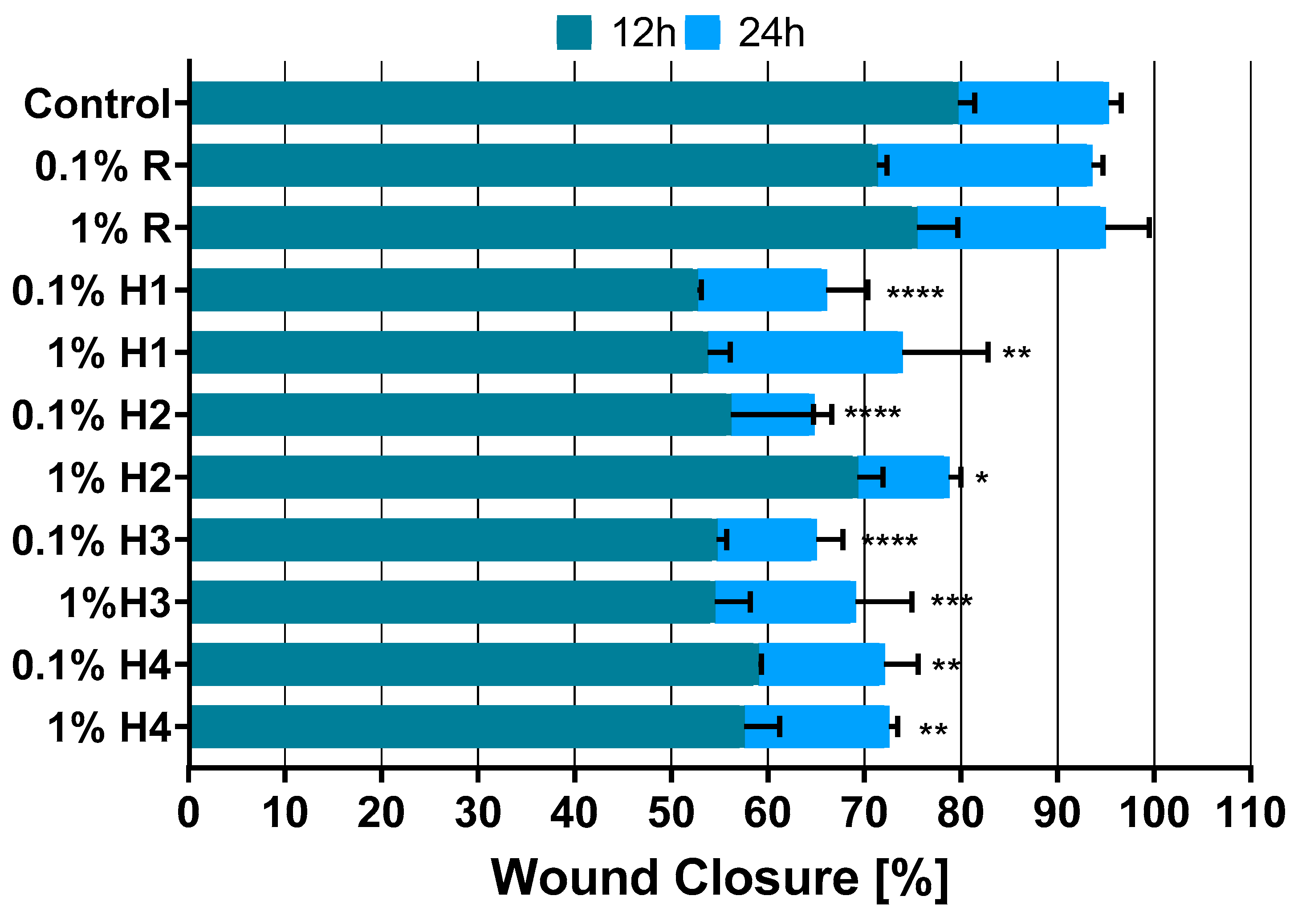
To evaluate the “wound-healing” activity of the samples, BJ and MCF-7 cells were subjected to a migration scratch assay. The effect of individual honey samples on the migration of normal and cancer cells was compared to the control migration of untreated cells. Based on the obtained microscopic images, the evolution of the gap created in the confluent cell monolayer in the presence of the samples was evaluated (Figure 2, Figure 3 and Figure 4). The quantification of the cell-free area was performed using ImageJ public domain software and data are expressed as the mean ± SE (Figure 2 and Figure 3).
The overall antimigration activity against cancer MCF-7 cells was highest for the 0.1% H2, 0.1% H3, and 0.1% H1 samples, as the wound closure values were 64.68%, 65.66%, and 65.98%, respectively (Figure 2). The lowest wound closure value was observed for H4 (71.99%). The rapeseed honey’s antimigration properties were only 1.17% weaker than the untreated control, which indicates its low inhibition of MCF-7 cells migration in comparison with the tested samples. The inhibition of cancer cell migration by honeydew honey may have resulted from its antioxidant activity [61,62]. An increase in the antioxidant status during apoptosis in hepatocellular carcinoma cells was observed previously [62]. The migration of normal fibroblasts was less affected by the tested samples; the degree of wound closure ranged from 79.47% (0.1% H1) to 92.89% (1% H4), while for rapeseed honey this value was only 3.39% lower than for the nontreated control. The inhibitory effect on MCF-7 cell migration was previously tested for propolis extracts, showing large differences depending on the origin of the sample [38]. However, it is not possible to directly compare the effects obtained for propolis and honey due to the much more complex composition of the propolis extract compared to honey. In the case of propolis, an isolated fraction of bioactive ingredients was used for research, while in the current study whole diluted honey was tested.
It is interesting that against both normal and cancer cell lines, the 0.1% concentrations of honeydew honey inhibited cell migration to a greater extent. Hence, the wound closure value was inversely proportional to the concentration of tested samples, including rapeseed honey. However, it was already described that a 0.1% v/v honey concentration supports wound healing in vitro under normoxia and hypoxia (3%), indicating that a honey dilution can be effective in hypoxic wound healing [63].

4. Conclusions

The research conducted concerned honeydew honey from coniferous honeydew, a product specific to the south-eastern region of Poland, whose medicinal properties have not yet been well-recognized. It has been shown that high-quality Podkarpackie honeydew honey is characterized by strong antioxidant and antibacterial properties compared to rapeseed honey. Among the tested bacteria, the most sensitive were K. pneumoniae and Streptococcus spp. strains. Moreover, it was demonstrated for the first time that honeydew honey inhibits the migration of cancer cells (MCF-7) and at the same time slightly affects the migration of normal fibroblasts. The attempt to use the scratch test using various cell lines demonstrated its usefulness in testing the therapeutic potential of honey. The obtained results indicate the great medicinal potential of this variety of honey, with the decisive factor being the quality of honey guaranteed by the Protected Designation of Origin label.

Author Contributions

Conceptualization, E.C. and M.D.; methodology, M.T. and E.C.; software, M.T.; validation, M.T., E.C. and K.L.-S.; formal analysis, M.D.; investigation, M.T., E.C., M.M. and K.L.-S.; resources, M.D.; data curation, M.M.; writing—original draft preparation, M.T., E.C. and M.M.; writing—review and editing, M.D.; visualization, M.M. and E.C.; supervision, M.D.; project administration, M.D.; funding acquisition, M.D. All authors have read and agreed to the published version of the manuscript.

Funding

This work was supported by the Polish Ministry of Science and Higher Education research project within the University of Rzeszów PB/ZCHTZ/2023.

Institutional Review Board Statement

Not applicable.

Informed Consent Statement

Not applicable.

Data Availability Statement

The data presented in this study are available in the article.

Conflicts of Interest

The authors declare no conflicts of interest.

References

  1. Shah, A.; Ahmed Zia, S.; Rafi, M.; Mehmood, A.; Aslam, S.; Chaudhry, M.T. Quantification of honeydew production caused by dubas bug on three date palm cultivars wasps fauna of Pakistan. J. Entomol. Zool. Stud. 2016, 4, 278–284. [Google Scholar]
  2. Fischer, M.K.; Völkl, W.; Hoffmann, K.H. Honeydew production and honeydew sugar composition of polyphagous black bean aphid, Aphis fabae (Hemiptera: Aphididae) on various host plants and implications for ant-attendance. Eur. J. Entomol. 2005, 102, 155–160. [Google Scholar] [CrossRef]
  3. Commission Regulation (EU) No 710/2010 of 6 August 2010 on entering a name in the register of protected designations of origin and protected geographical indications („Podkarpacki Miód Spadziowy” (PDO)). Off. J. Eur. Communities 2010, 208, 1–2.
  4. Dżugan, M.; Zaguła, G.; Wesołowska, M.; Sowa, P.; Puchalski, C. Levels of toxic and essential metals in varietal honeys from podkarpacie. J. Elem. 2017, 22, 1039–1048. [Google Scholar] [CrossRef]
  5. Tomczyk, M.; Bocian, A.; Sidor, E.; Miłek, M.; Zaguła, G.; Dżugan, M. The Use of HPTLC and SDS-PAGE Methods for Coniferous Honeydew Honey Fingerprinting Compiled with Mineral Content and Antioxidant Activity. Molecules 2022, 27, 720. [Google Scholar] [CrossRef]
  6. Tomczyk, M.; Zaguła, G.; Puchalski, C.; Dżugan, M. Transfer of some toxic metals from soil to honey depending on bee habitat conditions. Acta Univ. Cibiniensis Ser. E Food Technol. 2020, 24, 49–59. [Google Scholar] [CrossRef]
  7. Kaczmarek, A.; Muzolf-Panek, M.; Tomaszewska-Gras, J.; Konieczny, P. Predicting the botanical origin of honeys with chemometric analysis according to their antioxidant and physicochemical properties. Polish J. Food Nutr. Sci. 2019, 69, 191–201. [Google Scholar] [CrossRef]
  8. Seraglio, S.K.T.; Silva, B.; Bergamo, G.; Brugnerotto, P.; Gonzaga, L.V.; Fett, R.; Costa, A.C.O. An overview of physicochemical characteristics and health-promoting properties of honeydew honey. Food Res. Int. 2019, 119, 44–66. [Google Scholar] [CrossRef]
  9. Directive 2014/63/EU of the European Parliament and of the Council amending Council Directive 2001/110/EC relating tohoney. Off. J. Eur. Communities 2014, 164, 1–5.
  10. Zielińska, S.; Wesołowska, M.; Bilek, M.; Kaniuczak, J.; Dżugan, M. The saccharide profile of Polish honeys depending on their botanical origin. J. Microbiol. Biotechnol. Food Sci. 2014, 3, 387–390. [Google Scholar]
  11. Kuś, P.M.; Jerković, I.; Marijanović, Z.; Tuberoso, C.I.G. Screening of Polish Fir Honeydew Honey Using GC/MS, HPLC-DAD, and Physical-Chemical Parameters: Benzene Derivatives and Terpenes as Chemical Markers. Chem. Biodivers. 2017, 14, e1700179. [Google Scholar] [CrossRef]
  12. Recklies, K.; Peukert, C.; Kölling-Speer, I.; Speer, K. Differentiation of Honeydew Honeys from Blossom Honeys and According to Their Botanical Origin by Electrical Conductivity and Phenolic and Sugar Spectra. J. Agric. Food Chem. 2021, 69, 1329–1347. [Google Scholar] [CrossRef]
  13. Porcza, L.; Simms, C.; Chopra, M. Honey and Cancer: Current Status and Future Directions. Diseases 2016, 4, 30. [Google Scholar] [CrossRef]
  14. Puścion-Jakubik, A.; Karpińska, E.; Moskwa, J.; Socha, K. Content of Phenolic Acids as a Marker of Polish Honey Varieties and Relationship with Selected Honey-Quality-Influencing Variables. Antioxidants 2022, 11, 1312. [Google Scholar] [CrossRef]
  15. Pita-Calvo, C.; Vázquez, M. Differences between honeydew and blossom honeys: A review. Trends Food Sci. Technol. 2017, 59, 79–87. [Google Scholar] [CrossRef]
  16. Dżugan, M.; Tomczyk, M.; Sowa, P.; Grabek-Lejko, D. Antioxidant activity as biomarker of honey variety. Molecules 2018, 23, 2069. [Google Scholar] [CrossRef]
  17. Kędzierska-Matysek, M.; Stryjecka, M.; Teter, A.; Skałecki, P.; Domaradzki, P.; Florek, M. Relationships between the content of phenolic compounds and the antioxidant activity of polish honey varieties as a tool for botanical discrimination. Molecules 2021, 26, 1810. [Google Scholar] [CrossRef]
  18. Fernández-Estellé, M.; Hernández-González, V.; Saurina, J.; Núñez, O.; Sentellas, S. Characterization and Classification of Spanish Honeydew and Blossom Honeys Based on Their Antioxidant Capacity. Antioxidants 2023, 12, 495. [Google Scholar] [CrossRef]
  19. Tsiapara, A.V.; Jaakkola, M.; Chinou, I.; Graikou, K.; Tolonen, T.; Virtanen, V.; Moutsatsou, P. Bioactivity of Greek honey extracts on breast cancer (MCF-7), prostate cancer (PC-3) and endometrial cancer (Ishikawa) cells: Profile analysis of extracts. Food Chem. 2009, 116, 702–708. [Google Scholar] [CrossRef]
  20. Martiniakova, M.; Kovacova, V.; Mondockova, V.; Zemanova, N.; Babikova, M.; Biro, R.; Ciernikova, S.; Omelka, R. Honey: A Promising Therapeutic Supplement for the Prevention and Management of Osteoporosis and Breast Cancer. Antioxidants 2023, 12, 567. [Google Scholar] [CrossRef]
  21. Erejuwa, O.O. Effect of honey in diabetes mellitus: Matters arising. J. Diabetes Metab. Disord. 2014, 13, 23. [Google Scholar] [CrossRef] [PubMed]
  22. Jaganathan, S.K.; Mandal, M. Honey Constituents and their apoptotic effect in colon cancer cells. J. Apiprod. Apimed. Sci. 2009, 1, 29–36. [Google Scholar] [CrossRef]
  23. Mărgăoan, R.; Topal, E.; Balkanska, R.; Yücel, B.; Oravecz, T.; Cornea-Cipcigan, M.; Vodnar, D.C. Monofloral honeys as a potential source of natural antioxidants, minerals and medicine. Antioxidants 2021, 10, 1023. [Google Scholar] [CrossRef] [PubMed]
  24. Eteraf-Oskouei, T.; Najafi, M. Uses of Natural Honey in Cancer: An Updated Review. Adv. Pharm. Bull. 2022, 12, 248–261. [Google Scholar] [CrossRef] [PubMed]
  25. Sowa, P.; Grabek-Lejko, D.; Wesołowska, M.; Swacha, S.; Dżugan, M. Hydrogen peroxide-dependent antibacterial action of Melilotus albus honey. Lett. Appl. Microbiol. 2017, 65, 82–89. [Google Scholar] [CrossRef] [PubMed]
  26. Alygizou, A.; Grigorakis, S.; Gotsiou, P.; Loupassaki, S.; Calokerinos, A.C. Quantification of Hydrogen Peroxide in Cretan Honey and Correlation with Physicochemical Parameters. J. Anal. Methods Chem. 2021, 2021, 5554305. [Google Scholar] [CrossRef]
  27. Feknous, N.; Boumendjel, M. Natural bioactive compounds of honey and their antimicrobial activity. Czech J. Food Sci. 2022, 40, 163–178. [Google Scholar] [CrossRef]
  28. Mandal, M.D.; Mandal, S. Honey: Its medicinal property and antibacterial activity. Asian Pac. J. Trop. Biomed. 2011, 1, 154–160. [Google Scholar] [CrossRef]
  29. Farida, F.; Miftakhul Nizar, A.; Ode Nurlina, W.; Agus, R. Effectiveness of Honey Dressing on Healing of Diabetic Foot Ulcer: Systematic Review. Int. J. Psychosoc. Rehabil. 2020, 24, 2020. [Google Scholar]
  30. Dżugan, M.; Miłek, M.; Kielar, P.; Stępień, K.; Sidor, E.; Bocian, A. SDS-PAGE Protein and HPTLC Polyphenols Profiling as a Promising Tool for Authentication of Goldenrod Honey. Foods 2022, 11, 2390. [Google Scholar] [CrossRef]
  31. Ben Mansour, R.; Ksouri, W.M.; Cluzet, S.; Krisa, S.; Richard, T.; Ksouri, R. Assessment of Antioxidant Activity and Neuroprotective Capacity on PC12 Cell Line of Frankenia thymifolia and Related Phenolic LC-MS/MS Identification. Evid.-Based Complement. Altern. Med. 2016, 2016, 2843463. [Google Scholar] [CrossRef]
  32. Santos, J.S.; Alvarenga Brizola, V.R.; Granato, D. High-throughput assay comparison and standardization for metal chelating capacity screening: A proposal and application. Food Chem. 2017, 214, 515–522. [Google Scholar] [CrossRef]
  33. Dżugan, M.; Miłek, M.; Sidor, E.; Buczkowicz, J.; Hęclik, J.; Bocian, A. The Application of SDS-PAGE Protein and HPTLC Amino Acid Profiling for Verification of Declared Variety and Geographical Origin of Honey. Food Anal. Methods 2023, 16, 1157–1171. [Google Scholar] [CrossRef]
  34. Tanaka, T.; Kawase, M.; Tani, S. Urease inhibitory activity of simple α,β-unsaturated ketones. Life Sci. 2003, 73, 2985–2990. [Google Scholar] [CrossRef]
  35. Bocian, A.; Ciszkowicz, E.; Hus, K.K.; Buczkowicz, J.; Lecka-Szlachta, K.; Pietrowska, M.; Petrilla, V.; Petrillova, M.; Legáth, Ľ.; Legáth, J. Antimicrobial activity of protein fraction from Naja ashei venom against Staphylococcus epidermidis. Molecules 2020, 25, 293. [Google Scholar] [CrossRef]
  36. Miłek, M.; Ciszkowicz, E.; Sidor, E.; Hęclik, J.; Lecka-Szlachta, K.; Dżugan, M. The Antioxidant, Antibacterial and Anti-Biofilm Properties of Rapeseed Creamed Honey Enriched with Selected Plant Superfoods. Antibiotics 2023, 12, 235. [Google Scholar] [CrossRef]
  37. Zapała, L.; Kosińska, M.; Woźnicka, E.; Byczyński, Ł.; Ciszkowicz, E.; Lecka-Szlachta, K.; Zapała, W.; Chutkowski, M. Comparison of spectral and thermal properties and antibacterial activity of new binary and ternary complexes of Sm(III), Eu(III) and Gd(III) ions with N-phenylanthranilic acid and 1,10-phenanthroline. Thermochim. Acta 2019, 671, 134–148. [Google Scholar] [CrossRef]
  38. Miłek, M.; Ciszkowicz, E.; Tomczyk, M.; Sidor, E.; Zaguła, G.; Lecka-Szlachta, K.; Pasternakiewicz, A.; Dżugan, M. Poplar-Type Polish Propolis Considering Local Flora Diversity Breast Cancer Cells. Molecules 2022, 27, 725. [Google Scholar] [CrossRef]
  39. Balkanska, R.; Ignatova, M. Determination of Selected Physicochemical Parameters of Bulgarian Honeydew Honey. J. Mt. Agric. Balk. 2014, 17, 558–571. [Google Scholar]
  40. Atanassova, J.; Lazarova, M.; Yurukova, L. Significant parameters of Bulgarian honeydew honey. J. Cent. Eur. Agric. 2016, 17, 640–651. [Google Scholar] [CrossRef]
  41. Damto, T. A Review on Status of Honey Adulteration and Their Detection Techniques in Ethiopia. J. Nutr. Food Sci. 2021, 11, 180. [Google Scholar]
  42. Serin, S.; Turhan, K.N.; Turhan, M. Correlation between water activity and moisture content of Turkish flower and pine honeys. Food Sci. Technol. 2018, 38, 238–243. [Google Scholar] [CrossRef]
  43. Cabrera, M.; Perez, M.; Gallez, L.; Andrada, A.; Balbarrey, G. Colour, antioxidant capacity, phenolic and flavonoid content of honey from the Humid Chaco Region, Argentina. Phyton 2017, 86, 124–130. [Google Scholar] [CrossRef]
  44. Pauliuc, D.; Dranca, F.; Oroian, M. Antioxidant activity, total phenolic content, individual phenolics and physicochemical parameters suitability for Romanian honey authentication. Foods 2020, 9, 306. [Google Scholar] [CrossRef]
  45. Chis, A.M.; Purcarea, C.; Dżugan, M.; Teusdea, A. Comparative antioxidant content and antioxidant activity of selected Romanian and Polish honeydew honey. Rev. Chim. 2016, 67, 214–218. [Google Scholar]
  46. Wilczyńska, A. Phenolic content and antioxidant activity of different types of polish honey—A short report. Polish J. Food Nutr. Sci. 2010, 60, 309–313. [Google Scholar]
  47. Dor, G.O.L.M.; Mahomoodally, M.F. Chemical profile and in vitro bioactivity of tropical honey from Mauritius. Asian Pac. J. Trop. Dis. 2014, 4, S1002–S1013. [Google Scholar] [CrossRef]
  48. Montaser, M.; Ali, A.T.; Sayed, A.M.; Abdelmohsen, U.R.; Zidan, E.W.; Orfali, R.; Rateb, M.E.; Zaki, M.A.; Hassan, H.M.; Mohammed, R.; et al. 1H-NMR Metabolic Profiling, Antioxidant Activity, and Docking Study of Common Medicinal Plant-Derived Honey. Antioxidants 2022, 11, 1880. [Google Scholar] [CrossRef]
  49. Ankley, L.M.; Monteiro, M.P.; Camp, K.M.; O’Quinn, R.; Castillo, A.R. Manuka honey chelates iron and impacts iron regulation in key bacterial pathogens. J. Appl. Microbiol. 2020, 128, 1015–1024. [Google Scholar] [CrossRef]
  50. Sánchez-Chino, X.M.; Jiménez-Martínez, C.; Ramírez-Arriaga, E.; Martínez-Herrera, J.; Corzo-Ríos, L.J.; Godínez García, L.M. Actividad antioxidante y quelante de metales de las mieles de Melipona beecheii y Frieseomelitta nigra originarias de Tabasco, México. TIP Rev. Espec. Ciencias Quím.-Biol. 2019, 22, 1–7. (In Spanish) [Google Scholar] [CrossRef]
  51. Wesołowska, M.; Dżugan, M. Aktywność i stabilność termiczna diastazy występującej w Podkarpackich miodach odmianowych. Zywn. Nauk. Technol. Jakosc/Food. Sci. Technol. Qual. 2017, 24, 103–112. (In Polish) [Google Scholar] [CrossRef]
  52. Sidor, E.; Tomczyk, M.; Dzugan, M. Application of Ultrasonic or Microwave Radiation to Delay Crystallization and Liquefy Solid Honey. J. Apic. Sci. 2021, 65, 243–253. [Google Scholar] [CrossRef]
  53. Dżugan, M.; Grabek-Lejko, D.; Sidor, E.; Tomczyk, M. The impact of ultrasound decrystallization on enzymatic, antioxidant and antibacterial properties of honey. Innov. Food Sci. Emerg. Technol. 2021, 71, 102709. [Google Scholar] [CrossRef]
  54. Karabournioti, S.; Zervalaki, P. The effect of heating on honey HMF and invertase. Apiacta 2001, 36, 177–181. [Google Scholar]
  55. Zhang, G.Z.; Tian, J.; Zhang, Y.Z.; Li, S.S.; Zheng, H.Q.; Hu, F.L. Investigation of the maturity evaluation indicator of honey in natural ripening process: The case of rape honey. Foods 2021, 10, 2882. [Google Scholar] [CrossRef]
  56. Sahin, H. Honey as an apitherapic product: Its inhibitory effect on urease and xanthine oxidase. J. Enzyme Inhib. Med. Chem. 2016, 31, 490–494. [Google Scholar] [CrossRef] [PubMed]
  57. Rückriemen, J.; Klemm, O.; Henle, T. Manuka honey (Leptospermum scoparium) inhibits jack bean urease activity due to methylglyoxal and dihydroxyacetone. Food Chem. 2017, 230, 540–546. [Google Scholar] [CrossRef] [PubMed]
  58. Li, Y.; Ni, M. Regulation of biofilm formation in Klebsiella pneumoniae. Front. Microbiol. 2023, 14, 1238482. [Google Scholar] [CrossRef]
  59. Muselius, B.; Sukumaran, A.; Yeung, J.; Geddes-McAlister, J. Iron Limitation in Klebsiella pneumoniae Defines New Roles for Lon Protease in Homeostasis and Degradation by Quantitative Proteomics. Front. Microbiol. 2020, 11, 546. [Google Scholar] [CrossRef]
  60. Erejuwa, O.O.; Sulaiman, S.A.; Ab Wahab, M.S. Effects of honey and its mechanisms of action on the development and progression of cancer. Molecules 2014, 19, 2497–2522. [Google Scholar] [CrossRef]
  61. Dragan, S.R.; Nicola, T.; Ilina, R.Ş.; Ursoniu, S.; Kimar, A.; Nimade, S.; Nicola, T. Role of multi-component functional foods in the complex treatment of patients with advanced breast cancer. Rev. Med. Chir. Soc. Med. Nat. Iasi 2017, 111, 877–884. [Google Scholar]
  62. Hassan, M.I.; Mabrouk, G.M.; Shehata, H.H.; Aboelhussein, M.M. Antineoplastic effects of bee honey and nigella sativa on hepatocellular carcinoma cells. Integr. Cancer Ther. 2012, 11, 354–363. [Google Scholar] [CrossRef] [PubMed]
  63. Chaudhary, A.; Bag, S.; Barui, A.; Banerjee, P.; Chatterjee, J. Honey dilution impact on in vitro wound healing: Normoxic and hypoxic condition. Wound Repair Regen. 2015, 23, 412–422. [Google Scholar] [CrossRef] [PubMed]
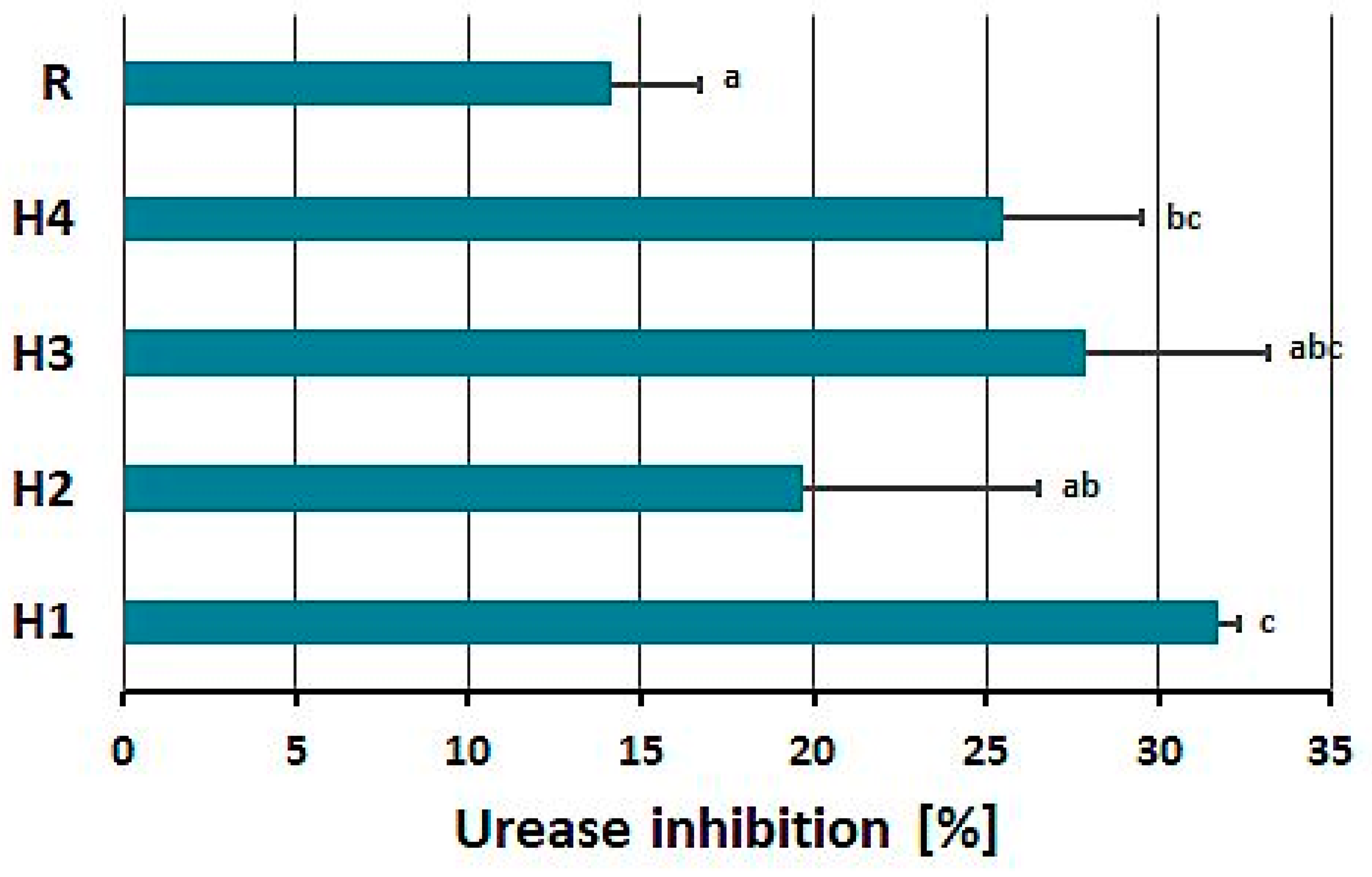
Figure 1. Jack beans urease inhibitory activity of 20% solutions of tested honeys. H1, H2, H3, H4—honeydew honeys, R—rapeseed honey. The error bars represent the standard deviation. a,b,c—means marked with different letters are significantly different (p < 0.05).
Applsci 14 00710 g001
Figure 2. Effect of honeydew honey treatment on breast cancer cells (MCF-7). The scratch wound area was measured 12 and 24 h post-treatment. The error bars present the standard deviation. Statistically significant differences between treated samples and untreated control are shown as follows: * p < 0.05, ** p < 0.001, *** p < 0.0005, and **** p < 0.0001.
Applsci 14 00710 g002
Figure 3. Effect of honeydew honey treatment on normal fibroblasts (BJ). The scratch wound area was assessed at 12, 24, 40, and 64 h post-treatment. Statistically significant differences between treated samples and untreated control are shown as follows: * p < 0.05, ** p < 0.001.
Applsci 14 00710 g003
Figure 4. Scratch assay to investigate the wound healing properties of tested honeys in 1% concentration on breast cancer cells (MCF-7). The area of the scratch was measured at 0, 12, and 24 h after the treatment.
Applsci 14 00710 g004
Table 1. Characteristics of tested honey samples.
Sample CodeHoney Type
/Variety
Apiary
Location
Organoleptic Evaluation
TextureColorSmellTaste
H1fir
honeydew
49.68 N, 21.81 Esticky, gummylight brownpleasant, specific, resinousslightly resinous, spicy
H249.48 N, 22.43 Eviscous liquiddark brown
with black tint
resinous, slightly acidicslightly resinous, caramel, spicy
H349.47 N, 22.07 Eviscous liquiddark brown
with black tint
resinous, slightly acidicslightly resinous, caramel, spicy
H449.34 N, 22.19 Eviscous liquiddark brown
with black tint
woody, smoky slightly resinous, spicy
Rrapeseed49.81 N, 21.54 Efully crystallizedpale yellowspecific for rape flowervery sweet, slightly, bland
Table 2. Basic physicochemical characterization of tested honey samples. The data are shown as mean ± SD (n = 3).
Honey Sample Water
Content [%]
Water ActivitypHFree Acidity [mval/kg]Conductivity [mS/cm]HMF
[mg/kg]
Color
[mm Pfund]
Color
A450 nm
H117.10 ± 0.10 b0.522 ± 0.006 a4.69 ± 0.01 d27.35 ± 0.07 b1.21 ± 0.00 d3.42 ± 0.71 a>150 b0.466 ± 0.011 b
H218.90 ± 0.10 d0.570 ± 0.003 c4.20 ± 0.02 b35.55 ± 0.07 c0.99 ± 0.01 b3.22 ± 0.51 a>150 b0.831 ± 0.025 c
H315.60 ± 0.10 a0.567 ± 0.001 c4.38 ± 0.00 c39.80 ± 0.14 d1.13 ± 0.00 c6.58 ± 0.67 b>150 b0.901 ± 0.009 d
H419.00 ± 0.30 d0.602 ± 0.002 d4.16 ± 0.02 b41.10 ± 0.14 e1.01 ± 0.00 b7.20 ± 0.53 b>150 b0.859 ± 0.022 cd
R17.60 ± 0.10 c0.533 ± 0.003 b4.10 ± 0.02 a8.55 ± 0.07 a0.14 ± 0.00 a<LOQ40 ± 0.3 a0.071 ± 0.010 a
a,b,c,d,e—means marked with different superscript letters within the column are significantly different (p < 0.05).
Table 3. Total phenolic content (TPC) and antioxidant capacity of tested honey samples. The data are shown as mean ± SD (n = 3).
Honey SampleTPC
[mg GAE/kg]
DPPH
[μmol TE/kg]
FRAP
[μmol TE/kg]
CUPRAC
[μmol TE/kg]
Fe2+ Chelating Ability
[mg EDTA/kg]
Cu2+ Chelating Ability
[mg EDTA/kg]
H1657.24 ± 80.91 c773.98 ± 48.26 bc1443.53 ± 33.72 b8243.59 ± 683.80 b2498.31 ± 224.56 c1123.91 ± 20.46 bc
H2823.91 ± 43.67 b710.36 ± 43.17 b1628.29 ± 18.32 c10666.67 ± 385.26 c969.39 ± 44.06 b1075.50 ± 31.44 b
H31001.98 ± 23.07 d941.73 ± 141.93 c2213.82 ± 47.44 d11160.26 ± 664.04 c2081.72 ± 297.54 c1164.75 ± 11.42 bc
H4980.65 ± 7.10 d865.03 ± 90.22 bc2126.10 ± 83.77 d11166.67 ± 122.13 c2038.96 ± 93.61 c1191.98 ± 14.59 c
R195.44 ± 2.27 a125.98 ± 13.27 a272.48 ± 2.51 a2237.18 ± 154.25 a441.63 ± 95.88 a582.37 ± 87.21 a
a,b,c,d—means marked with different superscript letters within the column are significantly different (p < 0.05).
Table 4. The protein content and enzymatic activity of tested honeydew honey samples compared to control rape honey. The data are shown as mean ± SD (n = 3).
Honey SampleProtein
[mg/100 g]
Diastase Number [DN]NAG
[mU/g]
β-GAL
[mU/g]
α-GLU
[mU/g]
H1110.71 ± 15.40 b14.64 ± 0.18 e3.61 ± 0.51 a12.51 ± 0.73 b13.42 ± 0.11 b
H2199.64 ± 12.83 c10.04 ± 0.10 a12.74 ± 0.17 c12.62 ± 1.57 b12.86 ± 1.12 b
H3276.77 ± 32.08 d11.92 ± 0.05 b15.64 ± 0.79 d17.79 ± 0.45 c18.54 ± 1.18 c
H4335.75 ± 10.27 e13.51 ± 0.213 c24.22 ± 0.67 e29.74 ± 0.73 d30.45 ± 0.62 d
R69.87 ± 3.85 a14.20 ± 0.13 d5.92 ± 0.28 b6.11 ± 0.34 a6.99 ± 0.22 a
a,b,c,d,e—means marked with different superscript letters within the column are significantly different (p < 0.05).
Table 5. Antibacterial properties of honeydew and rapeseed honeys from Podkarpackie Province.
Bacterial StrainEscherichia coli
ATCC 10536
Klebsiella pneumoniae
ATCC 13883
Listeria
monocytogenes
SLR 2249
Staphylococcus aureus
ATCC 6538
Staphylococcus epidermidis
ATCC 35984
Streptococcus agalactiae
DSM 2134
Streptococcus pyogenes
DSM 20565
MIC/MBC [% w/v]
H1ND/ND12.5/25.0ND/NDND/NDND/ND50.0/ND50.0/ND
H2ND/ND25.0/NDND/NDND/NDND/NDND/ND25.0/ND
H3ND/ND12.5/25.0ND/NDND/NDND/ND50.0/50.050.0/ND
H4ND/ND12.5/25.0ND/NDND/NDND/ND50.0/50.025.0/50.0
RND/NDND/NDND/NDND/NDND/NDND/NDND/ND
ND—not determined in the range of tested concentrations; the highest tested concentration was 50%.
Disclaimer/Publisher’s Note: The statements, opinions and data contained in all publications are solely those of the individual author(s) and contributor(s) and not of MDPI and/or the editor(s). MDPI and/or the editor(s) disclaim responsibility for any injury to people or property resulting from any ideas, methods, instructions or products referred to in the content.