Article

Doped Epoxy Resins as an Alternative to Luminescent Optical Sensors

by
Małgorzata Gil-Kowalczyk
* and
Paweł Mergo
Laboratory of Optical Fibers Technology, Institute of Chemical Science, Faculty of Chemistry, Maria Curie Sklodowska University in Lublin, M. Curies-Sklodowska Sq. 5, 20-031 Lublin, Poland
*
Author to whom correspondence should be addressed.
Appl. Sci. 2024, 14(14), 6170; https://doi.org/10.3390/app14146170
Submission received: 14 June 2024 / Revised: 7 July 2024 / Accepted: 12 July 2024 / Published: 16 July 2024

Abstract

The main objective of the study was to prepare and then subject to thorough analysis photosensitive materials to determine their usability as materials for the production of special polymer optical fibers. A comparison of the physicochemical properties of compositions containing commercially available fluorescein with compositions doped with 2.7-dihydroxy naphthalene with epichlorohydrin (2.7-NAF.EP) was made. The degree of copolymer conversion, which is one of the most important parameters confirming the effectiveness of the curing method, was calculated based on ATR-FT-IR spectra. Additionally, in order to check the processing capabilities of the obtained compositions, a thorough thermal and spectroscopic analysis was performed (the best method used for this purpose is the coupled analysis technique (TG-DSC-MS)). The obtained results indicate that the photoluminescent properties of the dopants used were not suppressed after their introduction into the matrix. Thermal and spectroscopic analysis allowed us to determine the polymerization conditions in which the physicochemical properties of the obtained materials are the best from the optical fiber-technology point of view.

1. Introduction

Polymer materials (i.e., blends, composites) have found widespread use thanks to their favorable properties and wide modification possibilities. They owe their popularity to valuable properties such as low density, high mechanical strength, durability, resistance to atmospheric factors, thermostability and good processability [1,2,3,4,5,6,7,8]. Conventionally, resins are cured using thermal, redox or, as described below, UV curing. Typically, in order to avoid a premature start of the polymerization process, the initiator is introduced just before the reaction begins. A more desirable curing process, however, would be a “cure on demand” system, in which the resin would not be cured until needed. Such a technique is photopolymerization, the reaction of which will not start until light in the range of wavelengths activating the added photoinitiator is supplied to the system. Photocuring technology has found many applications in various industrial sectors, where it outclasses other processing techniques, thanks to its advantages, namely, ultra-fast polymerization (for comparison, thermal polymerization can last from several minutes to several hours [9,10,11], and in the case of photopolymerization we are talking about seconds) of solvent-free preparations, which occurs selectively only in illuminated places [1,12,13,14,15,16,17,18]. It is worth emphasizing the usefulness of this method in microelectronics, where UV-curable resins are used not only as photoresistors in the imaging stage, but also as quick-drying adhesives, sealants and protective coatings [19,20,21]. Also, as protective coatings, derivatives of epoxy resins are used in optical fiber technology, and thanks to the ease of their modification, they can be used as well as a matrix for materials with dedicated properties used to produce special optical fibers. These fibers are used in sensors for detecting physical parameters and biochemical compounds, but due to the increasing demand for more selective and sensitive sensors, this branch of the industry is still very interesting for researchers and is constantly being developed [6,7,8,22,23,24,25,26]. The use of polymeric materials doped with luminophores poses a number of requirements regarding their composition and morphology. What has a major impact on the properties of the final product is the method of immobilizing the dopant in the polymer matrix. The dopant can connect to the matrix by a hydrogen bond, and then we are dealing with a physical connection, but when the dopant is connected by a covalent bond, we are dealing with a chemical connection. There are also materials exhibiting luminescent properties obtained by adsorption [27,28,29,30,31]. The luminescent composites obtained in this way are used to obtain thin films for sensing applications. These materials are characterized by quite a simple preparation methodology and, more importantly, their application on optical fibers or waveguides is equally simple. The method of obtaining the photosensitive element is that which distinguishes the work presented so far, from the research carried out at the Laboratory of Optical Fiber Technology, Maria Curie-Skłodowska University. Our goal is not to apply a photosensitive material to a fragment of a silica fiber, from which the protective coating has been removed, or to place this material on the tip of the fiber [32,33,34,35,36,37] but to obtain a fiber for which outer jacket will be photosensitive throughout its entire volume. Thanks to this, we will obtain the best effect of the light interaction from the luminophore with the guiding part (core). According to our technology, in the case of polymer fibers, the best material compatibility is achieved by co-producing both the core and the cladding of the optical fiber. This is best illustrated by the example we describe in the article [38], where it was proven that during the preform fabrication styrene diffuses into poly(methyl methacrylate), creating a transition layer. This layer ensures a good mechanical and optical connection between the core and cladding material. Therefore, the material we made is first subjected to thermal and spectroscopic analysis, which checks which conditions for the polymerization process are the best, and in the next step, preform is obtained. Then, a hole is drilled in the preform, into which core material is introduced and another polymerization is performed.
Therefore, in order to conduct a thorough analysis of conditions under which the prepared mixtures (the use of two different photosensitive substances, three different exposure times, the addition of an active solvent, and two epoxy resins with different viscosities) can be used in accordance with our technology, we carried out a series of tests using coupled TG/DSC/MS (thermogravimetry/differential scanning calorimetry/mass spectroscopy). Coupled methods provide comprehensive information about the changes that occur in the tested material under the influence of a given temperature regime. Additionally, the ATR/FT-IR (Attenuated Total Reflectance/Fourier-Transform Infrared Spectroscopy) technique made it possible to control the degree of double-bond conversion.

2. Materials and Methods

The set of investigated samples contained one of two derivatives of bisphenol A epoxy diacrylate EB150 or EB600 (CYTEC, Viesbaden, Germany) doped with photoluminescent dopants: fluorescein (ALDRICH, Warsaw, Poland) or 2.7-dihydroxy naphthalene with epichlorohydrin (the substance synthesized at the Department of Polymer Chemistry, UMCS, Lublin, Poland), was obtained by photopolymerization. The structures of all substances used are shown in Figure 1. The acrylates used vary in viscosity; therefore, it was possible to investigate the influence of a changing viscosity, by adding the active solvent N-vinyl-pyrrolidone (NVP) (Fluka AG, Buchs, Switzerland), on obtained material properties. The detailed information about the synthesis of 2.7-dihydroxy naphthalene with epichlorohydrin (2.7-NAF.EP) was presented in Refs. [39,40].
To obtain studied compositions, bisphenol A epoxy diacrylate derivative was dissolved in NVP. The process was conducted in the three-necked flask equipped with a stirrer and a water condenser. During the whole process, temperature (80 °C) was controlled by the thermometer placed in one of the flask neck. After the acrylate derivative was completely dissolved, a photoluminescent dopant was added. The concentration of dopant in all prepared samples was 1 wt.%. Detailed information about compositions used in the syntheses is given in Table 1 and Table 2.
All compositions were polymerized with UV light in the presence of 1 wt.% of 2,2-dimethoxy-2-phenylacetophenone (Irgacure 651, ALDRICH, Warsaw, Poland) for 15, 30 and 60 s. The curing process was carried out at room temperature in a chamber in which 8 UV lamps (OSRAM, DULUX L BL UVA 18 W/78, UVA radiation in the range of 315–400 nm) were placed. The atmosphere of the photopolymerization reaction is very important, which is why during the entire process the chamber was washed with inert gas, argon, to prevent oxygen inhibition. The oxygen molecule is a radical, which is why it is highly reactive. Atmospheric oxygen can extinguish radicals generated from the initiator at the very beginning of photopolymerization, i.e., at the initiation stage. In the next step, unreactive peroxyradicals may be formed. This causes the active part of the chain to “freeze” and thus generally inhibit the reaction. Moreover, radicals deactivated in this way are less subject to the reinitiation reaction. During photo crosslinking, this reaction determines the structure of the resulting polymer because it has a significant impact on the degree of entanglement of the polymer chains [1,12,13,14,15,16]. Therefore, also in this chamber, liquid compositions were poured between two glass plates, which were placed in a stand specially prepared for this purpose. The distance between the plates was 2 mm, which was possible thanks to the use of a rubber spacer, and the distance of the UV lamps from the assembled system was 10 cm on each side of the plates.
The attenuated total reflection–Fourier-transform infrared (ATR/FT-IR) spectra were obtained using a Bruker FTIR spectrophotometer TENSOR 27 (Bruker, Leipzig, Germany). The spectra were recorded in the frequency range of 4000–600 cm−1 with a resolution of 4 cm−1 and 128 scans.
The thermal properties were investigated based on results obtained from STA 449 Jupiter F1 (Netzsch, Selb, Germany). In our studies, the sample mass of about 5 mg was heated from 25 °C to 600 °C in a closed aluminum crucible and with a heating rate of 10 °C/min. The inert atmosphere of helium (50 mL/min) was used. The volatiles emitted during the heat increase were analyzed using the quadrupole mass spectrometer QMS 403 C Aëolos (Netzsch, Selb, Germany) coupled online to the STA instrument. The mass spectrometer was connected online to the STA instrument by a quartz capillary heated to 300 °C to avoid the condensation process of volatiles. The QMS was operated using an electron impact ionizer with the energy 70 eV. The measurements were performed in the scan mode for m/z, where m is the mass of the molecule and z is a charge of the molecule in the electron charge units in a range from 10 to 150 m/z.

3. Results and Discussion

3.1. Double-Bond Conversion

One of the most important values describing the quality of the obtained polymer, and more precisely indicating the correctness of the polymerization process, is the conversion of double bonds. This value provides information about the number of unreacted functional groups, the cross-linking density and the functionality of monomers used in polymerization. The double-bond conversion was calculated based on IR spectra obtained using the ATR technique using Equation (1). The data obtained in this way are presented in Table 3.
π = 1 A p 810 c m 1 A p 1720 c m 1 × A 0 1720 c m 1 A 0 810 c m 1 × 100 % ,
where A p 810 c m 1 is the band absorbance at the wavelength 810 cm−1 derived from the polymer, A p 1720 c m 1 is the band absorbance at the wavelength 1720 cm−1 derived from the polymer, A 0 810 c m 1 is the band absorbance at the wavelength 810 cm−1 derived from the monomer, and A 0 1720 c m 1 is the band absorbance at the wavelength 1720 cm−1 derived from the monomer.
The band at wavelength 1720 cm−1 was used as a reference. This band corresponds to the stretching vibrations of the carbonyl group and does not change during the polymerization process. The band at wavelength 810 cm−1, which corresponds to the -C=C- in the monomer, was used to calculate the double-bond conversion [41].
When designing the composition that would be best from the optical fiber-technology point of view, both resins, with different viscosities, various exposure times, and two photosensitive substances, were taken into account. The viscosity of EB150 given by the manufacturer and measured at a temperature of 25 °C is 1400 mPa*s, while for EB600 it reaches the value of 3000 mPa*s, but for this epoxy acrylate, measurements were performed at 60 °C. This information is important because it affects the course of the polymerization process and explains the use of the active solvent (NVP, viscosity 2.4 mPa*s measured at 20 °C). When comparing the double-bond conversion values for compositions based on EB150 resin, a significant increase in the discussed value can be observed with an increase in exposure time from 15 to 30 s. At the same time, this tendency is not observed when the exposure time is increased to 60 s (where the conversion most often remained at the same level). Such observations were made for samples No.1–4 and 6, while for samples No.5 and No.7–9, as time increases, the value of double bond conversion increases. It is worth noting that in the case of the highest conversion values, they always exceed 80%, with the exception of sample No.3 (75.9%). Comparing the compositions without the photosensitive dopant (samples No.1, 4, and 7), it can be clearly stated that the addition of an active solvent increases the degree of double-bond reaction. The addition of a photosensitive dopant has different effects depending on the starting composition. In the case of 75 wt.% EB150 content, the addition of fluorescein worsens the conversion rate by approximately 10% (samples No.3-60). A similar tendency is observed when we add 2.7-NAF.EP to the composition containing 67 wt.% of the resin (samples No.5-60). When the resin content is 50 wt.%, the addition of both photosensitive substances slightly improves the conversion of double bonds (samples No.8-60 and No.9-60). For samples based on EB600 resin, an increase in conversion rate is observed with increasing exposure time. The exceptions are samples No.14-60 and No.15-60, where a slight decrease in the discussed value is observed. It is noteworthy that the highest bond-conversion values for these samples exceed 90%. The exceptions are samples No.11 and No.12, where the resin content is the highest (75 wt.%) and where photosensitive compounds have been added. At this point, it is worth explaining why the influence of the active solvent on the double-bond conversion values is more pronounced in the case of compositions based on EB600 resin. Of course, this is related to the difference in the viscosity of the resins. EB150 resin is characterized by relatively low viscosity. The addition of NVP increases the diffusion of photoinitiator radicals and monomer molecules in the structure of the resulting polymer, but not to the same extent as for compositions based on EB600. There is a very significant increase in the conversion rate already after 15 and 30 s of UV exposure (for example, comparing samples No.10-15 with No.13-15). The addition of both photosensitive agents to the composition containing the smallest amount of NVP worsens the conversion of double bonds (by approximately 10% in the case of 2.7-NAF.EP, samples No.11-60, and by approximately 20% in the case of fluorescein, sample No.12-60). However, in the case of 33% of the solvent content, the conversion of double bonds remains at a similar level (samples No.13-15), and in the case of 50% of the solvent content it increases slightly (samples No.16–18). The highest values of double-bond conversion (100%, Table 3) were obtained for samples containing 50 wt.% NVP and the addition of 2.7-NAF.EP hardened for 30 (samples No.17-30) and 60 s (samples No.8-60 and No.17-60), and for the sample containing fluorescein, hardened for 30 (samples No.18-30) and 60 s (samples No.18-60).

3.2. Thermal and Spectroscopic Analysis

Two types of polymers are generally used in optical fiber technology. These are linear polymers, from which optical fibers are produced in the thermal processing process, and cross-linked polymers used as protective coatings for glass or polymer optical fibers and as a material for special fibers. In order to produce an optical fiber, in the first stage a preform is produced. Obtaining it from a linear polymer requires precise control of the thermopolymerization process. If the process is not carried out properly, it leads to heterogeneity of the polymer structure and additionally causes cross-linking of the system. As mentioned, cross-linked polymers are used as protective coatings and, as the name suggests, provide protection for brittle glass, and in the case of polymer optical fibers, are also materials that trap traveling light in the fiber core. Therefore, such material should have a lower refractive index than the material used to produce the core, in order to meet the principle of total internal reflection. To be able to properly protect the newly formed fiber, such coatings are applied immediately, using the photopolymerization process, after the fiber is drawn out. Thermal resistance and mechanical strength are some of the most important parameters checked for materials potentially useful as coatings. The presented research focused on thermal properties. Table 4 contains the all-important parameters related to thermal resistance, which were read based on data obtained from TG/DTG/DSC analyses [14,42,43,44,45,46]. The table shows the temperatures at 1, 3 and 5 wt.% mass loss. On this basis, it is possible to compare the polymerization conditions at which the temperature parameters are the most favorable. The maximum values presented for the DTG and DSC curves additionally allow for determining the thermal resistance of the obtained materials. Table 4 also includes information on the amount of remaining unreacted mass. Graphs from these analyzes are provided in the Supplementary Materials (Figure S1).
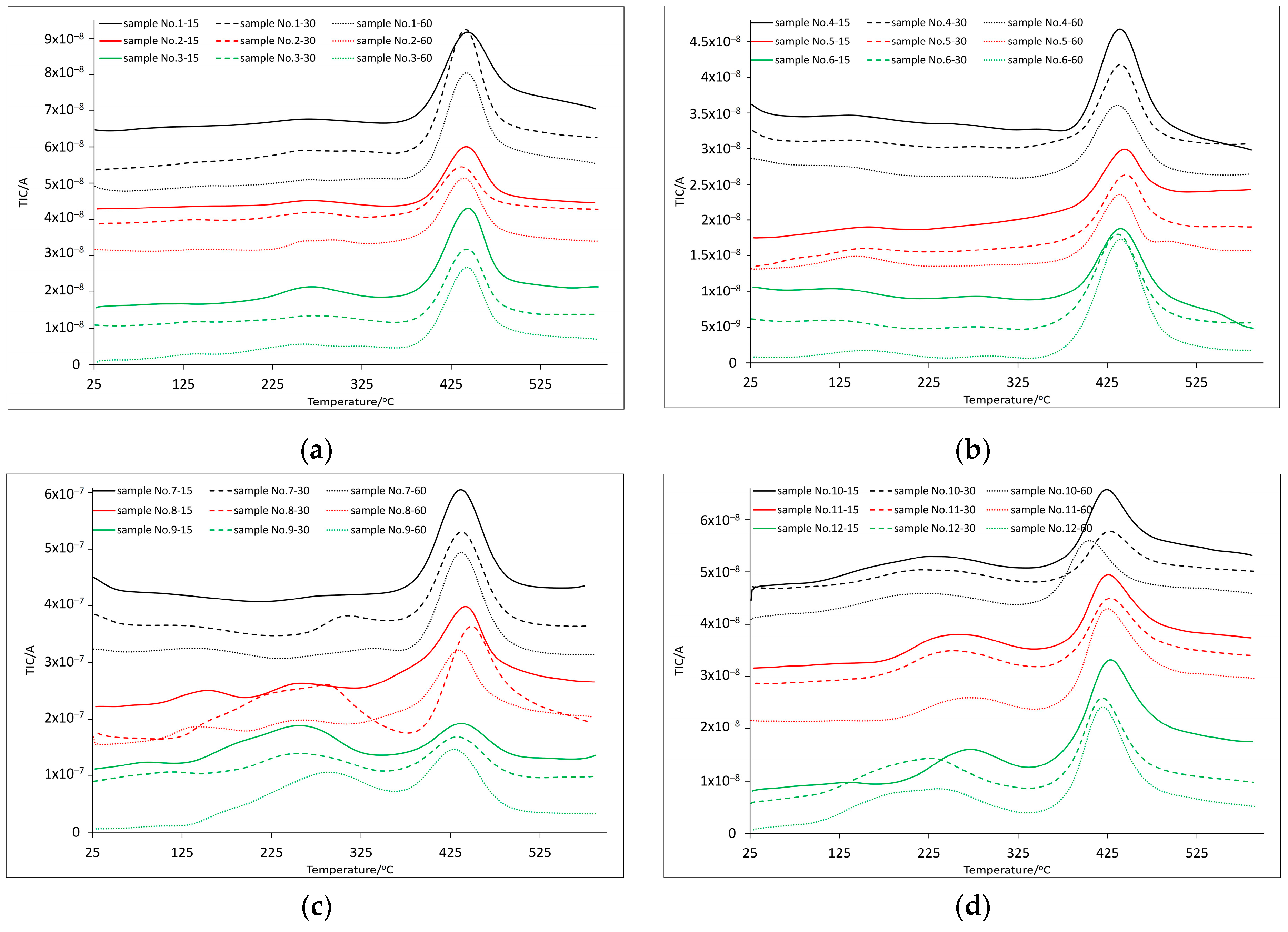
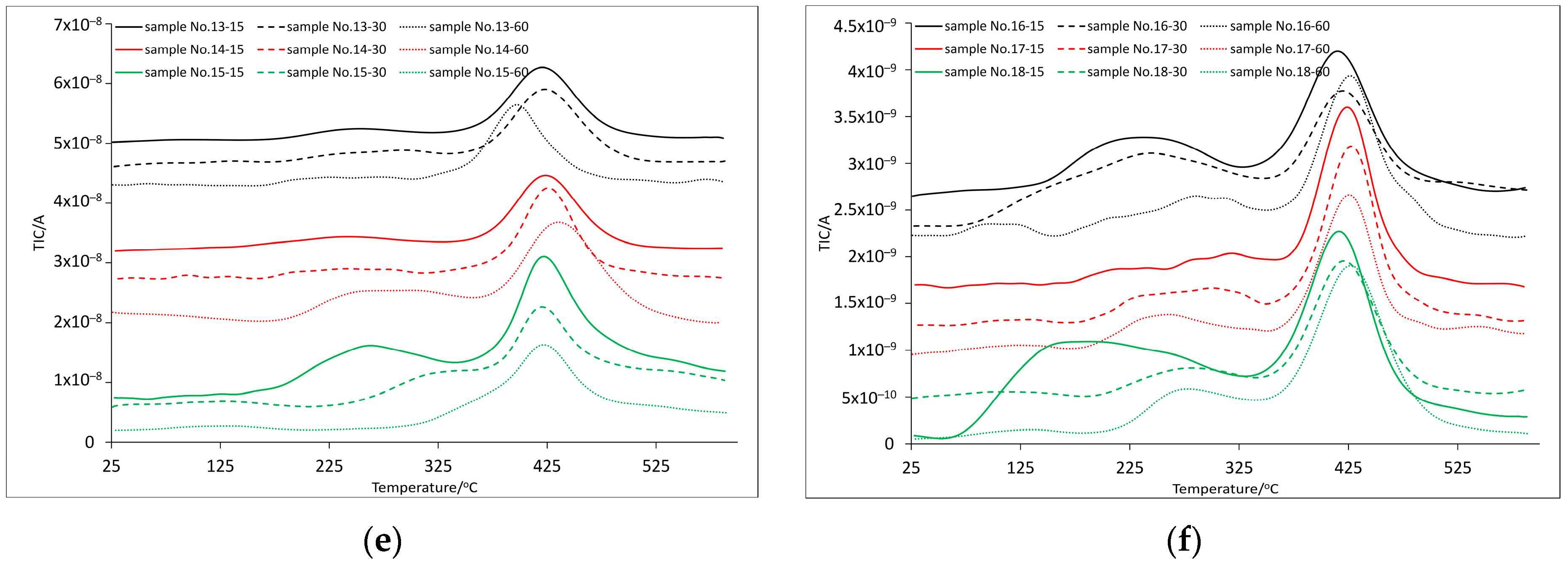
Analyzing the data for compositions based on EB150 resin with 25 wt.% NVP addition (samples No.1, No.2 and No.3), it can be concluded that the increase in exposure time does not significantly affect the improvement of thermal resistance. For samples No.1-15, No.1-30 and No.1-60, a noticeable mass loss (around 1 wt.%) was recorded around 120 °C, although the temperature values obtained for the sample exposed to 15 s of light are slightly higher. The same applies to the DTG curve values. The fact that 3–4 maxima are recorded on the DTG and DSC curves proves the complexity of the composite structure. The presence of one maximum (at the highest temperature) is obviously related to the proper decomposition of the sample. All the earlier ones may indicate incomplete polymerization of monomers, which are released from the tangled structure of the polymer at elevated temperatures, and their earlier decomposition occurs. The same applies to incompletely polymerized active solvent molecules. However, the initial mass loss and the corresponding maxima recorded on the DTG and DSC curves are most likely related to the presence of water physically adsorbed on the surface. The hygroscopic properties of the analyzed materials should be taken into account, which, based on the mass loss value, would indicate that the samples No.1-60 showed the greatest properties in question. Mass spectroscopy helps in the interpretation of changes that occur in a sample subjected to a temperature regime. Based on this, it is possible to determine what gases and at what temperature are released when the sample is heated (information about thermal decomposition pathways) [41,47,48]. Due to the multitude of ions appearing during the entire analysis, the article includes collective ion mass charts, i.e., the so-called ion current (Figure 2).
The analysis of spectroscopic spectra indicates that for samples No.1–15 up to a temperature of 170 °C, for the mass loss only water is responsible (as indicated by the presence of ions m/z = 17–19). Above this temperature, ions m/z = 26–29 begin to appear, responsible for the presence of CH2=C+, CH2=CH+, CH2=CH2+ (terminal vinyl group), CO or CH3CH2+. Above the mentioned temperature, ions m/z = 42–44 also appear, responsible for the presence of groups: CH2=CH-CH3+, CH2-C=O+, CH3-C=O+ and H2N-C=O+ (a carboxyl group is also possible). The mentioned ions are most likely related to the presence of an active solvent in the composition, which is the first to be released from the still relatively stiff polymer structure at elevated temperatures, and at first undergo thermal decomposition. The same situation is observed for samples No.1-30 and No.1-60 but in both cases from the temperature of 150 °C, which is consistent with the first DTG values (Table 4). In the case of DSC curves, all emerging effects are endothermic and their first maxima are slightly shifted towards lower temperatures compared to the DTG maxima. The addition of photosensitive substances (samples No.2 and No.3) shifts the first DTG maximum towards lower temperatures when comparing it to the pure matrix, and this should be carefully monitored, because each subsequent inclusion of additional substances usually disturbs the polymerization process, causing a deterioration in the quality of the obtained structures. The first maxima recorded by DSC, as before, remain around 120 °C, and are related to the endothermic thermal decomposition of the smallest molecules released from the structure, i.e., the active solvent. This is confirmed by mass spectroscopy and the presence of m/z ions: 27, 28, 29, 42, 43, 44 and m/z = 14 and 15 (methylene and methyl groups) which appear around the temperature of 130 °C. The exception are samples No.3-15, where the mentioned ions appear already around 100 °C, which indicates poor polymerization, confirmed by the double-bond conversion result (38.6%, Table 3). It is worth adding at this point that the temperature of 200 °C is the limit temperature in polymer optical fiber technology, and it is directly related to the technological process itself, because it is around 200 °C that polymer fibers are generally drawn out. In fact, it may shift towards higher temperatures, especially for cross-linked compositions, and due to the glass transition temperature of the matrix. But, during thermal tests, when we compare the processing possibilities of materials, we always assume a temperature of 200 °C as the temperature limiting the possibility of using the material. Therefore, when analyzing the data, the results are presented as those above and below the mentioned temperature. One more maximum (or two in the case of samples No.1-15, No.2-30 and No.2-60) is observed in the DTG and DSC curves before the final decomposition occurs. It appears within the temperature range of 230–240 °C. It is related (and read at the maximum visible on the ion current curve Figure 2a) to the presence of ions m/z = 14,15, 17–19 and ions m/z = 26–28 related to the vinyl group most likely from NVP (there are no other ions indicating the decomposition of the resin). There are also ions coming from the breakdown of the lactam ring m/z = 40–44 and 53–56: C2H2N+, C3H5+, C2H2O+, C2H3O+, H2NCO+, C3H3N+, C3H4N+, C3H3O+ and C3H4O+. Their presence confirms the further release of active solvent molecules and their decomposition. Based on the DTG curves, it was also possible to determine the temperature at the maximum decomposition and for all the samples discussed (i.e., containing 25 wt.% NVP addition) it is approximately 437 °C. In the case of DSC curves, it is worth noting that several maxima appeared in the temperature range where proper sample decomposition occurs. This indicates the complexity of the process related to the heterogeneity of the system, but the fact that these peaks occur at similar temperatures most likely indicates the same decomposition mechanism. In the case of the addition of fluorescein, these maxima are slightly shifted towards lower temperatures (sample series No.3). In addition to the ions mentioned so far, in the maximum of the ion current (at a temperature of approximately 440 °C, Figure 2), there are ions indicating the complete decomposition of the substances included in the tested materials. As is the case with compounds with a benzyl group, most often formed as a result of α-cleavage, in the mass spectra an ion with mass m/z = 91 is observed. In most cases, this ion quickly rearranges to the more stable tropylium ion. It, in turn, losing a neutral acetylene molecule (ion m/z = 26), decays to form the ion m/z = 65 (C5H5+) and in the next stage, also as a result of the loss of C2H2, it is transformed into the ion m/z = 39 (C3H3+). The presence of these ions proves that the resin used has decomposed. Other ions indicating the decomposition of benzene, i.e., the resin used and photosensitive dopants, are also present: m/z = 50 (C4H2+), m/z = 51 (C4H3+), m/z = 52 (C4H4+), m/z = 63 (C5H3+), m/z = 64 (C5H4+), m/z = 77 (C6H5+), m/z = 78 (C6H6+), m/z = 78 (C6H7+), m/z = 92 ((C6H5+)CH2+ H), m/z = 93 ((C6H5+)O), m/z = 94 ((C6H5+)O+H), m/z = 104 ((C6H5+)CHCH2), m/z = 105 ((C6H5+)CHCH3), and m/z = 111 (molecular ion of the active solvent). The remaining part of the mass that did not decompose up to the temperature of 600 °C ranges from 9 to 13 wt.% (Table 4) for the analyzed samples. There were no observations that would indicate that the initial composition or exposure time would affect the residual mass.
In the case of samples with a higher content of active solvent (samples No.4–9, where the NVP content is 33 wt.% and 50 wt.%), the first recorded mass loss (1 wt.% Table 4) is slightly shifted towards lower temperatures (values mostly below 100 °C) compared to the previously discussed samples. Analyzing the mass spectra, only the presence of ions responsible for water physically adsorbed on the surface was found (m/z = 17 and 18). The hygroscopic properties of the analyzed samples should be considered. The lowest temperatures were recorded for samples with the highest NVP content (sample No.7 with 50 wt.% NVP content and without the addition of photosensitive dopants). In the case of samples No.4-15, No.4-30 and No.4-60, the first maximum recorded on the DTG curve is in the temperature range 134–144 °C, and is, as mentioned, related to the presence of ions m/z = 17 and 18. The second maximum for the first two samples occurs at a temperature of approximately 188 °C and, based on the mass spectra, is related to the presence of ions m/z = 15, 17–19, 28, 29, 41–44, and 56. In the case of samples No.4-60 the same set of ions is observed, but it should be noted that the second maximum of the DTG curve is recorded at the temperature of 237.3 °C. Therefore, although the sample hardened for 60 s shows a similar mechanism of decomposition of the first molecules released from the polymer structure (most likely NVP), the process itself, seems to be slower, as indicated by the shift in the decomposition maximum towards higher temperatures. At the same time, the double-bond conversion of samples No.4-30 and No.4-60 is at a similar level, which may indicate that this does not affect the final structure of the obtained polymer. Of course, the appearance of the mentioned ions is related to the endothermic effects recorded on the DSC curves (temperatures slightly shifted towards lower values). It is worth noting that compared to the substances with 25 wt.% NVP content, compositions containing 33 wt.% addition of active solvent between temperatures of 100 °C and 200 °C are characterized by a maximum of mass loss of 2. Due to the assumption that the first one concerns only physically adsorbed water, it should be stated that compared to sample No.1, sample No.4 is characterized by higher thermal resistance. Additionally, samples No.4-60 register the maximum mass loss related to the decomposition of the substance well above 200 °C, so they can be considered as the best in the series. The situation is the same for the entire series of samples No.6, where at around 140 °C a mass loss is recorded only due to the presence of ions m/z = 17 and 18, and the next maximum is above 200 °C. In the case of sample No.5, no clear maximum related to the presence of ions m/z = 17 and 18 is recorded. The evaporation of water is so extended in time that in the temperature range of 100–200 °C only one maximum with ions m/z = 14–18, 28, 29, 42–44 is recorded (which corresponds to one endothermic effect, the maximum of which is at lower temperatures). Based on the results of mass loss and ions associated with it, it can be concluded that for samples doped with photosensitive substances, the addition of an active solvent improves thermal properties (comparing 25 wt.% with 33 wt.% NVP content). Above 200 °C, the discussed samples show two maxima (except for series No.5, where only one is recorded, related to the proper decomposition of the sample, based on which it can be concluded that this series has better thermal properties), the first one in the temperature range 230–242 °C, and the second 431–437 °C. Endothermic peaks appear in DSC curves in similar temperature ranges. Based on the results obtained from mass spectroscopy, it can be concluded that the mass ion profile for sample series No.4 and No.6 looks similar. Around the temperature of 240 °C, the presence of ions m/z = 15, 17, 18, 26–29, 30 (CH2NH2+), 31 (CH3NH2+), 41–44, 52–56, 68, 82–85, and 111 is recorded. Ions 68 (C4H4O+) and 82–85 (C4H4NO+, C4H5NO+, C4H6NO+, C4H7NO+) are also associated with NVP decomposition, but appear in the case of samples with higher solvent content (33 wt.% and 50 wt.%). Analyzing mass spectra at temperatures of 320–350 °C (i.e., in the range where subsequent endothermic effects are recorded), we find that no new ions appear. Only the change in their intensity is noticeable (Figure 2). In the case of samples in series No.5, a slightly different ion profile can be observed. Up to a temperature of approximately 275 °C (the DSC maximum shifted towards higher temperatures compared to the series of samples No.4 and No.6), only ions m/z = 14, 15, 17, 18, 27–29, 31, 40, and 44 are present. At the temperature of about 340 °C the ion m/z = 56 and m/z = 111 does appear. It should be concluded that the structure of the polymers obtained in this series is more homogeneous. Ions resulting from the NVP decomposition appear in mass spectra at higher temperatures, and the fact that fewer effects are recorded is an additional advantage. As a rule, the smaller the maximum on the DTG and DSC curves, the more homogeneous the structure. The greater complexity of the structure for series No.5 is evidenced by the multiple peaks on the DSC curve in the temperature range of the proper sample decomposition. But, from the optical fiber-technology point of view, this is much less important, because in this temperature range the polymer fibers are not drawn out. The ions recorded in the mass spectra within the proper sample decomposition for the series of samples No.4–6 are identical to those presented for samples No.1–3. The amount of residual mass ranges from 8.2 wt.% to 10.2 wt.%. Samples No.7-15, No.8-15 and No.9-15 show the lowest double-bond conversions among all the analyzed compositions (Table 3). This is most likely due to too-little UV exposure time and too much active solvent addition (50 wt.%). While NVP itself has a positive effect on the final structure of the polymer, longer exposure to UV radiation is necessary in some cases. The best proof of this is the conversion of double bonds at the level of 100% for samples No.8-60. Evidence of an improper polymerization process, or more precisely, its too rapid interruption, is the presence of ions m/z = 30, 31, 41–44, 56, and 111 at a temperature of about 125 °C for the mentioned samples. As was the case with the series of samples with the 33 wt.% NVP addition, in this case, for the sample with the 2.7-NAF.EP addition, one maximum is recorded on the DTG curve up to a temperature of 200 °C (shifted towards higher temperatures when comparing with samples No.7 and No.9). The next maximum is the one related to the proper sample decomposition. Mass spectroscopy confirms the presence of ions m/z = 14, 15, 17, 18, 26–31,and 40–44 appearing from the temperature of about 130 °C, but their release from the structure as a result of evaporation/decomposition of the substance is so slow and their intensity so small that only one maximum on the DTG curve is recorded. However, this does not apply to DSC curves, where several endothermic maximums can be observed in the discussed temperature range. The same ions are recorded for the rest of the samples but with the DTG maximum recorded at lower temperatures. Around the second maximum of mass loss for samples No.7 and No.9 the following ions were registered: m/z = 14, 15, 17–19, 26–30, 40–44, 52–56, 68, 82–85, and 111, which indicate the decomposition of the active solvent. However, no ion confirming the decomposition of the epoxy resin or any of the photosensitive additives was detected. Their presence was recorded only at the temperature of proper decomposition (ions in the mass range that were already presented). The maximum of decomposition does not differ significantly from the values for samples with 33 wt.% NVP content. Residual mass for the discussed samples is within the range of 7–12 wt.%.
A total of 1 wt.% mass loss for samples with 25 wt.% NVP content and based on EB600 resin is recorded just after exceeding the temperature of 100 °C (sample series No.10–12). And, based on spectroscopic spectra, it was determined that it is related to the evaporation of water physically adsorbed on the sample surface. The first maximum on the DTG curve varies between 160 and 190 °C. Its value shifts towards lower temperatures for samples doped with photosensitive compounds. The endothermic peak is present in the DSC curves, but significantly shifted towards lower temperatures compared to the DTG maximum (around 120–130 °C). Based on the mass spectra, it was possible to interpret the cause of mass changes in the considered temperature range. There are ions coming from the methylene, methyl and vinyl groups (m/z = 14, 15, 26–28), and from the decomposition of the lactam ring (m/z = 40–44, 53–56, 66 (C4H4N+), 67(C3HNO+), 68, 69(C4H5O+)), as well as indicating the presence of benzene ring fragments (m/z = 39, 50–52, 65, 77–79). This indicates the presence of matrix fragments even before the temperature of 200 °C. A small addition of active solvent (25 wt.%) does not sufficiently reduce the considerable viscosity of EB600, which means that not only NVP molecules, but also resin, are trapped during polymerization. As the temperature increases, the released elements are the first to be registered by the mass analyzer. Ion analysis at the second DTG maximum confirms this statement, where all ions typically present in the decomposition range are observed. The low conversion for these substances (Table 3) also confirms the assumption. DSC curves confirm the occurrence of endothermic processes, and their maximums fall approximately around the registration of 1 wt.% mass loss and the second maximum on the DTG curve. There are also maxima in the range of 300–380 °C, unrelated to the maximum on the DTG curve. But looking at Figure S1J–L, you can see that they are within the range of the sample decomposition. The multitude of these maxima indicates the complexity of the decomposition process related to the diversity of the obtained polymer structures. The situation is completely different in the case of compositions containing 33 wt.% of NVP (sample series No.13-15). For the No.13 and No.15 sample series, only around the second DTG maximum (around 230–240 °C), few ions indicating the presence of a vinyl group (m/z = 27) or an active solvent fragment (m/z = 56) are detected. In the case of series No.14, the first maximums recorded on the DTG curve are slightly above 200 °C, which proves that the addition of 2.7-NAF.EP does not negatively affect the thermal properties of the matrix. Also, in this case, few ions were recorded in the mass spectra (m/z = 14, 15, 27, 40, 41, 56). The second DTG maximum recorded for samples No.14-15 is located at a temperature of 260.6 °C (Table 4), and additional ions m/z = 28–31.43 appear in this region. For samples No.14-30, this maximum was determined at a temperature of 238.3 °C, and the mass spectra indicate the presence of ions m/z = 44, 54, and 55 (apart from those already mentioned). The second DTG maximum for samples No.14-60 was registered at 421.1 °C. This means that ions for which mass is related to the fragments of the EB600 appear only in the temperature range of the proper sample decomposition. This, in turn, allows us to assume that the discussed samples show the best thermal properties of those presented so far (the conversion of double bonds above 90%, Table 3). The profile of endothermic transformations is similar to analogous samples with 25 wt.% NVP content, except for those around the second DTG maximum, where two DSC maximums are observed. For the last series of samples (No.16–18), 1 wt.% mass loss was recorded in the range of 90–115 °C and it cannot be stated that exposure time or the started composition influenced this value. The first maximum on the DTG curve for samples No.16-15 and the first and second maximum for samples No.16-30 are associated with the presence of ions m/z = 17, 18, 27, 28, 29, 40–44, 54–56, 68, and 82, which indicates the relatively early appearance of active solvent fragments. For samples No.17-30, No.17-60, No.18-30 and No.18-60 (i.e., samples with the addition of photosensitive compounds), in the area of the first DTG maximum only the presence of ions m/z = 17, 18 was recorded. Taking this into account and the fact that the second DTG maximum exceeds 200 °C and, additionally, the fact that conversion value of double bonds is at the level of 100% (Table 3), we expect that these are the best materials from the polymer optical fiber-technology point of view. As in the case of samples with 33 wt.% NVP content, in the discussed series of samples, only at the proper decomposition temperature are ions indicating the decomposition of compounds containing benzene in their structure present. Residual mass for samples based on EB600 remains within the range of 9.8–16.9 wt.%. This value appears to decrease with increasing amounts of NVP added.
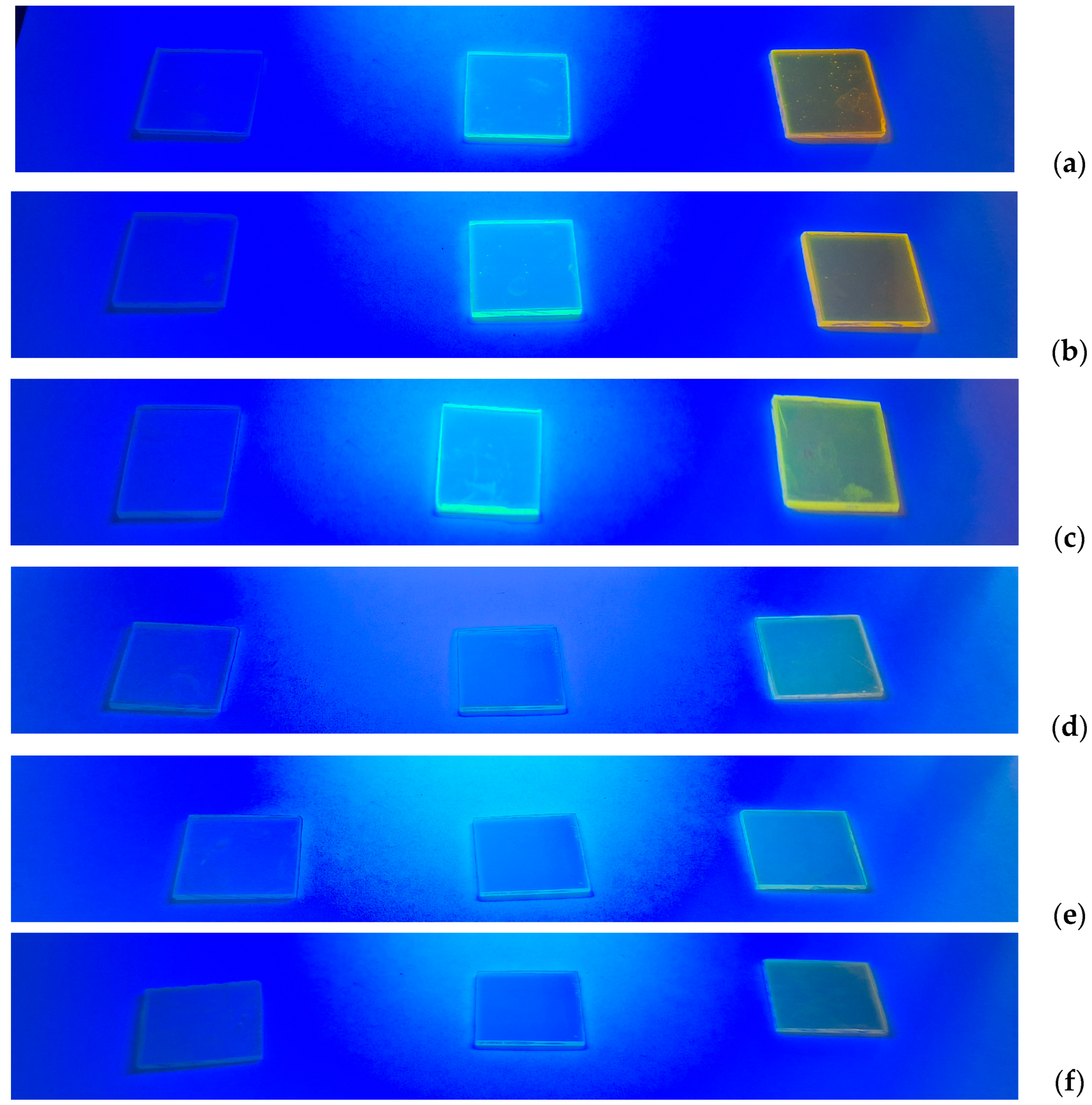
In the next stage, detailed tests of the absorption and emission of the obtained compositions will be carried out. Below are photos (Figure 3) taken under an 8W UV lamp.
It can be clearly stated that the incorporation or suspension of photosensitive substances into the structure of the resulting polymer did not affect their photoluminescent properties. There is a significant difference between the pure matrix and doped compositions. Moreover, higher glow intensity is observed in the case of compositions based on EB600 resin. It is also worth noting that this intensity changes with the change in the NVP content.

4. Conclusions

Based on the results obtained from ATR/FT-IR, it was found that the conversion of double bonds increases with increasing exposure time. This increase is more pronounced for compositions based on EB600. It can be additionally stated that increasing the NVP content in the starting composition also increases the value in question. However, in the case of compositions with EB600, the influence of the solvent is more pronounced, which is related to a decrease in viscosity and thus an increase in the diffusion of both initiator radicals and trapped monomers. It is significant that the highest values of double-bond conversion exceed 80% in the case of the EB150 matrix and 90% in the case of the EB600 matrix. The addition of photosensitive compounds to the composition with EB150 has different effects depending on the amount of active solvent added. The highest conversion value—100%—was obtained for the composition of 50 wt.% NVP with the addition of 2.7-NAF.EP. The addition of photosensitive compounds to compositions based on EB600 does not affect the conversion value, or slightly improves it (except for the composition with 25 wt.% NVP content, where their addition worsens the conversion of double bonds, and the effect of fluorescein is greater). Summarizing the results obtained from the coupled TG/DSC/MS methods, it can be concluded that the addition of photosensitive compounds to compositions containing 25 wt.% NVP and based on both EB150 and EB600 worsens their thermal properties. This is confirmed by mass spectra indicating the presence of active solvent fragments at temperatures slightly exceeding 100 °C. For compositions based on EB150, increasing the NVP content improves the properties in question, as ions indicating the presence of NVP begin to appear closer to the temperature of 200 °C. This confirms that a decrease in viscosity has a beneficial effect on the structure of the polymer, but if the solvent content is too high, the exposure time should be increased (sample series No.7–9). Additionally, it can be stated that compositions containing 2.7-NAF.EP show slightly better thermal properties than analogous samples with the addition of fluorescein (fewer number of maxima recorded on the DTG curve and their values shifted towards higher temperatures). In the case of compositions based on EB600, an increase in the NVP content significantly improves their thermal properties. Already, at 33 wt.% NVP, ions originating from the matrix appear only at temperatures exceeding 200 °C or only at the temperature of proper decomposition. The addition of photosensitive compounds does not worsen the properties discussed, and it can even be said that the admixture of 2.7-NAF.EP improves them. The most thermally stable samples, where fewer maxima are observed on the DTG and DSC curves, and where ions indicating evaporation/decomposition of the matrix exceed 200 °C and where the conversion of double bonds is 100% are samples No.17-30 and No.17-60 and samples No.18-30 and No.18-60.

Supplementary Materials

The following supporting information can be downloaded at: https://www.mdpi.com/article/10.3390/app14146170/s1, Figure S1: Thermal analysis (TG/DTG-DSC curves) for samples: (A) No.1; (B) No.2; (C) No.3; (D) No.4; (E) No.5; (F) No.6; (G) No.7; (H) No.8; (I) No.9; (J) No.10; (K) No.11; (L) No.12; (M) No.13; (N) No.14; (O) No.15; (P) No.16; (R) No.17; (S) No.18.

Author Contributions

Conceptualization, M.G.-K.; methodology, M.G.-K.; validation, M.G.-K. and P.M., formal analysis, M.G.-K. and P.M.; investigation, M.G.-K.; resources, M.G.-K. and P.M.; data curation, M.G.-K.; writing—original draft preparation, M.G.-K.; writing—review and editing, M.G.-K. and P.M.; visualization, M.G.-K.; supervision, M.G.-K. and P.M. All authors have read and agreed to the published version of the manuscript.

Funding

This research received no external funding.

Institutional Review Board Statement

Not applicable.

Informed Consent Statement

Not applicable.

Data Availability Statement

The original contributions presented in the study are included in the article/Supplementary Materials, further inquiries can be directed to the corresponding author/s.

Conflicts of Interest

The authors declare no conflicts of interest.

References

  1. Odian, G. Principles of Polymerization, 4th ed.; Wiley: Hoboken, NJ, USA, 2004. [Google Scholar]
  2. John, Ł.; Ejfler, J. A Brief Review on Selected Applications of Hybrid Materials Based on Functionalized Cage-like Silsesquioxanes. Polymers 2023, 15, 1452. [Google Scholar] [CrossRef] [PubMed]
  3. Garcia, O.; Garrido, L.; Sastre, R.; Costela, A.; Garcia-Moreno, I. Synthetic Strategies for Hybrid Materials to Improve Properties for Optoelectronic Applications. Adv. Funct. Mater. 2008, 18, 2017–2025. [Google Scholar] [CrossRef]
  4. Cernadas, T.; Santos, M.; Miquel, S.P.; Correia, I.J.; Alves, P.; Ferreira, P. Photocurable Polymeric Blends for Surgical Application. Materials 2020, 13, 5681. [Google Scholar] [CrossRef] [PubMed]
  5. Gil-Kowalczyk, M.; Łyszczek, R.; Jusza, A.; Piramidowicz, R. Thermal, Spectroscopy and Luminescent Characterization of Hybrid PMMA/Lanthanide Complex Materials. Materials 2021, 14, 3156. [Google Scholar] [CrossRef] [PubMed]
  6. Jusza, A.; Lipińska, L.; Baran, M.; Olszyna, A.; Jastrzębska, A.; Gil, M.; Mergo, P.; Piramidowicz, R. Praseodymium doped nanocrystals and nanocomposites for application in white light sources. Opt. Mater. 2019, 95, 109247. [Google Scholar] [CrossRef]
  7. Piramidowicz, R.; Jusza, A.; Lipińska, L.; Gil, M.; Mergo, P. RE3+:LaALO3 doped luminescent polymer composites. Opt. Mater. 2019, 87, 35–41. [Google Scholar] [CrossRef]
  8. Łyszczek, R.; Gil, M.; Głuchowska, H.; Podkościelna, B.; Lipke, A.; Mergo, P. Hybrid materials based on PEGDMA matrix and europium(III) carboxylates -thermal and luminescent investigations. Eur. Polym. J. 2018, 106, 318–328. [Google Scholar] [CrossRef]
  9. Retailleau, M.; Ibrahim, A.; Allonas, X. Dual-cure photochemical/thermal polymerization of acrylates: A photoassisted process at low light intensity. Polym. Chem. 2014, 5, 6503. [Google Scholar] [CrossRef]
  10. Kapłoń, Ł.; Kochanowski, A.; Molenda, M.; Moskal, P.; Wieczorek, A.; Bednarski, T.; Białas, P.; Czerwiński, E.; Korcyl, G.; Kowal, J.; et al. Plastic scintillators for positron emission tomography obtained by the bulk polymerization method. Bio-Algorithms Med-Syst. 2014, 10, 27–31. [Google Scholar] [CrossRef]
  11. Chen, M.; Zhang, Z.; Hu, P.; Jin, J.; Li, J.; Liu, J.; Lin, X.; Tan, X. Effect of thermal polymerization temperature and time of PQ/PMMA on the holographic data storage. Proc. SPIE 2022, 12231, 1223103. [Google Scholar] [CrossRef]
  12. Decker, C. Photoinitiated Crosslinking polymerization. Prog. Polym. Sci. 1996, 21, 593–650. [Google Scholar] [CrossRef]
  13. Decker, C. Kinetic Analysis and Performance of UV-Curable Coatings. In Radiation Curing. Science and Technology; Pappas, S.P., Ed.; Plenum Press: New York, NY, USA; London, UK, 1992; pp. 135–179. [Google Scholar]
  14. Jacobine, A.F.; Nakos, S.T. Photopolymerizable Silicone Monomers, Oligomers, and Resins. In Radiation Curing. Science and Technology; Pappas, S.P., Ed.; Plenum Press: New York, NY, USA; London, UK, 1992; pp. 181–240. [Google Scholar]
  15. Scott, T.F.; Cook, W.D.; Forsythe, J.S. Photo-DSC cure kinetics of vinyl ester resins. I. Influence of temperature. Polymer 2002, 43, 5839–5845. [Google Scholar] [CrossRef]
  16. Andrzejewska, E. Photopolymerization kinetics of multifunctional monomers. Prog. Polym. Sci. 2001, 26, 606–665. [Google Scholar] [CrossRef]
  17. Kim, D.S.; Seo, W.H. Ultraviolet-Curing Behavior and Mechanical Properties of a Polyester Acrylate Resin. J. Appl. Polym. Sci. 2004, 92, 3921–3928. [Google Scholar] [CrossRef]
  18. White, T.J.; Liechty, W.B.; Guymon, C.A. The Influence of N-vinyl Pyrrolidone on Polymerization Kinetics and Thermo-Mechanical Properties of Crosslinked Acrylate Polymers. J. Polym. Sci. A Polym. Chem. 2007, 45, 4062–4073. [Google Scholar] [CrossRef]
  19. Sysova, O.; Durin, P.; Gablin, C.; Leonard, D.; Teolis, A.; Trombotto, S.; Delair, T.; Berling, D.; Servin, I.; Tiron, R.; et al. Green deep-UV photoresist based on chitosan for microelectronics. J. Appl. Polym. Sci. 2023, 140, e54244. [Google Scholar] [CrossRef]
  20. Mendes-Felipe, C.; Rodrigues-Marinho, T.; Vilas, J.L.; Lanceros-Mendez, S. UV curable nanocomposites with tailored dielectric response. Polymer 2020, 196, 122498. [Google Scholar] [CrossRef]
  21. Lucchetti, L.; Gobbi, L.; Simoni, F. Analysis of the Phase Separation Process in UV Cured Polymer Dispersed Liquid Crystals for Optical Applications. Mol. Cryst. Liq. Cryst. Sci. Technol. Sect. A Mol. Crys. Liq. Cryst. 2001, 359, 89–96. [Google Scholar] [CrossRef]
  22. Carrillo-Betancourt, R.; Lopez-Camero, A.D.; Hernandez-Cordero, J. Luminescent Polymer Composites for Optical Fiber Sensors. Polymers 2023, 15, 505. [Google Scholar] [CrossRef] [PubMed]
  23. Jakubowski, K.; Huang, C.S.; Boesel, L.F.; Hufenus, R.; Heuberger, M. Recent advances in photoluminescent polymer optical fibers. Curr Opin. Solid State Mater. Sci. 2021, 25, 100912. [Google Scholar] [CrossRef]
  24. Arrospide, E.; IIlarramendi, M.A.; Ayesta, I.; Guarrotxena, N.; Garcia, O.; Zubia, J.; Durana, G. Effects of Fabrication Methods on the Performance of Luminescent Solar Concentrators Based on Doped Polymer Optical Fibers. Polymers 2021, 13, 424. [Google Scholar] [CrossRef]
  25. Jakubowski, K.; Kerkemeyer, W.; Perret, E.; Heuberger, M.; Hufenus, R. Liquid-core polymer optical fibers for luminescent waveguide applications. Mater. Des. 2020, 196, 109131. [Google Scholar] [CrossRef]
  26. Xu, S.; Xiang, H.; Wang, Z.; Tang, X.; Zhang, Y.; Zhan, X.; Chen, J. Conjugation of a phenanthrene-imidazole fluorophore with the chondroitin sulfate generated from Escherichia coli K4 polysaccharide. J. Appl. Polym. Sci. 2021, 138, 51538. [Google Scholar] [CrossRef]
  27. Jankowska, K.; Zdarta, J.; Grzywaczyk, A.; Kijeńska-Gawrońska, E.; Biadosz, A.; Jesionowski, T. Electrospun poly(methyl methacrylate)/polyaniline fibres as a support for laccase immobilisation and use in dye decolourisation. Environ. Res. 2020, 184, 109332. [Google Scholar] [CrossRef] [PubMed]
  28. Basabe-Desmonts, L.; Reinhoudt, D.N.; Crego-Calama, M. Design of fluorescent materials for chemical sensing. Chem. Soc. Rev. 2007, 36, 993–1017. [Google Scholar] [CrossRef] [PubMed]
  29. Ahumada, G.; Borkowska, M. Fluorescent Polymers Conspectus. Polymers 2022, 14, 1118. [Google Scholar] [CrossRef] [PubMed]
  30. Melnikov, P.; Bobrov, A.; Marfin, Y. On the Use of Polymer-Based Composites for the Creation of Optical Sensors: A Review. Polymers 2022, 14, 4448. [Google Scholar] [CrossRef] [PubMed]
  31. Machado, M.G.C.; Pound-Lana, G.; Oliveira, M.A.; Lanna, E.G.; Fialho, M.C.P.; Brito, A.C.F.; Barboza, A.P.M.; Aguiar-Soares, R.D.O.; Mosqueira, V.C.F. Labeling PLA-PEG nanocarriers with IR780: Physical entrapment versus covalent attachment to polylactide. Drug Deliv. Transl. Res. 2020, 10, 1626–1643. [Google Scholar] [CrossRef] [PubMed]
  32. Uttamlal, M.; Sloan, W.D.; Millar, D. Covalent immobilization of fluorescent indicators in photo- and electropolymers for the preparation of fibreoptic chemical sensors. Polym. Int. 2002, 51, 1198–1206. [Google Scholar] [CrossRef]
  33. Sloan, W.D.; Uttamlal, M. A fibre-optic calcium ion sensor using a calcein derivative. Luminescence 2001, 16, 179–186. [Google Scholar] [CrossRef]
  34. Wang, X.D.; Wolfbeis, O.S. Fiber-Optic Chemical Sensors and Biosensors (2008−2012). Anal. Chem. 2013, 85, 487–508. [Google Scholar] [CrossRef] [PubMed]
  35. Isaad, J.; El Achari, A. Azathia crown ether possessing a dansyl fluorophore moiety functionalized silica nanoparticles as hybrid material for mercury detection in aqueous medium. Tetrahedron 2013, 69, 4866–4874. [Google Scholar] [CrossRef]
  36. Dai, H.J.; Liu, F.; Gao, Q.Q. A highly selective fluorescent sensor for mercury ion (II) based on azathia-crown ether possessing a dansyl moiety. Luminescence 2011, 26, 523–530. [Google Scholar] [CrossRef] [PubMed]
  37. Kaewtong, C.; Niamsa, N.; Wanno, B.; Morakot, N.; Pulpoka, B.; Tuntulani, T. Optical chemosensors for Hg2+ from terthiophene appended rhodamine derivatives: FRET based molecular and in situ hybrid gold nanoparticle sensors. New J. Chem. 2014, 38, 3831–3839. [Google Scholar] [CrossRef]
  38. Mergo, P.; Martynkien, T.; Urbańczyk, W. Polymer optical microstructured fiber with birefringence induced by stress-applying elements. Opt. Lett. 2014, 39, 3018–3021. [Google Scholar] [CrossRef] [PubMed]
  39. Podkościelna, B.; Gawdzik, B. Influence of diluent compositions on the porous structure of methacrylate derivatives of aromatic diols and divinylbenzene. Appl. Surf. Sci. 2010, 256, 2462–2467. [Google Scholar] [CrossRef]
  40. Fila, K.; Gargol, M.; Goliszek, M.; Podkościelna, B. Synthesis of epoxy resins derivatives of naphthalene-2.7-diol and theircross-linked products. J. Therm. Anal. Calorim. 2019, 138, 4349–4358. [Google Scholar] [CrossRef]
  41. Silverstein, R.M.; Webster, F.X.; Kiemle, D.J. Spectrometric Identyfication of Organic Compounds, 7th ed.; John Wiley & Sons, Inc.: Hoboken, NJ, USA, 2005. [Google Scholar]
  42. Hoyle, C.E. Calorimetric Analysis of Photopolymerization. In Radiation Curing. Science and Technology; Pappas, S.P., Ed.; Plenum Press: New York, NY, USA; London, UK, 1992; pp. 57–133. [Google Scholar]
  43. Wunderlich, B. Thermal Analysis of Polymeric Materials; Springer: Berlin/Heidelberg, Germany; New York, NY, USA, 2005. [Google Scholar]
  44. Koga, N. Thermoanalytical Methods: Fundamental Principles and Features. In Thermal Analysis of Polymeric Materials: Methods and Developments, 1st ed.; Kindle, Edition; Pielichowski, K., Pielichowska, K., Eds.; Wiley: Hoboken, NJ, USA, 2022; pp. 1–39. [Google Scholar]
  45. Toda, A. Modulated Temperature Differential Scanning Calorimetry. In Thermal Analysis of Polymeric Materials: Methods and Developments, 1st ed.; Kindle, Edition; Pielichowski, K., Pielichowska, K., Eds.; Wiley: Hoboken, NJ, USA, 2022; pp. 41–73. [Google Scholar]
  46. Lin, S.Y.; Chen, L.C.; Cheng, W.T. Use of Different Temperature Control Techniques Coupled with FTIR Spectroscopy to Simultaneously Induce and Identify the Physical Properties, Chemical Reactions, and Thermal Degradation of Polymers. In Thermal Analysis of Polymeric Materials: Methods and Developments, 1st ed.; Kindle, Edition; Pielichowski, K., Pielichowska, K., Eds.; Wiley: Hoboken, NJ, USA, 2022; pp. 169–226. [Google Scholar]
  47. Johnstone, R.A. Mass Spectrometry for Organic Chemists (Cambridge Texts in Chemistry and Biochemistry); Cambridge University Press: London, UK; New York, NY, USA, 1972. [Google Scholar]
  48. Danikiewicz, W. Spektrometria Mas, 1st ed.; Wydawnictwo Naukowe PWN: Warszawa, Poland, 2020. [Google Scholar]
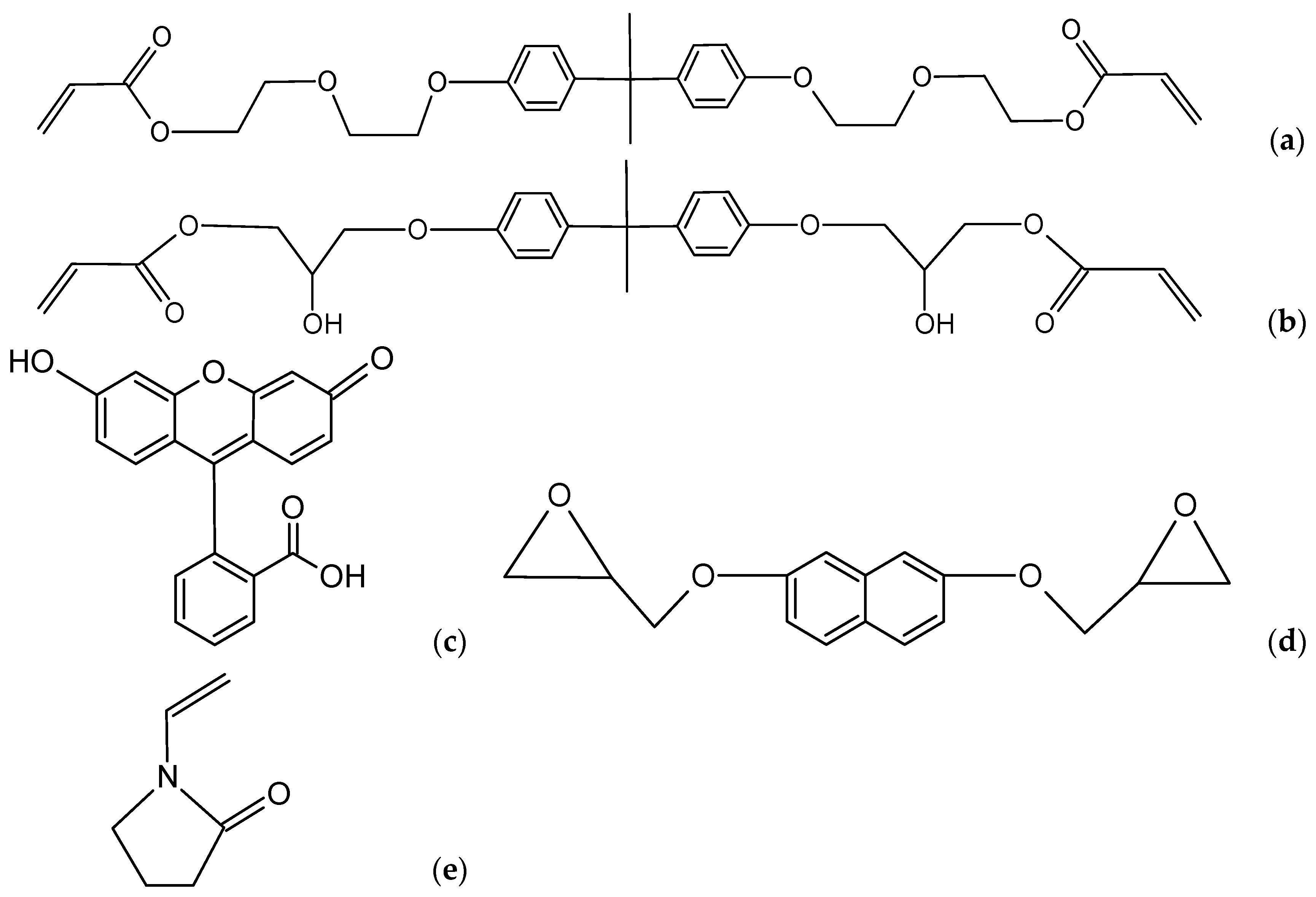
Figure 1. Substances used in the study: (a) EB150, (b) EB600, (c) Fluorescein, (d) 2.7-NAF.EP, (e) NVP.
Applsci 14 06170 g001
Figure 2. Ion current for: (a) sample series No.1–3; (b) sample series No.4–6; (c) sample series No.7–9; (d) sample series No.10–12; (e) sample series No.13–15; (f) sample series No.16–18.
Applsci 14 06170 g002aApplsci 14 06170 g002b
Figure 3. Compositions exposed to UV radiation: (a) samples No.10-60, No.11-60, No.12-60; (b) samples No.13-60, No.14-60, No.15-60; (c) samples No.16-60, No.17-60, No.18-60; (d) samples No.1-60, No.2-60, No.3-60; (e) samples No.4-60, No.5-60, No.6-60; (f) samples No.7-60, No.8-60, No.9-60.
Applsci 14 06170 g003
Table 1. Experimental parameters of the syntheses for EB150.
SampleEB150/wt.%NVP/wt.%Dopant/wt.%Polymerization Time/s
2.7-NAF.EPFluorescein
Sample No.1-157525--15
Sample No.1-307525--30
Sample No.1-607525--60
Sample No.2-1575251-15
Sample No.2-3075251-30
Sample No.2-6075251-60
Sample No.3-157525-115
Sample No.3-307525-130
Sample No.3-607525-160
Sample No.4-156733--15
Sample No.4-306733--30
Sample No.4-606733--60
Sample No.5-1567331-15
Sample No.5-3067331-30
Sample No.5-6067331-60
Sample No.6-156733-115
Sample No.6-306733-130
Sample No.6-606733-160
Sample No.7-155050--15
Sample No.7-305050--30
Sample No.7-605050--60
Sample No.8-1550501-15
Sample No.8-3050501-30
Sample No.8-6050501-60
Sample No.9-155050-115
Sample No.9-305050-130
Sample No.9-605050-160
Table 2. Experimental parameters of the syntheses for EB600.
SampleEB150/wt.%NVP/wt.%Dopant/wt.%Polymerization Time/s
2.7-NAF.EPFluorescein
Sample No.10-157525--15
Sample No.10-307525--30
Sample No.10-607525--60
Sample No.11-1575251-15
Sample No.11-3075251-30
Sample No.11-6075251-60
Sample No.12-157525-115
Sample No.12-307525-130
Sample No.12-607525-160
Sample No.13-156733--15
Sample No.13-306733--30
Sample No.13-606733--60
Sample No.14-1567331-15
Sample No.14-3067331-30
Sample No.14-6067331-60
Sample No.15-156733-115
Sample No.15-306733-130
Sample No.15-606733-160
Sample No.16-155050--15
Sample No.16-305050--30
Sample No.16-605050--60
Sample No.17-1550501-15
Sample No.17-3050501-30
Sample No.17-6050501-60
Sample No.18-155050-115
Sample No.18-305050-130
Sample No.18-605050-160
Table 3. Double-bond conversion.
SampleConversion/%SampleConversion/%
Sample No.1-1564.1Sample No.10-1573.9
Sample No.1-3086.5Sample No.10-3082.7
Sample No.1-6086.2Sample No.10-6092.1
Sample No.2-1576.1Sample No.11-1575.4
Sample No.2-3085.9Sample No.11-3080.4
Sample No.2-6088.5Sample No.11-6084.0
Sample No.3-1538.6Sample No.12-1546.9
Sample No.3-3075.2Sample No.12-3066.3
Sample No.3-6075.9Sample No.12-6070.3
Sample No.4-1575.4Sample No.13-1590.6
Sample No.4-3098.4Sample No.13-3092.4
Sample No.4-6096.9Sample No.13-6095.0
Sample No.5-1554.2Sample No.14-1591.7
Sample No.5-3072.4Sample No.14-3094.2
Sample No.5-6086.4Sample No.14-6090.4
Sample No.6-1579.4Sample No.15-1592.6
Sample No.6-3093.5Sample No.15-3095.3
Sample No.6-6095.5Sample No.15-6093.5
Sample No.7-1531.6Sample No.16-1581.5
Sample No.7-3084.3Sample No.16-3094.3
Sample No.7-6094.1Sample No.16-6096.2
Sample No.8-1523.7Sample No.17-1597.5
Sample No.8-3092.9Sample No.17-30100.0
Sample No.8-60100.0Sample No.17-60100.0
Sample No.9-1523.3Sample No.18-1583.3
Sample No.9-3060.7Sample No.18-30100.0
Sample No.9-6096.5Sample No.18-60100.0
Table 4. Temperature characteristics based on TG/DTG and DSC curves.
SampleTemperature at Mass Loss/°CDTGmax/°CDSCmax/°CResidual Mass/wt.%
1/wt.%3/wt.%5/wt.%IIIIIIIVIIIIIIIV
Sample No.1-15125.5188.0230.0166.0230.8349.0438.5113.4231.4- *400.9/435.3/
446.5
13.2
Sample No.1-30120.6164.3199.7147.7235.4- *439.6123.6234.2- *400.6/434.0/
442.8
10.7
Sample No.1-60112.2156.7198.4148.7237.1- *434.3124.1232.5- *406.9/431.6/
447.2
12.3
Sample No.2-15111.6177.6221.8137.6242.3- *437.4127.0240.0- *399.3/437.9/
448.0
9.6
Sample No.2-30109.2150.6180.9133.2236.6- *437.4121.2239.5334.1400.7/432.1/
441.2
9.7
Sample No.2-60127.1172.2207.3138.9238.6- *438124.8241.0343.1396.9/429.1/
436.5/444.8
10.1
Sample No.3-15147.3192.9222.2- *233.6- *439.1- *232.1- *394.4/434.2/
445.4
12.1
Sample No.3-30105.0146.6178.9132.5235.5- *437.6118.9234.0- *393.9/431.7/
443.4
10.9
Sample No.3-60116.9155.9187.5136.8229.9- *436.9120.3233.3- *431.1/441.713.0
Sample No.4-15102.3139.6168.7134.3188.4242.6431.5129.3/
166.7
240.6322.6/
351.1
429.38.8
Sample No.4-3096.5122.5170.9143.6187.6252.4432.4131.2/
170.9
250.1325.1/
352.5
428.310.2
Sample No.4-6092.8117.1170.8137.5-237.3431.5127.1255.2322.1/
352.8
428.410.2
Sample No.5-1592.6134.2156.4- *185.4- *436.7132.7273.6333.5431.7/444.59.7
Sample No.5-3098.7137.5159.6- *172.0- *436.1130.1279.0345.8414.7/429.7/
434.8/444.6
10.0
Sample No.5-60101.2140.0164.1- *193.3- *436.4127.9278.5- *427.9/436.48.2
Sample No.6-1595.7134.2166.3140.2- *235.8432.5130.3/
160.7
243.2323.0/
352.5
430.29.9
Sample No.6-3085.4135.6163.0138.1- *230.0432.8122.0/
174.2
239.2322.2/
350.8
430.210.0
Sample No.6-60100.4145.8175.9141.8- *231.2433.2128.9/
162.3
235.3321.5/
351.0
431.09.1
Sample No.7-1580.9130.2160.8162.7- *276.7433.3131.0/
163.2
- *284.6/
320.3/
350.6
431.110.2
Sample No.7-3080.2130.1160.4165.4- *277.0432.7131.2/
155.5
- *282.0/
323.9/
352.5
428.510.5
Sample No.7-6082.2129.0159.0153.9192.5239.4433.0147.0- *325.3/
353.6
427.311.5
Sample No.8-1589.7118.1133.1- *199.8- *435.5129.0199.3332.8429.2/440.310.2
Sample No.8-3096.1128.2143.6- *190.2- *434.4132.2/
167.8
193.4287.6428.6/437.68.8
Sample No.8-6098.9130.7.149.2- *186.6- *434.5.128.9173.0/
187.7
169.1430.9/439.212.3
Sample No.9-1585.9118.7137.1157.1- *222.2434.0- *159.3/
223.9
- *430.6/473.47.3
Sample No.9-3090.2131.8152.5147.1- *235.9435.2- *143.2/232.0- *434.67.5
Sample No.9-60101.3136.7158.7175.3- *239.6431.8127.8168.8/235.7- *429.5/436.88.8
Sample No.10-15103.0137.2162.8171.8- *227.2422.8127.4225.3296.0/
381.0
407.0/427.114.7
Sample No.10-3092.7136.1166.3183.9- *231.0423.2110.4225.3381.0421.8/428.916.9
Sample No.10-60113.8152.7180.5190.0- *230.7421.8129.8224.5298.0/
377.5
404.6/425.7/
432.3
16.1
Sample No.11-15112.7152.1180.0189.1- *- *421.8130.2222.4308.3/
377.8
399.9/407.2/
423.9
13.3
Sample No.11-30109.3147.0174.6173.4- *- *422.0129.2224.2323.4/
379.3
402.5/417.9/
427.2
9.6
Sample No.11-60102.9149.1183.6188.5- *236.0420.9128.2223.9374.9397.2/422.3/
430.6
11.5
Sample No.12-15106.0156.5186.4183.0- *223.5421.3119.3/
158.3
220.7369.5404.5/429.315.4
Sample No.12-30106.3145.5171.0174.4- *223.9421.4121.7218.3370.1401.5/415.9/
428.4
15.3
Sample No.12-60112.9146.9173.2162.9- *231.0421.5125.5222.6372.0402.0/418.7/
426.5
14.8
Sample No.13-15109.8152.4182.4189.7- *245.7420.3148.1197.9/
225.7
379.8424.413.0
Sample No.13-30100.9149.4181.4194.2- *241.6419.7125.7195.3/
223.8
366.6418.6/426.09.8
Sample No.13-60102.0148.4179.6191.4- *238.2420.6126.1195.3/
226.5
279.8379.8/402.6/
417.2/423.5
13.9
Sample No.14-15104.8142.0168.1- *206.8260.6421.8- *209.6/
247.1/
280.4
372.6396.4/425.614.6
Sample No.14-30112.4158.6190.7- *202.2238.3419.8- *208.2/
233.3/
266.3
378.2410.7/421.813.5
Sample No.14-6099.7141.6170.2- *200.1- *421.1123.4208.8/
246.7/
278.3
381.5406.5/418.6/
425.7
13.4
Sample No.15-1592.7133.4159.1183.7- *227.3421.9125.3182.7/
224.7
373.1398.5/426.811.4
Sample No.15-30109.1149.4179.1196.2- *239.8420.6129.1192.3/223.8375.5395.5/415.1/
424.3
11.8
Sample No.15-60102.1142.3171.9194.4- *234.7421.7129.6187.9/
224.2
376.6409.7/424.016.5
Sample No.16-15108.9145.7172.2171.7- *- *421.8189.5222.5/
278.7
368.4425.115.0
Sample No.16-3091.9132.4160.7- *- *230.8421.5122.8193.3/
229.5
- *425.510.2
Sample No.16-6092.4137.3171.3122.7195.4246.3422.6192.8226.5/
243.6
- *422.6/432.412.2
Sample No.17-1594.0135.1160.9- *- *200.5419.9- *206.9379.3409.3/424.513.6
Sample No.17-30106.8145.9173.1166.2- *207.5420.8210.8- *377.4400.9/422.413.0
Sample No.17-6090.9138.7172.9109.7- *216.3421.7200.3/
211.3
265.3- *406.7/421.8/
435.3
12.6
Sample No.18-1592.9129.9155.2188.6- *235.2421.1125.3188.7/
226.2
- *420.7/427.412.2
Sample No.18-30114.9159.4189.8130.3- *238.0420.6125.3193.7/
227.1
- *413.8/423.213.2
Sample No.18-6098.9146.0180.2148.6- *237.3421.7125.3191.7/234.1- *407.0/422.213.8
* “-”—no effect was observed.
Disclaimer/Publisher’s Note: The statements, opinions and data contained in all publications are solely those of the individual author(s) and contributor(s) and not of MDPI and/or the editor(s). MDPI and/or the editor(s) disclaim responsibility for any injury to people or property resulting from any ideas, methods, instructions or products referred to in the content.