Article

Evaluation of Radiation Doses Received by Physicians during Permanent 198Au Grain Implant Brachytherapy for Oral Cancer

1
Course of Radiological Technology, Health Sciences, Tohoku University Graduate School of Medicine, 2-1 Seiryo, Aoba-ku, Sendai 980-8575, Japan
2
Department of Radiation Disaster Medicine, International Research Institute of Disaster Science, Tohoku University, 468-1 Aramaki Aza-Aoba, Aoba-ku, Sendai 980-0845, Japan
3
Department of Radiation Oncology, Tohoku University Graduate School of Medicine, 1-1 Seiryo-chou, Aoba-ku, Sendai 980-8574, Japan
4
Department of Clinical Radiology, Tohoku University Hospital, 1-1 Seiryo-chou, Aoba-ku, Sendai 980-8574, Japan
*
Author to whom correspondence should be addressed.
Appl. Sci. 2024, 14(14), 6010; https://doi.org/10.3390/app14146010
Submission received: 25 May 2024 / Revised: 6 July 2024 / Accepted: 7 July 2024 / Published: 10 July 2024
(This article belongs to the Special Issue Advances in Diagnostic and Therapeutic Radiology — 2nd Edition)

Abstract

:

Featured Application

Importance of the evaluation of the physician’s eye exposure to radiation during 198Au grain brachytherapy, given that the eye dose limit has been reduced drastically from 150 to 20 mSv/year.

Abstract

Brachytherapy is a practical, effective procedure for the local treatment of cancer; it delivers a high radiation dose to a limited tissue volume while sparing the surrounding normal tissues. Although the clinical benefit of brachytherapy is clear, there have been very few studies on the radiation dose received by physicians during the procedure. Furthermore, no study has investigated the eye radiation dose received by physicians performing 198Au grain (seed) brachytherapy, using an eye dosimeter. Recently, the International Commission on Radiological Protection (ICRP) recommended significantly reducing the occupational lens dose limit, from 150 to 20 mSv/yr (100 mSv/5 years). Therefore, it has become essential to evaluate the eye radiation doses of medical workers. We evaluated the eye radiation dose of a brachytherapy physician performing 198Au permanent grain implantation for tongue cancer; this is the first study on this topic. The maximum eye dose was ~0.1 mSv/procedure, suggesting that it is unlikely to exceed the ICRP limit (20 mSv/yr) for the lens, unless many procedures are performed with inappropriate radiation protection. To reduce the dose of radiation received by 198Au grain brachytherapy physicians, it is necessary to use additional lead shielding equipment when preparing the treatment needles, i.e., when loading the grains. This study provides useful information on radiation exposure of physicians conducting 198Au permanent grain brachytherapy.

1. Introduction

Brachytherapy is a practical, effective procedure for the local treatment of cancer; it delivers a high radiation dose to a limited tissue volume while sparing the surrounding normal tissues [1,2,3,4,5,6,7,8,9,10]. Thus, it is frequently chosen as a method of cancer treatment to avoid the large tissue defects caused by surgery and to conserve good function [11,12]. Fine control during the procedure and postoperative quality of life are good with brachytherapy compared to surgery and chemo–radiation therapy [13,14,15]. Surgery causes tissue defects, and full-dose external radiation therapy is typically associated with mandibular osteoradionecrosis, xerostomia, and an impaired sense of taste after treatment [16,17,18]. These complications often lower patients’ post-treatment quality of life [19].
The incidence rate of tongue cancer with advanced age is increasing in Japan. Brachytherapy is typically chosen to treat oral cancer, particularly tongue cancer, because it preserves the shape and functions of the tongue [20,21,22]. Until recently, 192Ir hairpins and 137Cs needles were widely used for the treatment of tongue cancer via brachytherapy [23]. However, these methods have negative impacts on patients of advanced age and/or with underlying conditions such as dementia, diabetes, and apoplexy [24,25,26]. Hence, 198Au grains have gradually become more common as an alternative source of radiation [27]. As shown in Figure 1, these grains are small and can easily be inserted, permanently, under local anesthesia, without causing severe pain or discomfort during either the insertion or the subsequent treatment and therefore can be prescribed for patients who cannot tolerate the previously used procedures [28]. Furthermore, patients can eat immediately after implantation, unlike those who receive treatment using 192Ir or 137Cs. In addition, 198Au has a short half-life of 2.7 days, which shortens the hospital stay [29,30]. Therefore, 198Au permanent grain brachytherapy has become a very useful treatment for tongue cancer [31,32].
Although the clinical benefit of brachytherapy is clear, there have been very few studies on the radiation dose received by the physician during the procedure [33]. Furthermore, no study has investigated the eye dose received by physicians performing 198Au grain brachytherapy. In 2011, the International Commission on Radiological Protection (ICRP) recommended significantly reducing the occupational eye lens dose limit, from 150 to 20 mSv/yr (100 mSv/5 years) [34,35]. Therefore, it has become essential to evaluate the eye radiation doses received by medical workers. Furthermore, protecting against occupational doses, particularly in interventional radiology (IVR), has been deemed critical according to numerous studies [35,36,37,38,39,40]. However, no previous studies have investigated the occupational doses for physicians performing 198Au grain brachytherapy.
In this study, we evaluated the eye radiation dose received by a brachytherapy physician conducting 198Au grain implantation for oral cancer. Our studies were conducted during brachytherapy application on real persons, and no phantoms were used. Furthermore, we compared the eye doses measured via an eye dosimeter and a neck badge. In addition, we investigated the effective and the hand doses received by the physician.

2. Materials and Methods

In this study, 198Au permanent grain implant brachytherapy was performed by a highly experienced physician. The protocol for this retrospective study was approved by our Institutional Review Board.

2.1. 198Au Brachytherapy

Preparation: 198Au grains (Japan Radioisotope Association, Tokyo, Japan) were used for brachytherapy. The half-life of the nuclide is 2.7 days, and the energy of its main γ-emission is 412 keV. As shown in Figure 1, the treatment device consisted of a sealed platinum rod (2.5 mm long) with an external diameter of 0.8 mm surrounding the grains of the nuclide. The nominal activity of each grain was 185 MBq, although, of course, at the time of treatment, the activity had decayed from its nominal value at production.
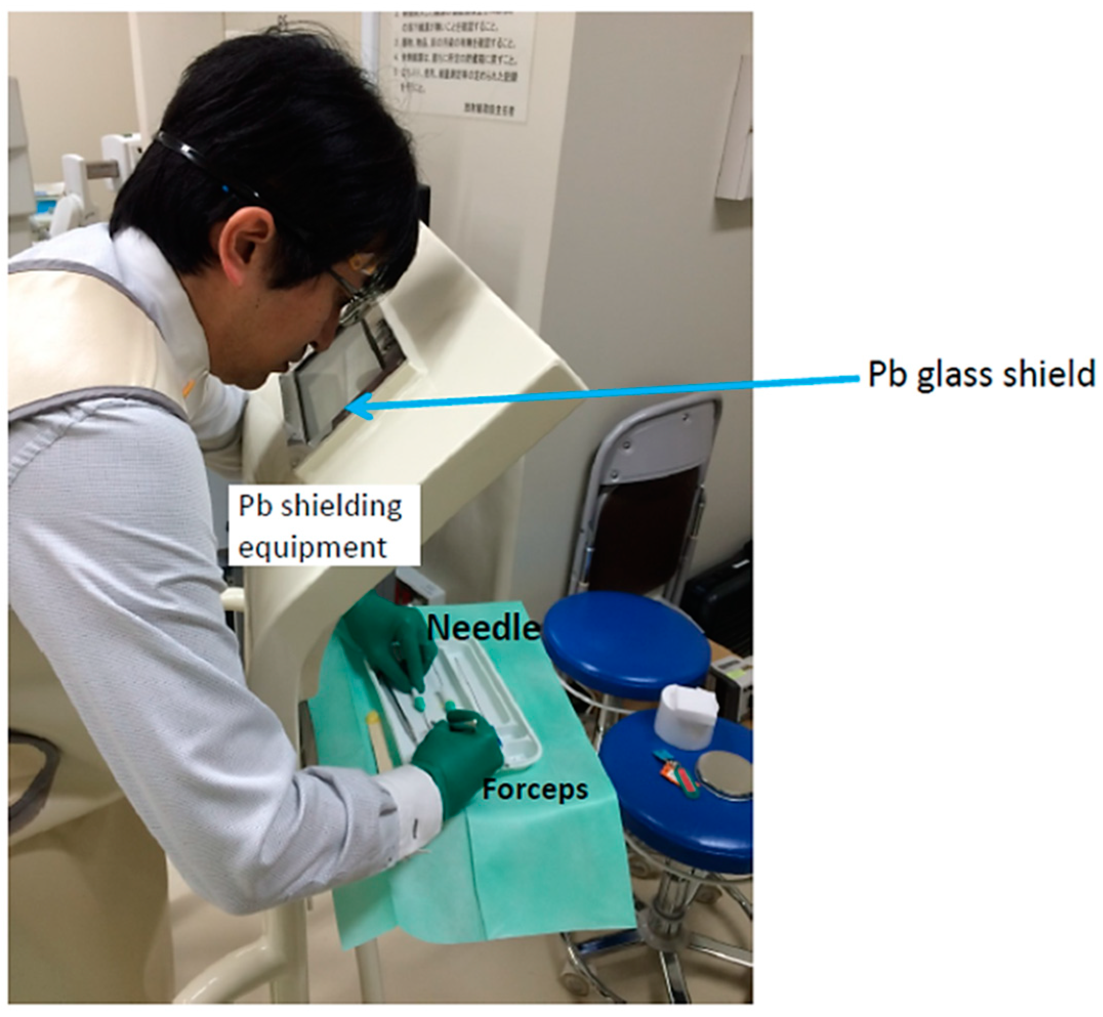
Physicians manually load a 198Au grain (seed) into a needle using a forceps, as shown in Figure 2, with one needle loaded only with one grain. During loading, the physician used an additional lead shielding device for radiation protection, as shown in Figure 3. In this study, typically 10–12 grains (i.e., 10–12 needles) were used, depending on the tumor size.
Treatment: During the procedure, the treating physician confirmed the presence of the tumor and controlled the direction in which the needle was inserted, by hand, under direct vision (Figure 4). The 198Au grains (seeds) were implanted into the tongue surface at a depth of ~5 mm. The physician wore a protective apron (usually 0.35 mm lead equivalent) but not leaded eyeglasses. However, significant shielding effects of the apron (0.35 mm lead equivalent) for 412 keV γ-ray may not have been achieved. The patients were hospitalized for approximately 3–5 days based on the number of 198Au grains (seeds) inserted.

2.2. Subjects

This retrospective study was conducted at Tohoku University Hospital. Eleven subjects, most of whom had tongue cancer, underwent consecutive 198Au grain brachytherapy insertions. Each procedure was conducted under local anesthesia.

2.3. Dosimetry

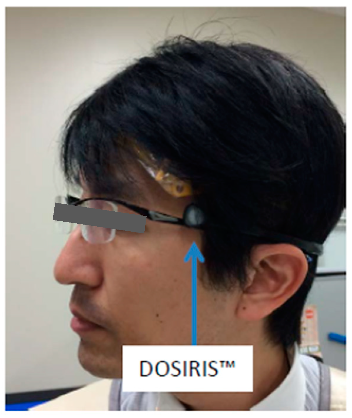
The brachytherapy physician used eye lens dosimeters (DOSIRIS™, IRSN, Croisy-sur-Seine, France) that specifically measured the eye radiation dose, specifically, the 3 mm dose equivalent [Hp(3)]. This dosimeter consists of a thermoluminescent sensor (7LiF:Mg,Ti), a plastic capsule, and a headset. The dosimeters were supplied by Chiyoda Technol Corporation (Tokyo, Japan). After each brachytherapy procedure, they were returned to the company to be read and for dose calibration. The natural background radiation for each procedure was subtracted. Dose calibration was performed before the dosimetry. The physician wore one of these dosimeters just lateral to the left eye, as shown in Figure 5, and for several procedures, also wore a dosimeter lateral to the right eye. The eye dosimeters were positioned to avoid interference with the physician’s work.
During each procedure, we also used a commercial silver-activated phosphate glass personal dosimeter (70 μm dose equivalent, Hp(0.07); Glass Badge, Chiyoda Technol Corporation), which was worn outside the lead apron to the left of the neck of the physician, as shown in Figure 6. These dosimeters were also returned to the company to be read. In addition, the physician also wore radiophotoluminescent glass dosimeters (RPLDs, measurement/readout system, Dose Ace FDG-1000, Chiyoda Technol Corporation) on the right and left hands, which were also used to evaluate Hp(0.07), with RPLD measurements converted to this value.
We assessed the relationship between the eye and the neck dosimeter readings, Hp(3) and Hp(0.07), respectively, to determine whether it was feasible to estimate the physician’s eye dose using a neck dosimeter. Furthermore, we investigated the correlations between the eye dosimeter readings and the total radiation activity.
The total radiation activity for each brachytherapy treatment, A, was calculated as follows:
A [MBq] = B × number of implanted 198Au grains
where B is the 198Au activity at implantation, corrected for decay since production.

2.4. Statistical Analyses

The Mann–Whitney U test was used to compare the two groups across different dose measurements, with statistical significance determined at p-value < 0.05. We analyzed the correlations between the eye dosimeter measurements and the total radiation activity via linear regression.

3. Results

3.1. Physician’s Eye/Neck Doses

Table 1 summarizes the results. The duration (mean ± standard deviation) of the procedure was 24 ± 8 min. Hp(3) was 0.06 mSv/procedure (maximum, ~0.1 mSv/procedure). The values of Hp(3) for the left and right eyes were almost equal (0.06 ± 0.02 and 0.05 ± 0.03 mSv/procedure, respectively, with no statistical significance). In several procedures, the neck badge dosimeter detected nothing: the dose was below its detection limit. In other words, the neck badge dosimeter underestimated the eye dose compared to the eye dosimeter. The Hp(0.07) values for the left and right neck areas were almost identical (0.03 ± 0.02 and 0.03 ± 0.02 mSv/procedure, respectively, with no statistical significance).

3.2. Physician’s Hand Doses

Doses were approximately 1.7 times higher for the right hand than for the left hand (518.2 ± 276.4 and 310.1 ± 156.7 μSv/procedure, respectively, p < 0.05).

3.3. Correlations among the Dose Measurements

A strong correlation was detected between the left and the right dose measurements for the eye, neck, and hand (Figure 7, Figure 8 and Figure 9). There was a correlational trend between the neck and the eye dose measurements (Figure 10 and Figure 11) and between the neck and the right-hand dose measurements (Figure 12 and Figure 13). There was also a correlation between A (total radiation activity) and each dose measurement (eye, neck, and hand, Figure 14, Figure 15 and Figure 16). There was also a correlational trend between the right-hand dose measurements and the left-eye dose measurements (Figure 17).

4. Discussion

It is important to evaluate the dose of radiation that medical workers receive, particularly to their eyes [41,42,43,44,45,46,47,48,49,50]. Many studies have been conducted on medical occupational radiation dose evaluation and protection, including by our research group [51,52,53,54,55,56,57,58,59,60]. However, no study to date has specifically investigated occupational radiation doses to the eyes in physicians performing 198Au permanent grain brachytherapy. In this study, we measured this to be ~0.1 mSv/procedure at most, implying that such physicians’ exposure will probably not exceed the regulatory eye dose limit. However, we believe that this dose cannot be ignored because physician exposure may exceed the limit if numerous procedures are performed with inadequate radioprotection. Interventional radiology (IVR) procedures are often associated with increased fluoroscopy time; consequently, they may expose IVR physicians to radiation-induced injuries. The eye dose per procedure for a 198Au brachytherapy physician was somewhat higher than that for an IVR physician [61,62]. However, IVR physicians may face a higher risk of radiation exposure because they typically perform a greater number of procedures.
Note that in this study, the physician loaded the needles while using an additional lead shielding device. Without it, the eye dose might have exceeded the limit; therefore, we strongly recommend that 198Au brachytherapy physicians use an additional lead shielding device while loading needles. Thus, educating the brachytherapy staff about radiation protection is important. The procedures in this study were performed by a highly experienced brachytherapy physician and show how the right training of workers is important, as suggested in many ICRP publications [34,63,64,65,66].
Regarding the IVR staff, particularly physicians, the left eye typically receives a higher radiation dose than the right one [62,67]. However, we found that the eye radiation dose was similar in both eyes for the physician administering 198Au grain brachytherapy, with a strong correlation between left- and right-eye doses. Therefore, wearing an eye dosimeter on either side is sufficient for 198Au grain brachytherapy physicians. Estimation of the occupational radiation dose to the eye often involves using a neck badge [68,69]. In IVR, a relation between the neck badge dose and the eye dosimeter dose has been reported, and these doses are somewhat correlated [68,70]. In this study, although there was a trend toward a correlation between the neck and the eye doses (Figure 10 and Figure 11), the association was not statistically significant. Therefore, the neck dose during brachytherapy treatment may be not a good indicator of the physician’s eye dose. We also found that the eye dose tended to be underestimated using a neck dosimeter; in many instances, the neck badge failed to detect any radiation during 198Au grain brachytherapy. The neck dosimeters measured very low levels, probably at the limit of reliable measures. This finding may be due to the fact that the neck badge was further from the source than the eye dosimeter, although we did not measure the distance between the eye and the neck dosimeters. Thus, estimating the eye dose in relation to the neck badge dose may be challenging for 198Au brachytherapy physicians. Therefore, we recommend that 198Au brachytherapy physicians wear an eye dosimeter (such as DOSIRIS) on either side of their head for an accurate assessment of the eye radiation dose.
We found a correlation between physician’s eye dose and total radiation activity (r = 0.65). Therefore, the total radiation activity during brachytherapy treatment may be an indicator for estimating a physician’s eye dose.
The hand dose was ~0.5 mSv/procedure in this study, which is roughly equivalent to other results for doses received by brachytherapy physicians, such as when using 125I seeds for prostate cancer [71,72]. According to our assessment, 198Au brachytherapy physicians are unlikely to exceed the regulatory hand dose limit (500 mSv/y), as it is unlikely that a physician will perform more than 1000 procedures annually. We found a higher dose in the right hand than in the left hand; as the physician was right-handed, this was probably because the right hand was nearer the radiation source for longer than the left hand.
The physician manually loaded the radiation sources in this study. To reduce the physician dose, manufacturers need to develop a more convenient, automatic system for loading the sources into the treatment needles.
During the implantation of the grains into the tongue, a brachytherapy physician cannot use additional lead shielding equipment because it would impede the work (Figure 4). To reduce the eye dose, we recommend that the brachytherapy physician wear leaded eyeglasses during implantation, if available.
Even if the clinical advantages of 198Au permanent grain brachytherapy for treating tongue cancer are evident, the assessment of the physician’s eye radiation dose during 198Au grain brachytherapy is important, particularly in consideration of the significant reduction in the occupational eye dose limit from 150 to 20 mSv/year. We evaluated and estimated eye doses for 198Au grain brachytherapy physicians using an eye dosimeter and a personal neck badge, and measured the hand dose received by the physician. The results demonstrated that the eye dose for physicians during 198Au brachytherapy warrants attention in the absence of complete radiation protection. An additional Pb shielding device is needed for radiation protection during 198Au brachytherapy. To date, no studies have investigated physicians’ eye doses in 198Au brachytherapy. Despite the limited scope of this study, the results contribute valuable insights into radiation safety for 198Au brachytherapy physicians. As this was a single-institutional study, further research, such as a multi-institutional studies involving a larger number of cases, will be necessary.

5. Conclusions

We evaluated the eye radiation dose of a brachytherapy physician performing 198Au permanent grain implantation for tongue cancer; this is the first study on this topic. The maximum eye dose was ~0.1 mSv/procedure, suggesting that it is unlikely to exceed the ICRP limit (20 mSv/yr) for the lens, unless many procedures are performed with inappropriate radiation protection. To reduce the dose of radiation received by brachytherapy physicians, it is necessary to use additional lead shielding equipment when preparing the treatment needles, i.e., when loading the grains.
The measured doses to each eye were almost the same, but the eye dose was underestimated when evaluated using a neck dosimeter. We recommend using an eye dosimeter to correctly evaluate the Hp(3) eye dose. The hand dose recorded in this study (~0.5 mSv/procedure) was similar to that of a 125I brachytherapy physician. This study provides useful information on radiation exposure to physicians conducting 198Au grain brachytherapy.

Author Contributions

Conceptualization, K.J., Y.I. and K.C.; methodology, K.J., Y.I. and K.C.; software, M.F., Y.I. and M.S.; validation, M.F., K.J., Y.I., M.S., M.Z. and K.C.; formal analysis, M.F., K.O., Y.I. and K.C.; investigation, M.F., K.O., H.I., T.K., Y.M. and K.J.; resources, K.J. and K.C.; data curation, M.F., K.O., H.I., T.K., Y.M. and Y.I.; writing—original draft preparation, Y.I. and M.F.; writing—review and editing, K.J., M.S., M.Z. and K.C.; visualization, M.F., Y.I. and M.S.; supervision, K.J., M.Z. and K.C.; project administration, K.J. and K.C.; funding acquisition, K.C. All authors have read and agreed to the published version of the manuscript.

Funding

This work was supported in part by a Grant-in-Aid for Scientific Research from the Japan Society for the Promotion of Science (23H03537) and by an Industrial Disease Clinical Research Grant (240401-02), Japan.

Institutional Review Board Statement

This retrospective was approved by the ethics committee of Tohoku University Hospital, Japan (approval number; 2023-1-984).

Informed Consent Statement

The protocol for this retrospective study was approved by the institutional review board. This study included de-identified patient data. Informed consent from the study participants was not needed.

Data Availability Statement

All data that support the findings of this study are included within the article.

Acknowledgments

We thank Yuto Oomori and Shuusei Maki, Tohoku University, Japan, for their invaluable assistance.

Conflicts of Interest

The authors declare no conflicts of interest.

References

  1. Chen, D.; Parsa, R.; Chauhan, K.; Lukovic, J.; Han, K.; Taggar, A.; Raman, S. Review of brachytherapy clinical trials: A cross-sectional analysis of ClinicalTrials.gov. Radiat. Oncol. 2024, 19, 22. [Google Scholar] [CrossRef]
  2. Zhang, S.; Zeng, N.; Yang, J.; He, J.; Zhu, F.; Liao, W.; Xiong, M.; Li, Y. Advancements of radiotherapy for recurrent head and neck cancer in modern era. Radiat. Oncol. 2023, 18, 166. [Google Scholar] [CrossRef]
  3. Neugebauer, J.; Blum, P.; Keiler, A.; Süß, M.; Neubauer, M.; Moser, L.; Dammerer, D. Brachytherapy in the Treatment of Soft-Tissue Sarcomas of the Extremities-A Current Concept and Systematic Review of the Literature. Cancers 2023, 15, 1133. [Google Scholar] [CrossRef]
  4. Poder, J.; Rivard, M.J.; Howie, A.; Carlsson Tedgren, Å.; Haworth, A. Risk and Quality in Brachytherapy From a Technical Perspective. Clin. Oncol. (R. Coll. Radiol.) 2023, 35, 541–547. [Google Scholar] [CrossRef]
  5. International Commission on Radiological Protection (ICRP). Prevention of High-dose-rate Brachytherapy Accidents. ICRP Publication 97. Ann. ICRP 2005, 35, 5–75. [Google Scholar]
  6. International Commission on Radiological Protection (ICRP). Radiation Safety Aspects of Brachytherapy for Prostate Cancer using Permanently—Implanted Sources. ICRP Publication 98. Ann. ICRP 2005, 35, 3–50. [Google Scholar]
  7. Fujita, M.; Hirokawa, Y.; Kashiwado, K.; Akagi, Y.; Kashimoto, K.; Kiriu, H.; Ohtani, K.; Wada, T. An analysis of mandibular bone complications in radiotherapy for T1 and T2 carcinoma of the oral tongue. Int. J. Radiat. Oncol. Biol. Phys. 1996, 34, 333–339. [Google Scholar] [CrossRef]
  8. Chargari, C.; Deutsch, E.; Blanchard, P.; Gouy, S.; Martelli, H.; Guérin, F.; Dumas, I.; Bossi, A.; Morice, P.; Viswanathan, A.N.; et al. Brachytherapy: An overview for clinicians. CA Cancer J. Clin. 2019, 69, 386–401. [Google Scholar] [CrossRef]
  9. Skowronek, J. Current status of brachytherapy in cancer treatment—Short overview. J. Contemp. Brachyther. 2017, 9, 581–589. [Google Scholar] [CrossRef]
  10. Kovács, G.; Martinez-Monge, R.; Budrukkar, A.; Guinot, J.L.; Johansson, B.; Strnad, V.; Skowronek, J.; Rovirosa, A.; Siebert, F.A. GEC-ESTRO Head & Neck Working Group. GEC-ESTRO ACROP recommendations for head & neck brachytherapy in squamous cell carcinomas: 1st update—Improvement by cross sectional imaging based treatment planning and stepping source technology. Radiother. Oncol. 2017, 122, 248–254. [Google Scholar] [CrossRef]
  11. Yang, J.; Xiong, X.; Liao, X.; Zheng, W.; Xu, H.; Yang, L.; Wei, Q. Nonsurgical salvage options for locally recurrent prostate cancer after primary definitive radiotherapy: A systematic review and meta-analysis. Int. J. Surg. 2024, 15, 81–88. [Google Scholar] [CrossRef]
  12. Gordon, K.; Smyk, D.; Gulidov, I.; Golubev, K.; Fatkhudinov, T. An Overview of Head and Neck Tumor Reirradiation: What Has Been Achieved So Far? Cancers 2023, 15, 4409. [Google Scholar] [CrossRef]
  13. Slevin, F.; Zattoni, F.; Checcucci, E.; Cumberbatch, M.G.K.; Nacchia, A.; Cornford, P.; Briers, E.; De Meerleer, G.; De Santis, M.; Eberli, D.; et al. A Systematic Review of the Efficacy and Toxicity of Brachytherapy Boost Combined with External Beam Radiotherapy for Nonmetastatic Prostate Cancer. Eur. Urol. Oncol. 2023. [Google Scholar] [CrossRef]
  14. Schaulin, M.S.; Delouya, G.; Zwahlen, D.; Taussky, D. Tracing the Evolution of Prostate Brachytherapy in the 20th Century. Oncology 2024, 102, 283–290. [Google Scholar] [CrossRef]
  15. Liang, Z.; Yuliang, C.; Zhu, M.; Zhou, Y.; Wu, X.; Li, H.; Fan, B.; Zhou, Z.; Yan, W. The direct prognosis comparison of. Eur. J. Med. Res. 2023, 28, 181. [Google Scholar] [CrossRef]
  16. Fionda, B.; Bussu, F.; Placidi, E.; Rosa, E.; Lancellotta, V.; Parrilla, C.; Zinicola, T.; De Angeli, M.; Greco, F.; Rigante, M.; et al. Interventional Radiotherapy (Brachytherapy) for Nasal Vestibule: Novel Strategies to Prevent Side Effects. J. Clin. Med. 2023, 12, 6154. [Google Scholar] [CrossRef]
  17. Yuan, M.; Wang, L.; Xiao, Y.; Guo, X.; Hu, Y. Iodine-125 seed brachytherapy combined with pembrolizumab for advanced non-small-cell lung cancer after failure of first-line chemotherapy: A report of two cases and literature review. J. Contemp. Brachyther. 2023, 15, 81–88. [Google Scholar] [CrossRef]
  18. Yamazaki, H.; Yoshida, K.; Yoshioka, Y.; Shimizutani, K.; Furukawa, S.; Koizumi, M.; Ogawa, K. High dose rate brachytherapy for oral cancer. J. Radiat. Res. 2013, 54, 1–17. [Google Scholar] [CrossRef]
  19. Harada, H.; Ishikawa, Y.; Tanaka, S.; Kishida, K.; Umezawa, R.; Yamamoto, T.; Takahashi, N.; Takeda, K.; Suzuki, Y.; Jingu, K. Brachytherapy for primary nasal vestibule cancer using Au-198 grains. Int. Cancer Conf. J. 2022, 11, 184–187. [Google Scholar] [CrossRef]
  20. Tuček, L.; Vošmik, M.; Petera, J. Is There Still a Place for Brachytherapy in the Modern Treatment of Early-Stage Oral Cancer? Cancers 2022, 14, 222. [Google Scholar] [CrossRef]
  21. Arboleda, L.P.A.; de Carvalho, G.B.; Santos-Silva, A.R.; Fernandes, G.A.; Vartanian, J.G.; Conway, D.I.; Virani, S.; Brennan, P.; Kowalski, L.P.; Curado, M.P. Squamous Cell Carcinoma of the Oral Cavity, Oropharynx, and Larynx: A Scoping Review of Treatment Guidelines Worldwide. Cancers 2023, 15, 4405. [Google Scholar] [CrossRef] [PubMed]
  22. Umeda, M.; Komatsubara, H.; Ojima, Y.; Minamikawa, T.; Shibuya, Y.; Yokoo, S.; Ishii, J.; Komori, T. A comparison of brachytherapy and surgery for the treatment of stage I-II squamous cell carcinoma of the tongue. Int. J. Oral Maxillofac. Surg. 2005, 34, 739–744. [Google Scholar] [CrossRef] [PubMed]
  23. Yoshimura, R.; Shibuya, H.; Miura, M.; Watanabe, H.; Ayukawa, F.; Hayashi, K.; Toda, K. Quality of life of oral cancer patients after low-dose-rate interstitial brachytherapy. Int. J. Radiat. Oncol. Biol. Phys. 2009, 73, 772–778. [Google Scholar] [CrossRef]
  24. Yamazaki, H.; Inoue, T.; Yoshida, K.; Yoshioka, Y.; Furukawa, S.; Kakimoto, N.; Shimizutani, K. Brachytherapy for early oral tongue cancer: Low dose rate to high dose rate. J. Radiat. Res. 2003, 44, 37–40. [Google Scholar] [CrossRef] [PubMed]
  25. Kakimoto, N.; Inoue, T.; Murakami, S.; Furukawa, S.; Yoshida, K.; Yoshioka, Y.; Yamazaki, H.; Tanaka, E.; Shimizutani, K. Results of low- and high-dose-rate interstitial brachytherapy for T3 mobile tongue cancer. Radiother. Oncol. 2003, 68, 123–128. [Google Scholar] [CrossRef]
  26. Khalilur, R.; Hayashi, K.; Shibuya, H. Brachytherapy for tongue cancer in the very elderly is an alternative to external beam radiation. Br. J. Radiol. 2011, 84, 747–749. [Google Scholar] [CrossRef]
  27. Okazawa, K.; Yuasa-Nakagawa, K.; Yoshimura, R.; Shibuya, H. Permanent interstitial re-irradiation with Au-198 seeds in patients with post-radiation locally recurrent uterine carcinoma. J. Radiat. Res. 2013, 54, 299–306. [Google Scholar] [CrossRef]
  28. Konishi, M.; Fujita, M.; Takeuchi, Y.; Kubo, K.; Imano, N.; Nishibuchi, I.; Murakami, Y.; Shimabukuro, K.; Wongratwanich, P.; Verdonschot, R.G.; et al. Treatment outcomes of real-time intraoral sonography-guided implantation technique of 198Au grain brachytherapy for T1 and T2 tongue cancer. J. Radiat. Res. 2021, 62, 871–876. [Google Scholar] [CrossRef]
  29. Konishi, M.; Takeuchi, Y.; Imano, N.; Kubo, K.; Nishibuchi, I.; Murakami, Y.; Shimabukuro, K.; Wongratwanich, P.; Kakimoto, N.; Nagata, Y. Brachytherapy with 198Au grains for cancer of the floor of the mouth: Relationships between radiation dose and complications. Oral. Radiol. 2022, 38, 105–113. [Google Scholar] [CrossRef]
  30. Horiuchi, J.; Takeda, M.; Shibuya, H.; Matsumoto, S.; Hoshina, M.; Suzuki, S. Usefulness of 198Au grain implants in the treatment of oral and oropharyngeal cancer. Radiother. Oncol. 1991, 21, 29–38. [Google Scholar] [CrossRef]
  31. Konishi, M.; Shimabukuro, K.; Hirokawa, J.; Sadatoki, T.; Katsuta, T.; Imano, N.; Nishibuchi, I.; Murakami, Y.; Kakimoto, N. Radiation doses of medical radiation workers performing low-dose-rate brachytherapy with 198Au grains and 192Ir pins for patients with oral cancers. Oral Radiol. 2023, 40, 234–241. [Google Scholar] [CrossRef] [PubMed]
  32. Ryu, Y.; Shibuya, H.; Hayashi, K. 198Au grain implantation for early tongue cancer in patients of advanced age or poor performance status. J. Radiat. Res. 2013, 54, 1125–1130. [Google Scholar] [CrossRef] [PubMed]
  33. Penfold, S.N.; Marcu, L.; Lawson, J.M.; Asp, J. Evaluation of physician eye lens doses during permanent seed implant brachytherapy for prostate cancer. J. Radiol. Prot. 2012, 32, 339–347. [Google Scholar] [CrossRef] [PubMed]
  34. International Commission on Radiological Protection (ICRP). Statement on Tissue Reactions. Available online: https://www.icrp.org/page.asp?id=123 (accessed on 10 March 2024).
  35. International Atomic Energy Agency. Radiation Protection and Safety of Radiation Sources: International Basic Safety Standards; General Safety Requirements Part 3; IAEA: Vienna, Austria, 2014. [Google Scholar]
  36. Chida, K. What are useful methods to reduce occupational radiation exposure among radiological medical workers, especially for interventional radiology personnel? Radiol. Phys. Technol. 2022, 15, 101–115. [Google Scholar] [CrossRef] [PubMed]
  37. Matsuzaki, S.; Moritake, T.; Morota, K.; Nagamoto, K.; Nakagami, K.; Kuriyama, T.; Kunugita, N. Development and assessment of an educational application for the proper use of ceiling-suspended radiation shielding screens in angiography rooms using augmented reality technology. Eur. J. Radiol. 2021, 143, 109925. [Google Scholar] [CrossRef] [PubMed]
  38. Sato, T.; Eguchi, Y.; Yamazaki, C.; Hino, T.; Saida, T.; Chida, K. Development of a New Radiation Shield for the Face and Neck of IVR Physicians. Bioengineering 2022, 9, 354. [Google Scholar] [CrossRef] [PubMed]
  39. Chida, K.; Takahashi, T.; Ito, D.; Shimura, H.; Takeda, K.; Zuguchi, M. Clarifying and visualizing sources of staff-received scattered radiation in interventional procedures. AJR Am. J. Roentgenol. 2011, 197, W900–W903. [Google Scholar] [CrossRef] [PubMed]
  40. Zuguchi, M.; Chida, K.; Taura, M.; Inaba, Y.; Ebata, A.; Yamada, S. Usefulness of non-lead aprons in radiation protection for physicians performing interventional procedures. Radiat. Prot. Dosim. 2008, 131, 531–534. [Google Scholar] [CrossRef] [PubMed]
  41. Fujibuchi, T. Radiation protection education using virtual reality for the visualisation of scattered distributions during radiological examinations. J. Radiol. Prot. 2021, 41, S317–S328. [Google Scholar] [CrossRef]
  42. Inaba, Y.; Hitachi, S.; Watanuki, M.; Chida, K. Radiation Eye Dose for Physicians in CT Fluoroscopy-Guided Biopsy. Tomography 2022, 8, 438–446. [Google Scholar] [CrossRef]
  43. Osanai, M.; Sato, H.; Sato, K.; Kudo, K.; Hosoda, M.; Hosokawa, S.; Kitajima, M.; Tsushima, M.; Fujita, A.; Hosokawa, Y.; et al. Occupational Radiation Dose, Especially for Eye Lens: Hp(3), in Medical Staff Members Involved in Computed Tomography Examinations. Appl. Sci. 2021, 11, 4448. [Google Scholar] [CrossRef]
  44. Imai, S.; Akahane, M.; Ogata, Y.; Tanki, N.; Sato, H.; Tameike, K. Occupational eye lens dose in endoscopic retrograde cholangiopancreatography using a dedicated eye lens dosimeter. J. Radiol. Prot. 2021, 41, 579–589. [Google Scholar] [CrossRef] [PubMed]
  45. Hamada, N. Ionizing radiation sensitivity of the ocular lens and its dose rate dependence. Int. J. Radiat. Biol. 2017, 93, 1024–1034. [Google Scholar] [CrossRef] [PubMed]
  46. Hamada, N.; Azizova, T.V.; Little, M.P. An update on effects of ionizing radiation exposure on the eye. Br. J. Radiol. 2020, 93, 20190829. [Google Scholar] [CrossRef] [PubMed]
  47. Haskal, Z.J. Interventional radiology carries occupational risk for cataracts. RSNA News 2004, 14, 5–6. [Google Scholar]
  48. Vañó, E.; Gonzalez, L.; Fernández, J.M.; Haskal, Z.J. Eye lens exposure to radiation in interventional suites: Caution is warranted. Radiology 2008, 248, 945–953. [Google Scholar] [CrossRef] [PubMed]
  49. International Commission on Radiological Protection (ICRP). Avoidance of Radiation Injuries from Medical Interventional Procedures; ICRP Publication 85; Pergamon: Oxford, UK, 2000; Volume 30. [Google Scholar]
  50. IAEA. Implications for Occupational Radiation Protection of the New Dose Limit for the Lens of the Eye; IAEA: Vienna, Austria, 2013; Volume 1731, pp. 1–34. [Google Scholar]
  51. Kato, M.; Chida, K.; Munehisa, M.; Sato, T.; Inaba, Y.; Suzuki, M.; Zuguchi, M. Non-Lead Protective Aprons for the Protection of Interventional Radiology Physicians from Radiation Exposure in Clinical Settings: An Initial Study. Diagnostics 2021, 11, 1613. [Google Scholar] [CrossRef] [PubMed]
  52. Endo, M.; Haga, Y.; Sota, M.; Tanaka, A.; Otomo, K.; Murabayashi, Y.; Abe, M.; Kaga, Y.; Inaba, Y.; Suzuki, M.; et al. Evaluation of novel X-ray protective eyewear in reducing the eye dose to interventional radiology physicians. J. Radiat. Res. 2021, 62, 414–419. [Google Scholar] [CrossRef] [PubMed]
  53. Matsubara, K.; Takei, Y.; Mori, H.; Kobayashi, I.; Noto, K.; Igarashi, T.; Suzuki, S.; Akahane, K. A multicenter study of radiation doses to the eye lenses of medical staff performing non-vascular imaging and interventional radiology procedures in Japan. Phys. Medica 2020, 74, 83–91. [Google Scholar] [CrossRef] [PubMed]
  54. Inaba, Y.; Hitachi, S.; Watanuki, M.; Chida, K. Occupational Radiation Dose to Eye Lenses in CT-Guided Interventions Using MDCT-Fluoroscopy. Diagnostics 2021, 11, 646. [Google Scholar] [CrossRef]
  55. Shindo, R.; Ohno, S.; Yamamoto, K.; Konta, S.; Inaba, Y.; Suzuki, M.; Zuguchi, M.; Chida, K. Comparison of shielding effects of over-glasses-type and regular eyewear in terms of occupational eye dose reduction. J. Radiol. Prot. 2024, 44, 023501. [Google Scholar] [CrossRef] [PubMed]
  56. Yokoyama, S.; Suzuki, S.; Toyama, H.; Arakawa, S.; Inoue, S.; Kinomura, Y.; Kobayashi, I. Evaluation of eye lens dose of interventional cardiologists. Radiat. Prot. Dosim. 2017, 173, 218–222. [Google Scholar] [CrossRef] [PubMed]
  57. Koenig, A.; Maas, J.; Viniol, S.; Etzel, R.; Fiebich, M.; Thomas, R.; Mahnken, A. Scatter radiation reduction with a radiation-absorbing pad in interventional radiology examinations. Eur. J. Radiol. 2020, 132, 109245. [Google Scholar] [CrossRef] [PubMed]
  58. Haga, Y.; Chida, K.; Kimura, Y.; Yamanda, S.; Sota, M.; Abe, M.; Kaga, Y.; Meguro, T.; Zuguchi, M. Radiation eye dose to medical staff during respiratory endoscopy under X-ray fluoroscopy. J. Radiat. Res 2020, 61, 691–696. [Google Scholar] [CrossRef] [PubMed]
  59. Cornelis, F.H.; Razakamanantsoa, L.; Ammar, M.B.; Lehrer, R.; Haffaf, I.; El-Mouhadi, S.; Gardavaud, F.; Najdawi, M.; Barral, M. Ergonomics in interventional radiology: Awareness is mandatory. Medicina 2021, 57, 500. [Google Scholar] [CrossRef] [PubMed]
  60. Ikezawa, K.; Hayashi, S.; Takenaka, M.; Yakushijin, T.; Nagaike, K.; Takada, R.; Yamai, T.; Matsumoto, K.; Yamamoto, M.; Omoto, S.; et al. Occupational radiation exposure to the lens of the eyes and its protection during endoscopic retrograde cholangiopancreatography. Sci. Rep. 2023, 13, 7824. [Google Scholar] [CrossRef] [PubMed]
  61. Kato, M.; Chida, K.; Ishida, T.; Toyoshima, H.; Yoshida, Y.; Yoshioka, S.; Moroi, J.; Kinoshita, T. Occupational Radiation Exposure of the Eye in Neurovascular Interventional Physician. Radiat. Prot. Dosim. 2019, 185, 151–156. [Google Scholar] [CrossRef] [PubMed]
  62. Haga, Y.; Chida, K.; Kaga, Y.; Sota, M.; Meguro, T.; Zuguchi, M. Occupational eye dose in interventional cardiology procedures. Sci. Rep. 2017, 7, 569. [Google Scholar] [CrossRef] [PubMed]
  63. ICRP. Education and Training in Radiological Protection for Diagnostic and Interventional Procedures. ICRP Publication 113. Ann. ICRP 2009, 39, 7–68. [Google Scholar]
  64. International Commission on Radiological Protection (ICRP). Occupational radiological protection in brachytherapy. ICRP Publication 149. Ann. ICRP 2021, 50, 5–75. [Google Scholar] [CrossRef]
  65. International Commission on Radiological Protection (ICRP). Occupational radiological protection in interventional procedures. ICRP Publication 139. Ann. ICRP 2018, 47, 1–118. [Google Scholar] [CrossRef] [PubMed]
  66. International Commission on Radiological Protection (ICRP). Radiological Protection in Fluoroscopically Guided Procedures outside the Imaging Department. ICRP Publication 117. Ann. ICRP 2010, 40, 1–102. [Google Scholar] [CrossRef] [PubMed]
  67. Fujisawa, M.; Haga, Y.; Sota, M.; Abe, M.; Kaga, Y.; Inaba, Y.; Suzuki, M.; Meguro, T.; Hosoi, Y.; Chida, K. Evaluation of Lens Doses among Medical Staff Involved in Nuclear Medicine: Current Eye Radiation Exposure among Nuclear-Medicine Staff. Appl. Sci. 2023, 13, 9182. [Google Scholar] [CrossRef]
  68. Ishii, H.; Chida, K.; Satsurai, K.; Haga, Y.; Kaga, Y.; Abe, M.; Inaba, Y.; Zuguchi, M. Occupational eye dose correlation with neck dose and patient-related quantities in interventional cardiology procedures. Radiol. Phys. Technol. 2022, 15, 54–62. [Google Scholar] [CrossRef] [PubMed]
  69. Chida, K.; Kaga, Y.; Haga, Y.; Kataoka, N.; Kumasaka, E.; Meguro, T.; Zuguchi, M. Occupational dose in interventional radiology procedures. AJR Am. J. Roentgenol. 2013, 200, 138–141. [Google Scholar] [CrossRef]
  70. Martin, C.J. Personal dosimetry for interventional operators: When and how should monitoring be done? Br. J. Radiol. 2011, 84, 639–648. [Google Scholar] [CrossRef]
  71. Fujii, K.; Ko, S.; Nako, Y.; Tonari, A.; Nishizawa, K.; Akahane, K.; Takayama, M. Dose measurement for medical staff with glass dosemeters and thermoluminescence dosemeters during 125I brachytherapy for prostate cancer. Radiat. Prot. Dosim. 2011, 144, 459–463. [Google Scholar] [CrossRef]
  72. Aronowitz, J.N.; Connock, G.; Haq, R.; Morin, M.J. Radiation exposure from permanent prostate brachytherapy without fluoroscopy. Nowotwory 2009, 59, 184–187. [Google Scholar]
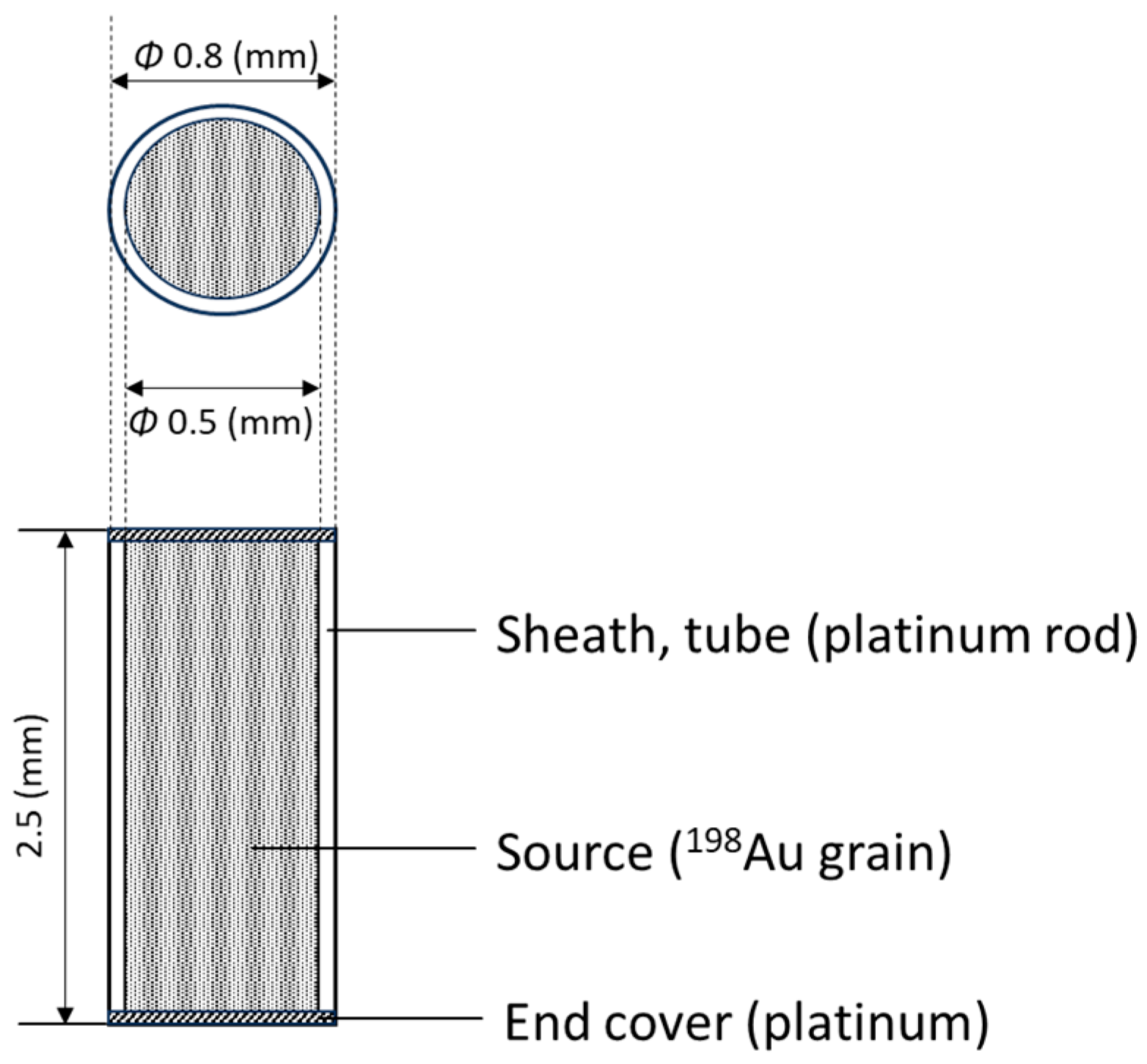
Figure 1. Schematic of 198Au equipment for brachytherapy. The nominal activity of each grain (seed) was 185 MBq (although during brachytherapy treatment, radiation activity obviously decays based on the half-life of 198Au).
Applsci 14 06010 g001
Figure 2. Puncture needles and forceps used in the described 198Au brachytherapy for source (198Au grain) loading. Physicians load the source into the needles using the forceps.
Applsci 14 06010 g002
Figure 3. Loading of the needles, i.e., inserting a 198Au permanent grain (seed) into a puncture needle, was performed by a brachytherapy physician by hand (non-automatic), using a forceps. The physician performed the loading of the needles behind additional Pb shielding equipment (mobile Pb shielding equipment) including a lead glass shield.
Applsci 14 06010 g003
Figure 4. 198Au grain brachytherapy. During the procedure, after the physician visualized/confirmed the tumor, 198Au grains (loaded into needles) were inserted in the tumor by the physician.
Applsci 14 06010 g004
Figure 5. DOSIRIS™ was worn lateral to the left eye of the physician. (The physician wore non-Pb eyeglasses).
Applsci 14 06010 g005
Figure 6. The physician also wore personal dosimeters (glass badge, neck dosimeter) outside the Pb apron to the left and right of the neck. Hand doses were also evaluated using RPLDs.
Applsci 14 06010 g006
Figure 7. Relationship between physician’s right- and left-eye doses (DOSIRIS™) during 198Au grain brachytherapy.
Applsci 14 06010 g007
Figure 8. Relationship between physician’s right and left neck doses (personal glass badges) during 198Au grain brachytherapy.
Applsci 14 06010 g008
Figure 9. Relationship between physician’s right- and left-hand doses (RPLD) during 198Au grain brachytherapy.
Applsci 14 06010 g009
Figure 10. Relationship between physician’s neck and eye doses during 198Au grain brachytherapy. (Left).
Applsci 14 06010 g010
Figure 11. Relationship between physician’s neck and eye doses during 198Au grain brachytherapy. (Right).
Applsci 14 06010 g011
Figure 12. Relationship between physician’s left neck and right-hand doses during 198Au grain brachytherapy.
Applsci 14 06010 g012
Figure 13. Relationship between physician’s right neck and right-hand doses during 198Au grain brachytherapy.
Applsci 14 06010 g013
Figure 14. Relationship between physician’s left-eye dose and total radiation activity at implantation during 198Au grain brachytherapy.
Applsci 14 06010 g014
Figure 15. Relationship between physician’s left neck dose and total radiation activity at implantation during 198Au grain brachytherapy.
Applsci 14 06010 g015
Figure 16. Relationship between physician’s right-hand dose and total radiation activity at implantation during 198Au grain brachytherapy.
Applsci 14 06010 g016
Figure 17. Relationship between physician’s right-hand dose and left-eye dose (DOSIRIS™) during 198Au grain brachytherapy.
Applsci 14 06010 g017
Table 1. Summary of the results for the subjects participating in this study.
PatientType of Cancer Number of Au Grains Total Radiation Activity (MBq)DOSIRIS (mSv)Personal Badge (mSv)RPLD (mSv)
Left EyeRight EyeLeft NeckRight NeckLeft HandRight Hand
1Tongue1120350.08---0.3800.576
2Tongue1221150.07---0.2410.599
3Tongue814260.040.030.010.020.3400.609
4Tongue1017350.050.050.020.030.3100.221
5Tongue814880.030.030.020.020.0740.150
6Tongue1719470.060.060.050.06-0.527
7Tongue1221500.070.060.030.040.5080.763
8Oral1526590.010000.4610.980
9Nose916300.090.100.060.100.1420.243
10Tongue913220.050.06000.1430.221
11Tongue1524530.060.040.040.040.5020.809
Average 11.51905.50.060.050.030.030.3100.518
Standard deviation 3.1428.90.020.030.020.030.1570.276
-, No measuring device was used; 0, below detection limit.
Disclaimer/Publisher’s Note: The statements, opinions and data contained in all publications are solely those of the individual author(s) and contributor(s) and not of MDPI and/or the editor(s). MDPI and/or the editor(s) disclaim responsibility for any injury to people or property resulting from any ideas, methods, instructions or products referred to in the content.