Article

Three-Dimensional Pulp Volume Analysis in Lip and Palate Cleft Population

by
Inês Francisco
1,2,3,4,5,*,
Raquel Travassos
1,2,4,5,
Filipa Marques
1,
Madalena Prata Ribeiro
1,
Mariana Rodrigues
1,
Patrícia Quaresma
1,
Francisco Caramelo
2,3,4,5,6,
Carlos Miguel Marto
2,3,4,5,7,8,
Anabela Baptista Paula
1,2,3,4,5,7,
Catarina Nunes
1,2,4,5 and
Francisco Vale
1,2,3,4,5
1
Institute of Orthodontics, Faculty of Medicine, University of Coimbra, 3000-075 Coimbra, Portugal
2
Coimbra Institute for Clinical and Biomedical Research (iCBR), Area of Environment Genetics and Oncobiology (CIMAGO), Faculty of Medicine, University of Coimbra, 3000-075 Coimbra, Portugal
3
Laboratory for Evidence-Based Sciences and Precision Dentistry, University of Coimbra, 3000-075 Coimbra, Portugal
4
Centre for Innovative Biomedicine and Biotechnology (CIBB), University of Coimbra, 3000-075 Coimbra, Portugal
5
Clinical Academic Center of Coimbra (CACC), Hospitais da Universidade de Coimbra, 3004-561 Coimbra, Portugal
6
Laboratory of Biostatistics and Medical Informatics (LBIM), Faculty of Medicine, University of Coimbra, 3004-531 Coimbra, Portugal
7
Institute of Integrated Clinical Practice, Faculty of Medicine, University of Coimbra, 3004-531 Coimbra, Portugal
8
Institute of Experimental Pathology, Faculty of Medicine, University of Coimbra, 3004-531 Coimbra, Portugal
*
Author to whom correspondence should be addressed.
Appl. Sci. 2023, 13(6), 3728; https://doi.org/10.3390/app13063728
Submission received: 14 February 2023 / Revised: 1 March 2023 / Accepted: 14 March 2023 / Published: 15 March 2023
(This article belongs to the Special Issue Advanced Dental Biomaterials: Technologies and Applications)

Abstract

:
Aim: Cleft lip and palate (CLP) patients have a greater predisposition to tooth malformation, which could affect pulp volume. The aim of this study is to evaluate the dental pulp volume of central incisors in 3D images between individuals with and without CLP. Materials and Methods: This retrospective case-control study is single-centered and was recruited between January 2016 and October 2022. Ninety-four patients who were followed in the Institute of Orthodontics were evaluated and divided into two groups: a control group of patients without CLP and a test group of patients with CLP. The 3D data were imported by 3D image semi-automatic segmenting software named ITK-SNAP to calculate tooth pulp volume. Results: The dental pulp volume for both groups, control and CLP, did not show statistically significant differences. In the cleft group, when comparing the pulp volume between the cleft side and the non-cleft side, the cleft side showed a smaller volume. Regarding age and sex, no statistically significant differences were observed. Conclusions: Although there are no differences in mean pulp volume between patients with and without CLP, there is a pulp volume reduction in the teeth on the cleft side when compared to the unaffected contralateral side.

1. Introduction

Once the eruption of permanent dentition is complete, dental age can be predicted by observing the different physiological age-related changes in dental tissues. One of these physiological changes is the deposition of secondary dentin, which is carried out by odontoblasts during a gradual and continuous process [1,2,3,4]. As a result, there is a progressive narrowing of the pulp chamber that can be correlated to age and subsequently used as an indicator of chronological age [1,5,6,7].
The measurement of secondary dentin deposition by dental radiographs is a simple, non-invasive and reproducible method that can be used, in both ante and post-mortem periods, to estimate the age of an individual [8]. In contrast, destructive methods involving tooth section for microscopic analysis are deemed unacceptable for forensic purposes as they result in the loss of evidence [9].
Kvaal et al. (1995) [7] developed a pioneering work using radiographic images and evaluating the deposition of secondary dentin to estimate age. However, limitations of this analysis include structure overlap and information suppression caused by the transformation of a three-dimensional (3D) structure into a two-dimensional (2D) image [10]. In recent years, technological advances in 3D imaging have allowed for the 3D evaluation of dental structures and the subsequent correlation of these with age using radiographic exams such as cone beam computed tomography (CBCT) [11].
There are several methods available to determine age, and they vary depending on the purpose, the type of sample and the accuracy required. Some of the most used methods are:
(1)
Radiocarbon dating [12]: radiocarbon dating is a technique used to determine the age of organic materials by measuring the amount of carbon-14 present;
(2)
Dental analysis [13]: dental analysis is a method of determining age by examining the development and wear of teeth. The teeth develop in a predictable pattern, and the amount of wear on the teeth can give an indication of the person’s age;
(3)
Bone analysis [14]: the bones of the body develop and fuse at different ages, and by examining the bones, the age of the individual can be estimated;
(4)
Radiographic analysis [15]: radiographic analysis is a technique used to examine the internal structure of the body. By examining X-rays or other imaging techniques, the age of the individual can be estimated based on the development of certain bones or the presence of certain features;
(5)
Hormone analysis [16]: Hormone levels change as a person ages, and by examining the levels of certain hormones, the age of the individual can be estimated.
Each method has its advantages and disadvantages, and the choice of method depends on the accuracy required and the type of sample available.
Age estimation plays a significant role in human identification in legal justice systems, law enforcement and criminal investigation cases such as homicides, suicides and massive humanitarian disasters [17,18,19]. The current global socio-political context demands a significant increase in access to age estimation methods in living individuals, in order to clarify criminal and civil liability as well as additional underlying social issues [20]. Some age estimation methods, as described before, include the macroscopic and radiographic analysis of the maturation and development phases of the bone and dental structures [17]. Teeth are known to be the most reliable age indicator as they are resistant to deterioration even under extreme conditions due to the presence of a superficial enamel layer. During the maturation phase, teeth are less affected by endocrine diseases, dietary deficiencies and environmental changes, ensuring complete mineralization and subsequent eruption. This same resilience is observed even during the postmortem period, when tissues are subjected to increased microbiological activity, mechanical forces and fluctuations in pH, humidity and temperature [1,17,19].
Some studies have considered pulpal volume as a parameter for age estimation, but none have focused on a population with cleft lip and palate (CLP) [9,11,21]. Cleft lip and palate is the most common craniofacial malformation, representing 1 in every 500 to 1000 births worldwide. It has been reported in the literature that teeth close to the cleft region are likely to present some degree of malformation (in size, shape or number) and are commonly associated with developmental delays or asymmetric formation timing, which could affect pulp volume [22]. According to Assis et al. [23], CLP patients often present a greater number of teeth with an enlarged pulp volume when compared to non-CLP individuals. Moreover, the dental anomalies present in these CLP patients may serve as clinical markers for sub-phenotypes. As these markers are more accurately identified, clinicians will be more alert to follow up with genetic testing [24,25,26].
The awareness of pulp volume in CLP patients is essential to execute some clinical procedures, as well as age estimation of this population. As it stands, the aim of this study is to determine the changes in the pulp volume of maxillary central incisors in patients with and without CLP. The null hypothesis refers to the non-existence of differences in the pulp volume of patients with and without CLP.

2. Materials and Methods

2.1. Study Design

The present retrospective case-control study was single-centered and conducted in accordance with the Declaration of Helsinki. The Ethics Committee of the Faculty of Medicine of the University of Coimbra approved the protocol (CE-086/2020) after obtaining informed consent from each participant or their legal representative.

2.2. Selection Criteria

The study sample was created from existing records of patients who are followed in the Institute of Orthodontics of the Faculty of Medicine of the University of Coimbra, Coimbra, Portugal. The selection period extended from 1 January 2016 to 31 October 2022.
The following inclusion criteria were selected: (a) healthy Caucasian individuals of both genders before orthodontic treatment; (b) unilateral cleft lip and palate Caucasian individuals; (c) fully formed and completely erupted permanent upper central incisors (11 and 21 teeth as the FDI World Dental Federation two-digit notation—ISO3950); (d) patients who had performed a high-quality CBCT involving these teeth; (e) patients without dental trauma history. All the individuals presented dental caries lesions, external or internal root resorptions, excessive tooth wear (Tooth Wear Index > 2 points according to Smith and Knight’s tooth wear index score) [27], restorations, prosthetic appliances, root canal treatments, periapical lesions, coronary or radicular fractures, dental pulp calcifications and pathological processes were excluded from the sample.
The sample size was calculated assuming that the average difference in the pulpal volume was 0.5 mm3 with a standard deviation of 0.5 mm3. This sample was assessed using an independent and bilateral sample Student-T test. An allocation ratio of 1:1 was presumed, which means that both groups have the same number of subjects. The G*Power 3.1.9.2 software was used to determine the sample size, evaluate the different statistical significance values (α = 0.01, α = 0.05 e α = 0.10) and test power (1 − β = 0.80, 1 − β = 0.85, 1 − β = 0.90).
The following table (Table 1) shows the sample size values for each group and for the different scenarios. Since the budget was sufficient, the most rigorous values were chosen (α = 0.01; 1 − β = 0.90).
The selected sample was divided into two groups: a control group that included healthy Caucasian individuals and an experimental group, constituted by unilateral cleft lip and palate patients. Data information of each added patient included pulp volume of upper right and left central incisors, sex, cleft type, date of birth and exam acquisition to calculate the actual age. All the CBCT images were taken for diagnosis or treatment purposes; thus, no additional or unnecessary radiation exposure was applied to these patients.

2.3. Image Acquisition and Segmentation

All scans were acquired using an i-CAT Vision scanner (Imaging Sciences International, Hatfield, PA, USA). All the images were obtained with the same parameters: 120 kVp, 5 mA and 16 × 10 cm field of view (FOV). The scanning process takes approximately four seconds.
The acquired images were subsequently exported as Digital Imaging and Communications in Medicine (DICOM) files and reconstructed with a voxel size of 0.3 mm3 and an axial layer thickness of 1 mm. Thereafter, the obtained data were imported by a 3D image semi-automatic segmenting software named ITK-SNAP, version 3.8.0 (open-source software: www.itksnap.org, accessed on 13 February 2023) to calculate the tooth pulp volume.
The segmentation begins with the selection of the region of interest (ROI) and defining the tooth limits to be examined using multiplanar reconstructions. For the main measurement of pulp volume, the endpoints were the most apical tooth root and at the cementoenamel junction. An additional measure was considered, due to the anatomical variability that patients with CLP present, from the cementoenamel junction to the middle of the clinical crown, since the dental pulp can extend to this region (Figure 1). Then, an adequate threshold interval is selected by an interactive method and a 3D reconstruction of the pulp cavity is obtained according to the method of Andrade et al. The volume of the segmented structure is obtained in cubic millimeters (mm3).

2.4. Inter- and Intra-Observer Agreement

Firstly, inter-observer accordance was tested by analyzing a sample of 7 teeth (upper central incisors) included in the study sample. After both observers (M.R., P.Q.) obtained inter-observer agreement, the measurements were taken separately, with a 1-month interval. To test the intra-observer agreement, a random sample of 7 teeth was re-examined, after 6 weeks. During these processes, the observers did not have access to the patient’s identification nor to the first measurements that were taken.

2.5. Statistical Analysis

The description of sex was assessed with absolute and relative frequencies. The comparison between the two groups, control and CLP, was performed using Fisher’s exact test. For the description of age and pulp volume, it was opted to use the mean, median, standard deviation and the percentiles 25 and 75. For the comparison between the control and study groups, and between the groups formed by gender, the mean of teeth 11 and 21 was calculated and compared between the two groups using the Mann–Whitney test, after having verified the violation of the normality assumption by the Shapiro–Wilk test. The distribution of the values of the pulpal volumes was also evaluated using extreme and quartile graphs.
The pulpal volume comparison between teeth 11 and 21 (paired groups) was performed using the student’s t-test for paired samples or the Wilcoxon test according to the verification of the normality assumption. The accordance of the pulpal volumes was performed by the intraclass correlation coefficient and by visual inspection of the scatter plot of the variables in question and its comparison with the direct proportionality y = x.
The relationship between mean pulpal volume and age was evaluated through simple linear regression.
Statistical analysis was performed in IBM® SPSS® v26 with a significance level of 0.05.
The accordance analysis was performed using an iota index calculation using the irr package of the R (R Core Team, 2014). Visually, the results were represented on a Bland–Altman graph, which was attained via IBM® SPSS® v26.
The chosen statistical significance level was 0.05.

3. Results

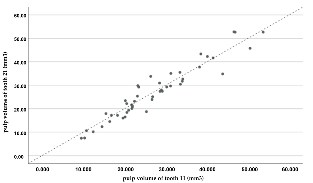
3.1. Interobserver Agreement

Taking into account the two observers/researchers, the accordance between the observations is quite elevated, with an iota index of 0.992.

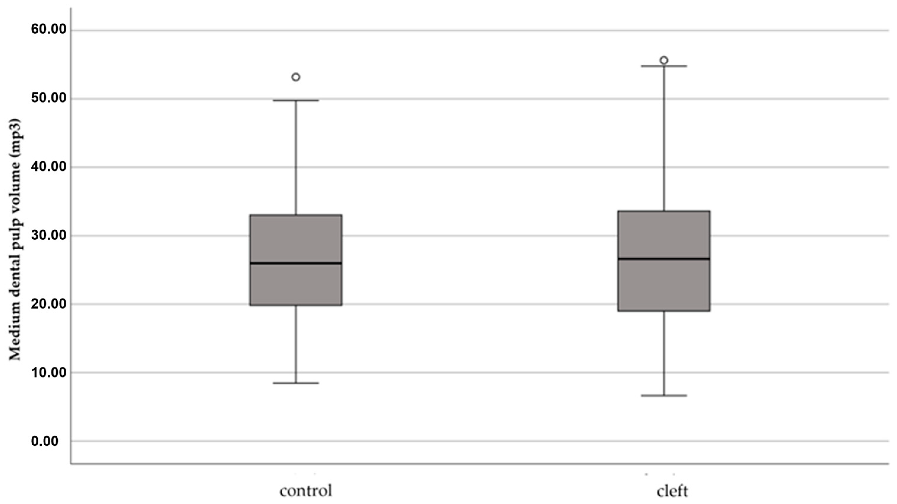
3.2. Pulp Volume Comparison between Patients with and without CLP

This study sample consisted of 94 patients (33 females and 61 males), of which 47 patients had CLP and 47 were healthy patients.
The Table 2 describes the sample in both groups, control and CLP, in regard to sex and age. The distribution of these variables is homogenous between both groups (p > 0.05).
The median dental pulp volume for both groups, control and CLP, did not show statistically significant results (Figure 2), with a p = 0.757. In the control group, the mean pulp volume was 27.0, with a median of 26, a value of 19.5 for the 25th percentile and a value of 33.2 for the 75th percentile. In the sample group, a mean value of 28.2 was obtained, with a median of 27 and values of 18.9 and 33.8 for the 25th and 75th percentile, respectively.
Regarding the dental pulp volume in the control group for both upper central incisors, the results were also not statistically significant, with a p = 0.664. Regarding the analysis of tooth 11, a mean value of 27.1 was obtained, with a median of 25, a 25th percentile value of 20.2 and a 75th percentile value of 33.6. Tooth 21, on the other hand, had a mean value of 25.4, a median of 11 and 75th and 25th percentile values of 33.6 and 26.9, respectively.
The dental pulp volume, however, showed statistically significant results for the study group of the cleft side and non-cleft side (p < 0.001). On the non-cleft side, teeth presented a mean volume of 30.4, with a median of 28 and values of 21.3 for the 25th percentile and 28.5 for the 75th percentile. On the cleft side, the pulpal volume assessment of the teeth presented a mean of 28.0, a median of 13 and 25th and 75th percentiles of 26.0 and 38.5, respectively.
It can be observed in Figure 3 that the points are in proximity with the line, which translates into an accordance between the pulp volumes of the teeth on both sides. This concordance is corroborated by the intraclass correlation coefficient that has a value of 0.977 (p < 0.001). In Figure 4, on the other hand, it is possible to observe that the points are more distant of the line, and that the teeth on the cleft side present a lower pulpal volume. The concordance between the volume values and the intraclass correlation coefficient is very strong, presenting a value of 0.917 (p < 0.001).

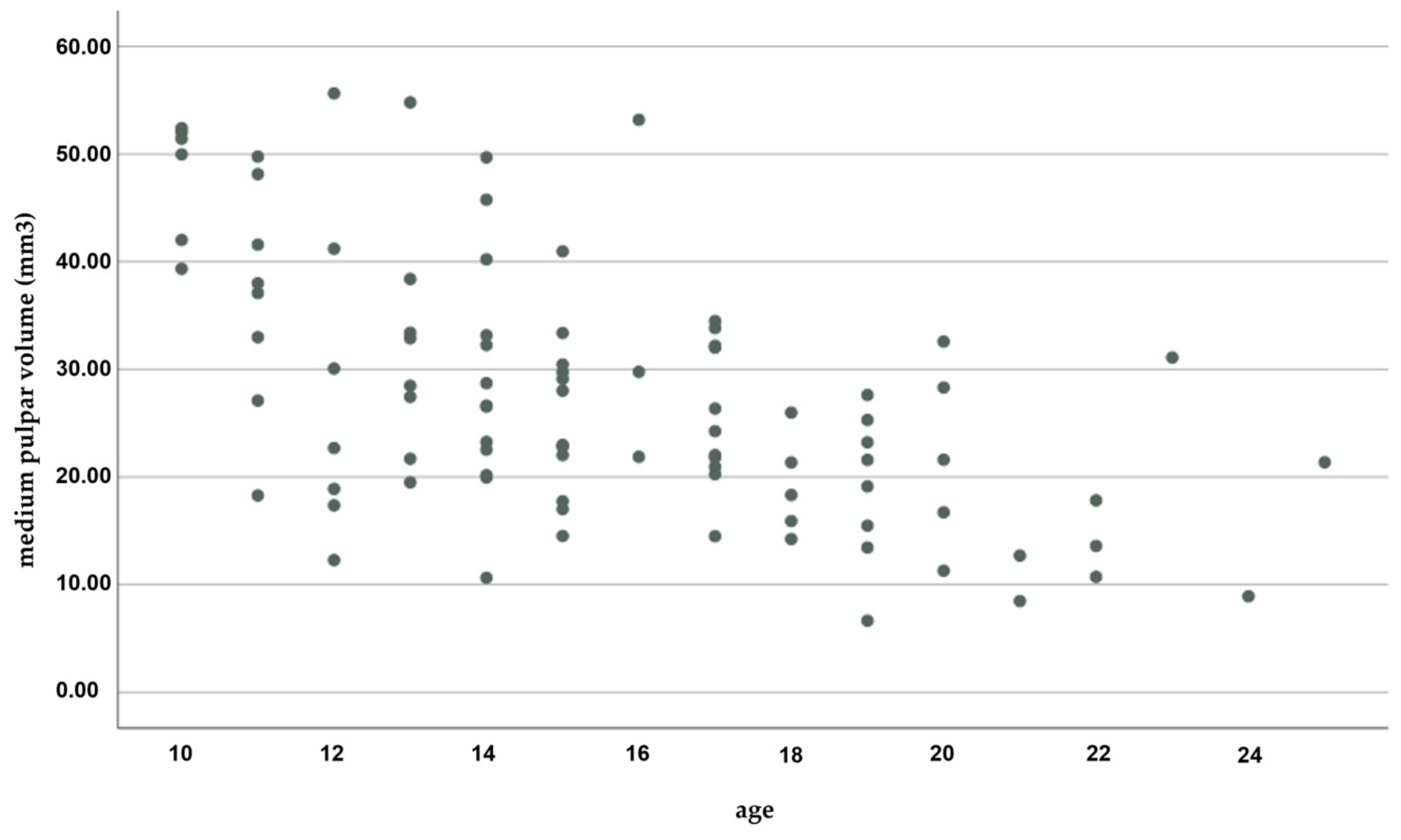
3.3. Comparison of Pulp Volume Regarding Sex and Age

The average dental pulp volume value in both sexes, regardless of group, is presented in the following Table 3. It can be concluded that the values were statistically significant, with a p < 0.001 and mean values of 30.8 for males and 21.4 for females.
The mean dental pulp volume for both sexes in the control group also presented a statistically significant value, with the pulp volume in males significantly higher than in females (p = 0.001). In males, there was a mean of 30.2, a median value of 30, with a 75th percentile value of 34.5 and a 25th percentile value of 21.8. In females, there was a mean of 20.8, with a median value of 19 and 25th and 75th percentile values of 15.1 and 22.4, respectively.
Similarly, the mean dental pulp volume for both genders in the CLP group also presented a statistically significant value, p = 0.007. The mean value for males was 31.4, with a median value of 29 (±12.5) and values of 22.5 for the 25th percentile and 40.2 for the 75th percentile. Females presented a mean value of 21.9, with a median value of 20 (±11.2) and values of 14.4 and 25.9 for the 25th and 75th percentiles, respectively.
A statistically significant linear regression can be observed (p < 0.001) between the medium pulpal volume and age, with age explaining approximately 32% of the pulpal volume variance. Age represents a regression coefficient of −1.919, which means that for each additional year a medium decrease of 1.919 mm3 in pulpal volume is observed. The dispersion graph shows the relationship between age and medium pulpal volume (Figure 5).

4. Discussion

The main goal of the present study was to compare the medium pulpal volume of central incisors in CLP patients and a control group using a 3D radiography technique (CBCT), to understand if CLP patients present significative pulp volume changes compared with healthy patients. The central incisors were chosen for this study since these teeth present with the least internal anatomical variations and due to the fact that it is easier to isolate the pulpal area of a tooth with a single canal [28].
Regarding the evaluation of gender and age within the range of 12 and 19 years, there were no significant differences between control and study groups. In the literature, it is described that the correlation between pulpal volume and age is inverse, meaning that as a person’s age increases, the pulpal volume tends to decrease [9,28]. The results obtained differ from the literature, since a sample with a reduced age range was chosen. This sample made it possible to compare pulp volume between the two studied groups, reducing the risk of bias associated with changes in pulp volume due to age. In this study, it was verified that the pulp volume does not change between groups considering the same chronological age.
Regarding the sexual dimorphism of pulp volume, in a general manner males present a larger volume than females. The sexual dimorphism of teeth, analyzed from a genetic point of view, is reported in the literature with the Y chromosome being associated with an increased tooth growth due to a greater development of amelogenesis and dentinogenesis through increased mitotic activity [9,23,29,30].
It should be noted that patients with CLP often present dental anomalies in terms of shape and size associated with the cleft. Embryologically, this is explained by barely overlapping timings of germen formation and cleft defect development [31,32,33]. In the study group, when comparing the cleft and no cleft sides, differences were observed in dental pulp volume between the two, with the first group recording significantly smaller sizes. These results are in line with studies described by Estalim et al. and Akcam et al., who stated that most dental anomalies found were associated with the cleft side [31,34].
Considering that many CLP patients are subjected to orthodontic treatment, changes in the pulp volume of patients subjected to this type of therapy should be studied in the foreseeable future. During orthodontic treatment, significant forces, albeit controlled, are applied to teeth in order to promote tooth movement. These forces may cause pulpal reactions, pulp hyperemia, releasing inflammatory mediators that in turn increase blood circulation. Under normal circumstances, increased blood circulation decreases inflammatory mediators, but the pulp is limited by dentin, which impedes the occurrence of this decrease. Subsequently, there is a stimulation of the reparative responses of odontoblasts, leading to the accumulation of tertiary dentin. Studies described in the literature confirm that orthodontic forces reduce the pulp volume, which can vary within a range of 3 to 55.9 mm3 [35,36,37]. In the same way that orthodontic forces promote a volumetric increase in the pulp because of inflammation, periodontal disease, which is highly prevalent in CLP patients, can also generate this effect cascading from gingival inflammation [38,39]. To date, only a few studies that compare dental pulp volume between CLP and non-CLP patients have been found, which hinders any possible comparisons between the obtained results and the current available literature.
The present study is posed with some limitations as it was not possible to perform subgroup analysis in the study sample, namely according to etiology, phenotype, cleft severity and size. Another limitation is the existing discrepancy between the real and the pulp volume calculated from the radiological image. However, the use of the same image acquisition protocol for the studied groups makes it possible to reduce the impact of this limitation. The chosen voxel can also influence the accurate identification of the pulp volume. There are other radiological exams with greater accuracy, such as computerized axial tomography. However, these methods present a higher radiation dose than CBCT. As patients with CLP are consecutively exposed to radiation due to the numerous tests required for cleft closure procedures, the choice of CBCT is the most cost-effective choice.
To tackle present limitations, future studies should select samples according to cleft phenotype, diversify the type of tooth evaluated, evaluate the pulp volume in other age groups as well as study the effects of orthodontic appliances.

5. Conclusions

Despite pulpal volume differences between the sample and control groups not being statistically significant, it was observed that the incisors located on the cleft side presented overall inferior pulpal volume values when compared to their counterparts.

Author Contributions

Conceptualization, F.V. and I.F.; methodology, M.R., P.Q. and F.C.; software, M.R. and P.Q.; validation, F.C., I.F. and C.M.M.; formal analysis, F.C. and C.M.M.; investigation, M.R., P.Q., R.T. and C.N.; data curation, I.F.; writing—original draft preparation, M.R. and F.M.; writing—review and editing, I.F., A.B.P. and M.P.R.; visualization, A.B.P., R.T. and C.N.; supervision, F.V.; project administration, F.V. and I.F; Funding acquisition, F.V. All authors have read and agreed to the published version of the manuscript.

Funding

This research received no external funding.

Institutional Review Board Statement

The Ethics Committee of the Faculty of Medicine of the University of Coimbra approved the protocol (CE-086/2020), after obtaining informed consent from each participant or their legal representative.

Informed Consent Statement

Informed consent was obtained from all subjects involved in the study.

Data Availability Statement

The data presented in this study are available on request from the corresponding author.

Conflicts of Interest

The authors declare no conflict of interest.

References

  1. Gustafson, G.; Malmö, D.O. Age Determinations on Teeth. J. Am. Dent. Assoc. 1950, 41, 45–54. [Google Scholar] [CrossRef] [PubMed]
  2. Arora, J.; Talwar, I.; Sahni, D.; Rattan, V. Secondary Dentine as a Sole Parameter for Age Estimation: Comparison and Reliability of Qualitative and Quantitative Methods among North Western Adult Indians. Egypt. J. Forensic Sci. 2016, 6, 170–178. [Google Scholar] [CrossRef] [Green Version]
  3. Maples, W.R. An Improved Technique Using Dental Histology for Estimation of Adult Age. J. Forensic Sci. 1978, 23, 764–770. [Google Scholar] [CrossRef] [PubMed]
  4. Solheim, T. A New Method for Dental Age Estimation in Adults. Forensic Sci. Int. 1993, 59, 137–147. [Google Scholar] [CrossRef] [PubMed]
  5. Agematsu, H.; Someda, H.; Hashimoto, M.; Matsunaga, S.; Abe, S.; Kim, H.-J.; Koyama, T.; Naito, H.; Ishida, R.; Ide, Y. Three-Dimensional Observation of Decrease in Pulp Cavity Volume Using Micro-CT: Age-Related Change. Bull. Tokyo Dent. Coll. 2010, 51, 1–6. [Google Scholar] [CrossRef] [PubMed] [Green Version]
  6. Gupta, P. Human Age Estimation from Tooth Cementum and Dentin. J. Clin. Diagn. Res. 2014, 8, ZC07. [Google Scholar] [CrossRef] [PubMed]
  7. Kvaal, S.I.; Kolltveit, K.M.; Thomsen, I.O.; Solheim, T. Age Estimation of Adults from Dental Radiographs. Forensic Sci. Int. 1995, 74, 175–185. [Google Scholar] [CrossRef]
  8. Agarwal, N.; Ahuja, P.; Sinha, A.; Singh, A. Age Estimation Using Maxillary Central Incisors: A Radiographic Study. J. Forensic Dent. Sci. 2012, 4, 97. [Google Scholar] [CrossRef] [Green Version]
  9. Andrade, V.M.; Fontenele, R.C.; de Souza, A.C.; de Almeida, C.A.; Vieira, A.C.; Groppo, F.C.; Freitas, D.Q.; Junior, E.D. Age and Sex Estimation Based on Pulp Cavity Volume Using Cone Beam Computed Tomography: Development and Validation of Formulas in a Brazilian Sample. Dentomaxillofac. Radiol. 2019, 48, 20190053. [Google Scholar] [CrossRef]
  10. Lee, S.-M.; Oh, S.; Kim, J.; Kim, Y.-M.; Choi, Y.-K.; Kwak, H.H.; Kim, Y.-I. Age Estimation Using the Maxillary Canine Pulp/Tooth Ratio in Korean Adults: A CBCT Buccolingual and Horizontal Section Image Analysis. J. Forensic Radiol. Imaging 2017, 9, 1–5. [Google Scholar] [CrossRef]
  11. Ge, Z.; Yang, P.; Li, G.; Zhang, J.; Ma, X. Age Estimation Based on Pulp Cavity/Chamber Volume of 13 Types of Tooth from Cone Beam Computed Tomography Images. Int. J. Leg. Med. 2016, 130, 1159–1167. [Google Scholar] [CrossRef]
  12. Ubelaker, D.H. Radiocarbon Analysis of Human Remains: A Review of Forensic Applications. J. Forensic Sci. 2014, 59, 1466–1472. [Google Scholar] [CrossRef] [PubMed]
  13. Hillson, S. Dental Pathology. In Biological Anthropology of the Human Skeleton; John Wiley & Sons, Inc.: Hoboken, NJ, USA, 2018; pp. 293–333. [Google Scholar]
  14. Buikstra, J.E.; Ubelaker, D.H. Standards for Data Collection from Human Skeletal Remains; Arkansas Archeological Survey Research Series; University of Arkansas: Fayetteville, AR, USA, 2012; Volume 44. [Google Scholar]
  15. Manini, A.; Savino, G.; Calliada, F. Radiographic Anatomy; Springer: Berlin/Heidelberg, Germany, 2020. [Google Scholar]
  16. Vermeulen, A.; Kaufman, J.M. Ageing of the Hypothalamo-Pituitary-Testicular Axis in Men. Horm. Res. 1995, 43, 25–28. [Google Scholar] [CrossRef] [PubMed]
  17. Corral, C.; García, F.; García, J.; León, P.; Herrera, A.; Martínez, C.; Moreno, F. Chronological versus Dental Age in Subjects from 5 to 19 Years: A Comparative Study with Forensic Implications. Colomb. Médica 2010, 41, 215–223. [Google Scholar] [CrossRef]
  18. Vodanović, M.; Dumančić, J.; Galić, I.; Savić Pavičin, I.; Petrovečki, M.; Cameriere, R.; Brkić, H. Age Estimation in Archaeological Skeletal Remains: Evaluation of Four Non-Destructive Age Calculation Methods. J. Forensic Odontostomatol. 2011, 29, 14–21. [Google Scholar] [PubMed]
  19. Pereira, C.P.; Caldas, R.; Pestana, D. Legal Medical Age Estimation in Portuguese Adult Cadavers: Evaluation of the Accuracy of Forensic Dental Invasive and Non-Invasive Methods. J. Forensic Sci. Criminol. 2013, 1, 1. [Google Scholar] [CrossRef] [Green Version]
  20. Afify, M.M. Age Estimation from Pulp/Tooth Area Ratio in Three Mandibular Teeth by Panoramic Radiographs: Study of an Egyptian Sample. J. Forensic Res. 2014, 5, 2. [Google Scholar] [CrossRef]
  21. Ge, Z.; Ma, R.; Li, G.; Zhang, J.; Ma, X. Age Estimation Based on Pulp Chamber Volume of First Molars from Cone-Beam Computed Tomography Images. Forensic Sci. Int. 2015, 253, 133.e1–133.e7. [Google Scholar] [CrossRef]
  22. Tannure, P.N.; Oliveira, C.A.G.R.; Maia, L.C.; Vieira, A.R.; Granjeiro, J.M.; de Castro Costa, M. Prevalence of Dental Anomalies in Nonsyndromic Individuals with Cleft Lip and Palate: A Systematic Review and Meta-Analysis. Cleft Palate-Craniofacial J. 2012, 49, 194–200. [Google Scholar] [CrossRef]
  23. de Assis, I.O.; de Lavôr, J.R.; Cavalcante, B.G.N.; Lacerda, R.H.W.; Vieira, A.R. Pulp Enlargement in Individuals Born with Cleft Lip and Palate Pulp, a Radiographic Study from the Cleft Lip and Palate Service of Paraiba, Brazil. Eur. Arch. Paediatr. Dent. 2021, 22, 1101–1106. [Google Scholar] [CrossRef]
  24. Küchler, E.C.; da Motta, L.G.; Vieira, A.R.; Granjeiro, J.M. Side of Dental Anomalies and Taurodontism as Potential Clinical Markers for Cleft Subphenotypes. Cleft Palate-Craniofacial J. 2011, 48, 103–108. [Google Scholar] [CrossRef] [PubMed]
  25. Jamal, G.A.A.; Hazza’a, A.M.; Rawashdeh, M.A. Prevalence of Dental Anomalies in a Population of Cleft Lip and Palate Patients. Cleft Palate-Craniofacial J. 2010, 47, 413–420. [Google Scholar] [CrossRef]
  26. Cassolato, S.F.; Ross, B.; Daskalogiannakis, J.; Noble, J.; Tompson, B. Treatment of Dental Anomalies in Children with Complete Unilateral Cleft Lip and Palate at Sickkids Hospital, Toronto. Cleft Palate-Craniofacial J. 2009, 46, 166–172. [Google Scholar] [CrossRef] [PubMed]
  27. Smith, B.G.; Knight, J.K. An Index for Measuring the Wear of Teeth. Br. Dent. J. 1984, 156, 435–438. [Google Scholar] [CrossRef] [PubMed]
  28. Biuki, N.; Razi, T.; Faramarzi, M. Relationship between Pulp-Tooth Volume Ratios and Chronological Age in Different Anterior Teeth on CBCT. J. Clin. Exp. Dent. 2017, 9, e688–e693. [Google Scholar] [CrossRef] [Green Version]
  29. Alvesalo, L.; Tammisalo, E.; Townsend, G. Upper Central Incisor and Canine Tooth Crown Size in 47,XXY Males. J. Dent. Res. 1991, 70, 1057–1060. [Google Scholar] [CrossRef] [PubMed]
  30. de Angelis, D.; Gibelli, D.; Gaudio, D.; Cipriani Noce, F.; Guercini, N.; Varvara, G.; Sguazza, E.; Sforza, C.; Cattaneo, C. Sexual Dimorphism of Canine Volume: A Pilot Study. Leg. Med. 2015, 17, 163–166. [Google Scholar] [CrossRef]
  31. Eslami, N.; Majidi, M.R.; Aliakbarian, M.; Hasanzadeh, N. Prevalence of Dental Anomalies in Patients with Cleft Lip and Palate. J. Craniofacial Surg. 2013, 24, 1695–1698. [Google Scholar] [CrossRef]
  32. Konstantonis, D.; Alexandropoulos, A.; Konstantoni, N.; Nassika, M. A Cross-Sectional Analysis of the Prevalence of Tooth Agenesis and Structural Dental Anomalies in Association with Cleft Type in Non-Syndromic Oral Cleft Patients. Prog. Orthod. 2017, 18, 20. [Google Scholar] [CrossRef] [Green Version]
  33. Tsai, T.-P.; Huang, C.-S.; Huang, C.-C.; See, L.-C. Distribution Patterns of Primary and Permanent Dentition in Children with Unilateral Complete Cleft Lip and Palate. Cleft Palate-Craniofacial J. 1998, 35, 154–160. [Google Scholar] [CrossRef]
  34. Akcam, M.O.; Evirgen, S.; Uslu, O.; Memikoglu, U.T. Dental Anomalies in Individuals with Cleft Lip and/or Palate. Eur. J. Orthod. 2010, 32, 207–213. [Google Scholar] [CrossRef] [PubMed]
  35. Guler, A.Y.; Isik, B.K.; Esen, A.; Menziletoglu, D. Assessment of Pulp Volume Changes after Surgically Assisted Rapid Palatal Expansion. J. Stomatol. Oral Maxillofac. Surg. 2021, 122, 263–266. [Google Scholar] [CrossRef] [PubMed]
  36. Popp, T.W.; Årtun, J.; Linge, L. Pulpal Response to Orthodontic Tooth Movement in Adolescents: A Radiographic Study. Am. J. Orthod. Dentofac. Orthop. 1992, 101, 228–233. [Google Scholar] [CrossRef] [PubMed]
  37. Venkatesh, S.; Ajmera, S.; Ganeshkar, S.V. Volumetric Pulp Changes after Orthodontic Treatment Determined by Cone-Beam Computed Tomography. J. Endod. 2014, 40, 1758–1763. [Google Scholar] [CrossRef]
  38. Terlemez, A.; Alan, R.; Gezgin, O. Evaluation of the Periodontal Disease Effect on Pulp Volume. J. Endod. 2018, 44, 111–114. [Google Scholar] [CrossRef] [Green Version]
  39. Gaggl, A.; Schultes, G.; Kärcher, H.; Mossböck, R. Periodontal Disease in Patients with Cleft Palate and Patients with Unilateral and Bilateral Clefts of Lip, Palate, and Alveolus. J. Periodontol. 1999, 70, 171–178. [Google Scholar] [CrossRef]
Figure 1. Pulp volume measurement using the ITK_SNAP software. X red: endpoints of the measurements; Red line: Cement-enamel junction.
Applsci 13 03728 g001
Figure 2. Diagram of extremes and quartiles shows the value distribution of the medium pulpal volume for both the control and CLP groups.
Applsci 13 03728 g002
Figure 3. Comparison of the values for the pulp volume of the upper central incisors.
Applsci 13 03728 g003
Figure 4. Comparison of the pulp volume values on the teeth on the cleft side and the non-cleft side.
Applsci 13 03728 g004
Figure 5. Relationship between age and medium pulpal volume.
Applsci 13 03728 g005
Table 1. Sample size values for each group. β: Power; α: Significance level.
α = 0.01α = 0.05α = 0.10
1 − β = 0.801076
1 − β = 0.851186
1 − β = 0.901397
β = 0.80
β = 0.85
β = 0.90
Table 2. Control and CLP group for sex and age.
Control (47)CLP (47)p
sex (M/F)31/16 (66.0%/34.0%)31/16 (66.0%/34.0%)1.000 £
Age16.1; 16 (3.6) 13.0/19.014.8; 14 (3.5) 12.0/17.00.065 §
£ Exact Fisher Test; § Mann–Whitney Test.
Table 3. The medium dental pulp volume value in both sexes.
Masculine (61)Feminine (33)p
Volume (mm3) 30.8; 29 (11.3) 22.5/37.121.4; 19 (10.6) 14.4/22.9<0.001 £
£ Mann–Whitney Test.
Disclaimer/Publisher’s Note: The statements, opinions and data contained in all publications are solely those of the individual author(s) and contributor(s) and not of MDPI and/or the editor(s). MDPI and/or the editor(s) disclaim responsibility for any injury to people or property resulting from any ideas, methods, instructions or products referred to in the content.

