Article

Anchorage Loss Evaluation during Maxillary Molars Distalization Performed by Clear Aligners: A Retrospective Study on 3D Digital Casts

1
Department of Systems Medicine, University of Rome “Tor Vergata”, Viale Oxford 81, 00133 Rome, Italy
2
Department of Health Sciences, Uni-Camillus Saint Camillus International University, 00131 Rome, Italy
3
Department of Dentistry, Universiteti Katolik “Zoja e Këshillit të Mirë”, 1026 Tirana, Albania
*
Author to whom correspondence should be addressed.
Appl. Sci. 2023, 13(6), 3646; https://doi.org/10.3390/app13063646
Submission received: 13 February 2023 / Revised: 2 March 2023 / Accepted: 9 March 2023 / Published: 13 March 2023
(This article belongs to the Special Issue Biotechnology Applied to Dentistry and Oral Maxillofacial Surgery)

Abstract

:
The purpose of the research was to analyze the premolar and canine anchorage loss observed during maxillary molars distalization in subjects with Class II malocclusion treated with clear aligners. A total of 49 subjects (27 females, 22 males, mean age 14.9 ± 6 years) derived from the Department of Orthodontics of the University of Rome “Tor Vergata” were selected according to the following inclusion criteria: Caucasian ancestry, Class I or minor Class II skeletal malocclusion, Class II occlusal relationship, permanent dentition with fully erupted second molars, and good compliance with aligners. Each patient underwent the same non-extractive molar distalization protocol with no auxiliaries other than attachments and Class II elastics. Prior to treatment (T1) and at the end of the first maxillary molars distalization movement (T2), digital dental casts were taken by using an iTero intraoral scanner. Linear measurements were performed in order to evaluate the anchorage loss at the level of the second and first premolars and the canines. The statistical comparison of T2 and T1 values was obtained using a paired t-test (p < 0.05). A statistically significant distalization of the maxillary first permanent molars (2.5 mm) was observed; a slight and not significant anchorage loss of the first and second premolars was assessed, while a statistically significant mesial movement of upper canines (1.33 mm) was highlighted. Clear aligner treatment was effective in obtaining a molar distalization movement. However, during molar distalization, an anchorage loss at the level of upper canines was observed.

1. Introduction

Maxillary molar distalization is one of the most common types of treatment for the resolution of Class II molar malocclusion [1], and it is defined as the backward movement of the upper teeth that increases the length of the dental arch and corrects the molar relationship [2]. This kind of treatment is indicated in growing and adult patients presenting with maxillary dentoalveolar protrusion or minor skeletal Class II discrepancies [3,4].
In the literature, many traditional appliances to distalize upper molars have been described. Usually, they include an extra-oral anchorage device that is removable and needs the collaboration of the patient and an intra-oral anchorage system, which are fixed appliances. The amount of distalization that can be obtained depends on the characteristics of the device and the severity of the malocclusion [5,6].
One of the most successful extra-oral appliances is the conventional extra-oral headgear, which is used for the maxillary molar distalization and the control of the forward growth of the maxilla. However, despite its effectiveness and results’ predictability, it requires considerable patient compliance, which is an essential factor in obtaining a satisfying dental relationship [7]. Therefore, through the years, clinicians have developed different intra-oral fixed distalization devices with dental anchorage. Among them, the most widely known are the Hilgers’ Pendulum, the Distal Jet, the Jones Jig, and the First Class. Each of these structures is composed of a passive unit, usually an acrylic Nance button, and an active unit, such as superelastic nickel-titanium (NiTi) archwires [8], coil springs on a continuous archwire or one sectional archwire [5,9], springs in beta titanium alloy [10], and vestibular screws associated with palatal NiTi coil springs [6]. Their strength is the absence of patient effort, but some negative effects may result during the therapy [5,6]. The main disadvantages encountered are dental anchorage loss, the flaring of the maxillary incisors, the mesialization of premolars, the distal tipping of maxillary molars, the clockwise rotation of the mandible, and an increase in the lower anterior facial height due to the distal tipping of maxillary molars. These adverse effects are principally due to the use of the dental unit as an anchorage unit during the molars’ movement.
In order to reduce these unwanted dental secondary movements, temporary anchorage devices (TADs) were recently introduced to orthodontic treatment, which use the bone instead the teeth as the anchorage unit. Various authors [11,12,13,14] have reported favorable results using this system as a support in the distalization mechanics of maxillary molars. However, also in the use of TADs, it is necessary to consider some risks and complications related to the structure of the device, such as a screw fracture. Additionally, pain, discomfort, and invasiveness for the patients cause a lack of collaboration, as well as the clinician’s learning curve for the placement of TADs and root proximity [11,12,13,14].
In recent years, an increase in demand for aesthetic, minimally invasive, and more comfortable orthodontic solutions has been seen in adults and growing subjects. Therefore, the orthodontic treatment with removable clear aligners (clear aligners treatment (CAT)) has become a competitive and more hygienic alternative to conventional fixed appliances [15]. CAT is today used also for maxillary distalization movement with the advantage of planning the molar backward movement and teeth alignment simultaneously, thus reducing treatment duration [16]. Several articles [17,18,19] in the literature analyzed the molar distalization performed by CAT, reporting predictable distalization movement up to 3 mm. Among them, Ravera et al. [17] showed that CAT is appropriate for maxillary dentition distalization up to 2–3 mm with no significant mesiodistal tipping movement when it is followed the protocol of 50% sequential distalization, combined with Class II elastics and rectangular vertical attachments on upper molars and premolars.
However, as CAT uses dental anchorage, undesirable effects can happen in the same way as with conventional intraoral appliances. In the literature, the premolar and incisor anchorage loss obtained during molar distalization performed with traditional intraoral devices is widely described [3,20,21]. More than 20% of the space gained between the first upper molars and premolars during the activation of these devices derives from the mesialization of the anterior anchorage unit [22].
On the contrary, to our latest knowledge, very few studies [1,17,18,23,24] analyzed the anchorage loss obtained during the upper-molar distalization movement with CAT, mainly evaluating this side effect at the end of treatment. Among them, Liu et al. [23] described mesial and labial proclination in maxillary anterior teeth, identifying the center of rotation at the intersection of the apical and middle third of the roots, with fewer unwanted movements determined by the application of Class II elastics. Saif et al. [24] evaluated a group of 38 patients with a mean age of 25.4 years, and they observed a statistically significant relationship between the amount of central- and lateral-incisor anchorage loss and the total maxillary molar distal movement. Non-significant correlation between the amount of molar distal movement and mesial shift of the canine was found.
None of these studies analyzed the anchorage loss at the level of upper premolars and canines at the end of the upper first molar distalization before the retraction of the anterior segment. Therefore, the present retrospective study aims to analyze, using three-dimensional (3D) digital casts, the premolar and canine anchorage loss occurring during maxillary molar distalization in young adults presenting with Class II occlusal relationship treated by CAT.

2. Materials and Methods

This study project was accepted by the ethical committee of the University of Rome “Tor Vergata” (Protocol Number: 257/21), and all the adult subjects or minor subjects’ parents signed a consent form. The study group involved a sample of 49 patients (27 females, 22 males, with a mean age of 14.9 ± 6 years) who received CAT at the Department of Orthodontics of the University of Rome “Tor Vergata” from January 2021 to January 2022.
Recruited subjects met the following inclusion criteria: caucasian ancestry; permanent dentition stage; fully erupted second molars; Class II occlusal relationship; slight skeletal Class II malocclusion due to maxillary protrusion assessed on lateral cephalograms (4° < ANB° < 7°), anterior crowding less than 4 mm; and good compliance with aligners. Exclusion criteria were severe skeletal discrepancies, supernumerary or agenesis teeth, cleft lip and/or palate, and other periodontal diseases.
Each subject underwent the same non-extraction protocol comprising the application of CA (Align Technology Inc., Santa Clara, CA, USA) and the use of Invisalign attachments and Class II elastics only as auxiliaries.
For each patient, the ClinCheck was planned by the same operator with the same standardized distalization protocol, which consisted of 50% sequential distalization until the achievement of the Class I molar and premolars relationships, mesial out of the upper first molars simultaneously with distalization movements, and retraction “en masse” of the anterior group. The sequential distalization of the upper arch protocol consists of moving one tooth at a time, beginning at the upper second molars. When the second molars have completed two-thirds of their way, the first molars move back, and so on, until the “en masse” retraction of the incisors group is finished. Class II elastics were used to support distal movements and provided from the start of treatment.
Each patient was recommended to wear the aligners full-time, removing them only during meals and oral hygiene, and to change their aligners every 7 days. Class II elastics were applied on aligners using precision-cut hooks positioned at the level of the first lower molars and upper canines. Each subject was instructed to wear the elastics (1/4″—4.5 oz) full-time. In each appointment every 4 weeks, the operator checked aligner fitting and attachment positions.
A single operator, through two different interviews, appraised the patient’s compliance with aligners and with elastics. Compliance was assessed on a 3-point Likert-type scale (poor, moderate, good) [25]. Compliance was rated poor when the patient wore the aligners for less than 16 h per day, moderate when the use was between 16 and 20 h per day, and good when the patient wore the aligners full-time, as recommended by the clinicians. The same scale was used to assess the cooperation with elastics. If the aligners lost their fitness because of poor cooperation, new scans were necessary, but the prescription form of the therapy was set up to continue treatment until the same final position decided in the first approved ClinCheck was achieved.

2.1. Measurement Protocol

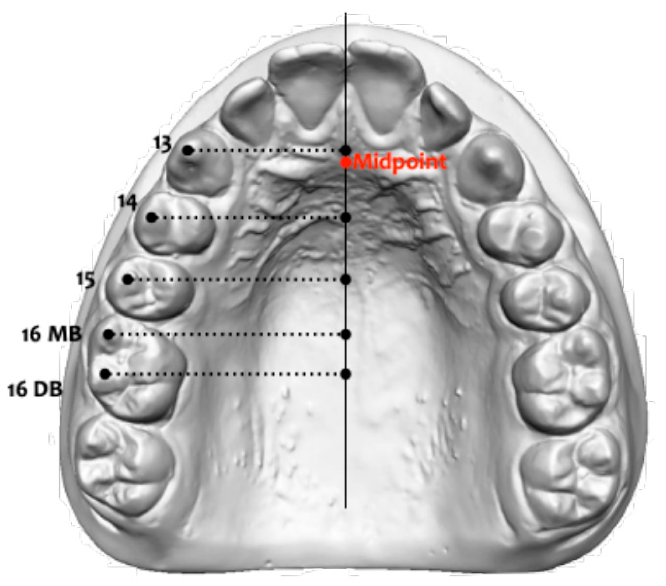
Prior to treatment (T1) and at the end of the first maxillary molars distalization movement (T2), digital dental casts (.stl files) created from an intraoral scanner iTero® Orthodontic ver. 5.2.1.290 (Align Technology Inc., Santa Clara, CA, USA) were analyzed. The average time between the T2–T1 scans was 5.5 months (Figure 1). Palatal rugae were used as landmarks for the evaluation of molar, premolar, and canine movements and compared using Viewbox 4.0 software (dHAL software, Kifissia, Greece). In particular, the midpalatal raphe was used as a reference axis, and the distance of each orthogonal dental projection was measured from the midpoint of the first palatine ruga. On each digital cast, the following sagittal measurements were evaluated at both observation times [26]:
-
First Molar Mesio Buccal Sagittal (1.6 MBS/2.6 MBS): the amount of space between the mid-point of the first palatal ruga and the projection of the mesiobuccal cusp of the first right and left permanent molars on the mid-palatal raphe;
-
First Molar Disto Buccal Sagittal (1.6 DBS/2.6 DBS): the amount of space between the mid-point of the first palatal ruga and the projection of the distobuccal cusp of the first right and left permanent molars on the mid-palatal raphe;
-
Second Premolar Buccal Sagittal (1.5 PBS/2.5 PBS): the amount of space between the mid-point of the first palatal ruga and the projection of the cusp of the second right and left premolars on the mid-palatal raphe;
-
First Premolar Buccal Sagittal (1.4 PBS/2.4 PBS): the amount of space between the mid-point of the first palatal ruga and the projection of the cusp of the first right and left premolars on the mid-palatal raphe;
-
Canine Sagittal (1.3 CS/2.3 CS): the amount of space between the mid-point of the first palatal ruga and the projection of the cusp of the right and left canines on the mid-palatal raphe (Figure 2).

2.2. Statistical Analysis

The power of the project was analyzed based on a minimum sample size of 45 subjects with an effect size equal to 1. The power was 0.81 at an alpha level of 0.05. A unique operator (S.L.) performed all the measurements. To assess intra-operator reliability, the same examiner re-performed all measurements of the entire sample 2 weeks after the first analysis. The reliability of the measurements was evaluated by means of an interclass correlation coefficient (ICC).
Exploratory statistics showed that all variables were normally distributed (Kolmogorov–Smirnov test) with equality of variances (Levene test). Descriptive statistics (means and standard deviations), 95% confidence intervals, and statistical comparisons using paired t-tests were calculated for the T1 to T2 cephalometric changes in the TG. All statistical computations were performed with SPSS software (version 12; SPSS, Chicago, IL, USA). The level of significance was set at 5%.

3. Results

During the therapy, all the treated subjects showed adequate cooperation with the use of aligners. In particular, moderate cooperation was reported only in 5 patients, and the remaining 44 patients had good compliance. No one wore the aligners less than 16 h per day. As a result, cooperation with aligners was good in 89.8% of the patients. Regarding the Class II elastics collaboration, 16 patients (30% of the whole sample) reported they were not consistent in their use, and they showed poor cooperation.
The ICC test showed almost perfect agreement, with a score of 0.96 for the linear measurements evaluated. The random error varied from 0.18 mm (1.6 DBS value) to 0.28 mm (1.3 CS value).
The intragroup comparison between T2–T1 values showed a statistically significant improvement in the sagittal position of the first molars, with an average distalization movement of 2.5 mm (1.6 DBS = 3 mm, p = 0.0001; 1.6 MBS = 2.4 mm, p = 0.0001; 2.6 DBS = 2.2 mm, p = 0.0008; 2.6 MBS = 2.4 mm, p = 0.0006). From the analysis of the sagittal movement of the dental anchorage unit, a significant anchorage loss was detected at the level of both upper canines with an average of 1.3 mm of mesialization movement (1.3 CS = −1.5 mm, p = 0.0001; 2.3 CS = −1.15 mm, p = 0.008). No statistically significant differences in the sagittal positions of the first and the second premolars were observed (Table 1).

4. Discussion

Although CAT is widely used in orthodontic clinical practice, knowledge about side effects in many types of movements obtained with this technique has not yet been clearly described and supported by scientific evidence.
Therefore, the aim of the present investigation is to focus on the maxillary dental distalization treatment through CAT and, in particular, to evaluate the anchorage loss at the level of upper premolars and canines at the end of the upper first molars’ distal movement as unwanted effects of this therapy.
As we all know, the molar distalization procedure requires an anchoring unit usually represented by the anterior part of the arch. Biomechanically, the posterior force applied on the molars generates an equal and opposite reciprocal force, which is discharged on the anterior teeth, particularly in the area of the incisors, which ultimately leads to their flaring and anchorage loss [27]. The resistance of a tooth to a given force depends on the following factors: root area and shape, bone density and periodontal health, muscular force, facial biotype, oral habits, and type of movement applied. Moreover, each dental element has an anchor value, which depends mainly on its root surface. It is very important to take the anchor value into consideration when planning orthodontic movements, especially during distalization movements.
In the literature, many authors [3,7,21,28,29,30] described the anchorage loss, analyzing the millimeter and angular variation through cephalometric analysis on lateral cephalograms during distalization treatment with traditional intra-oral fixed appliances, and they highlighted that mutual forces resulted in a loss of anterior anchorage that was responsible for some of the space that appeared. Among them, Bussik and McNamara [31] found that 24% of the total space opening anterior during maxillary molar distalization was due to the reciprocal anchorage loss of the maxillary premolars with the slightly mesial movement of the upper incisors, whereas the bodily movement of maxillary first molar contributed to 76%.
Additionally, Bolla et al. [3], in their study about the comparison between the upper molars distalization obtained with a Distal Jet and other traditional methods, highlighted that at the end of the backward molar movement, 71% of the 4.5 mm of space obtained between the upper first molar and the first premolar was due to the distal movement of the molar crown on each side, while the remaining 29% resulted from the loss of anchorage of the premolars.
On the contrary, as described by Chiu et al. [32], distal tipping of the first premolar of −1.7° was observed during distalization treatment performed with a pendulum. Thus, there is no consensus on the direction in which premolars tip during the movement of molar distalization with these popular appliances.
Furthermore, Class II elastics are frequently used in conventional fixed-multibracket therapy for anchorage support and sagittal correction during maxillary molar distalization. Although widely employed, some researchers have found that the unwanted effects of Class II elastics are very habitual, increasing the facial height, owing to the rotation of the mandible and the consequent worsening of smile aesthetics [33].
In recent years, CAT has become an alternative to conventional fixed appliances, and therefore, they are also used for molars distalization with a specific protocol, defined as the standardized 50% sequential distalization protocol. This strategy is particularly advantageous because it does not involve the movement of more than two teeth at the same time. In this way, the arch is divided into two units, in which the support one has more dental anchoring mass than the active segment that performs the movement. This standardized 50% sequential distalization protocol makes the movements of the teeth in the posterior part of the arch more predictable, which have a higher anchoring value than the single-rooted elements of the anterior sector.
Recent studies [18,34] evaluated the accuracy of upper-molar bodily distal movement with aligners. Verma and George [34], in their systematic review, reported a significant amount of distalization of nearly 2–3 mm in all the selected studies and suggested this complex movement could be performed using CAT with good control over vertical craniofacial parameters, mesio-distal angulation of molars, and anchorage loss.
Other studies analyzed the anchorage loss obtained during the upper-molar distalization movement with CAT. Ravera et al. [17] and Caruso et al. [1] observed no anchorage loss at the end of the therapy on upper incisors during distalization movements performed with aligners, sequential movement protocol, and early Class II elastics. On the contrary, Liu et al. [23], using three-dimensional finite element models, found a worsening of anterior anchorage during first molar distalization, with a mesial palatal relapse tendency of the second molar. However, they reported that the use of Class II elastics was effectively able to reinforce the anchorage of both anterior teeth and molars.
The configuration of the precision cuts for elastics also deserves particular attention. There are no clinical guidelines for the selection of an application for Class II elastics, including the bonding of buttons directly onto the tooth surface and the precise cutting of clear aligners [23]. In our protocol, in the upper arch, the intra-oral elastic hooked directly to the aligner (on canines or first premolars) exerts a distal force on the entire arch reducing the overjet. This principle is also applicable during the sequential distalization mechanics to better express the posterior movement, especially where it is necessary to correct a Class II malocclusion with proclined maxillary incisors. Also in the lower arch, according to our protocol, it is suitable to use precision cuts (on first molars) in order to work by associating all the dental elements and generating a homogeneous force on the entire arch, which thus functions as a passive anchoring segment. Moreover, the application of a precision button cut on the lower molars causes a direct force to be applied to the tooth. Since our protocol provides for the use of full-time elastics, the clinical preference of a precision cut avoids the expression of any extrusive force vectors and, thus, causes a clockwise movement of the mandible.
CAT has also been used to improve vertical dimension control, rendering this a superior alternative for the treatment of patients with hyperdivergent or open bites [1]. In fact, when programming the distalization movements, it is possible to associate an intrusive force vector able to control any extrusion, simultaneously improving the fitting of the aligner and promoting the bodily movement of the dental elements.
Some authors [35,36] suggested that it could even be the structure of the aligner that promotes an intrusive movement. The results obtained indicate that the occlusal force applied in association with the thickness of the aligners promotes intrusion force and, therefore, controls the clockwise rotation of the occlusal plane during distalization.
Staderini et al. [37] found that CAT could prevent the expression of an undesired vertical movement thanks to its structure, which covers the occlusal surface. The extrusive force vector resulting from using Class II elastics is opposed by the biting force and, therefore, keeps the vertical dimension unchanged.
The limitation of these previous studies is that they measured the accuracy of the tooth movements at the final stage and not after achieving distal molar movement, so the final planning movement of the anterior teeth could influence their results.
Therefore, the purpose of our study was to investigate the immediate effect of completed maxillary molar distalization on premolar and canine anchorage loss using 3D digital dental casts, and to our knowledge, only Saif et al. [24] evaluated a similar clinical condition.
Our chosen measurements were defined by landmarks located on palatal anatomical structures represented by the palatal rugae area, so they were not influenced by the anterior tooth movement [38,39].
The decision to use the palatal rugae area in our study was based on the ability to evaluate the precision position of all dental elements at any given moment of the therapy, without the need to submit the patients to X-ray exposure and the possibility to use invariable landmarks for the entire treatment time. Moreover, the possibility of obtaining digital models of the patient during each orthodontic check-up allows for an accurate and rapid collection of records during treatment, without the need to request additional, more invasive, and expensive examinations.
Regarding the CAT efficacy in achieving the distalization movement, our results agreed with those of previous studies [1,17,18,24,34,40,41], showing a significant improvement in the sagittal position of first molars, with an average distalization movement of 2.5 mm.
On the contrary, a significant anchorage loss was detected at the level of both upper canines with an average of 1.3 mm of mesialization movement, while no statistically significant differences for the first and second premolars were observed. This result was also found by Saif et al. [24], reporting that the anchorage loss involved the central incisors (39.9%), the lateral incisors (37.4%), and canines (22.7%) during molar distalization movement.
According to the other studies presented in the literature [1,17,18,24,34,40,41], also in our research, no statistically significant movements were detected at the premolar level. Slight differences were found at the level of the second premolars, attributable to the start of distal movements, as required by the protocol, but nonetheless not significant.
In our study as well as in the study by Saif et al. [24], the major loss of anchorage was detected in patients who had poor compliance with the use of Class II elastics. This indicates that the use of Class 2 full-time elastics was able to generate an equal and opposite force to the reaction force in the anterior sector, supporting the distalization movement, strengthening the anterior anchorage, and counteracting the unwanted side effects.
The significant mesial movement of upper canines could also be due to the mechanics of the device. In fact, CA is a closed system of forces, and the applied distalization movement could produce a plastic deformation of its structure, which “stretches” distally and produces a reaction force in the anterior part of the arch [42].

Limitation

The main limitation of our study was the retrospective study design. In order to obtain more suitable information about the unwanted effects of this protocol treatment, further research is necessary with a larger sample size. Moreover, the absence of a comparison between CAT therapy and conventional distalization treatment as a control group can be considered a limitation of the study. Another limitation of this study was the analysis applied that did not consider the exact root positions and the degree of crown tipping.

5. Conclusions

Upper-molar distalization achieved with clear aligner therapy allows the correction of Class II relationship due to maxillary dentoalveolar protrusion or moderate skeletal discrepancies. However, during the distal movement of the upper molars, side effects on the anchorage teeth were present; in particular, a significant mesial movement of the upper canines was noticed.

Author Contributions

Conceptualization, P.C. and R.L.; methodology, V.P. and C.P.; software, V.P.; validation, P.C., R.L. and C.P.; formal analysis, V.P.; investigation, S.L.; resources, C.P. and R.L.; data curation, S.L.; writing—original draft preparation, S.L. and R.L. All authors have read and agreed to the published version of the manuscript.

Funding

This research received no external funding.

Institutional Review Board Statement

The study was conducted according to the guidelines of the Declaration of Helsinki and approved by the Ethics Committee of the University of Rome “Tor Vergata” (protocol number 257/21).

Informed Consent Statement

Informed consent was obtained from all participants’ parents included in the study.

Data Availability Statement

The data presented in this study are available on request from the corresponding author.

Conflicts of Interest

The authors declare no conflict of interest.

Abbreviations

CA: Clear Aligners; CAT: Clear Aligners Treatment; 3D: three-dimensional; ICC: Interclass Correlation Coefficient; MBS: First Molar Mesio Buccal Sagittal; DBS: First Molar Disto Buccal Sagittal; PBS: Premolar Buccal Sagittal; CS: Canine Sagittal; T1: pre-treatment phase; T2: at the end of the first maxillary molars distalization movement; IC (confidence interval); SD (standard deviation); diff: mean difference.

References

  1. Caruso, S.; Nota, A.; Ehsani, S.; Maddalone, E.; Ojima, K.; Tecco, S. Impact of molar teeth distalization with clear aligners on occlusal vertical dimension: A retrospective study. BMC Oral Health 2019, 19, 182. [Google Scholar] [CrossRef] [PubMed] [Green Version]
  2. Alogaibi, Y.A.; Al-Fraidi, A.A.; Alhajrasi, M.K.; Alkhathami, S.S.; Hatrom, A.; Afify, A.R. Distalization in Orthodontics: A Review and Case Series. Case Rep. Dent. 2021, 8843959. [Google Scholar] [CrossRef] [PubMed]
  3. Bolla, E.; Muratore, F.; Carano, A.; Bowman, S.J. Evaluation of maxillary molar distalization with the distal jet: A comparison with other contemporary methods. Angle Orthod. 2002, 72, 481–494. [Google Scholar] [PubMed]
  4. Lione, R.; Franchi, L.; Laganà, G.; Cozza, P. Effects of cervical headgear and pendulum appliance on vertical dimension in growing subjects: A retrospective controlled clinical trial. Eur. J. Orthod. 2015, 37, 338–344. [Google Scholar] [CrossRef] [Green Version]
  5. Carano, A.; Testa, M. The distal jet for upper molar distalization. J. Clin. Orthod. 1996, 30, 374–380. [Google Scholar]
  6. Fortini, A.; Lupoli, M.; Giuntoli, F.; Franchi, L. Dentoskeletal effects induced by rapid molar distalization with the first class appliance. Am. J. Orthod. Dentofac. Orthop. 2004, 125, 697–704. [Google Scholar] [CrossRef]
  7. Taner, T.U.; Yukay, F.; Pehlivanoglu, M.; Cakirer, B. A comparative analysis of maxillary tooth movement produced by cervical headgear and pend-x appliance. Angle Orthod. 2003, 73, 686–691. [Google Scholar]
  8. Locatelli, R.; Bednar, J.; Dietz, V.S.; Gianelly, A.A. Molar distalization with superelastic NiTi wire. J. Clin. Orthod. 1992, 26, 277–279. [Google Scholar]
  9. Jones, R.D.; White, J. Rapid Class II molar correction with an open-coil jig. J. Clin. Orthod. 1992, 26, 661–664. [Google Scholar]
  10. Hilgers, J.J. The pendulum appliance for Class II non-compliance therapy. J. Clin. Orthod. 1992, 26, 706–714. [Google Scholar]
  11. Kinzinger, G.S.; Gülden, N.; Yildizhan, F.; Diedrich, P.R. Efficiency of a skeletonized distal jet appliance supported by miniscrew anchorage for noncompliance maxillary molar distalization. Am. J. Orthod. Dentofac. Orthop. 2009, 136, 578–586. [Google Scholar] [CrossRef]
  12. Maino, B.G.; Gianelly, A.A.; Bednar, J.; Mura, P.; Maino, G. MGBM system: New protocol for Class II non-extraction treatment without cooperation. Prog. Orthod. 2007, 8, 130–143. [Google Scholar]
  13. Suzuki, E.Y.; Suzuki, B. Placement and removal torque values of orthodontic miniscrew implants. Am. J. Orthod. Dentofac. Orthop. 2011, 139, 669–678. [Google Scholar] [CrossRef] [PubMed]
  14. Soheilifar, S.; Mohebi, S.; Ameli, N. Maxillary molar distalization using conventional versus skeletal anchorage devices: A systematic review and meta-analysis. Int. Orthod. 2019, 17, 415–424. [Google Scholar] [CrossRef] [PubMed]
  15. Miller, D.B. Invisalign in TMD treatment. Int. J. Orthod. Milwaukee 2009, 20, 15–19. [Google Scholar] [PubMed]
  16. Kravitz, N.; Kusnoto, B.; BeGole, E.; Obrez, A.; Agran, B. How well does Invisalign work? A prospective clinical study evaluating the efficacy of tooth movement with Invisalign. Am. J. Orthod. Dentofac. Orthop. 2009, 135, 27–35. [Google Scholar] [CrossRef]
  17. Ravera, S.; Castroflorio, T.; Garino, F.; Daher, S.; Cugliari, G.; Deregibus, A. Maxillary molar distalization with aligners in adult patients: A multicenter retrospective study. Prog. Orthod. 2016, 17, 12. [Google Scholar] [CrossRef] [Green Version]
  18. Simon, M.; Keilig, L.; Schwarze, J.; Jung, B.A.; Bourauel, C. Treatment outcome and efficacy of an aligner technique--regarding incisor torque, premolar derotation and molar distalization. BMC Oral Health 2014, 14, 68. [Google Scholar] [CrossRef] [Green Version]
  19. Rossini, G.; Parrini, S.; Castroflorio, T.; Deregibus, A.; Debernardi, C.L. Efficacy of clear aligners in controlling orthodontic tooth movement: A systematic review. Angle Orthod. 2015, 85, 881–889. [Google Scholar] [CrossRef] [Green Version]
  20. Joseph, A.A.; Butchart, J.C. An evaluation of the pendulum distalizing appliance. Semin. Orthod. 2000, 6, 129–135. [Google Scholar] [CrossRef]
  21. Ghosh, J.; Nanda, R.S. Evaluation of an intraoral maxillary molar distalization technique. Am. J. Orthod. Dentofac. Orthop. 1996, 110, 639–646. [Google Scholar] [CrossRef] [PubMed]
  22. Fontana, M.; Cozzani, M.; Caprioglio, A. Non-compliance maxillary molar distalizing appliances: An overview of the last decade. Prog. Orthod. 2012, 13, 173–184. [Google Scholar] [CrossRef] [PubMed]
  23. Liu, X.; Cheng, Y.; Qin, W.; Fang, S.; Wang, W.; Ma, Y.; Jin, Z. Effects of upper-molar distalization using clear aligners in combination with Class II elastics: A three-dimensional finite element analysis. BMC Oral Health 2022, 22, 546. [Google Scholar] [CrossRef]
  24. Saif, B.S.; Pan, F.; Mou, Q.; Han, M.; Bu, W.; Zhao, J.; Guan, L.; Wang, F.; Zou, R.; Zhou, H.; et al. Efficiency evaluation of maxillary molar distalization using Invisalign based on palatal rugae registration. Am. J. Orthod. Dentofac. Orthop. 2022, 161, e372–e379. [Google Scholar] [CrossRef]
  25. Arnold, W.E.; McCroskey, J.C.; Prichard, S.V.O. The Likert-type scale. Todays Speech 1967, 15, 31–33. [Google Scholar] [CrossRef]
  26. Lione, R.; Paoloni, V.; De Razza, F.C.; Pavoni, C.; Cozza, P. The Efficacy and Predictability of Maxillary First Molar Derotation with Invisalign: A Prospective Clinical Study in Growing Subjects. Appl. Sci. 2022, 12, 2670. [Google Scholar] [CrossRef]
  27. Bondemark, L.; Kurol, J. Distalization of maxillary first and second molars simultaneously with repelling magnets. Eur. J. Orthod. 1992, 14, 264–272. [Google Scholar] [CrossRef]
  28. Byloff, F.K.; Darendeliler, M.A. Distal molar movement using the pendulum appliance. Part 1: Clinical and radiological evaluation. Angle Orthod. 1997, 67, 249–260. [Google Scholar]
  29. Caprioglio, A.; Beretta, M.; Lanteri, C. Maxillary molar distalization: Pendulum and Fast-Back, comparison between two approaches for Class II malocclusion. Prog. Orthod. 2011, 12, 8–16. [Google Scholar] [CrossRef]
  30. Fuziy, A.; Rodrigues de Almeida, R.; Janson, G.; Angelieri, F.; Pinzan, A. Sagittal, vertical, and transverse changes consequent to maxillary molar distalization with the pendulum appliance. Am. J. Orthod. Dentofac. Orthop. 2006, 130, 502–510. [Google Scholar] [CrossRef]
  31. Bussick, T.J.; McNamara, J.A., Jr. Dentoalveolar and skeletal changes associated with the pendulum appliance. Am. J. Orthod. Dentofac. Orthop. 2000, 117, 333–343. [Google Scholar] [CrossRef] [PubMed]
  32. Chiu, P.P.; McNamara, J.A., Jr.; Franchi, L. A comparison of two intraoral molar distalization appliances: Distal jet versus pendulum. Am. J. Orthod. Dentofac. Orthop. 2005, 128, 353–365. [Google Scholar] [CrossRef]
  33. Janson, G.; Sathler, R.; Fernandes, T.M.; Branco, N.C.; Freitas, M.R. Correction of class II malocclusion with class II elastics: A systematic review. Am. J. Orthod. Dentofac. 2013, 143, 383–392. [Google Scholar] [CrossRef]
  34. Verma, P.; George, A.M. Efficacy of clear aligners in producing molar distalization: Systematic review. APOS Trends Orthod. 2021, 11, 317–324. [Google Scholar] [CrossRef]
  35. Garino, F.; Castroflorio, T.; Daher, S.; Ravera, S.; Rossini, G.; Cugliari, G.; Deregibus, A. Effectiveness of composite attachments in controlling upper-molar movement with aligners. J. Clin. Orthod. 2016, 50, 341–347. [Google Scholar] [PubMed]
  36. Gomez, J.P.; Peña, F.M.; Martínez, V.; Giraldo, D.C.; Cardona, C.I. Initial force systems during bodily tooth movement with plastic aligners and composite attachments: A three-dimensional finite element analysis. Angle Orthod. 2015, 85, 454–460. [Google Scholar] [CrossRef]
  37. Staderini, E.; Ventura, V.; Meuli, S.; Maltagliati, L.A.; Gallenzi, P. Analysis of the changes in occlusal plane inclination in a Class II deep bite “teen” patient treated with clear aligners: A case report. Int. J. Environ. Res. Public Health 2022, 19, 651. [Google Scholar] [CrossRef]
  38. Van der Linden, F.P. Changes in the position of posterior teeth in relation to ruga points. Am. J. Orthod. 1978, 74, 142–161. [Google Scholar] [CrossRef]
  39. Lanteri, V.; Cossellu, G.; Farronato, M.; Ugolini, A.; Leonardi, R.; Rusconi, F. Assessment of the Stability of the palatal rugae in a 3D-3D superimposition technique following Slow Maxillary expansion (SMe). Sci. Rep. 2020, 10, 2676. [Google Scholar] [CrossRef] [Green Version]
  40. Lione, R.; Balboni, A.; Di Fazio, V.; Pavoni, C.; Cozza, P. Effects of pendulum appliance versus clear aligners in the vertical dimension during Class II malocclusion treatment: A randomized prospective clinical trial. BMC Oral Health 2022, 22, 441. [Google Scholar] [CrossRef]
  41. Lione, R.; Paoloni, V.; De Razza, F.C.; Pavoni, C.; Cozza, P. Analysis of Maxillary First Molar Derotation with Invisalign Clear Aligners in Permanent Dentition. Life 2022, 12, 1495. [Google Scholar] [CrossRef] [PubMed]
  42. Bichu, Y.M.; Alwafi, A.; Liu, X.; Andrews, J.; Ludwig, B.; Bichu, A.Y.; Zou, B. Advances in orthodontic clear aligner materials. Bioact. Mater. 2022, 22, 384–403. [Google Scholar] [CrossRef] [PubMed]
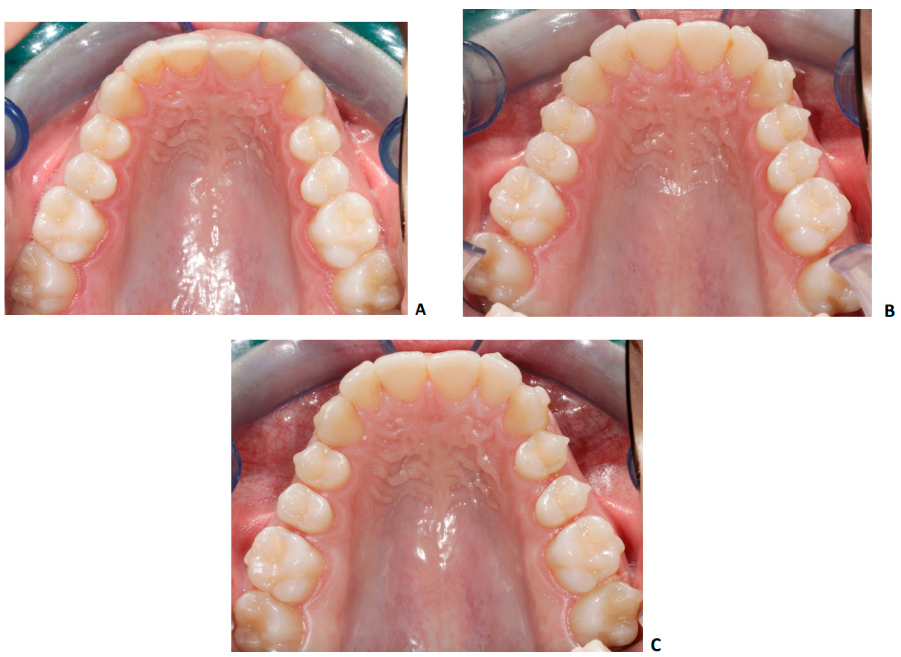
Figure 1. Pre-treatment—T1 (A), 50% sequential distalization protocol (B), at the end of distalization of first upper molars—T2 (C).
Applsci 13 03646 g001
Figure 2. Sagittal measurements performed at T1 and at T2 on digital dental casts: 1.6 MBS/2.6 MBS (the amount of space between the mid-point of the first palatal ruga and the projection of the mesiobuccal cusp of the first right or/and left permanent molar on the mid-palatal raphe); 1.6 DBS/2.6 DBS (the amount of space between the mid-point of the first palatal ruga and the projection of the distobuccal cusp of the first right or/and left permanent molar on the mid-palatal raphe); 1.5 PBS/2.5 PBS (the amount of space between the mid-point of the first palatal ruga and the projection of the cusp of the second first right or/and left premolar on the mid-palatal raphe); 1.4 PBS/2.4 PBS (the amount of space between the mid-point of the first palatal ruga and the projection of the cusp of the second first right or/and left premolar on the midpalatal raphe); 1.3 CS/2.3 CS (the amount of space between the mid-point of the first palatal ruga and the projection of the cusp of the right or/and left canine on the mid-palatal raphe).
Applsci 13 03646 g002
Table 1. Descriptive statistics and statistical comparison of the T2–T1 changes with paired t-test.
VariablesT1 (n = 49 27F; 22M) T2 (n = 49 27F; 22M)T2–T1
Measurements (mm)Mean T1SD Mean T2SD Diff95% CIp Value
1.3 CS3.01.021.51.03−1.50.932 to 2.068*** (0.0001)
2.3 CS2.051.40.91.1−1.150.366 to 1.934** (0.008)
1.4 PBS7.73.38.02.70.3−1.358 to 0.758NS (0.5454)
2.4 PBS 7.92.77.82.4−0.1−1.317 to 1.483NS (0.8981)
1.5 PBS 14.23.714.73.00.5−1.396 to 0.329NS (0.20)
2.5 PBS14.72.715.03.00.3−1.046 to 0.513NS (0.47)
1.6 DBS25.23.728.23.23.0−3.938 to −2.029*** (0.0001)
1.6 MBS 20.63.623.03.02.4−3.341 to −1.492*** (0.0001)
2.6 DBS 25.23.327.43.72.2−3.258 to −1.142*** (0.0008)
2.6 MBS 20.53.222.93.22.4−3.440 to −1.260*** (0.0006)
NS: not significant, ** p < 0.01, *** p < 0.001. CI: confidence of interval; SD: standard deviation.
Disclaimer/Publisher’s Note: The statements, opinions and data contained in all publications are solely those of the individual author(s) and contributor(s) and not of MDPI and/or the editor(s). MDPI and/or the editor(s) disclaim responsibility for any injury to people or property resulting from any ideas, methods, instructions or products referred to in the content.