Article

Protective Effect of Juglone (5-Hydroxy-1,4-naphthoquinone) against Iron- and Zinc-Induced Liver and Kidney Damage

1
Department of Nutrition and Dietetics, Faculty of Health Sciences, Suleyman Demirel University, Isparta 32260, Turkey
2
Department of Bioengineering, Institute of Science and Technology, Suleyman Demirel University, Isparta 32260, Turkey
*
Author to whom correspondence should be addressed.
Appl. Sci. 2023, 13(4), 2203; https://doi.org/10.3390/app13042203
Submission received: 20 January 2023 / Revised: 2 February 2023 / Accepted: 3 February 2023 / Published: 8 February 2023
(This article belongs to the Section Environmental Sciences)

Abstract

:
Although heavy metals are naturally occurring elements that are found throughout the Earth’s crust, most environmental contamination and human exposure result from anthropogenic activities, such as mining and smelting operations, industrial production and use, and the domestic and agricultural use of metals and metal-containing compounds. The accumulation of heavy metals eventually produces reactive oxygen species that can cause oxidative stress, which may lead to the production of various diseases. The aim of this study was to evaluate the possible effects of iron and zinc on kidney and liver tissues and the positive effects of juglone (5-hydroxy-1,4-naphthoquinone) antioxidant activity, using an immunohistochemical technique. The animals under study were randomly divided into five groups (seven in each group): group I, control; group II, iron (Fe) (600 ppm); group III, zinc (Zn) (400 ppm); group IV, Fe + antioxidant juglone; and group V, Zn + antioxidant juglone. Hematoxylin-eosin (H&E) was applied to determine the histological sides of the damage caused by the heavy metals in the liver and kidney tissues and the effects of the administration of juglone on reducing these damages. Furthermore, the immunohistochemical TUNEL method was applied to determine the DNA damages in the cells. The density of the damage in the liver and kidney tissues of the iron group was higher than in the other groups.
Keywords:
iron; juglone; kidney; liver; zinc

1. Introduction

Heavy metals are naturally occurring elements that have a high atomic weight and a density at least five times greater than that of water. Their multiple industrial, domestic, agricultural, medical and technological applications have led to their wide distribution in the environment, raising concerns over their potential effects on human health, as well as the environment itself. Their toxicity depends on several factors, including the dose, route of exposure, and chemical species, as well as the age, gender, genetics, and nutritional status of exposed individuals. Heavy metals, metal compounds and various minerals, which constitute an important portion of pollutants, are widely spread in lakes, rivers, oceans and their sediments. Heavy metals spread in waters as a result of natural and anthropogenic influences [1]. In addition to their toxic, acute, chronic and direct effects, heavy metals have indirect physiological effects on aquatic organisms, particularly on the eggs and young individuals of these organisms, on whom these effects are more severe. Heavy-metal pollution is rapidly increasing, which presents many environmental problems. These heavy metals are mainly accumulated in soil and are transferred to the food chain through plant grown on these soils [2,3,4].
Chromium (Cr), manganese (Mn), cobalt (Co), copper (Cu), zinc (Zn), molybdenum (Mo), mercury (Hg), nickel (Ni), tin (Sn), lead (Pb), cadmium (Cd), antimony (Sb), etc., are the main elements of heavy metals. Three kinds of heavy metal are of concern: toxic metals (such as, Hg, Cr, Pb, Zn, Cu, Ni, Cd, As, Co, Sn, etc.), precious metals (such as Pd, Pt, Ag, Au, Ru, etc.) and radionuclides, such as uranium (U), thorium (Th), radium (Ra), americium (Am). Heavy metals are natural components of the Earth’s crust. They cannot be destroyed or degraded. However, most of these heavy metals become toxic at high concentrations due to their ability to accumulate in living tissues. The removal of heavy metals from industrial wastewater is of primary importance. Cadmium, zinc, copper, nickel, lead, mercury, vanadium and chromium are often detected in industrial wastewaters [1,5,6,7].
Iron is a functional part of hemoglobin found in the structure of red blood cells in humans. It is an element of vital importance and it is also found in the structure of cytochrome peroxidase, catalase enzymes and myoglobin. Surplus iron accumulates in the bone marrow, liver and spleen [8]. While there is a control mechanism in the human body for iron absorption, there is no control mechanism for the removal of iron from the body. When taken into the body in excessive amounts, iron may cause damage to the digestive system and enter the circulatory system, through which it may reach and cause damage to organs such as the liver and heart. Organs are damaged due to prolonged exposure to iron, which may result in death. Iron, found in colloidal form in water environments, may cause death in fish by densely accumulating in their gills. The iron accumulated in bodies of the fish is transferred to the human body as a result of the consumption of fish as food and may negatively affect human health [9,10]. Iron toxicity is classified as corrosive or cellular. Ingested iron can cause direct caustic injury to the gastrointestinal mucosa, resulting in nausea, vomiting, abdominal pain, and diarrhea. Significant fluid and blood loss can lead to hypovolemia. Hemorrhagic necrosis of the gastrointestinal mucosa can lead to hematemesis, perforation and peritonitis. At the cellular level, iron impairs cellular metabolism in the heart, liver and central nervous system. Free iron enters cells and concentrates in the mitochondria. This disrupts oxidative phosphorylation, catalyzes lipid peroxidation, forms free radicals and ultimately leads to cell death [11].
Insufficient zinc intake negatively affects more than 300 enzymes, whereas excessive amounts may cause various forms of damage to organisms. Zinc chloride (ZnCl₂) toxicity has been reported to cause acute renal failure. Excessive zinc intake has been shown to cause copper deficiency in experimental animals. These two elements are biological antagonists. The excessive intake of zinc and cadmium (Cd) blocks copper absorption and reduces copper levels. Bone is the most sensitive tissue to zinc accumulation. The excessive intake of zinc through foods may lead to poisoning. Zinc is non-toxic in element form. Its toxicity is caused by soluble salts. Zinc toxicity has been reported to be associated with lung symptoms, mental disorders and renal failure in humans. Decreased appetite and activity of the immune system, late wound healing, extreme skin sensitivity and cholesterol elevation are general problems observed in humans due to excessive zinc intake. Zinc in different cell lines causes oxidative stress, such as lipid peroxidation, cell membrane leakage, oxidative DNA damage, increases in intracellular calcium and even antiproliferative activity. Different researchers have reported many adverse effects, but not cancer. Toxicologically, zinc is known to cause less damage compared to arsenic, cadmium, chromium, copper and lead. The interaction of toxic metals with food, industry and the environment may affect the availability of essential elements in the organism (cells, tissues, organs, molecules) [12,13].
The antioxidant used in this study, juglone, was obtained from walnut (Juglans regia L.) The walnut tree (Juglans regia L.) is naturally found in southeastern Europe, Asia, and India. The walnut tree naturally grows in every region of Turkey. Walnut is rich in essential fatty acids and tocopherols. Linoleic acid, oleic acid, linolenic acid, palmitic acid and stearic acid lead to a decrease in LDL cholesterol and an elevation in HDL cholesterol and provide protection against cardiovascular diseases. In addition, thanks to the herbal sterols, folate, tannin and polyphenols in its content, walnut has a very important place in nutrition. Juglone is also known to cause an apoptotic/necrotic effect when applied to colon cancer HT29 cell lines in different doses. It was reported in the literature that apoptosis occurred in lung-cancer cells, colon-cancer cells and HaCaT keratinocyte cells stimulated with juglone. Juglone has an apoptotic effect on leukemia HL-60 cells. Juglone is an important natural component obtained from the green fruit coat and leaves of walnut. In recent studies, it was shown that juglone had anticancer, antibacterial and antiviral effects [14,15,16,17,18].
The purpose of this study is to determine the effect of the consumption of foods and waters with high heavy-metal contents in tissues, especially the liver and kidneys. In this study, the effects of iron and zinc on the kidney and liver tissues of Wistar albino rats and the positive effect of the antioxidant, juglone, when applied to reduce these effects were studied by using histological and immunohistochemical methods.

2. Materials and Methods

2.1. Experimental Design and Animals

Thirty-five Wistar-albino-type male adult rats were used in the study. Permission was obtained from the local ethics committee (Süleyman Demirel University Experimental Animals Research Unit, ethics number: 21438139-338). There were 5 groups, each consisting of 7 adult rats. The 35 rats were cared for and fed under the same environmental conditions, with a constant temperature of 20 ± 2 °C, a humidity of 55–60% and a 12/12 h light/dark cycle. The heavy-metal and antioxidant applications were performed with the gavage method on a daily basis for 30 days.
First group was control group (1 mL water), second was given iron (600 ppm), third was given zinc (400 ppm), fourth was given Fe (600 ppm) + antioxidant juglone, and fifth was given Zn (400 ppm) + antioxidant juglone through the gavage method [19,20]. All methods were performed in accordance with the relevant guidelines and regulations.

2.2. Surgical Procedure

At the end of 30 days, liver and kidney tissues were removed under ketamine–xylazine anesthesia and fixed in 10% formalin solution. Tissues were washed under tap water for a night in order to remove fixatives.

2.3. Histological Analysis

The routine histological tissue-tracking procedure was performed. The tissues were buried in paraffin and 4–5 µ sections were taken from paraffin blocks. Hematoxylin–eosin (H&E) staining was used for histopathological evaluations and the TUNEL method was utilized for staining apoptotic cells [21]. The preparations were examined and evaluated under a light microscope (Leica DM 500) and photographing was performed. In the evaluation of hematoxylin–eosin staining, damage to tissues was assessed. Damage to tissues was scored semi-quantitatively with numbers from 0 to +3 (0: none, +1: slight, +2: moderate, +3: severe).

2.4. Immunohistochemical Analysis

The TUNEL method (TdT-mediated dUTP-digoxigenin nick end labeling), which determines DNA fragmentation, was applied in order to stain apoptotic cells. In accordance with the instructions from the manufacturer, cells that underwent apoptosis were determined using ApopTag Plus Peroxidase In Situ Apoptosis Detection Kit (Chemicon, cat. no.: QIA33, Rolling Meadows, IL, USA). Tissues were deparaffinized with xylene, dehydrated with alcohol series and washed with PBS. Next, tissues were incubated with 5% proteinase K for 10 min and with 3% hydrogen peroxide for 5 min in order to prevent endogenous peroxidase activity. After washing with PBS, tissues were incubated with equilibration buffer for 20 min and kept in a humid environment at 37 °C. The TdT enzyme was applied in a humidified chamber at 37 °C for 90 min. Tissues were kept in block buffer for 10 min and then in stop/wash buffer for another 10 min and incubated with anti-digoxigenin-peroxidase for 30 min. Apoptotic cells were visualized with diaminobenzidine (DAB) substrate. Apoptotic cells were identified by a brown core. Apoptotic cells were counted under a microscope. The mean values and standard deviations of apoptotic cells were determined for each group.

3. Results

It was found that the histological structures of the kidney tissues removed from the control group rates had a normal appearance. Tubular dilation, hydropic degeneration in the tubular cells, shrinkage in the glomeruli and congestion were observed in the kidney tissues removed from the rats that were administered iron. Although similar findings were obtained in the zinc group, congestion was not observed. The histopathological changes were similar in the groups which were administered juglone together with Fe and Zn, although the tubular dilation and degeneration in the tubular cells were not as severe. When the severity of the damage was scored, the highest level of damage was in the Fe group (+++), followed by the Fe–juglone (++) and Zn (++) groups. It was found that there was mild damage in the Zn–juglone group. In the immunohistochemical examinations, the apoptotic cell count was found to be 69.50 ± 4.20 on average in the Fe group, 41.25 ± 2.75 on average in the Zn group, 43.0 ± 3.55 on average in the Fe–juglone group, 26.25 ± 3.55 on average in the Zn–juglone group, and 14.75 ± 2.55 on average in the control group (Figure 1). The conformity of the parameters in all the study groups to the normal distribution was evaluated with the Kolmogorov–Smirnov test. The changes observed in the relevant parameters of the study groups were made using one-way analysis of variance (one-way ANOVA) and multiple comparisons were made according to the Bonferroni test. Values of p < 0.05 were considered statistically significant. The apoptotic-cell-count data and the statistical-analysis results (Table 1) are presented. The difference between the groups in terms of the total apoptotic-cell count was statistically significant (p = 0.001). The difference between the control group, Zn–juglone group, Zn group, Fe–juglone group and Fe group was statistically significant and the apoptotic-cell count increased in the Fe group (p = 0.001). The difference between the groups containing juglone and the Fe group and Zn group were statistically significant and a decrease in the apoptotic-cell count was detected in the groups containing juglone (p = 0.001).
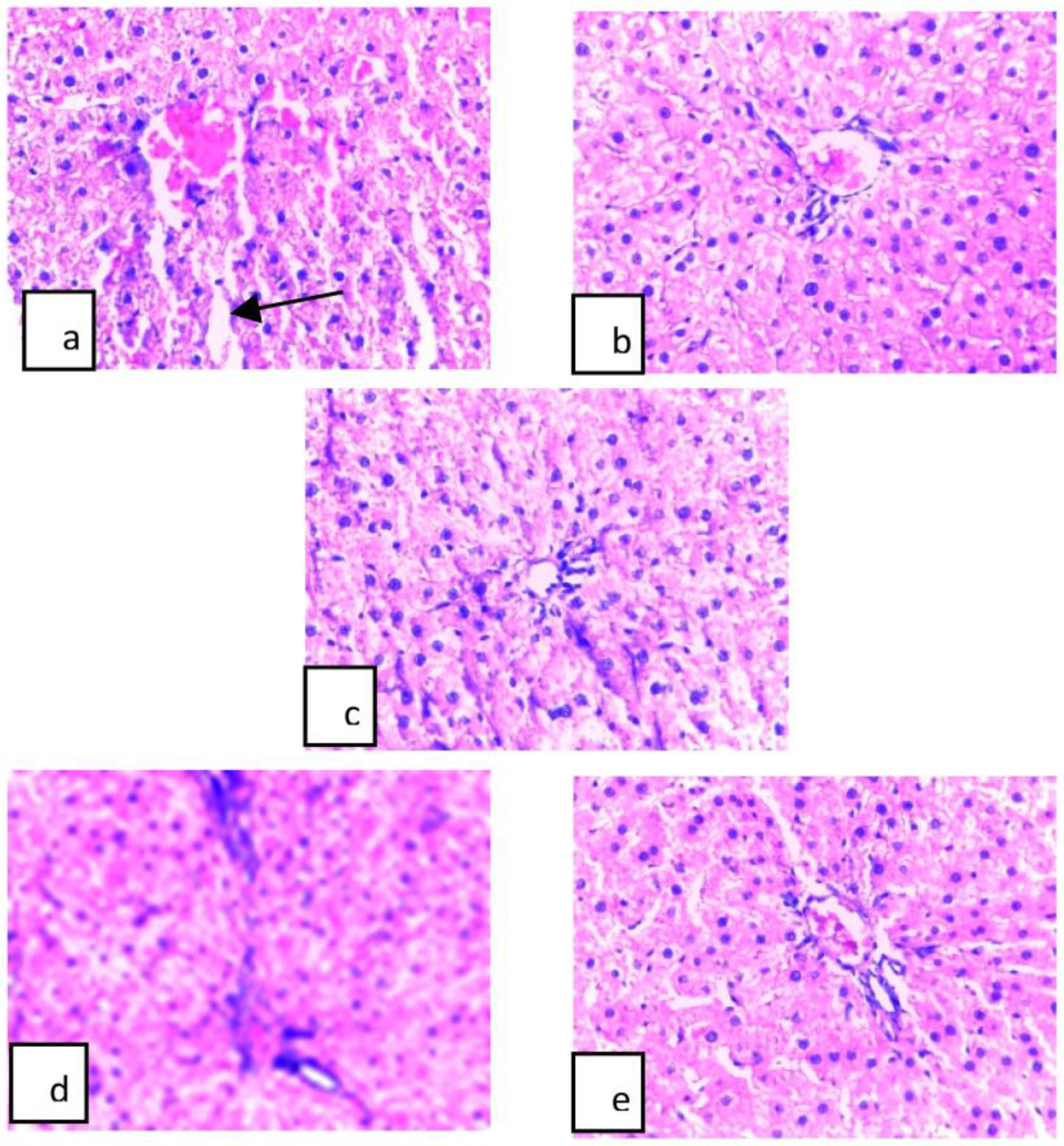
It was found that the histological structure of the liver tissues removed from the control group rats had a normal appearance. Hydropic vacuolar degeneration, mononuclear cell infiltration and dilation in the sinusoids were observed in the liver tissues of the rats that were administered Fe and Zn, while focal necrosis was observed in the Fe group. When the severity of the damage was scored, the highest level of damage was in the Fe group (+++), followed by the Zn and Zn–juglone groups (++). It was found that there was a mild damage in the Zn–juglone group (Figure 2). In terms of the apoptotic cell count, the Fe group (51.66 ± 2.91) demonstrated the highest levels, while the Zn group (50.60 ± 2.40) demonstrated the second-highest levels. It was found that there no significant difference between the Fe–juglone group (14.00 ± 3.80), the Zn–juglone group (13.60 ± 3.04) and the control group (12.60 ± 1.81) (Table 2 and Figure 3). The obtained results were expressed as arithmetic mean (X) and standard deviation (SD) and the statistical analysis was performed using the SPSS 20.0 program. The conformity of the parameters in all the study groups to the normal distribution was evaluated with the Kolmogorov–Smirnov test. The changes observed in the relevant parameters of the study groups were determined using one-way analysis of variance (one-way ANOVA) and multiple comparisons were made according to the Bonferroni test. Values of p < 0.05 were considered statistically significant. The apoptotic-cell-count data and the statistical-analysis results (Table 2) are presented. The difference between the groups in terms of the total apoptotic-cell count was statistically significant (p = 0.001). The difference between the groups containing juglone, Zn and Fe was statistically significant, and a decrease in the apoptotic-cell count was detected in the groups containing juglone (p = 0.001).

4. Discussion

Toxic metals, to a large extent, are dispersed in the environment through industrial effluents, organic wastes, refuse burning and transport and power generation. They can be carried to locations many miles away from their sources by wind, depending upon whether they are in gaseous or particulate form. Metallic pollutants are ultimately washed out of the air onto land or the surfaces of waterways. Thus, air is also a route for the pollution of environment. Metals containing industrial effluents constitute major sources of metallic pollution of the hydrosphere [22].
Heavy metal is a broad term used to describe a group of naturally occurring metallic elements of high molecular weight and density when compared to water. At low concentrations, certain heavy metals, such as iron (Fe), zinc (Zn), copper (Cu) and manganese (Mn), are essential for human survival, but they can become toxic agents at higher concentrations. Other heavy metals, such as arsenic (As), cadmium (Cd), lead (Pb), thallium (Tl) and mercury (Hg), serve no biological purpose. However, they inevitably enter the human body due to their presence in the environment. Similarly to the essential metals, they induce toxicity once certain concentrations are reached [23].
Specifically, heavy metals induce oxidative stress by generating free radicals and reducing antioxidant levels. Heavy metals also alter the confirmation of protein and DNA and inhibit their function. Chelation therapy is commonly used to treat metal toxicity. Chelation is a chemical process that occurs when the interaction between a central metal atom/ion and a ligand leads to the formation of a complex ring-like structure. The ligand has a donor ion/molecule, which has a lone pair of electrons and may be monodentate to polydentate. Since each metal has a different reactivity with a ligand, a specific chelation agent is required for each metal. Combination therapy with a chelating agent and an antioxidant led to improved outcomes [24].
Aluminum-oxide nanoparticles (Al2O3NPs) and zinc -oxide nanoparticles (ZnONPs) are involved in many industries and they are extensively abundant in many aspects of human life [25]. Zinc is a very common pollutant in the environment; its occurrence may influence water-based ecological environments. Consequently, many studies have been directed toward the spread of zinc in water environments. Anthropogenic activities, including municipal wastewater release and coal-burning power plants, industrial methods involving metals and atmospheric outcomes are the main sources of zinc contamination. The extreme discharge of zinc contaminated the surface water and subsurface environment and contributed to groundwater pollution [26].
Organ damage arising from chronic iron overload is remarkable for the range of tissues affected (both replicative and nonreplicative) and for the often slow and insidious onset of organ dysfunction. Foremost amongst the organs and cell types affected by iron overload are the liver, heart and pancreatic beta cells [27].
In the study conducted by Özden et al. [28], it was reported that hydropic degeneration, karyomegaly, steatosis, lobular inflammation, focal necrosis, twinning cell plates, cholestasis, portal area inflammation and Kupffer-cell hyperplasia were observed in rats after fluoxetine. In this study, we obtained similar results in the Fe and Zn groups.
The nitric-oxide and lipid-peroxidation levels were determined to investigate the effects of antioxidants, such as taurine, melatonin and acetylcysteine in preventing cadmium-induced liver damage. It was observed that the glutathione levels and the superoxide-dismutase and glutathione-peroxidase-enzyme activities decreased, while thiobarbituric-acid reactive substances, nitric oxide and immunohistochemically inducible nitric-oxide-synthase-positive cells increased in the liver tissues of the groups administered only cadmium [29]. In our study, we observed that the damage was greater in the iron group and that the number of apoptotic cells was higher in this group. Although similar results were obtained in the zinc group to those in the the iron group, the number of apoptotic cells was considerably reduced in the other groups.
Amifostine was observed to decrease the liver and kidney toxicity associated with radiation. It was found that renal scintigraphy formed diffuse atrophy in the renal tubuli and led to an increase in diffuse connective tissue between the tubuli, while there was focal atrophy in the renal tubuli and an increase in loose fibrous connective tissue between the tubuli in the amifostine + radiation group. In the radiation group, there was diffuse degeneration and fibrous-connective-tissue formation around robust hepatocyte clusters in the liver tissues. The liver structure was protected, the central vein had a regular structure and regular portal areas were observed in the amifostine + radiation group [30]. In our study, similar histopathological results were obtained in the iron and zinc groups.
The melatonin treatment ameliorated the Cd-induced histopathological variations in the liver tissue, which was confirmed by the biochemical and molecular data. It is clear from the results of this study that melatonin exerts a hepatoprotective effect by improving the redox state and suppressing inflammatory reactions and cell apoptosis, as well as ameliorating the performance of liver-tissue histopathology, which is eventually reflected by the improvement in liver function in mice [31]. In this study, the juglone antioxidant reduced the effects of the heavy metals.
The negative effects in the proximal tubuli of the nephrons in the kidney tissues of rats administered cadmium in drinking water were histopathologically investigated. It was found that these effects partially diminished in the group administered melatonin together with cadmium, but did not completely disappear [32]. Similar findings were obtained from the iron and zinc groups of kidney tissue in this study. As with the melatonin, the juglone reduced these effects in this study.
Based on histopathological evaluations, it was reported that beta-glucan had positive effects on kidney damage induced by experimental conditions [33]. It was observed that sevoflurane caused damage in the kidney and liver tissues of rats. It was reported that expansion in the portal area and hepatic-vein tributaries and mild–moderate lymphocytic inflammatory cellular reactions were found in the liver, whereas mild focal inflammation induced by lymphocytes and plasma cells in the interstitial area was evident in kidneys [34]. In this study, the iron and zinc heavy metals also caused damage in the liver and kidney tissues.
It was found that the administration of cyclosporine A administration caused shrinkage in the glomeruli, necrosis, bleeding and degeneration in the tubular cells in kidney tissues of rats and that the administration of omega 3 and sesame oil together with cyclosporine A reduced damage in kidney tissues [35]. As with omega 3 and sesame oil, the juglone reduced these effects in this study.
No microscopic lesion was found in the cortex and medulla in the experimental groups in a study conducted in order to histologically evaluate the antioxidant effects of lycopene in the kidney tissues of diabetic rats [36].
It was reported that Cisplatin, a potent and widely used antitumoral compound, has the notable side effect of nephrotoxicity, inducing oxidative stress and apoptosis in the kidneys. An experiment was conducted to evaluate whether Ginkgo biloba can ameliorate Cisplatin-induced renal tubular damage. Proteinaceous effusion, the sloughing of the proximal tubular epithelium, tubular vacuolar degeneration, and necrosis were evident in the Cisplatin group. As expected, less histological damage was observed in the renal tubules in the Cisplatin-plus-Ginkgo-biloba group. The tubular-necrosis scores in the Ginkgo-biloba group reduced dramatically from 2.65 ± 0.24 to 0.96 ± 0.31 after Cisplatin treatment [37]. In this study, we found similar results from the juglone-plus-Fe and Juglone-plus-Zn groups.
In order to observe the effects of endosulfan and cypermethrin on liver and kidney tissues, these substances were applied in different concentrations by the oral gavage method. Different doses of endosulfan and cypermethrin caused significant structural changes in the liver and kidney tissues. Tubular dilation, degeneration in the tubular epithelium and bleeding in the cortical and medulla regions of the kidneys were observed. Tissue damage was observed in kidney tissues, depending on the dose [38]. In this study, we obtained similar results from the Fe and Zn groups.

5. Conclusions

Juglone, a phenolic compound found in walnuts, has been shown to exert both oxidant and antioxidant activities, to act as an inhibitor of Pin1 and to modulate cell signaling. These diverse actions may confer the possible health benefits of walnuts. Further, juglone may be useful as a therapeutic agent to combat various diseases and to improve health. Juglone may have either pro- or antioxidant characteristics, depending on the concentration [39]. It was reported that juglone reduced oxidative stress in brain and liver tissues [40]. In this study, juglone was found to reduce the effects of iron and zinc.
Hydropic vacuolar degeneration, mononuclear cell infiltration and dilation in the sinusoids were observed in the liver tissues of rats that were administered Fe and Zn, while focal necrosis was observed in the Fe group in this study. When the severity of the damage was scored, the highest level of damage was in the Fe group (+++), followed by the Zn and Zn–juglone groups (++). It was found that there was mild damage in the Zn–juglone group. Tubular dilation, hydropic degeneration in the tubular cells, shrinkage in the glomeruli and congestion were observed in the kidney tissues removed from the rats that were administered iron. Although similar findings were obtained in the zinc group, congestion was not observed. The histopathological changes were similar in the groups that were administered juglone together with iron and zinc, although the tubular dilation and the degeneration in the tubular cells were not as severe. In conclusion, although juglone had a protective effect, it was found that the dose administered did not fully prevent damage, but only reduced it.

Author Contributions

N.Ş. and M.Ş. conceived and planned the study. N.Ş. drafted and revised the manuscript. N.Ş. collected the data. N.Ş. and M.Ş. analyzed the data. All authors have read and agreed to the published version of the manuscript.

Funding

This work was supported by grants from The Scientific and Technical Research Council of Turkey (TÜBİTAK), contract no. 214O699.

Institutional Review Board Statement

The study was conducted in accordance with the Declaration of Helsinki, and approved by the Institutional Ethics Committee of Süleyman Demirel University Animal Experiments Local Ethics Committee Chairman Turkey (protocol code 21438139-338 and date of approval).

Informed Consent Statement

Not applicable.

Data Availability Statement

All the research data related to this manuscript will be available upon reasonable request from the corresponding authors.

Conflicts of Interest

The authors declare no potential conflicts of interest with respect to the research, authorship, and/or publication of this article.

References

  1. Sharfalddin, A.A.; Al-Younis, I.M.; Mohammed, H.A.; Dhahri, M.; Mouffouk, F.; Abu Ali, H.; Anwar, M.J.; Qureshi, K.A.; Hussien, M.A.; Alghably, M.; et al. Therapeutic properties of vanadium complexes. Inorganics 2022, 10, 244. [Google Scholar] [CrossRef]
  2. Savassi, L.A.; Arantes, F.P.; Gomes, M.V.T.; Bazzoli, N. Heavy metals and histopathological alterations in Salminus franciscanus (Lima & Britski, 2007) (Pisces: Characiformes) in the paraopeba river, minas gerais, brazil. Bull. Environ. Contam. Toxicol. 2016, 96, 478–483. [Google Scholar] [CrossRef] [PubMed]
  3. Vymazal, J. Concentration is not enough to evaluate accumulation of heavy metals and nutrients in plants. Sci. Total Environ. 2016, 544, 495–498. [Google Scholar] [CrossRef] [PubMed]
  4. Adrees, M.; Ali, S.; Rizwan, M.; Zia-ur-Rehman, M.; Ibrahim, M.; Abbas, F.; Farid, M.; Qayyum, M.F.; Irshad, M.K. Mechanisms of silicon-mediated alleviation of heavy metal toxicity in plants: A review. Ecotoxicol. Environ. Saf. 2015, 119, 186–197. [Google Scholar] [CrossRef]
  5. Abbas, S.H.; Ismail, I.M.; Mostafa, T.M.; Sulaymon, A.H. Biosorption of heavy metals: A review. J. Chem. Sci. Technol. 2014, 3, 74–102. [Google Scholar]
  6. Kapahi, M.; Sachdeva, S. Bioremediation options for heavy metal pollution. J. Health Pollut. 2019, 9, 191203. [Google Scholar] [CrossRef]
  7. Ayangbenro, A.S.; Babalola, O.O. A new strategy for heavy metal polluted environments: A review of microbial biosorbents. Int. J. Environ. Res. Public Health 2017, 14, 94. [Google Scholar] [CrossRef]
  8. Mohammed, H.A.; Ali, H.M.; Qureshi, K.A.; Alsharidah, M.; Kandil, Y.I.; Said, R.; Mohammed, S.A.A.; Al-Omar, M.S.; Al-Rugaie, O.; Abdellatif, A.A.H.; et al. Comparative phytochemical profile and biological activity of four major medicinal halophytes from Qassim flora. Plants 2021, 10, 2208. [Google Scholar] [CrossRef]
  9. Lo, B. The requirement of iron transport for lymphocyte function. Nat. Genet. 2016, 48, 10–11. [Google Scholar] [CrossRef]
  10. Pietrangelo, A. Iron and the liver. Liver Int. 2016, 36, 116–123. [Google Scholar] [CrossRef]
  11. Aalikhani, M.; Aalikhani, M.; Jahanshahi, M.; Elyasi, L.; Khalili, M. Berberine Is a Promising Alkaloid to Attenuate Iron Toxicity Efficiently in Iron-Overloaded Mice. Nat. Prod. Commun. 2022, 17, 1934578X211029522. [Google Scholar] [CrossRef]
  12. Gonzalez-Estrella, J.; Gallagher, S.; Sierra-Alvarez, R.S.; Field, J.A. Iron sulfide attenuates the methanogenic toxicity of elemental copper and zinc oxide nanoparticles and their soluble metal ion analogs. Sci. Total Environ. 2016, 548, 380–389. [Google Scholar] [CrossRef] [PubMed]
  13. Ziemińska, E.; Strużyńska, L. Zinc modulates nanosilver-induced toxicity in primary neuronal cultures. Neurotox. Res. 2016, 29, 325–343. [Google Scholar] [CrossRef] [PubMed]
  14. Paulsen, M.T.; Ljugman, M. The natural toxin junglone causes degradation of p53 and induces rapid H2AX phosphorylation and cell death in human fibroblasts. Toxicol. Appl. Pharmacol. 2005, 209, 1–9. [Google Scholar] [CrossRef]
  15. Oliveira, I.; Sousa, A.; Ferreira, I.C.; Bento, A.; Estevinho, L.; Pereira, J.A. Total phenols, antioxidant potential and antimicrobial activity of walnut (Juglans regia L.) green husks. Food Chem. Toxicol. 2008, 46, 2326–2331. [Google Scholar] [CrossRef]
  16. Xu, H.L.; Yu, X.F.; Qu, S.C.; Zhang, R.; Qu, X.R.; Chen, Y.P.; Ma, X.Y.; Sui, D.Y. Anti-proliferative effect of Juglone from Juglans mandshurica Maxim on human leukemia cell HL-60 by inducing apoptosis through the mitochondria-dependent pathway. Eur. J. Pharmacol. 2010, 645, 14–22. [Google Scholar] [CrossRef] [PubMed]
  17. Ji, Y.B.; Qu, Z.Y.; Zou, X. Juglone-induced apoptosis in human gastric cancer SGC-7901 cells via the mitochondrial pathway. Exp. Toxicol. Pathol. 2011, 63, 69–78. [Google Scholar] [CrossRef]
  18. Kviencinski, M.R.; Pedrose, J.A.; Felipe, K.B.; Farias, M.S.; Glorieux, C.; Valenzuela, M.; Sid, B.; Benites, J.; Valderramade, J.A.; Verrax, J.; et al. Inhibition of cell proliferation and migration by oxidative stress from ascorbate-driven juglone redox cycling in human bladder-derived T24 cells. Biochem. Bioph. Res. Commun. 2012, 421, 268–273. [Google Scholar] [CrossRef] [PubMed]
  19. Sugie, S.; Okamoto, K.; Rahman, K.W.; Tanaka, T.; Kawai, K.; Yamahare, J.; Mori, H. Inhibitory effects of plumbagin and juglone on azoxymethane-induced intestinal carcinogenesis in rats. Cancer Lett. 1998, 127, 177–183. [Google Scholar] [CrossRef]
  20. Omidi, A.; Riahinia, N.; Torbati, M.B.; Behdani, M.A. Evaluation of protective effect of hydroalcoholic extract of saffron petals in prevention of acetaminophen-induced renal damages in rats. Vet. Sci. Dev. 2015, 5, 5821. [Google Scholar] [CrossRef]
  21. Bancroft, J.D.; Stevens, A.; Turner, D.R. Theory and Practice of Histological Techniques; Churchill Livingstone: London, UK, 1996; 129p. [Google Scholar]
  22. Mahurpawar, M. Effects of heavy metals on human health. Int. J. Res. Granthaalayah 2015, 530, 1–7. [Google Scholar] [CrossRef]
  23. Fu, Z.; Xi, S. The effects of heavy metals on human metabolism. Toxicol. Mech. Methods 2020, 30, 167–176. [Google Scholar] [CrossRef] [PubMed]
  24. Kim, J.J.; Kim, Y.S.; Kumar, V. Heavy metal toxicity: An update of chelating therapeutic strategies. J. Trace Elem. Med. Biol. 2019, 54, 226–231. [Google Scholar] [CrossRef] [PubMed]
  25. Yousef, M.I.; Mutar, T.F.; Kamel, M.A.E.N. Hepato-renal toxicity of oral sub-chronic exposure to aluminum oxide and/or zinc oxide nanoparticles in rats. Toxicol. Rep. 2019, 6, 336–346. [Google Scholar] [CrossRef] [PubMed]
  26. Sankhla, M.S.; Kumar, R.; Prasad, L. Zinc impurity in drinking water and its toxic effect on human health. Indian Internet J. Forensic Med. Toxicol. 2019, 17, 84–87. [Google Scholar] [CrossRef]
  27. Eaton, J.W.; Qian, M. Molecular bases of cellular iron toxicity. Free. Radic. Biol. Med. 2002, 32, 833–840. [Google Scholar] [CrossRef]
  28. Elgebaly, H.A.; Mosa, N.M.; Allach, M.; El-Massry, K.F.; El-Ghorab, A.H.; Al Hroob, A.M.; Mahmoud, A.M. Olive oil and leaf extract prevent fluoxetine-induced hepatotoxicity by attenuating oxidative stress, inflammation and apoptosis. Biomed. Pharmacother. 2018, 98, 446–453. [Google Scholar] [CrossRef]
  29. Aydoğdu, N.; Kanter, M.; Erbaş, H.; Kaymak, K. The effects of taurine, melatonin and acetylcysteine on nitric oxide, lipid peroxidation and some antioxidants in cadmium induced liver injury. Erciyes. Med. J. 2007, 29, 89–096. [Google Scholar]
  30. Uğuzalp-Kaldır, M.; Yürüt-Çaloğlu, V.; Coşar-Alas, R.; Çermik, T.F.; Altaner, Ş.; Eskiocak Saynak, M.; İbiş, K.; Çaloğlu, M.; Tokatlı, F.; Koçak, Z.; et al. Prevention of radiation-induced liver and kiney toxicity: A role for amifostine. Turk. J. Oncol. 2007, 22, 105–117. [Google Scholar]
  31. Yang, Z.; He, Y.; Wang, H.; Zhang, Q. Protective effect of melatonin against chronic cadmium-induced hepatotoxicity by suppressing oxidative stress, inflammation, and apoptosis in mice. Ecotoxicol. Environ. Saf. 2021, 228, 112947. [Google Scholar] [CrossRef]
  32. Kaplan, M.; Atakan, İ.H.; Aydoğdu, N.; Aktoz, T.; Puyan, F.Ö.; Şeren, G.; Tokuç, B.; İnci, O. The effect of melotonin on cadmium-induced renal injury in chronically exposed rats. Turk. J. Urol. 2009, 35, 139–147. [Google Scholar]
  33. Koc, E.; Reis, K.A.; Ebinc, F.A.; Pasaoglu, H.; Demirtas, C.; Omeroglu, S.; Boztepe Derici, U.; GUz, G.; Erten, Y.; Bali, M.; et al. Protective effect of beta-glucan on contrast induced-nephropathy and a comparison of beta-glucan with nebivolol and N-acetylcysteine in rats. Clin. Nephrol. 2011, 15, 658–665. [Google Scholar] [CrossRef] [PubMed]
  34. Kanbak, M.; Erdem, N.; Ayhan, B.; Ozkaya, O.; Bayındı, E.; Sarıcaoğlu, F. Sevoflurane elimination and histopathological changes in lung, liver, kidney and brain tissues. Turk. Clin. J. Med. Sci. 2010, 30, 94. [Google Scholar]
  35. Goksu Erol, A.Y.; Avcı, G.; Sevimli, A.; Ulutas, E.; Ozdemir, M. The protective effects of omega 3 fatty acids and sesame oil against cyclosporine A-induced nephrotoxicity. Drug Chem. Toxicol. 2013, 36, 241–248. [Google Scholar] [CrossRef]
  36. Yegin, S.Ç.; Yur, F. The effect of lycopene application on the antioxidant activity in liver and kidney tissues of diabetic rats. Atatürk Univ. J. Vet. Sci. 2019, 14, 119–128. [Google Scholar] [CrossRef]
  37. Song, J.; Liu, D.; Feng, L.; Zhang, Z.; Jia, X.; Xiao, W. Protective effect of standardized extract of Ginkgo biloba against cisplatin-induced nephrotoxicity. Evid.-Based Complement. Altern. Med. 2013, 11, 846126. [Google Scholar] [CrossRef]
  38. Raj, J.; Ray, R.; Dogra, T.D.; Raina, A. Acute oral toxicity and histopathological study of combination of endosulfan and cypermethrin in wistar rats. Toxicol. Int. 2013, 20, 61–67. [Google Scholar] [CrossRef]
  39. Ahmad, T.; Suzuki, Y.J. Juglone in oxidative stress and cell signaling. Antioxidants 2019, 8, 91. [Google Scholar] [CrossRef]
  40. Cintesun, S.; Ozman, Z.; Kocyigit, A.; Mansuroglu, B.; Kocacalıskan, I. Effects of walnut (Juglans regia L.) kernel extract and juglone on dopamine levels and oxidative stress in rats. Food Biosci. 2022, 51, 102327. [Google Scholar] [CrossRef]
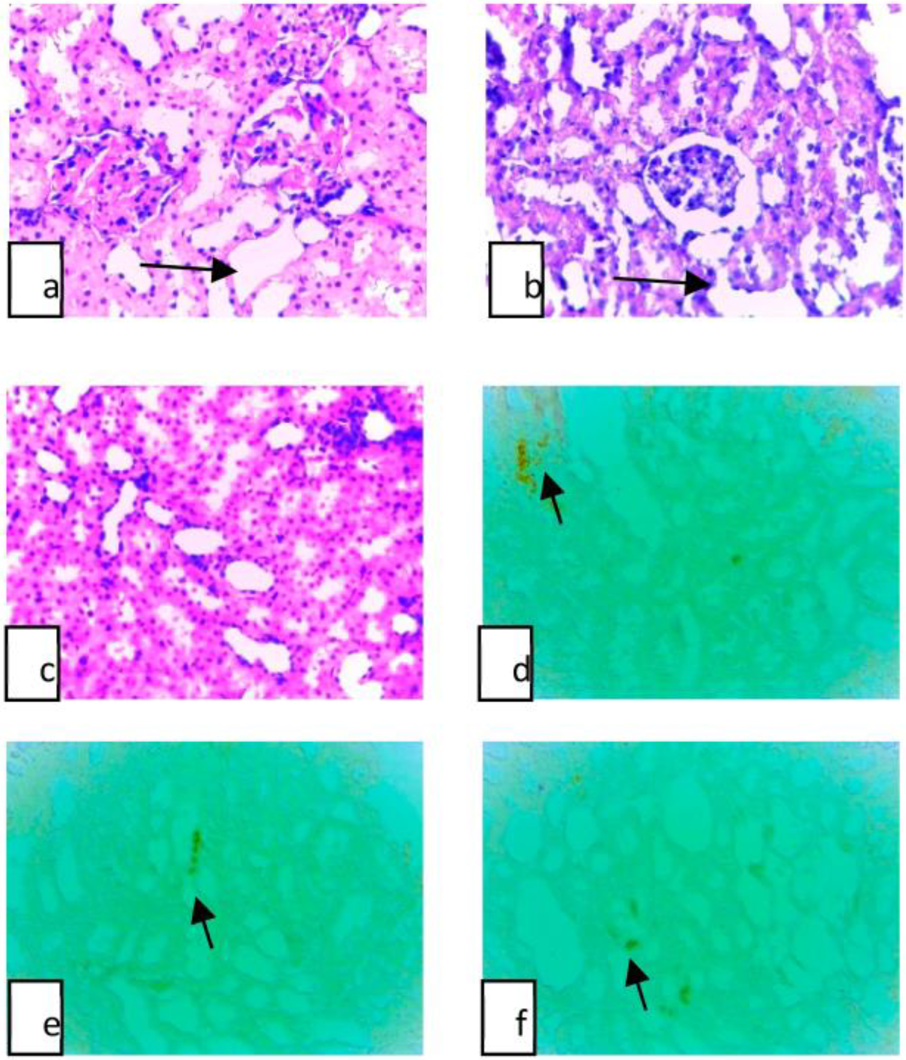
Figure 1. (a) Fe group, kidney, H&E, tubular dilation (arrow), ×40; (b) Zn group, kidney, H&E, tubular dilation (arrow), ×40; (c) Control group, Kidney, H&E, ×40; (d) Fe group, apoptotic cells in kidney (arrow), TUNEL, ×40; (e) Fe–juglone group, apoptotic cells in kidney (arrow), TUNEL, ×40; (f) Zn group, apoptotic cells in kidney (arrow),TUNEL, ×40.
Applsci 13 02203 g001
Figure 2. (a) Fe group, liver, H&E, dilation in sinusoids (arrow), ×40; (b) Zn group, liver, H&E, ×40; (c) control group, liver, H&E, ×40; (d) Zn–juglone group, liver, H&E, ×40; (e) Fe–juglone group, liver, H&E, ×40.
Applsci 13 02203 g002
Figure 3. (a) Fe group, apoptotic cells in liver, TUNEL, ×40; (b) Zn group, apoptotic cells in liver, TUNEL, ×40; (c) control group, apoptotic cells in liver, TUNEL, ×40.
Applsci 13 02203 g003
Table 1. Mean number of apoptotic cells in groups of kidney tissue.
GroupsFe GroupZn GroupFe–Juglone GroupZn–Juglone GroupControl Group
apoptotic cell count69.50 ± 4.20 *41.25 ± 2.75 **43.0 ± 3.55 *26.25 ± 3.55 **14.75 ± 2.55 */**
* There is a significant difference between the Fe group and the control and Fe–juglone group, p = 0.001. ** There is a significant difference between the Zn group and the control and Zn–juglone group, p = 0.001.
Table 2. Mean number of apoptotic cells in groups of liver tissue.
GroupsFe GroupZn GroupFe–Juglone GroupZn–Juglone GroupControl Group
apoptotic cell count51.66 ± 2.91 *50.60 ± 2.40 **14.00 ± 3.80 *13.60 ± 3.049 **12.60 ± 1.81 */**
* There is a significant difference between the Fe group and the control and Fe–juglone group, p = 0.001. ** There is a significant difference between the Zn group and the control and Zn–juglone groups, p = 0.001.
Disclaimer/Publisher’s Note: The statements, opinions and data contained in all publications are solely those of the individual author(s) and contributor(s) and not of MDPI and/or the editor(s). MDPI and/or the editor(s) disclaim responsibility for any injury to people or property resulting from any ideas, methods, instructions or products referred to in the content.