Article

Evaluation of the Failure Rate and Clinical Efficacy of Orthodontic Brackets Indirect Bonding with Computer-Aided Transfer Jig System: An In Vivo Study

1
Department of Orthodontics, Graduate School of Dentistry, Kyung Hee University, Seoul 02447, Republic of Korea
2
Department of Orthodontics, Kyung Hee University Dental Hospital, Seoul 02447, Republic of Korea
*
Author to whom correspondence should be addressed.
These authors contributed equally to this work.
Appl. Sci. 2023, 13(3), 1668; https://doi.org/10.3390/app13031668
Submission received: 21 December 2022 / Revised: 21 January 2023 / Accepted: 25 January 2023 / Published: 28 January 2023
(This article belongs to the Section Applied Dentistry and Oral Sciences)

Abstract

:
This study aimed to evaluate the failure rates and relevant factors of bonded orthodontic brackets with a computer-aided design and manufacturing (CAD/CAM)-based 3D-printed indirect bonding jig system (IDBS) using 2531 natural teeth selected from 99 orthodontic patients. Ceramic self-ligating brackets and metal tubes were used in this study. Proportion analysis was used to calculate the total bracket re-bonding rate and respective proportions of bonding failure and position error. Crossover frequency analysis was used to analyze the total bracket re-bonding, bonding failure, and position error rates in different tooth positions of the maxilla and mandible. Multiple linear regression analysis was used to evaluate the association between dependent variables (age, sex, treatment stage, skeletal divergence, and tooth position) and the bracket bonding failure rate. Pearson’s Chi-square test was used to test the difference between the maxilla and mandible for each variable. The total bracket re-bonding rate was 22.64%, and the bonding failure rate and position error rates accounted for 15.09% and 7.55%, respectively. The bonding failure rate was higher in the mandibular teeth than in the maxillary teeth (p < 0.05). Anterior teeth had a higher position error rate, and mandibular anterior teeth had a higher bonding failure rate. The accuracy of IDBS was higher in the premolars and molars. Sex, age, and treatment stage were affected by IDBS. Patients with hyperdivergent skeletal patterns had higher rates of bracket bonding failure. The results of this study can provide practical guidelines for placing brackets with 3D-printed IDBS on the entire dentition to ensure the precision and accuracy of their use during orthodontic treatments.

1. Introduction

As the actuating apparatus and force transmission devices of orthodontic treatment, brackets need to be placed in an opportune position to provide mechanical effects with treatment wires. Impertinent placement of brackets may lead to unnecessary tooth movement, including impertinent torque, rotation, inclination, or intrusion/extrusion [1]. Direct bonding (DB) is the basic technique for fixed appliance placement and is used worldwide. However, the accuracy of direct bracket bonding is limited by clinical experience and professional skills in orthodontics, including concentration and tiredness [2]. With the development of digital technology and materials, the indirect bracket bonding system (IDBS) was introduced in 1972 by Silvermann et al. [3]. Computer-aided design and computer-aided manufacturing (CAD/CAM) technology-assisted indirect bonding were presented in 2006. Moreover, stereolithography, a type of rapid prototyping method started in 2011, is used to create transfer trays, and described as “jigs” [4]. Compared with the DB technique, IDBS allows orthodontists to place the brackets accurately and individually at the ideal position for each tooth in the laboratory environment, where various uncontrollable factors such as malformation and defection of teeth and a limited field of vision affect the working conditions of orthodontists [5]. CAD/CAM technology assists IDBS in reducing the clinical treatment time by simplifying the bracket-bonding steps and optimizing the preprocessing process [6]. During clinical treatment, only three steps are required to complete a single bracket bonding: the tooth surface process, transfer trays/jigs placement, and adhesive curing. Czolgosz et al. found that the chair time of brackets bonded with the CAD/CAM-assisted IDBS was reduced by 4 min for every half of the mouth compared with the DB [6]. Furthermore, IDBS can effectively control bacterial plaque accumulation, reduce the onset of white spots, and reduce inflammation by minimizing the adhesive residual and enamel etching area [6]. This study also found that plaque accumulation around the brackets in IDBS was significantly lower than that in the DB during the first 4 months, which is the most difficult time to clean after bracket bonding, and the onset of white spots was significantly reduced during orthodontic treatment [7].
Several studies have demonstrated the positive effects of IDBS [8,9]. Sabbagh et al. described bracket transfer accuracy in a systematic review; the overall linear mean transfer errors in the mesiodistal, vertical, and buccolingual directions were 0.08 mm, 0.09 mm, and 0.14 mm, respectively. The overall angular mean transfer errors for the angulation, rotation, and torque were 1.13°, 0.93°, and 1.11°, respectively [8]. Park et al. showed that IDBS was in a clinically acceptable range of ±0.05 mm and 2.0° for linear and angular measurements, according to the American Board of Orthodontics Objective Grading System [9]. However, most previous studies have focused on evaluating the success rate and accuracy of IDBS in laboratory environments, that is, in vitro studies. In actual clinical practice, bracket transfer and bonding occur in the patient’s mouth with exposure to saliva and different stressors. Several factors may influence the success rate and accuracy of bracket bonding. However, the bonding failure rate of IDBS in clinical practice has not yet been investigated.
This clinical study aimed to evaluate the failure rates and relevant factors of the indirect bonding of orthodontic brackets with CAD/CAM-based 3D-printed IDB transfer jigs and to suggest practical guidelines for the clinical application of IDBS.

2. Materials and Methods

2.1. Participants

Participants who were treated using ceramic self-ligation orthodontic brackets (0.022-in Quicklear®, Forestadent, Pforzheim, Germany) and metal tubes (3M Unitek, Monrovia, CA, USA) with Tweemac prescription were selected, and 99 orthodontic patients (62 women and 37 men) were treated with fixed orthodontic appliances from 2018 to 2021 at the Kyung Hee University Dental Hospital. For these patients, the brackets were bonded to a total of 2596 teeth with CAD/CAM-based 3D-printed transfer jigs. For sample size calculation (G*Power version 3.1.9.2; Heinrich-Heine-Universität Düsseldorf, Düsseldorf, Germany), a multiple linear regression analysis was performed. This was necessary for a significant fit for the regression model (with a power of at least 0.95) based on the six independent variables (age, sex, treatment stage, vertical, and sagittal skeletal divergence, and tooth position) and α error of 0.05. The results reveal that the minimum required sample size was 146 teeth. All operators were board-certified orthodontists at the Kyung Hee University Dental Hospital Biocreative Orthodontic Center. This retrospective study was based on the medical records of patients at the Department of Orthodontics at Kyung Hee University Dental Hospital and was approved by the Institutional Review Board of Kyung Hee University Dental Hospital (KH-DT22001).

2.2. Inclusion and Exclusion Criteria

(1) Patients planned to be treated with IDBS; (2) Permanent dentitions; (3) Natural teeth excluding prosthetic teeth. The final sample size was 2531 teeth from 99 orthodontic patients (62 women and 37 men).

2.3. Study Procedure

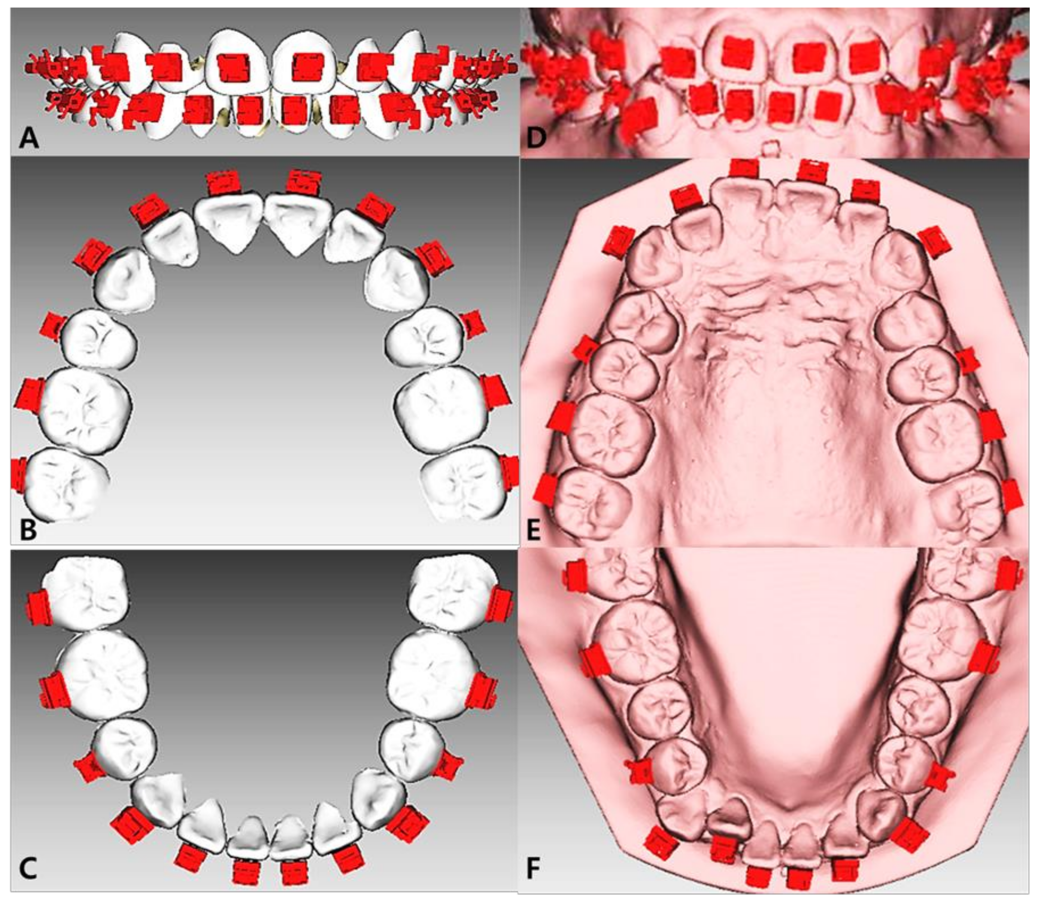
The clinical work procedure for the IDBS is shown (Figure 1). Intra-oral scan data were obtained from the patients using an intra-oral scanner (Medit I-700, Medit Corp., Seoul, Republic of Korea) and converted to stereolithography (STL) files. The virtual set-up and placement of the orthodontic brackets on each tooth were performed using 3Txer software (ver. 2.5; CENOS Co., Indeokwon, Gyeonggi-do, Republic of Korea) (Figure 2). The IDB Bracket transfer jigs were designed using CAD/CAM software and fabricated using a polyjet-type 3D printer (Projet MJP 3600, 3D Systems Co., Rock Hill, SC, USA) (Figure 3A). Ceramic self-ligation orthodontic brackets and metal tubes with Tweemac prescription were adapted into the fabricated transfer jigs. Teeth surfaces were etched with phosphoric acid gel and Transbond XT primers (Figure 3B). After the light-curing resin adhesive (Transbond™ XT Light Cure Adhesive, 3M Unitek, Monrovia, CA, USA) was applied to the bracket base, the IDB jig with the bracket was bonded to the tooth using a light-curing device (VALO, Ultradent, South Jordan, UT, USA (Figure 3C–E)). Premature contact spots were checked and ground after the jig removal (Figure 3G–I).

2.4. Measurements

The reasons for bracket re-bonding were divided into bonding failure (BF) and position error (PE, intentional removal of brackets due to position error). The total bracket re-bonding rate and respective proportions of BF and PE in the total bracket re-bonding were evaluated in the maxilla and mandible.
Treatment stages were classified into the initial, middle, and late stages based on the treatment period: The initial stage was 0–12 months after bonding brackets; the middle stage was 12–24 months after bonding brackets; and the late stage was >24 months after the initial bonding day.

2.5. Statistical Analysis

Proportion analysis was used to calculate the total bracket re-bonding rate and respective proportions of bonding failure and position error. Crossover frequency analysis was used to analyze the total bracket re-bonding, BF, and PE rates in different tooth positions of the maxilla and mandible, and Pearson’s Chi-square test was used to test the difference between the maxilla and mandible of each variable.
Multiple linear regression analysis was used to determine the association between dependent variables (age, sex, treatment stage, skeletal divergence, and tooth position) and the bracket bonding failure rate. All statistical analyses were performed using the SAS software (version 9.4, SAS Institute Inc., Cary, NC, USA). Statistical significance was set at p < 0.05, and p < 0.001 was considered statistically highly significant.

3. Results

The total bracket re-bonding rate was 22.64%. Among the total bracket re-bonding rates, the BF and PE rates were 15.09% and 7.55%, respectively (Table 1).
Statistically significant differences were observed in the BF rate between the maxilla and the mandible (p < 0.05). The BF rates of the anteriors, premolars, and molars were 11.26%, 11.78%, and 14.66% in the maxilla and 32.98%, 12.83%, and 16.49% in the mandible, respectively. However, there was no statistically significant difference between the maxilla and mandible in the PE and total bracket re-bonding rates (Table 2).
Statistically significant differences were found in the total re-bonding rate, BF, and PE according to sex and age (p < 0.05). Men had higher bracket re-bonding failure rates than women (p < 0.05). Teenagers had more re-bonded brackets than older patients (>20 years). Meanwhile, no statistically significant difference was found in the bracket re-bonding rate according to the skeletal classification in the sagittal plane. However, compared to patients with other vertical skeletal patterns, patients with hyperdivergent skeletal patterns showed higher BF and total re-bonding rates (p < 0.05) (Table 3).
The most frequent stage for the bracket re-bonding rate was the middle stage (p < 0.05). The BF, PE, and total re-bonding rates were higher at the initial stage than at the late stage. The BF rate was not statistically different among the three tooth position categories. However, PE was most common in the anterior teeth. The total bracket re-bonding rate was higher in the anterior teeth than in the premolars, and there was no significant difference between the anterior teeth and molars (Table 4).

4. Discussion

Bracket adhesion failure is directly related to the efficiency and effects of orthodontic treatment. Skidmore et al. found that the treatment time would be extended by 0.3 months for each bracket adhesion failure, and three or more bracket adhesion failures would extend the treatment time by up to 1.5 months [10]. Analysis and control of the bracket re-bonding rate were consistent with the benefits of orthodontic treatment for both patients and clinicians.
The precision and accuracy of an IDBS are influenced by digital technology and human factors. Digital technology factors come from the processes of intra-oral scanning, 3D modeling, and bracket visual setup, which assist in the fabrication of transfer jigs. These factors are easy to evaluate and analyze in laboratory environments. However, the “Human factors”, which come from clinicians’ and patients’ variability, were not easy to simulate and analyze in the laboratory. Hence, it was more accurate and realistic to evaluate the failure rate of bracket bonding through in vivo studies, which are influenced by multiple factors in clinical practice. This study found that the total bracket re-bonding rate was 22.64% in clinical practice, which was higher than in some previous studies [9,11]. However, it should be pointed out that failure rate of IDB has been demonstrated to be similar as the DB technique in the other study [12]. Operators should take this important factor into careful consideration before clinical choices. This discrepancy might be partly due to the gap between bases on the brackets and tooth surface. In the IDBS of brackets, the thickness of the resin between the bracket and tooth surface is inevitably greater than that of DBS [13]. According to the finite element analysis, the farther the applied force is from the bonding interface, the higher the momentum [14]. Moreover, the thicker the adhesive, the greater the shear stress on the brackets. Knox et al. found that adhesive thickness affected the distribution of major principal stresses within the adhesive [15]. Another study showed that the failure rate at the tooth-adhesive interface increased with increased adhesive thickness in shear testing [16]. The total bracket re-bonding rate was further divided into subjective and objective reasons for group discussions, which are position error (7.55%) and bonding failure (15.09%). In this study, BF (15.09%) was the main reason for the total bracket re-bonding rate compared to PE (7.55%).
The BF rates between the maxilla and mandible were significantly different (p < 0.05). Mandibular teeth showed a higher BF rate than maxillary teeth and were concentrated in the anterior teeth (Table 2). This was impressive because it suggested that the position most prone to bracket bonding failure was the anterior mandibular teeth. Kulkarni, et al. reported similar results [17]. It is speculated to be related to bite depth, arch wire force, movement of surrounding tissues, and excessive salivation. Bite depth has been reported to directly affect the bracket bonding failure rate, and more bracket failures occur in patients with deep bites than in those with an average or open bite [18], and the addition of bite blocks could effectively reduce the bonding failure rate [19]. The orthodontic arch wire moves the tooth by generating force through wire deformation, and bracket bonding failure occurs when the wire force exceeds the shear bond strength of the adhesive. Severe crowding is common in the lower anterior teeth, and the archwire deforms to a greater extent and exerts more force on the lower anterior brackets, eventually causing bracket-bonding failure. Most failed brackets were reported for leveling and alignment stage (0.016″ NiTi) wires [19]. Furthermore, the mandibular anterior teeth are covered by the lower lip and close to the tongue on the lingual side. Unexpected movement of soft tissues around the anterior teeth affects bracket bonding. The sublingual gland duct orifice is located on the lingual side of the lower anterior teeth, and excessive salivation would contaminate the bonding interface. The study found that saliva contamination can significantly reduce the shear bond strength of brackets bonded with hydrophobic bond systems [19]. Combined with previous studies, the results of this study suggest that paying attention to the patient’s bite depth, removing the premature contact spots of brackets in time, and controlling the force of the archwire within the shear bond strength of the adhesive may help to decrease the BF rate in IDBS, particularly for the mandibular anterior tooth.
Sex, age, and treatment stage were highly related to the re-bonding rate. In terms of sex, men showed higher total bracket re-bonding, which included both BF and PE, than women (Table 3). Sex has long been reported to be associated with bond failure; however, the results of previous studies remain controversial [20,21]. This may be influenced by the different dietary structures, masticatory strength, and pH of the saliva between men and women. It is difficult to exclude all confounding factors and only analyze the impact of sex. Teenagers had more re-bonded brackets than those over 20 years of age (Table 3). Treatment motivation helps explain this result. Treatment motivation can be divided into external (which is influenced by other people or the social environment) or internal (which is a personal desire) [22]. Barbosa et al. found that adult patients showed lower bracket breakage rates because they asked for orthodontic treatment according to their own desires, while adolescents do it because they are compelled by parents or other factors [23]. In addition, improving patient compliance could effectively reduce bracket failure rates [24]. However, only 48% of adolescent patients follow the treatment instructions [25]. This result suggests that orthodontists need to closely observe patient compliance. Subsequently, they should lead the patients to actively cooperate with the treatment and guide the patients to properly manage the orthodontic appliances by changing their diet and eating habits to reduce the total bracket re-bonding rate. For the treatment period, the most frequent stage for the bracket re-bonding rate was the middle stage (Table 4). The high BF rate may be influenced by the aging of the orthodontic adhesive. The aging of the adhesive is influenced by temperature fluctuations, enzymatic degradation, salivary exposure, and pH changes [26]. Dudás et al. compared the bond failures of different orthodontic materials and found that bond failure occurred owing to material aging, which peaks at 12–18 months [27]. Khan et al. also found a similar result, with 69% of bracket failures occurring in the first 6 months after boding [19]. The higher position error is speculated to be associated with the clinician’s tendency to evaluate the bracket position after the leveling and alignment of teeth, which would be hard to expect in the virtual set-up stage only with dental crowns.
Interestingly, the anterior teeth showed a higher position error rate than the premolars and molars (Table 4). In this study, we used a one-body transfer jig system based on CAD/CAM technology. The 3D-printed transfer trays had higher bracket transfer accuracy than the other methods [28]. Moreover, the one-body transfer jig system accommodated only one bracket, which eliminated interference from adjacent teeth, and the transfer accuracy was verified to be within a clinically acceptable level for anterior teeth [9]. Excluding appliance errors based on previous studies, the position error of brackets might occur during the process of virtual set-up and clinical transfer. Anterior teeth have sharp incisal edges and cusps, which make it difficult to obtain a smooth scan of these areas. Additionally, overlapping areas caused by dental crowding obstruct the entry of the scan ray. This affected the accuracy of the virtual dentition model [29]. In contrast, clinical transfer errors also need to be considered. During bracket transfer by one-body transfer jigs, pressure from the occlusal and buccal directions must be applied to ensure the accuracy and stability of the bracket position [30]. The shape of the occlusal surface of molars and premolars is polygonal in a large contact area with a transfer jig, which makes it easy to apply a stable occlusal pressure without any sliding. However, the anterior incisal edge is in linear or point contact with the transfer jig, which is usually accompanied by sliding and shaking during bracket transfer. Additionally, the molars and premolars are more upright in the jaws, which makes it easier for occlusal pressure to pass through the rotation center of the jig and the long axis of the tooth to maintain the stability of the transfer jigs. However, anterior teeth are often accompanied by crowding and inclination, which makes it difficult to apply a vertical force along the direction of the long axis of the tooth from the jig at the occlusal surface [31]. Considering the occlusal anatomy and smaller buccolingual inclination of molars and premolars, it is easier to apply a steady and sustained force to transfer the brackets more precisely for molars and premolars (Figure 3). A similar difference in which the bracket position between the actual and virtual models was the largest in the anterior teeth has been reported [1].
The vertical skeletal pattern was significantly associated with BF and the total re-bonding rate. Compared to patients with vertical skeletal patterns, patients with hyperdivergent skeletal patterns showed a higher total re-bonding rate, which was caused by the higher bonding failure rate. This result is assumed to be affected by muscle force. Patients with hyperdivergent skeletal patterns had a lower maximum voluntary bite force, which means that, when chewing the same food, hyperdivergent patients need to chew more times because of the lower bite force [32]. Additionally, research has found higher resting temporal muscle activity in patients with hyperdivergent skeletal patterns [33]. This causes the brackets to receive a certain shear force even in the resting state. In other words, brackets in patients with hyperdivergent skeletal patterns received more frequency and sustained cyclic occlusal forces than patients with other skeletal patterns. Mansour et al. found that cyclic loading, including occlusal forces, could reduce bond strength [34]. Another survival analysis of bracket bonds also supports this hypothesis. A high mechanical loading level significantly affects the bracket bond failure [35]. The results of this study show that patients with hyperdivergent skeletal patterns are more prone to bracket bonding failure due to the influence of muscle force and occlusal force cyclic loading when compared with normodivergent and hypodivergent patients.
To discuss the factors influencing the IDBS failure rate, orthodontic adhesives must be considered. A Transbond XT (3M Unitek, Monrovia, CA, USA) was used in this study. This is a high inorganic filler-contented direct bonding light-curing resin, considered the “gold standard” of orthodontic DB adhesives [36]. IDB orthodontic adhesives have the characteristics of low viscosity, nanometric filler particles, and modified properties. IDBS adhesives can maintain their shape after placement until light-curing without draining around the bracket during the bonding procedure [37]. Although the use of IDBS adhesives was an option, an in vivo study of failure rates of different resins found that the use of Transbond XT for the IDB was acceptable in clinical application [38]. Therefore, the use of Transbond XT in this study is reasonable and would not affect the research results.
Finally, the failure rate of the IDBS has been demonstrated to be higher than that of the DB technique during long-term follow-up (12–15 months) in previous studies [36]. The results of this study show that age, sex, treatment stage, vertical skeletal divergence, and tooth position are risk factors for bracket bonding failure. Orthodontists should carefully control these risk factors as much as possible to reduce the bracket bonding failure rate in the clinic.
Based on the results of this study, the following suggestions are provided for clinical guidance:
1.
Considering that mandibular teeth, especially mandibular anterior teeth, had a higher bonding failure rate, it is recommended to control the thickness of the resin adhesive as much as possible during the IDB process. For mandibular anterior teeth, the unexpected movement of the surrounding soft tissue must be limited, and contamination of the bonding interface caused by excessive salivation must be controlled. A small archwire was chosen for overcrowded cases to avoid bonding failure due to excessive orthodontic force. Additionally, care should be taken to check and remove premature contact spots. Resin bite blocks can be added when necessary to open the bite;
2.
Considering that the anterior teeth had a higher position error rate, the application of stable and steady occlusal pressure along the long axis of the tooth on the transfer jigs in the IDBS is suggested;
3.
The effects of sex, age, and vertical skeletal pattern warranted careful follow-up of male patients under 20 years of age with hyperdivergent skeletal patterns, provided they followed the treatment instructions. Moreover, patients should be advised to change their dietary habits.
This study has some limitations. Bracket bonding was not performed by the same operator. Although the technical sensitivity of different operators may have affected the results, the operators in this study were professional orthodontists who had received the same years of training from a standardized resident training institution to minimize operator error as much as possible. We, therefore, divided the reasons for bracket re-bonding into two factors. A more precise and specific investigation of the samples bonded by a single operator should be conducted in a future study.

5. Conclusions

This was the first study to evaluate the actual bracket re-bonding rate and related factors for IDBS with CAD/CAM-based transfer jig system. The results showed that:
1.
Anterior teeth had a higher position error rate. Mandibular anterior teeth had a higher bonding failure rate. IDBS had higher accuracy in the premolars and molars;
2.
Sex, age, and treatment periods are the factors affecting IDBS;
3.
Patients with hyperdivergent skeletal patterns had higher brackets bonding failure rates, while IDBS had higher strength in patients with normodivergent and hypodivergent skeletal patterns.

Author Contributions

Conceptualization, J.B., H.-J.L. and S.-H.K.; methodology, H.-J.L. and S.-H.K.; resources, J.B. and S.-H.K.; data curation, J.B. and H.-J.L.; writing—original draft preparation, J.B. and H.-J.L.; writing—review and editing, J.B. and S.-H.K.; visualization, S.-H.K.; supervision, S.-H.K.; project administration, S.-H.K. All authors have read and agreed to the published version of the manuscript.

Funding

This research received no external funding.

Institutional Review Board Statement

The study was conducted according to the guidelines of the Declaration of Helsinki and approved by the Institutional Review Board of Kyung Hee University Dental Hospital (protocol code KH-DT22001, 7 March 2022).

Informed Consent Statement

Informed consent was obtained from all the participants involved in the study.

Data Availability Statement

Not applicable.

Acknowledgments

The authors offer special thanks to Hyo-Won Ahn and Jin-Young Choi, Faculties, Department of Orthodontics, Kyung Hee University School of Dentistry for assisting manuscript preparation; and Seung-Woo Kang, General Manager of CENOS Co., Indeokwon, Gyeonggido, Republic of Korea for supporting the manuscript preparation with the figure image work; and Gyu-Beom Ko, CEO of Medit company, for supporting the manuscript preparation with the intra oral scanning (Medit I700, Medit Corp., Seoul, Republic of Korea).

Conflicts of Interest

The authors declare no conflict of interest.

References

  1. Park, J.H.; Choi, J.Y.; Oh, S.H.; Kim, S.H. Three-Dimensional Digital Superimposition of Orthodontic Bracket Position by Using a Computer-Aided Transfer Jig System: An Accuracy Analysis. Sensors 2021, 21, 5911. [Google Scholar] [CrossRef] [PubMed]
  2. Gange, P. The evolution of bonding in orthodontics. Am. J. Orthod. Dentofac. Orthop. 2015, 147 (Suppl. S4), S56–S63. [Google Scholar] [CrossRef]
  3. Silverman, E.; Cohen, M.; Gianelly, A.A.; Dietz, V.S. A universal direct bonding system for both metal and plastic brackets. Am. J. Orthod. 1972, 62, 236–244. [Google Scholar] [CrossRef]
  4. Kanashiro, L.K.; Robles-Ruíz, J.J.; Ciamponi, A.L.; Medeiros, I.S.; Dominguez, G.C.; de Fantini, S.M. Effect of adhesion boosters on indirect bracket bonding. Angle Orthod. 2014, 84, 171–176. [Google Scholar] [CrossRef] [PubMed] [Green Version]
  5. Panayi, N.C.; Tsolakis, A.I.; Athanasiou, A.E. Digital assessment of direct and virtual indirect bonding of orthodontic brackets: A clinical prospective cross-sectional comparative investigation. Int. Orthod. 2020, 18, 714–721. [Google Scholar] [CrossRef] [PubMed]
  6. Czolgosz, I.; Cattaneo, P.M.; Cornelis, M.A. Computer-aided indirect bonding versus traditional direct bonding of orthodontic brackets: Bonding time, immediate bonding failures, and cost-minimization. A randomized controlled trial. Eur. J. Orthod. 2021, 43, 144–151. [Google Scholar] [CrossRef]
  7. Dalessandri, D.; Dalessandri, M.; Bonetti, S.; Visconti, L.; Paganelli, C. Effectiveness of an indirect bonding technique in reducing plaque accumulation around braces. Angle Orthod. 2012, 82, 313–318. [Google Scholar] [CrossRef] [Green Version]
  8. Sabbagh, H.; Khazaei, Y.; Baumert, U.; Hoffmann, L.; Wichelhaus, A.; Janjic Rankovic, M. Bracket Transfer Accuracy with the Indirect Bonding Technique—A Systematic Review and Meta-Analysis. J. Clin. Med. 2022, 11, 2568. [Google Scholar] [CrossRef]
  9. Park, J.H.; Choi, J.Y.; Kim, S.H.; Kim, S.J.; Lee, K.J.; Nelson, G. Three-dimensional evaluation of the transfer accuracy of a bracket jig fabricated using computer-aided design and manufacturing to the anterior dentition: An in vitro study. Korean J. Orthod. 2021, 51, 375–386. [Google Scholar] [CrossRef]
  10. Skidmore, K.J.; Brook, K.J.; Thomson, W.M.; Harding, W.J. Factors influencing treatment time in or-thodontic patients. Am. J. Orthod. Dentofac. Orthop. 2006, 129, 230–238. [Google Scholar] [CrossRef]
  11. Huang, X.H.; Xu, L.; Lin, S. Effects of double transparent pressure diaphragm transfer tray on indirect bonding. Shanghai J. Stomatol. 2016, 25, 734–737. [Google Scholar]
  12. Menini, A.; Cozzani, M.; Sfondrini, M.; Scribante, A.; Cozzani, P.; Gandini, P. A 15-month evaluation of bond failures of ortho-dontic brackets bonded with direct versus indirect bonding technique: A clinical trial. Prog. Orthod. 2014, 15, 70. [Google Scholar] [CrossRef] [PubMed] [Green Version]
  13. Hassan, M.S.; Abdelsayed, F.A.; Abdelghany, A.H.; Morse, Z.; Aboulfotouh, M.H. For Indirect Orthodontic Attachment Place-ment, adding a Custom Composite Resin Base Is Not Beneficial: A Split-Mouth Randomized Clinical Trial. Int. J. Dent. 2022, 2022, 9059697. [Google Scholar] [CrossRef]
  14. Benaissa, A.; Merdji, A.; Bendjaballah, M.Z.; Ngan, P.; Mukdadi, O.M. Stress influence on orthodontic system components under simulated treatment loadings. Comput. Methods Programs Biomed. 2020, 195, 105569. [Google Scholar] [CrossRef]
  15. Knox, J.; Kralj, B.; Hübsch, P.F.; Middleton, J.; Jones, M.L. An evaluation of the influence of orthodontic adhesive on the stresses generated in a bonded bracket finite element model. Am. J. Orthod. Dentofac. Orthop. 2001, 119, 43–53. [Google Scholar] [CrossRef]
  16. Arici, S.; Caniklioglu, C.M.; Arici, N.; Ozer, M.; Oguz, B. Adhesive thickness effects on the bond strength of a light-cured resin-modified glass ionomer cement. Angle Orthod. 2005, 75, 254–259. [Google Scholar] [PubMed]
  17. Kulkarni, N.B.; Goyal, S. Comparison of Bracket Failure Rate between Two Different Materials Used to Fabricate Transfer Trays for Indirect Orthodontic Bonding. J. Contemp. Dent. Pract. 2022, 23, 307–312. [Google Scholar]
  18. Ahangar Atashi, M.H.; Shahamfar, M. Long-term evaluation of clinical performance of direct-bonded brackets: An epidemio-logic survey. J. Contemp. Dent. Pract. 2013, 14, 738–742. [Google Scholar] [CrossRef] [PubMed]
  19. Khan, H.; Mheissen, S.; Iqbal, A.; Jafri, A.R.; Alam, M.K. Bracket Failure in Orthodontic Patients: The Incidence and the Influence of Different Factors. BioMed Res. Int. 2022, 2022, 5128870. [Google Scholar] [CrossRef]
  20. Hitmi, L.; Muller, C.; Mujajic, M.; Attal, J.P. An 18-month clinical study of bond failures with resin-modified glass ionomer cement in orthodontic practice. Am. J. Orthod. Dentofac. Orthop. 2001, 120, 406–415. [Google Scholar] [CrossRef]
  21. Moninuola, A.E.; Costa, O.O.; Isiekwe, M.C. A review of orthodontic bond failure using a chemical cure adhesive. Odontostomatol. -Trop. Trop. Dent. J. 2010, 33, 35–40. [Google Scholar]
  22. Feldens, C.A.; Nakamura, E.K.; Tessarollo, F.R.; Closs, L.Q. Desire for orthodontic treatment and associated factors among adolescents in Southern Brazil. Angle Orthod. 2015, 85, 224–232. [Google Scholar] [CrossRef] [Green Version]
  23. Barbosa, I.V.; Ladewig, V.M.; Almeida-Pedrin, R.R.; Cardoso, M.A.; Santiago, J.F., Jr.; Conti, A.C.C.F. The association between patient’s compliance and age with the bonding failure of orthodontic brackets: A cross-sectional study. Prog. Orthod. 2018, 19, 11. [Google Scholar] [CrossRef] [PubMed]
  24. Wright, N.S.; Fleming, P.S.; Sharma, P.K.; Battagel, J. Influence of supplemental written information on adolescent anxiety, motivation and compliance in early orthodontic treatment. Angle Orthod. 2010, 80, 329–335. [Google Scholar] [CrossRef] [PubMed]
  25. Larsson, B.W.; Bergström, K. Adolescents’ perception of the quality of orthodontic treatment. Scand. J. Caring Sci. 2005, 19, 95–101. [Google Scholar] [CrossRef]
  26. Iliadi, A.; Baumgartner, S.; Athanasiou, A.E.; Eliades, T.; Eliades, G. Effect of intraoral aging on the setting status of resin com-posite and glass ionomer orthodontic adhesives. Am. J. Orthod. Dentofac. Orthop. 2014, 145, 425–433. [Google Scholar] [CrossRef]
  27. Dudás, C.; Czumbel, L.M.; Kiss, S.; Gede, N.; Hegyi, P.; Mártha, K.; Varga, G. Clinical bracket failure rates between different bonding techniques: A systematic review and meta-analysis. Eur. J. Orthod. 2022, cjac050. [Google Scholar] [CrossRef] [PubMed]
  28. Niu, Y.; Zeng, Y.; Zhang, Z.; Xu, W.; Xiao, L. Comparison of the transfer accuracy of two digital indirect bonding trays for labial bracket bonding. Angle Orthod. 2021, 91, 67–73. [Google Scholar] [CrossRef] [PubMed]
  29. Kwon, M.; Cho, Y.; Kim, D.W.; Kim, M.; Kim, Y.J.; Chang, M. Full-arch accuracy of five intraoral scanners: In vivo analysis of trueness and precision. Korean J. Orthod. 2021, 51, 95–104. [Google Scholar] [CrossRef]
  30. Montasser, M.A. Effect of applying a sustained force during bonding orthodontic brackets on the adhesive layer and on shear bond strength. Eur. J. Orthod. 2011, 33, 402–406. [Google Scholar] [CrossRef]
  31. Hoffmann, L.; Sabbagh, H.; Wichelhaus, A.; Kessler, A. Bracket transfer accuracy with two different three-dimensional printed transfer trays vs. silicone transfer trays. Angle Orthod. 2022, 92, 364–371. [Google Scholar] [CrossRef] [PubMed]
  32. Sathyanarayana, H.P.; Premkumar, S.; Manjula, W.S. Assessment of maximum voluntary bite force in adults with normal occlusion and different types of malocclusions. J. Contemp. Dent. Pract. 2012, 13, 534–538. [Google Scholar] [PubMed]
  33. Cha, B.K.; Kim, C.H.; Baek, S.H. Skeletal sagittal and vertical facial types and electromyographic activity of the masticatory muscle. Angle Orthod. 2007, 77, 463–470. [Google Scholar] [CrossRef] [PubMed]
  34. Mansour, A.Y.; Drummond, J.L.; Evans, C.A.; Bakhsh, Z. In vitro evaluation of selfetch bonding in orthodontics using cyclic fatigue. Angle Orthod. 2011, 81, 783–787. [Google Scholar] [CrossRef] [Green Version]
  35. Mansour, A.Y.; Bamashmous, M. Survival analysis of an orthodontic bracket bond subjected to cyclic tensile and shear forces. J. Orthod. Sci. 2018, 7, 1. [Google Scholar] [CrossRef]
  36. Dos Santos, A.L.; Wambier, L.M.; Wambier, D.S.; Moreira, K.M.; Imparato, J.C.; Chibinski, A.C. Orthodontic bracket bonding techniques and adhesion failures: A systematic review and metaanalysis. J. Clin. Exp. Dent. 2022, 14, e746–e755. [Google Scholar] [CrossRef]
  37. Inatomi, K.; Saito, H.; Endo, T. Bond strength of indirect bonded brackets in orthodontic adhesives with different viscosities. Dent. Mater. J. 2021, 40, 439–445. [Google Scholar] [CrossRef]
  38. Pamukcu, H.; Ozsoy, O.P.; Dagalp, R. In vitro and in vivo comparison of orthodontic indirect bonding resins: A prospective study. Niger. J. Clin. Pract. 2018, 21, 614–623. [Google Scholar] [CrossRef]
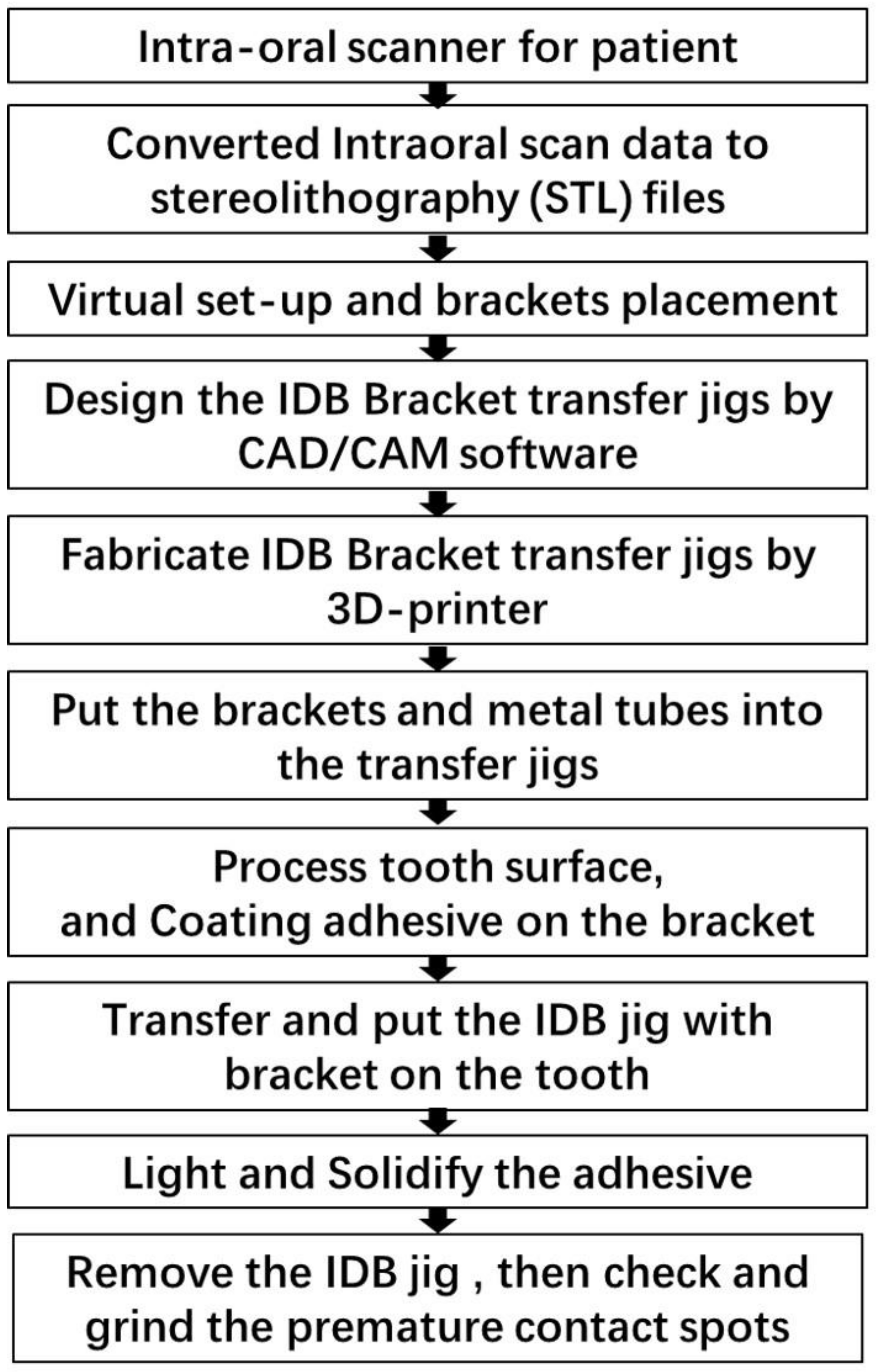
Figure 1. Clinical workflow list for indirect bonding jig system (IDBS) procedure. It takes 10 days to receive jigs after intraoral scanning and ordering. CAD/CAM, computer-aided design, and manufacturing; 3D, three-dimensional.
Applsci 13 01668 g001
Figure 2. An example of an indirect bonding jig system (IDBS) virtual set-up procedure. (AC): bracket placement on the virtual digital model; (DF): bracket position on the initial scanned model.
Applsci 13 01668 g002
Figure 3. An example of an indirect bonding jig system (IDBS) transfer procedure. (A) Brackets transferred to the model by indirect bonding procedure using the jig system; (B) teeth surface etching; (C) placement of the orthodontic adhesive on the bracket; (D) placement of the bracket on the tooth and application of sustained and steady occlusal pressure; (E) curing the adhesive using light; (F) removal of the transfer jig; (G,H) bracket transfer using a one-body jig on the incisors and premolars, respectively; (I) checking and grinding the premature contact spots.
Applsci 13 01668 g003
Table 1. Descriptive statistics of bracket re-bonding rate.
ItemsBracket Bonded TeethBonding FailurePosition ErrorTotal Re-Bonding
N%N%N%
Total2531 38215.09 1917.55 57322.64
Maxilla127714437.709750.7824142.06
Mandible125423862.309449.2133257.94
Male930 16617.85 9710.43 26328.28
Female1601 21613.49 945.87 31019.36
<20 years1183 21718.34 12310.40 34028.74
>20 years1348 16512.24 685.04 23317.28
Skeletal Class I782 11414.58 557.03 16921.61
Skeletal Class III1093 16515.10 837.59 24822.69
Skeletal Class III656 10315.70 538.08 15623.78
Hyperdivergency779 14118.10 486.16 18924.26
Normodivergency1066 14913.98 857.97 23421.95
Hypodivergency686 9213.41 588.45 15021.87
Initial stage2531 1706.72 160.63 1867.35
Middle stage2531 1756.91 1335.25 30812.17
Late stage2531 371.46 421.66 793.12
Incisors and canines127516913.2513710.7530624.00
Premolars652 9414.42284.2912218.71
Molars707 11916.83263.6814520.51
Table 2. Frequency statistics of bracket re-bonding rate in the maxilla and mandible.
ItemsBonding FailurePosition ErrorTotal Re-Bonding
SiteTotalSiteTotalSiteTotal
AnteriorPremolarMolarAnteriorPremolarMolarAnteriorPremolarMolar
ArchMaxillaCount434556144751210971185766241
Expected count63.7135.4344.8614469.5814.2213.2097128.7051.3160.99241
Proportion
within an arch (%)
29.8631.2538.89100.0077.3212.3710.31100.0048.9623.6527.39100.00
Proportion
within site (%)
25.4447.8747.0637.7054.7442.8638.4650.7938.5646.7245.5242.06
The proportion of the total (%)11.2611.7814.6637.7039.276.285.2450.7920.599.9511.5242.06
MandibleCount1264963238621616941886579332
Expected count105.2958.5774.1423867.4213.7812.7994177.2970.6984.01332
Proportion
within an arch (%)
52.9420.5926.47100.0065.9617.0217.02100.0056.6319.5823.80100.00
Proportion
within site (%)
74.5652.1352.9462.3045.2657.1461.5449.2161.4453.2854.4857.94
The proportion of the total (%)32.9812.8316.4962.3032.468.388.3849.2132.8111.3413.7957.94
TotalCount169941193821372826191306122145573
Expected count169941193821372826191306122145573
Proportion
within an arch (%)
44.2424.6131.15100.0071.7314.6613.61100.0053.4021.2925.31100.00
Proportion
within site (%)
100.00100.00100.00100.00100.00100.00100.00100.00100.00100.00100.00100.00
The proportion of the total (%)44.2424.6131.15100.0071.7314.6613.61100.0053.4021.2925.31100.00
Pearson Chi-Square 0.000 ** 0.208 0.189
** p < 0.01.
Table 3. Multiple linear regression analysis with dependent variables of sex, age, skeletal classification, and skeletal divergency.
VariablesBonding FailurePosition FailureTotal Re-Bonding
Estimate95% CIp-ValueEstimate95% CIp-ValueEstimate95% CIp-Value
SexMale0.00 0.00 0.00 .0.00 0.00 0.00 .0.00 0.00 0.00 .
Female−0.28 −0.51 −0.05 0.0150 * −0.62 −0.93 −0.30 0.0001 ** −0.39 −0.58 −0.21 <0.0001 **
Age<20 years0.00 0.00 0.00 .0.00 0.00 0.00 .0.00 0.00 0.00 .
>20 years−0.49 −0.70 −0.28 <0.0001 **−0.79 −1.10 −0.48 <0.0001 **−0.58 −0.76 −0.41 <0.0001 **
Skeletal
classification
I0.00 0.00 0.00 .0.00 0.00 0.00 .0.00 0.00 0.00 .
II0.04 −0.20 0.28 0.7675 0.09 −0.25 0.44 0.5988 0.05 −0.14 0.25 0.5860
III0.18 −0.09 0.45 0.2012 0.19 −0.19 0.58 0.3295 0.18 −0.04 0.40 0.1119
DivergencyHyperdivergency0.00 0.00 0.00 .0.00 0.00 0.00 .0.00 0.00 0.00 .
Normodivergency−0.33 −0.58 −0.08 0.0087 ** 0.01 −0.37 0.40 0.9446 −0.23 −0.43 −0.02 0.0322 *
Hypodivergency−0.30 −0.60 −0.01 0.0439 *0.14 −0.29 0.58 0.5133 −0.16 −0.40 0.08 0.1882
* p < 0.05, ** p < 0.01.
Table 4. Multiple linear regression analysis with dependent variables of treatment stage and tooth position.
VariablesBonding FailurePosition FailureTotal Re-Bonding
Estimate95% CIp-ValueEstimate95% CIp-ValueEstimate95% CIp-Value
Initial stage0.000 ...0.000 ...0.000 ...
Middle stage0.012 0.007 0.018 <0.0001 **0.024 0.020 0.028 <0.0001 **0.036 0.030 0.043 <0.0001 **
Late stage−0.015 −0.021 −0.009 <0.0001 **0.006 0.002 0.010 0.0039 **−0.009 −0.016 −0.002 0.0070 **
Incisors and Canine0.000 ...0.000 ...0.000 ...
Premolars0.000 −0.009 0.008 0.9057 −0.010 −0.015 −0.005 0.0001 ** −0.011 −0.020 −0.002 0.0222 **
Molars0.004 −0.004 0.012 0.3651 −0.011 −0.016 −0.006 <0.0001 **−0.008 −0.017 0.002 0.1058
** p < 0.01.
Disclaimer/Publisher’s Note: The statements, opinions and data contained in all publications are solely those of the individual author(s) and contributor(s) and not of MDPI and/or the editor(s). MDPI and/or the editor(s) disclaim responsibility for any injury to people or property resulting from any ideas, methods, instructions or products referred to in the content.