Systematic Review

Impact of Peri-Implant Inflammation on Metabolic Syndrome Factors: A Systematic Review

1
Centre for Oral Clinical and Translational Sciences, Faculty of Dentistry, Oral and Craniofacial Sciences, King’s College London, London WC2R 2LS, UK
2
Centre for Host Microbiome Interactions, Faculty of Dentistry, Oral and Craniofacial Sciences, King’s College London, London WC2R 2LS, UK
*
Author to whom correspondence should be addressed.
Appl. Sci. 2023, 13(21), 11747; https://doi.org/10.3390/app132111747
Submission received: 14 September 2023 / Revised: 22 October 2023 / Accepted: 25 October 2023 / Published: 27 October 2023
(This article belongs to the Special Issue Oral Microbial Communities and Oral Health (3rd Edition))

Abstract

:
This systematic review aims to evaluate the impact of peri-implantitis on the components of metabolic syndrome, and to provide suggestions on the management of peri-implantitis patients with metabolic disorders. A search for relevant records was performed in MEDLINE, EMBASE, and Global Health on 1st September 2023. Clinical trials, cohort studies, cross-sectional studies, and case-control studies containing comparisons of metabolic factors between patients with and without peri-implantitis were considered eligible. Study quality was assessed using the Newcastle–Ottawa scale. Out of 1158 records identified, 5 cross-sectional studies were eligible for final inclusion. Two studies reported significant differences in the lipid profile of patients with peri-implantitis, one of which reported higher total cholesterol and LDL cholesterol levels, while the other reported higher triglyceride levels. Another study reported significantly higher HbA1c levels in patients with peri-implantitis. The remaining two studies containing comparisons of BMI between patients with and without peri-implantitis indicated no significant differences. Overall, there are suggestions that peri-implantitis is associated with altered metabolic factors, including lipid profile and HbA1c level. However, there is not enough evidence to support these clinical implications due to the paucity of related literature and the low evidence level of the included studies. More investigations with stronger evidence levels are needed to narrow this gap of knowledge.

1. Introduction

Ever since the concept of osseointegration was introduced in the 1970s [1], dental implants have been widely used to replace missing teeth. This is attributed to their masticatory and biological advantages compared to conventional prostheses [2], and the ability to preserve adjacent teeth in partially dentate cases [3]. It is estimated that the prevalence of dental implants will be as high as 26% by the year 2026 [4].
Despite the increasingly promising results provided by dental implants, a common complication after implant placement is peri-implantitis. Peri-implantitis is a pathological condition where inflammation affects the peri-implant connective tissue and supporting alveolar bone [5]. Untreated peri-implantitis results in progressive alveolar bone loss around the implant, eventually leading to implant loss. The estimated prevalence of peri-implantitis ranges from 19.83% up to 47% based on various reports [6,7,8], underlying a heavy burden on oral health worldwide.
There is existing evidence indicating that several systemic conditions, including cardiovascular diseases (CVD), obesity, and hyperglycemia, may influence the progression and prognosis of peri-implantitis.
A high likelihood of comorbidity has been found between a history of cardiovascular diseases and a diagnosis of peri-implantitis [9,10]. Patients with CVD also present with increased peri-implant marginal bone loss [11]. There are shared serum risk markers between CVD and peri-implantitis, including high triglyceride and uric acid levels [12].
Obesity is also considered to increase the risk of developing peri-implantitis [13]. It compromises the efficiency of the immune response in the periodontium by increasing the systemic inflammatory burden [14]. Obese patients exhibited worse clinical parameters including plaque index, bleeding on probing, and probing depth, and significantly higher proinflammatory biomarkers such as interleukin-1β (IL-1β), interleukin-6 (IL-6), and C-reactive protein (CRP) [15,16,17,18,19].
Hyperglycemia in poorly controlled diabetes is associated with significantly higher peri-implant probing depth and crestal bone loss [20,21,22,23]. Impairment of bone healing after implant placement has also been reported in animal models of diabetes mellitus [24].
A recent retrospective cohort study assessed 216 implants with a mean duration of maintenance of 7 years and 4 months [25]. They found a significant difference in the onset of peri-implantitis between patients with and without systemic disorders. More specifically, they found that the presence of hypertension, diabetes mellitus, or osteoporosis have significant impacts on the onsets of peri-implantitis. A possible mechanism for the link between systemic disorders and peri-implantitis is via the reduced vascular supply and the emergence of cellular dysfunction, which further leads to the decreased immune robustness towards oral pathogens [26].
The above systemic conditions are all related to metabolic syndrome (MetS), which refers to the co-existence of several clinical metabolic findings including hypertension, hyperglycemia, abdominal obesity, and dyslipidemia [27,28]. The concept of MetS was formalized in 2001 [29]. According to the National Cholesterol Education Program (NCEP) Adult Treatment Panel Ⅲ (ATPⅢ) in the United States, MetS can be diagnosed when 3 of the following criteria are met:
  • Waist circumference (WC) ≥102 cm for males or ≥99 cm for females.
  • High-density lipoprotein cholesterol (HDL-C) <1.0 mmol/L (40 mg/dL) for males or <1.3 mmol/L (50 mg/dL) for females.
  • Triglyceride ≥ 1.69 mmol/L (150 mg/dL).
  • Fasting plasma glucose ≥ 110 mg/dL (6.1 mmol/L).
  • Blood pressure ≥ 130/85 mmHg.
MetS is highly prevalent worldwide, with an estimated prevalence of up to 24% in males and 46% in female population [30,31]. Although by definition MetS is not a disease itself, it does implicate higher risks for several cardiometabolic diseases. Patients with MetS have a two-fold increase in the risk of having CVD over 5 to 10 years [32,33], and at least a five-fold increase in the risk of having diabetes [27]. It is also significantly associated with nonalcoholic fatty liver disease (NFLD) via increased WC, triglycerides, and blood glucose [34].
Chronic inflammation is considered one of the potential mechanisms of MetS and extensively contributes to the related pathological outcomes [35,36]. As a chronic inflammatory disease, peri-implantitis has been investigated for how MetS and its components may influence clinical peri-implant parameters [19,37]. However, the impact of peri-implantitis on metabolic indicators remains unclear. It is reported that treating periodontitis, a chronic inflammation condition affecting the supporting tissues of teeth, reduced the proinflammatory cytokines [38] and improved serum lipid concentrations as well as HbA1c levels [39,40,41], underlining the potential impact of oral chronic inflammation on systemic and metabolic factors.
Therefore, the aim of this systematic review was to evaluate the effect of peri-implantitis on the factors related to MetS including waist circumference, body mass index (BMI), HDL cholesterol, LDL cholesterol, triglyceride, blood glucose, HbA1c level, and blood pressure, and to provide clinical implications on the management of peri-implantitis patients with metabolic disorders.

2. Materials and Methods

This systematic review was registered on PROSPERO under protocol CRD-42021269438, and was conducted in accordance with Preferred Reporting Items for Systematic Reviews and Meta-Analyses (PRISMA) statement (see Supplementary Materials) [42].

2.1. Focused Question

Based on the PICO format (Population, Intervention, Comparison, and Outcome), this systematic review aims to address a focused question: What is the effect of peri-implant diseases on metabolic syndrome factors?
  • Population: Patients with osseointegrated dental implants.
  • Intervention (exposure): Patients affected by peri-implantitis.
  • Comparison: Patients with healthy peri-implant conditions.
  • Outcome: Systemic metabolic factors in relation to MetS and its components.

2.2. Eligible Criteria and Study Selection

The inclusion criteria for this systematic review were defined in accordance with the PICO model and the focused question raised above. Included studies were prospective or retrospective cohort studies, observational cross-sectional studies, matched or non-matched case-control studies, or randomized and non-randomized control clinical trials. The target population comprised implant patients with and without the presence of peri-implantitis. The findings included measurement of metabolic factors within the components of MetS according to NCEP ATP Ⅲ criteria. The evaluation of other systemic factors in relation to MetS such as body mass index (BMI) or serum concentration of low-density lipoprotein cholesterol were also eligible for this review. Only studies published in the English language were included.
Exclusion criteria were: studies without full text; non-comparative case reports, case series, conference proceedings, or editorials; participants with other oral chronic inflammatory diseases such as progressing periodontitis; participants with comorbidities other than related metabolic diseases to MetS components.

2.3. Information Source and Search Strategy

An electronic literature search was conducted independently by 3 reviewers (Y.Z., E.L., and S.N.) on several databases using OVID interface for reports published up to 1st September 2023. Included databases were MEDLINE (1946 onwards), EMBASE (1974 onwards) and Global Health (1973 onwards). A detailed search strategy is shown in Table 1. In addition, a bibliography search of the related literature was conducted manually to identify more eligible studies. The International Clinical Trials Registry Platform Search was searched for ongoing or recently completed trials. Similarly, PROSPERO was searched for ongoing or recently completed reviews.

2.4. Data Management, Selection Process, and Data Synthesis

Three independent reviewers (Y.Z., E.L., and S.N.) independently screened studies for eligibility on the inclusion criteria to minimize bias, and any discrepancy was solved by discussion. Duplicate removal was performed using both EndNote 20 software (Clarivate Analytics, London, UK) and manual removal. The remaining studies were then screened by titles, abstracts, and keywords to filter out the irrelevant ones. Full texts of the included studies were then reviewed to extract the relevant information including study design, group settings, clinical characteristics, and parameters for outcome measurement. Authors of the eligible studies were contacted in case of any missing or unclear data. A predetermined sheet was used for data extraction to ensure consistency among reviewers on all included studies.

2.5. Quality and Risk of Bias Assessment

The quality of the included studies was evaluated using an adopted Newcastle–Ottawa scale for assessing the quality of cross-sectional studies [43,44,45]. The scale assessed the quality of studies from the perspectives of selection, comparability, and outcome with a maximum of 8 stars in total.
To be more specific, a maximum of 4 stars could be rated in terms of selection. One star was given if the selection of the exposed group could represent the average in the target population (Representativeness of Exposed Group); one star was given if the sample size was justified clearly (Sample Size); one star was given if the measurement of exposure was validated or described (Ascertainment of Exposure); and one star was given if the non-exposed group was reasonably selected and comparable to the exposed group (Selection of Non-exposed Group).
In terms of comparability, one star was given if the study controlled or adjusted for metabolic factors within the MetS components; one more star was given if the study controlled or adjusted for other confounding factors such as age and gender.
Finally, for outcome, one star was given if the outcome measurement was clearly described (Outcome Measurement); one more star was given if the statistical analysis was clearly described and appropriate (Statistical Test).

3. Results

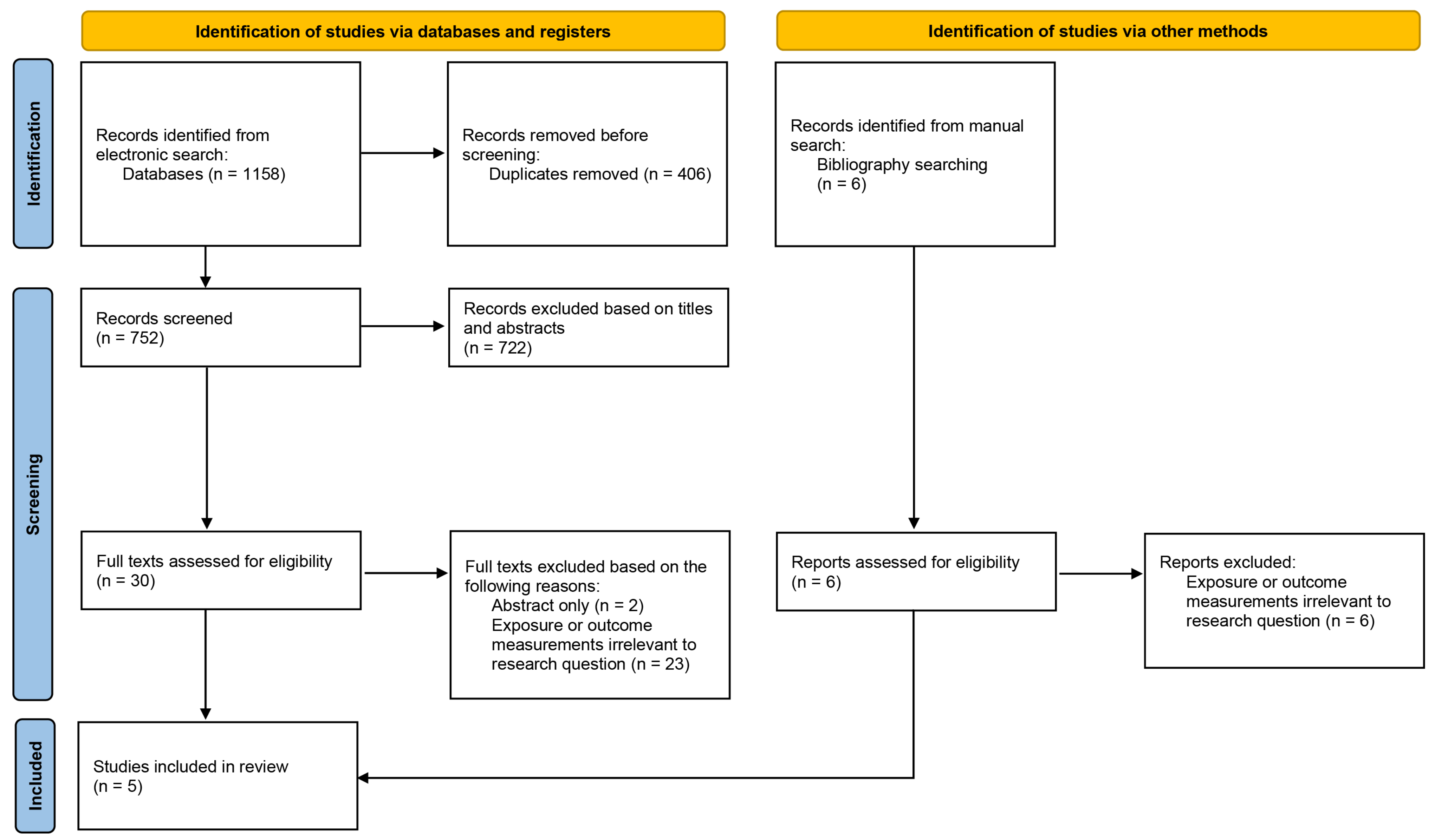
3.1. Study Selection

The flow chart for study selection is shown in Figure 1 [46]. A total of 1164 records were identified based on our search strategy (1058 from electronic databases, 6 from other sources). From these, 752 studies were selected after the removal of duplicates. Subsequent to initial screening based on title and abstract, 30 studies were identified as potentially eligible. Full-text assessment was conducted, and five studies were included for data extraction. The detailed reasons for exclusion after full text assessment are shown in Figure 1.

3.2. Study Characteristics

Table 2 demonstrates the characteristics of the five included studies [12,47,48,49,50]. These studies all followed a cross-sectional design, and all recruited patients with and without peri-implantitis for comparison. Among them, two studies [12,49] also recruited patients diagnosed with peri-implant mucositis, one study [47] included the presence of type-2 diabetes for more sub-group comparison, whilst one study [50] also recruited healthy subjects without implants or periodontal conditions as blank controls. No limitations were set to the diagnostic criteria for peri-implant diseases in this review. The criteria used in the four studies for diagnosing peri-implantitis and peri-implant mucositis were slightly different (Table 2). In terms of outcome measures, two studies [12,48] reported parameters within the definition of MetS components (HDL-C and Triglyceride) as well as factors relevant to MetS (Total cholesterol, LDL-C), while the other three studies [47,49,50] reported only factors relevant to MetS (BMI and HbA1c).
All studies included were cross-sectional. The quality of these studies was assessed in Table 3. Despite the low evidence level in terms of study types, these studies were identified as being of good quality for cross-sectional designs. Due to the lack of relevant literature and heterogeneity amongst included studies, quantitative analysis was not performed, and this review was carried out narratively.

3.3. Peri-Implantitis and Dyslipidemia

Blanco et al. [48] and Ustaoglu et al. [12] evaluated the association between peri-implantitis and dyslipidemia. Although they both found that peri-implantitis may alter the lipid profile in patients, their findings were not completely consistent.
In Blanco’s work [48], peri-implantitis was associated with a significantly higher total cholesterol level (mean 212 mg/dL in peri-implantitis group versus mean 156 mg/dL in controls, p < 0.001) and LDL cholesterol level (mean 120 mg/dL in peri-implantitis group versus mean 91 mg/dL in controls, p < 0.001) after adjusting for history of periodontitis. Together with other findings on proinflammatory markers, they concluded that patients with peri-implantitis exhibited increased low-grade systemic inflammation and dyslipidemia.
Nonetheless, Ustaoglu’s study [12], where peri-implant mucositis was additionally included as an individual group, suggested that total cholesterol and LDL cholesterol levels were not significantly different in patients diagnosed with peri-implant mucositis and peri-implantitis relative to healthy individuals (Table 2). Their results showed that level of triglycerides was significantly higher in patients with peri-implantitis than in the peri-implant health group (median 148 mg/dL versus median 95 mg/dL, p < 0.001). However, as an intermediate stage, levels of triglycerides in patients diagnosed with peri-implant mucositis (median 125 mg/dL) were not significantly different to either peri-implantitis or peri-implant health group.

3.4. Peri-Implantitis and Hyperglycemia

The study by Al-Aksar et al. [47] revealed that in patients both with and without type-2 diabetes, the presence of peri-implantitis was associated with significantly higher HbA1c levels (p < 0.001). In the non-diabetic group, HbA1c levels in patients without peri-implantitis were 4.3 ± 0.3%, while those in patients with peri-implantitis were 5.2 ± 0.1%. Meanwhile, in the type-2 diabetic group, HbA1c levels were 4.7 ± 0.1% in patients without peri-implantitis. This value increased significantly to 9.3 ± 1.5% in those diagnosed with peri-implantitis (p < 0.001).

3.5. Peri-Implantitis and BMI

Two studies [49,50] included performed comparison of BMI between patients with and without peri-implantitis. One of them, conducted by Lucarini et al. [49], compared BMI among patients diagnosed with peri-implant health (29.44 ± 4.34), peri-implant mucositis (32.56 ± 5.88), and peri-implantitis (33.94 ± 5.71). Although BMI was higher in value in the peri-implantitis group, the authors did not find any significant differences between groups (p = 0.61). Similarly, study by Sanchez-Siles et al. [50] also showed that there were no significant differences in BMI among patients with peri-implantitis, patients without peri-implantitis, and healthy controls with no implant (p = 0.378).

3.6. Peri-Implantitis and Hypertension

Based on our search strategy, we did not identify any records reporting the effect of peri-implantitis on blood pressure or severity of hypertension.

4. Discussion

To the best of the authors’ knowledge, this is the first systematic review assessing the potential impact of peri-implantitis on MetS or its components. There is some suggestion, based on the included studies, that peri-implantitis is associated with altered metabolic factors of MetS, including serum lipid profile and HbA1c levels. However, based on the cross-sectional design and the heterogeneity of the included studies, there is not enough evidence to support any clinical implications.
The relationship between MetS and peri-implantitis is very complex. Multiple studies have revealed that components of MetS are linked to the risk of developing peri-implantitis [37,51]. On the other hand, some studies suggest that chronic inflammation may represent as a triggering factor in the initiation of MetS [52], underlining a bilateral association between MetS and oral chronic inflammation.
As an analogue of peri-implantitis, periodontitis is reported to influence systemic conditions, affecting both inflammatory mediators [53,54] and metabolic indicators [55]. Therefore, as a chronic inflammatory disease in the oral cavity, peri-implantitis, which also influences systemic levels of pro-inflammatory cytokines, is intuitively inferred to have a potential impact on MetS and its components.
Furthermore, there is solid evidence provided by a systematic review suggesting that hyperglycemia and peri-implantitis are associated [21]. The risk of developing peri-implantitis in hyperglycemia subjects is 1.21- and 2.46- fold statistically significantly higher than that in normoglycemia subjects. In addition, obesity is found to be associated with elevated pro-inflammatory cytokines and worsened clinical parameters in subjects with peri-implantitis [56,57]. The mechanism behind the association between peri-implantitis and hyperglycemia/obesity lies in the reduction of vascular supply caused by microangiopathies, cellular dysfunction due to toxic microbial metabolites, and decreased immune robustness towards oral pathogens [26]. To this point, a reasonable inference can be made that there might be a bi-directional correlation between peri-implantitis and MetS components, linked by systemic inflammatory burdens. However, it is acknowledged that there is currently a gap of knowledge on whether the presence of peri-implantitis is associated with degree of hyperglycemia/dyslipidemia or severity of obesity.
In this systematic review, we found five related studies to address this question, all of which were cross-sectional in nature.
Al-Askar’s results [47] showed that peri-implantitis is associated with significantly higher levels of HbA1c both in T2DM and non-T2DM subjects. Nonetheless, all participants included in this study were males, which consequently led to potential bias given the hormonal differences, and the evidence that gender has been identified as a predictive factor for peri-implant bone levels [58]. Further, the statistical analysis for HbA1c level was not adjusted by other confounding factors such as age, history of periodontitis, or smoking habits. The observation of significantly higher HbA1c levels confirmed the link between hyperglycemia and peri-implantitis, but the cause-and-effect relationship needs to be further confirmed by longitudinal cohort studies.
In contrast, Blanco’s study [48] reported that blood glucose was not significantly different between peri-implantitis and controls. This is probably due to the relatively small sample size in this study along with the inclusion of other confounding factors such as history of periodontitis and current smokers, both of which can have an impact on glycemic control [59,60].
The primary finding of Blanco’s study was that peri-implantitis is associated with dyslipidemia, in agreement with Ustaoglu’s study [12]. However, it should be noted that the findings of these two studies were not completely consistent. Among the four indicators for lipid profile (total cholesterol, LDL cholesterol, HDL cholesterol, and triglycerides), Blanco et al. identified significantly higher total cholesterol and LDL cholesterol levels in peri-implantitis patients compared with peri-implant health group, whereas in Ustaoglu’s study, significant differences were found in triglycerides instead of total cholesterol and LDL. Notably, the two studies both had limited sample sizes, and both did not exclude periodontitis or smoking habits as confounding factors. Thus, further research with a larger sample size and greater exclusion of potential confounding factors is required to fully confirm the relationship between peri-implantitis and dyslipidemia. Due to their cross-sectional design, the results of the two studies were also influenced by inter-individual differences. To overcome this shortage, cohort studies are needed to track the prognosis of peri-implantitis and meanwhile monitor the changes in the lipid profile.
In Lucarini’s study [49], BMI was recorded in the clinical and demographic characteristics of the patients, and it was not significantly different among peri-implantitis, peri-implant mucositis, and peri-implant health groups. It was the same in the study conducted by Sanchez-Siles [50]. One possible reason is that the severity of obesity is affected by various factors. The indirect effects from the elevated inflammatory burden caused by peri-implantitis were covered by other more direct effects including genetic or dietary factors. This shortage can also be overcome by cohort design, where individuals are studied case-wise.
Another limitation of this systematic review is that the diagnostic criteria for peri-implantitis followed by the included studies were not consistent. According to the 2017 World Workshop on the Classification of Periodontal and Peri-implant Diseases [61], radiographic bone loss of ≥3 mm in combination with BOP and probing depth ≥6 mm is indicative of peri-implantitis. Two of the included studies [12,45] followed this criterion while the others [44,46,47] had different definitions on the presence of peri-implantitis. This might hamper the generalizability of our findings.

5. Conclusions

Overall, based on the literature reviewed, there are some indications of an association between the presence of peri-implantitis and MetS components. Nonetheless, it should be mentioned that some of the included studies had limited sample sizes, and the quality of these studies varies based on the Newcastle–Ottawa scale, which might compromise the generalizability of our findings. Due to the limited evidence at this stage, no direct clinical implications can be drawn. Given the prevalence of dental implants worldwide, and the rising prevalence of MetS, it remains a priority to clarify the relationship between peri-implant diseases and MetS. To this end, we suggest designing longitudinal studies with more specific objectives, more detailed exclusion criteria, higher evidence level, and larger sample sizes to further elucidate the cause-and-effect relationship between peri-implantitis and metabolic disorders.

Supplementary Materials

The following supporting information can be downloaded at: https://www.mdpi.com/article/10.3390/app132111747/s1, PRISMA checklists.

Author Contributions

Conceptualization, E.M.-C.L. and S.A.N.; methodology, Y.Z., E.M.-C.L. and S.A.N.; writing—original draft preparation, Y.Z.; writing—review and editing, E.M.-C.L., D.M. and S.A.N.; supervision, E.M.-C.L. and S.A.N.; project administration, S.A.N. All authors have read and agreed to the published version of the manuscript.

Funding

This research received no external funding.

Institutional Review Board Statement

Not applicable.

Informed Consent Statement

Not applicable.

Data Availability Statement

No new data were created during the conduction of this narrative systematic review.

Conflicts of Interest

The authors declare no conflict of interest.

References

  1. Branemark, P.I.; Adell, R.; Breine, U.; Hansson, B.O.; Lindstrom, J.; Ohlsson, A. Intra-osseous anchorage of dental prostheses. I. Experimental studies. Scand. J. Plast. Reconstr. Surg. 1969, 3, 81–100. [Google Scholar] [CrossRef]
  2. Buser, D.; Sennerby, L.; De Bruyn, H. Modern implant dentistry based on osseointegration: 50 years of progress, current trends and open questions. Periodontology 2017, 73, 7–21. [Google Scholar] [CrossRef] [PubMed]
  3. Jivraj, S.; Chee, W. Rationale for dental implants. Br. Dent. J. 2006, 200, 661–665. [Google Scholar] [CrossRef] [PubMed]
  4. Elani, H.W.; Starr, J.R.; Da Silva, J.D.; Gallucci, G.O. Trends in Dental Implant Use in the U.S., 1999-2016, and Projections to 2026. J. Dent. Res. 2018, 97, 1424–1430. [Google Scholar] [CrossRef]
  5. Schwarz, F.; Derks, J.; Monje, A.; Wang, H.L. Peri-implantitis. J. Clin. Periodontol. 2018, 45 (Suppl. S20), S246–S266. [Google Scholar] [CrossRef] [PubMed]
  6. Lee, C.T.; Huang, Y.W.; Zhu, L.; Weltman, R. Prevalences of peri-implantitis and peri-implant mucositis: Systematic review and meta-analysis. J. Dent. 2017, 62, 1–12. [Google Scholar] [CrossRef]
  7. Koldsland, O.C.; Scheie, A.A.; Aass, A.M. Prevalence of peri-implantitis related to severity of the disease with different degrees of bone loss. J. Periodontol. 2010, 81, 231–238. [Google Scholar] [CrossRef]
  8. Derks, J.; Tomasi, C. Peri-implant health and disease. A systematic review of current epidemiology. J. Clin. Periodontol. 2015, 42 (Suppl. S16), S158–S171. [Google Scholar] [CrossRef] [PubMed]
  9. Renvert, S.; Aghazadeh, A.; Hallstrom, H.; Persson, G.R. Factors related to peri-implantitis—A retrospective study. Clin. Oral. Implant. Res. 2014, 25, 522–529. [Google Scholar] [CrossRef] [PubMed]
  10. Ardakani, M.S.Z.; Salari, A.M.; Nasre-Esfahani, M. The relationship between cardiovascular diseases and peri-implantitis in patients with failed dental implants; a retrospective study. J. Dent. Med. 2020, 33, 152–157. [Google Scholar]
  11. Krennmair, S.; Weinlander, M.; Forstner, T.; Krennmair, G.; Stimmelmayr, M. Factors affecting peri-implant bone resorption in four Implant supported mandibular full-arch restorations: A 3-year prospective study. J. Clin. Periodontol. 2016, 43, 92–101. [Google Scholar] [CrossRef] [PubMed]
  12. Ustaoglu, G.; Erdal, E. Relationship between risk markers for cardiovascular disease and peri-implant diseases. Int. J. Implant. Dent. 2020, 6, 73. [Google Scholar] [CrossRef] [PubMed]
  13. Abduljabbar, T.; Al-Sahaly, F.; Kellesarian, S.V.; Kellesarian, T.V.; Al-Anazi, M.; Al-Khathami, M.; Javed, F.; Vohra, F. Comparison of peri-implant clinical and radiographic inflammatory parameters and whole salivary destructive inflammatory cytokine profile among obese and non-obese men. Cytokine 2016, 88, 51–56. [Google Scholar] [CrossRef] [PubMed]
  14. Huang, X.; Yu, T.; Ma, C.; Wang, Y.; Xie, B.; Xuan, D.; Zhang, J. Macrophages Play a Key Role in the Obesity-Induced Periodontal Innate Immune Dysfunction via Nucleotide-Binding Oligomerization Domain-Like Receptor Protein 3 Pathway. J. Periodontol. 2016, 87, 1195–1205. [Google Scholar] [CrossRef] [PubMed]
  15. Alasqah, M.N.; Al-Shibani, N.; Al-Aali, K.A.; Qutub, O.A.; Abduljabbar, T.; Akram, Z. Clinical indices and local levels of inflammatory biomarkers in per-implant health of obese and nonobese individuals. Clin. Implant Dent. Relat. Res. 2019, 21, 80–84. [Google Scholar] [CrossRef] [PubMed]
  16. Alkhudhairy, F.; Vohra, F.; Al-Kheraif, A.A.; Akram, Z. Comparison of clinical and radiographic peri-implant parameters among obese and non-obese patients: A 5-year study. Clin. Implant Dent. Relat. Res. 2018, 20, 756–762. [Google Scholar] [CrossRef]
  17. Brignardello-Petersen, R. Patients who are obese probably have worse peri-implant outcomes than those who are not, but there is not sufficient evidence that the implant survival is different. J. Am. Dent. Assoc. 2019, 150, e123. [Google Scholar] [CrossRef] [PubMed]
  18. Elsadek, M.F.; Almoajel, A. Assessment of clinical and radiographic peri-implant parameters in the obese and non-obese, along with destructive pro-inflammatory cytokines IL-1beta—And IL-6 in patients treated with Photodynamic therapy. Photodiagnosis Photodyn. Ther. 2022, 39, 102844. [Google Scholar] [CrossRef] [PubMed]
  19. Vohra, F.; Alkhudhairy, F.; Al-Kheraif, A.A.; Akram, Z.; Javed, F. Peri-implant parameters and C-reactive protein levels among patients with different obesity levels. Clin. Implant Dent. Relat. Res. 2018, 20, 130–136. [Google Scholar] [CrossRef]
  20. Dreyer, H.; Grischke, J.; Tiede, C.; Eberhard, J.; Schweitzer, A.; Toikkanen, S.E.; Glockner, S.; Krause, G.; Stiesch, M. Epidemiology and risk factors of peri-implantitis: A systematic review. J. Periodontal Res. 2018, 53, 657–681. [Google Scholar] [CrossRef]
  21. Monje, A.; Catena, A.; Borgnakke, W.S. Association between diabetes mellitus/hyperglycaemia and peri-implant diseases: Systematic review and meta-analysis. J. Clin. Periodontol. 2017, 44, 636–648. [Google Scholar] [CrossRef]
  22. Al-Sowygh, Z.H.; Ghani, S.M.A.; Sergis, K.; Vohra, F.; Akram, Z. Peri-implant conditions and levels of advanced glycation end products among patients with different glycemic control. Clin. Implant Dent. Relat. Res. 2018, 20, 345–351. [Google Scholar] [CrossRef]
  23. Dioguardi, M.; Cantore, S.; Quarta, C.; Sovereto, D.; Zerman, N.; Pettini, F.; Muzio, L.L.; Cosola, M.D.; Santacroce, L.; Ballini, A. Correlation Between Diabetes Mellitus and Peri-implantitis: A Systematic Review. Endocr. Metab. Immune Disord Drug Targets 2022, 23, 596–608. [Google Scholar] [CrossRef] [PubMed]
  24. Coelho, P.G.; Pippenger, B.; Tovar, N.; Koopmans, S.J.; Plana, N.M.; Graves, D.T.; Engebretson, S.; van Beusekom, H.M.M.; Oliveira, P.; Dard, M. Effect of Obesity or Metabolic Syndrome and Diabetes on Osseointegration of Dental Implants in a Miniature Swine Model: A Pilot Study. J. Oral. Maxillofac. Surg. 2018, 76, 1677–1687. [Google Scholar] [CrossRef] [PubMed]
  25. Seki, K.; Hasuike, A.; Hagiwara, Y. Clinical Evaluation of the Relationship Between Systemic Disease and the Time of Onset of Peri-Implantitis: A Retrospective Cohort Study. J. Oral. Implant. 2023, 49, 55–61. [Google Scholar] [CrossRef] [PubMed]
  26. de Oliveira, P.G.F.P.; Bonfante, E.A.; Bergamo, E.T.P.; de Souza, S.L.S.; Riella, L.; Torroni, A.; Benalcazar Jalkh, E.B.; Witek, L.; Lopez, C.D.; Zambuzzi, W.F.; et al. Obesity/Metabolic Syndrome and Diabetes Mellitus on Peri-implantitis. Trends Endocrinol. Metab. 2020, 31, 596–610. [Google Scholar] [CrossRef] [PubMed]
  27. Samson, S.L.; Garber, A.J. Metabolic syndrome. Endocrinol. Metab. Clin. N. Am. 2014, 43, 1–23. [Google Scholar] [CrossRef] [PubMed]
  28. Huang, P.L. A comprehensive definition for metabolic syndrome. Dis. Model. Mech. 2009, 2, 231–237. [Google Scholar] [CrossRef]
  29. Expert Panel on Detection, Evaluation; Treatment of High Blood Cholesterol in Adults. Executive Summary of the Third Report of the National Cholesterol Education Program (NCEP) Expert Panel on Detection, Evaluation, and Treatment of High Blood Cholesterol in Adults (Adult Treatment Panel III). JAMA 2001, 285, 2486–2497. [Google Scholar] [CrossRef]
  30. Cameron, A.J.; Shaw, J.E.; Zimmet, P.Z. The metabolic syndrome: Prevalence in worldwide populations. Endocrinol. Metab. Clin. N. Am. 2004, 33, 351–375, table of contents. [Google Scholar] [CrossRef] [PubMed]
  31. Saklayen, M.G. The Global Epidemic of the Metabolic Syndrome. Curr. Hypertens. Rep. 2018, 20, 12. [Google Scholar] [CrossRef]
  32. Wilson, P.W.; Meigs, J.B. Cardiometabolic risk: A Framingham perspective. Int. J. Obes. 2008, 32 (Suppl. S2), S17–S20. [Google Scholar] [CrossRef] [PubMed]
  33. Wilson, P.W.; D′Agostino, R.B.; Parise, H.; Sullivan, L.; Meigs, J.B. Metabolic syndrome as a precursor of cardiovascular disease and type 2 diabetes mellitus. Circulation 2005, 112, 3066–3072. [Google Scholar] [CrossRef] [PubMed]
  34. Smits, M.M.; Ioannou, G.N.; Boyko, E.J.; Utzschneider, K.M. Non-alcoholic fatty liver disease as an independent manifestation of the metabolic syndrome: Results of a US national survey in three ethnic groups. J. Gastroenterol. Hepatol. 2013, 28, 664–670. [Google Scholar] [CrossRef] [PubMed]
  35. McCracken, E.; Monaghan, M.; Sreenivasan, S. Pathophysiology of the metabolic syndrome. Clin. Dermatol. 2018, 36, 14–20. [Google Scholar] [CrossRef] [PubMed]
  36. Monteiro, R.; Azevedo, I. Chronic inflammation in obesity and the metabolic syndrome. Mediat. Inflamm. 2010, 2010, 289645. [Google Scholar] [CrossRef]
  37. Papi, P.; Murro, B.D.; Pranno, N.; Bisogni, V.; Saracino, V.; Letizia, C.; Polimeni, A.; Pompa, G. Prevalence of peri-implant diseases among an Italian population of patients with metabolic syndrome: A cross-sectional study. J. Periodontol. 2019, 90, 1374–1382. [Google Scholar] [CrossRef]
  38. D′Aiuto, F.; Ready, D.; Tonetti, M.S. Periodontal disease and C-reactive protein-associated cardiovascular risk. J. Periodontal Res. 2004, 39, 236–241. [Google Scholar] [CrossRef]
  39. Fu, Y.W.; Li, X.X.; Xu, H.Z.; Gong, Y.Q.; Yang, Y. Effects of periodontal therapy on serum lipid profile and proinflammatory cytokines in patients with hyperlipidemia: A randomized controlled trial. Clin. Oral. Investig. 2016, 20, 1263–1269. [Google Scholar] [CrossRef]
  40. Pussinen, P.J.; Vilkuna-Rautiainen, T.; Alfthan, G.; Palosuo, T.; Jauhiainen, M.; Sundvall, J.; Vesanen, M.; Mattila, K.; Asikainen, S. Severe periodontitis enhances macrophage activation via increased serum lipopolysaccharide. Arterioscler. Thromb. Vasc. Biol. 2004, 24, 2174–2180. [Google Scholar] [CrossRef]
  41. Borgnakke, W.S. IDF Diabetes Atlas: Diabetes and oral health—A two-way relationship of clinical importance. Diabetes Res. Clin. Pract. 2019, 157, 107839. [Google Scholar] [CrossRef] [PubMed]
  42. Moher, D.; Shamseer, L.; Clarke, M.; Ghersi, D.; Liberati, A.; Petticrew, M.; Shekelle, P.; Stewart, L.A.; Group, P.-P. Preferred reporting items for systematic review and meta-analysis protocols (PRISMA-P) 2015 statement. Syst. Rev. 2015, 4, 1. [Google Scholar] [CrossRef] [PubMed]
  43. Borgnakke, W.S.; Ylostalo, P.V.; Taylor, G.W.; Genco, R.J. Effect of periodontal disease on diabetes: Systematic review of epidemiologic observational evidence. J. Clin. Periodontol. 2013, 40 (Suppl. S14), S135–S152. [Google Scholar] [CrossRef]
  44. Moskalewicz, A.; Oremus, M. No clear choice between Newcastle-Ottawa Scale and Appraisal Tool for Cross-Sectional Studies to assess methodological quality in cross-sectional studies of health-related quality of life and breast cancer. J. Clin. Epidemiol. 2020, 120, 94–103. [Google Scholar] [CrossRef] [PubMed]
  45. Wells, G.A.; Shea, B.; O’Connell, D.; Peterson, J.; Welch, V.; Losos, M.; Tugwell, P. The Newcastle-Ottawa Scale (NOS) for Assessing the Quality of Nonrandomized Studies in Meta-Analyses. 2000. Available online: https://www.ohri.ca/programs/clinical_epidemiology/oxford.asp (accessed on 24 October 2023).
  46. Page, M.J.; McKenzie, J.E.; Bossuyt, P.M.; Boutron, I.; Hoffmann, T.C.; Mulrow, C.D.; Shamseer, L.; Tetzlaff, J.M.; Akl, E.A.; Brennan, S.E.; et al. The PRISMA 2020 statement: An updated guideline for reporting systematic reviews. BMJ 2021, 372, n71. [Google Scholar] [CrossRef]
  47. Al-Askar, M.; Ajlan, S.; Alomar, N.; Al-Daghri, N.M. Clinical and Radiographic Peri-Implant Parameters and Whole Salivary Interleukin-1beta and Interleukin-6 Levels among Type-2 Diabetic and Nondiabetic Patients with and without Peri-Implantitis. Med. Princ. Pract. 2018, 27, 133–138. [Google Scholar] [CrossRef] [PubMed]
  48. Blanco, C.; Linares, A.; Dopico, J.; Pico, A.; Sobrino, T.; Leira, Y.; Blanco, J. Peri-implantitis, systemic inflammation, and dyslipidemia: A cross-sectional biochemical study. J. Periodontal Implant. Sci. 2021, 51, 342–351. [Google Scholar] [CrossRef] [PubMed]
  49. Lucarini, G.; Zizzi, A.; Rubini, C.; Ciolino, F.; Aspriello, S.D. VEGF, Microvessel Density, and CD44 as Inflammation Markers in Peri-implant Healthy Mucosa, Peri-implant Mucositis, and Peri-implantitis: Impact of Age, Smoking, PPD, and Obesity. Inflammation 2019, 42, 682–689. [Google Scholar] [CrossRef] [PubMed]
  50. Sanchez-Siles, M.; Lucas-Azorin, J.; Salazar-Sanchez, N.; Carbonell-Meseguer, L.; Camacho-Alonso, F. Salivary Concentration of Oxidative Stress Biomarkers in a Group of Patients with Peri-Implantitis: A Transversal Study. Clin. Implant Dent. Relat. Res. 2016, 18, 1015–1022. [Google Scholar] [CrossRef]
  51. Papi, P.; Letizia, C.; Pilloni, A.; Petramala, L.; Saracino, V.; Rosella, D.; Pompa, G. Peri-implant diseases and metabolic syndrome components: A systematic review. Eur. Rev. Med. Pharmacol. Sci. 2018, 22, 866–875. [Google Scholar]
  52. Esposito, K.; Giugliano, D. The metabolic syndrome and inflammation: Association or causation? Nutr. Metab. Cardiovasc. Dis. 2004, 14, 228–232. [Google Scholar] [CrossRef] [PubMed]
  53. Saito, T.; Murakami, M.; Shimazaki, Y.; Matsumoto, S.; Yamashita, Y. The extent of alveolar bone loss is associated with impaired glucose tolerance in Japanese men. J. Periodontol. 2006, 77, 392–397. [Google Scholar] [CrossRef]
  54. Saito, T.; Murakami, M.; Shimazaki, Y.; Oobayashi, K.; Matsumoto, S.; Koga, T. Association between alveolar bone loss and elevated serum C-reactive protein in Japanese men. J. Periodontol. 2003, 74, 1741–1746. [Google Scholar] [CrossRef] [PubMed]
  55. Pirih, F.Q.; Monajemzadeh, S.; Singh, N.; Sinacola, R.S.; Shin, J.M.; Chen, T.; Fenno, J.C.; Kamarajan, P.; Rickard, A.H.; Travan, S.; et al. Association between metabolic syndrome and periodontitis: The role of lipids, inflammatory cytokines, altered host response, and the microbiome. Periodontology 2021, 87, 50–75. [Google Scholar] [CrossRef]
  56. Assery, N.M.; Jurado, C.A.; Assery, M.K.; Afrashtehfar, K.I. Peri-implantitis and systemic inflammation: A critical update. Saudi Dent. J. 2023, 35, 443–450. [Google Scholar] [CrossRef] [PubMed]
  57. Gasmi Benahmed, A.; Gasmi, A.; Tippairote, T.; Mujawdiya, P.K.; Avdeev, O.; Shanaida, Y.; Bjorklund, G. Metabolic Conditions and Peri-Implantitis. Antibiotics 2023, 12, 65. [Google Scholar] [CrossRef]
  58. Papantonopoulos, G.; Gogos, C.; Housos, E.; Bountis, T.; Loos, B.G. Peri-implantitis: A complex condition with non-linear characteristics. J. Clin. Periodontol. 2015, 42, 789–798. [Google Scholar] [CrossRef] [PubMed]
  59. Preshaw, P.M.; Alba, A.L.; Herrera, D.; Jepsen, S.; Konstantinidis, A.; Makrilakis, K.; Taylor, R. Periodontitis and diabetes: A two-way relationship. Diabetologia 2012, 55, 21–31. [Google Scholar] [CrossRef] [PubMed]
  60. Haire-Joshu, D.; Glasgow, R.E.; Tibbs, T.L. Smoking and diabetes. Diabetes Care 1999, 22, 1887–1898. [Google Scholar] [CrossRef]
  61. Renvert, S.; Persson, G.R.; Pirih, F.Q.; Camargo, P.M. Peri-implant health, peri-implant mucositis, and peri-implantitis: Case definitions and diagnostic considerations. J. Periodontol. 2018, 89 (Suppl. S1), S304–S312. [Google Scholar] [CrossRef]
Figure 1. Flow chart of study selection.
Applsci 13 11747 g001
Table 1. Detailed search strategy on OVID interface (Selected databases: MEDLINE, EMBASE, and Global Health).
No.Query
1(peri-implant disease* OR peri-implant mucositis OR peri-implantitis OR peri-implant infection OR peri-implant inflammation).mp.
2exp peri-implantitis/
31 OR 2
4exp metabolic syndrome/
5exp metabolic diseases/
6exp diabetes mellitus/OR exp diabetes/OR exp blood glucose/OR exp hyperglycaemia/ exp hyperglycemia/OR exp HbA1c/OR hyperglyc?emia.mp. OR diabetes.mp.
7exp hypertension/OR exp blood pressure/
8exp obesity/OR exp obesity abdominal/OR exp central obesity/OR exp abdominal obesity/OR abdominal obesity.mp. OR exp body mass index/
9exp triglycerides/OR high triglycerides.mp.
10exp dyslipidemia/OR exp Cholesterol/OR exp Cholesterol, HDL/OR exp HDL/OR low HDL.mp. OR exp LDL/OR LDL.mp.
11exp cardiovascular diseases/OR chronic heart disease*.mp. OR peripheral arterial disease.mp. OR exp cerebrovascular disease/OR cerebrovascular disease*.mp. OR cardiovascular event*.mp. OR exp stroke/
12metaboli*.mp.
13OR/4-12
143 AND 13
Table 2. Characteristics of the included studies.
StudyParticipant PopulationExclusion CriteriaGroup SettingsDiagnostic CriteriaOutcome ParametersResults
Sanchez-Siles et al. 2016 [47]
Cross-sectional
Participants were classified into three groups: patients with implants affected by peri-implantitis, patients with healthy implants, and healthy subjects without implants or any periodontal diseases.Peri-implant mucositis; periodontal diseases; age over 70 years; antioxidant-based dietary supplements.Peri-implantitis (PI, n = 30)
Peri-implant health (PH, n = 30)
Control without implants (C, n = 10)
Based on the consensus report of the sixth European workshop on periodontologyBMI (mean ± SD)PI: 27.47 ± 4.05
PH: 27.52 ± 4.81
C: 25.47 ± 2.34

p = 0.378
Al-Askar et al. 2018 [44]
Cross-sectional
Type-2 diabetic and nondiabetic patients with and without peri-implantitis
All male population
Systemic diseases; antibiotic and/or steroid intake within the previous 3 months; alcohol consumption; periodontal treatment within the previous 3 months; tobacco smoking for at least 12 months.Nondiabetic with peri-implantitis (NP, n = 39)
Nondiabetic without peri-implantitis (NH, n = 52)
Type-2 diabetic with peri-implantitis (DP, n = 35)
Type-2 diabetic without peri-implantitis (DH, n = 45)
Peri-implantitis:
PPD ≥ 5 mm, MBL ≥ 2 mm
HbA1c (%, mean ± SD) NP: 5.2 ± 0.1
NH: 4.3 ± 0.3
p < 0.001
DP: 9.3 ± 1.5
DH: 4.7 ± 0.1
p < 0.001
Lucarini et al. 2019 [46]
Cross-sectional
Patients with healthy implants as well as those with at least one implant in need of peri-implant treatment for inflammatory reasons.Presence of an important disease (HIV+, HCV+, allergies, etc.); inadequate plaque control; administration of antibiotics or corticosteroids within previous 6 months; chronic periodontitis.Peri-implantitis (PI, n = 16)
Peri-implant mucositis (PM, n = 16)
Peri-implant health (PH, n = 16)
Peri-implantitis:
PPD ≥ 5 mm with evidence of MBL
Peri-implant mucositis:
Presence of BOP without evidence of MBL
BMI (mean ± SD)PI: 33.94 ± 5.71
PM: 32.56 ± 5.88
PH: 29.44 ± 4.34

p = 0.61
Ustaoglu et al. 2020 [12]
Cross-sectional
Patients with healthy implants and those diagnosed with peri-implant mucositis and peri-implantitisSystemic administration of antibiotics for the last 3 months; pregnancy or breast feeding; diabetes mellitus; history of malignancy, radiotherapy, chemotherapy, or immuno-deficiency within the last 4 years.Peri-implantitis (PI, n = 58)
Peri-implant mucositis (PM, n = 49)
Peri-implant health (PH, n = 49)
Based on the 2017 World Workshop on the Classification of Periodontal and Peri-implant Diseases
Peri-implantitis:
PPD ≥ 6 mm and/or MBL ≥ 3 mm
Peri-implant mucositis:
Presence of BOP and/or suppuration without evidence of MBL
T-C (mg/dL, mean ± SD)PI: 213.4 ± 39.1
PM: 198.6 ± 44.3
PH: 202 ± 41.3
p = 0.221 †
Triglycerides (mg/dL, median, IQR)PI: 148 (107.5)
PM: 125 (93)
PH: 95 (61)
p < 0.001 †
HDL-C (mg/dL, median, IQR)PI: 47 (20.5)
PM: 52 (27.2)
PH: 54 (19)
p = 0.081 †
LDL-C (mg/dL, mean ± SD)PI: 130.2 ± 31.4
PM: 119.5 ± 38.5
PH: 120.1 ± 37.5
p = 0.674 †
Blanco et al. 2021 [45]
Cross-sectional
Individuals diagnosed with peri-implantitis and subjects with peri-implant health as controlsNot specified.
Patients identified as current smokers or had previous history of periodontitis were also included.
Peri-implantitis (PI, n = 16)
Controls (n = 31)
Based on the 2017 World Workshop on the Classification of Periodontal and Peri-implant Diseases
Peri-implantitis:
PPD ≥ 6 mm and/or MBL ≥ 3 mm
T-C (mg/dL, mean)PI: 212.0
Controls: 156.0
p < 0.001 *
Triglycerides (mg/dL, mean)PI: 80.0
Controls: 66.0
p = 0.119 *
HDL-C (mg/dL, mean)PI: 61.0
Controls: 55.0
p = 0.112 *
LDL-C (mg/dL, mean)PI: 120.0
Controls: 91.0
p < 0.001 *
Glucose (mg/dL, mean)PI:83.0
Controls: 81.0
p = 0.420 *
PPD, Peri-implant Probing Depth; MBL, Marginal Bone Loss. T-C, Total Cholesterol; HDL-C, High-Density Lipoprotein Cholesterol; LDL-C, Low-Density Lipoprotein Cholesterol. † p-value adjusted by subject age. * p-value for linear mixed model analysis after adjusting by history of periodontitis.
Table 3. Adopted Newcastle–Ottawa scale for assessing the quality of cross-sectional studies.
StudySelection
(Max. 3 Stars)
Comparability
(Max. 2 Stars)
Outcome
(Max. 1 Star)
RepresentativenessSample SizeSelection of Non-Exposed GroupAscertainment of ExposureComparability of Exposed and Non-Exposed Groups Outcome MeasurementStatistical Test
Sanchez-Siles et al. 2016 [47]* *****
Al-Askar et al. 2018 [44] ******
Lucarini et al. 2019 [46]* *****
Ustaoglu et al. 2020 [12]* ******
Blanco et al. 2021 [45]********
Disclaimer/Publisher’s Note: The statements, opinions and data contained in all publications are solely those of the individual author(s) and contributor(s) and not of MDPI and/or the editor(s). MDPI and/or the editor(s) disclaim responsibility for any injury to people or property resulting from any ideas, methods, instructions or products referred to in the content.

