Abstract
Air-polishing powders are used to remove stains from the enamel and various restorative materials, but their effect on the discoloration of CAD/CAM blocks remains scarce. Therefore, this study investigated the effect of various air-polishing powders on the color changes in different CAD/CAM blocks to predict the esthetic outcomes. Specimens were prepared from CAD/CAM blocks (Vita Mark II, Paradigm MZ100, Lava Ultimate, Cerasmart, Vita Enamic) and divided into five groups (n = 10) according to the air-polishing powder: sodium bicarbonate; aluminum trihydroxide; calcium carbonate; glycine; and erythritol. Color parameters were measured with a spectrophotometer before and after air-polishing. The color difference was calculated with the ΔE00 formula. Data were statistically evaluated with one-way ANOVA, Tukey, and two-way ANOVA tests (α = 0.05). The CAD/CAM block type and the air-polishing powder type significantly influenced the ΔE00 value, whereas their interactions did not affect it significantly. Calcium carbonate and aluminum trihydroxide significantly increased the ΔE00 values of Lava Ultimate and Cerasmart. Although none of the groups exceeded the acceptability threshold (ΔE00 = 1.8), most exceeded the perceptibility threshold (ΔE00 = 0.8). Consequently, dentists should avoid air-polishing or should repolish with care, depending on restorative material knowledge, to maintain color stability when uncertain about the material encountered clinically.
1. Introduction
In recent years, patients’ interest and expectations about prosthetic restorations have increased tremendously. In order to meet these expectations, the application of metal-free fixed restorations became very popular due to their superior biocompatibility, chemical stability, high color durability, lower surface roughness, and esthetical advantages [1,2]. As a result, there has been a significant advance in material development. One of these advances is CAD/CAM systems, which offer a faster and more compatible indirect prosthetic treatment [3], and another one is the diversification of tooth-colored CAD/CAM blocks to develop the ideal material. Consequently, manufacturers introduced CAD/CAM restorative materials of glass ceramics, and feldspar ceramic was the first member of this group with its good aesthetic properties, high hardness, and low thermal conductivity [1]. Then, to strengthen the physical properties of feldspar ceramics, zirconia-, leucite-, or lithium disilicate-reinforced ceramics have become prominent. However, glass ceramics have many disadvantages, including wearing the antagonist tooth, the necessity of firing for adequate strength, the difficulty of occlusal adjustment, and chipping [4,5]. Therefore, CAD/CAM blocks of resin-matrix ceramics, which overcome these disadvantages and have further advantages, are produced [2]. For instance, they are easier to process, polish, and repair, have better marginal quality, and are more elastic than dental ceramics [6,7,8]. Resin-matrix ceramic blocks typically consist of methacrylate-based polymer matrices with a high ceramic content, including glasses, porcelains, and ceramics, thereby being categorized as a type of ceramics by the American Dental Association [9]. They can be categorized into two primary classes related to their micro arrangement: (1) resin with scattered fillers and (2) polymer-infiltrated ceramic networks [2,10]. The first class consists of resin composite blocks, manufactured by integrating inorganic fillers, such as silica, zirconium, and barium, into an organic matrix composed of methacrylate monomers. The second class consists of blocks with polymer-infiltrated ceramic networks, which include mainly inorganic parts and the polymers infiltrated into them [2,7,10,11,12].
The aesthetic appearance of the restorative materials is very important for the patients, and color stability is crucial for an aesthetic smile. Most patients treated prosthodontically regularly undergo professional teeth cleaning, in which the stains and the plaque deposits are removed [13]. During this procedure, polishing is usually performed to sustain color stability and to make a more glossy and smoother surface after initial periodontal therapy [14,15]. In addition, polishing is also applied to remove the biofilm layer during supportive periodontal therapy (SPT) [16]. SPT includes all parts of a typical dental recall examination, periodontal re-evaluation, removal of bacterial plaque and calculus, and re-treatment of any parts with recurrent disease [17], and patients with high risk are advised to be recalled three to four times a year for preventive care [15,18]. During these recall appointments, apart from conventional techniques like using ultrasonic scalers and curette, air-polishing can be applied to remove bacterial plaque and stains [16,19]. Air-polishing provides a safer, faster, more compatible, and non-heat-producing option for patients [16,20,21,22]. Air-polishing devices work with air pressure, water, and abrasive powders [14]. When these abrasive air-polishing powders (APPs) are sprayed using the air-polishing instrument, dental plaque and stains are removed with kinetical energy [23,24,25].
Among the APPs, sodium bicarbonate, with a particle size of up to 250 µm, was the first to be introduced to the market [26]. However, according to the manufacturer’s instructions, its use should be avoided in patients with kidney disease and those who have inconveniences with a salty diet, and its taste is not pleasant. Therefore, aluminum trihydroxide, with an 80–325 µm particle size, was introduced as an alternative for removing heavy stains and the biofilm layer [24]. However, due to their great particle sizes, both APPs were abrasive and destructive for surfaces of both the enamel and the dental restorations [27]. To overcome the negative effects of these APPs, calcium carbonate, glycine, sodium bicarbonate with reduced particle size, and erythritol-based APPs were introduced [28]. Calcium carbonate, used as an abrasive in dentifrices, exists naturally on rocks, eggshells, pearls, and seashells [24]. In addition, this APP is in the form of small spheres in clusters, while sodium bicarbonate and glycine powders are in the form of grains [29]. Glycine (aminoacetic acid) is the smallest of the non-essential amino acids found in proteins. It has two powder forms with different sizes of grains to be used, either supragingival or subgingival [24]. Furthermore, it is indicated for removing medium-light discolorations along with the APP composed of erythritol, a water-soluble sugar alcohol with very fine, dense particles [30].
There are studies about the efficiency of the APPs in removing discolorations and the staining susceptibility of restorative materials that are exposed to air-polishing and staining solutions afterward [31,32,33]. However, to the best of the authors’ knowledge, there is no research on how these APPs themselves affect the color stability of indirect restorative materials such as resin matrix ceramics. Dentists need to know how these APPs might affect the color of restorative materials to predict aesthetic outcomes. Therefore, this study aimed to investigate the effect of five different APPs on five different CAD/CAM restorative materials regarding color change. As a result, it is hypothesized that (1) applying different APPs does not affect the color parameters of CAD/CAM restorative materials and (2) the color change after air-polishing is not affected by the type of CAD/CAM restorative material.
2. Materials and Methods
Five different types of CAD/CAM blocks were tested in this study. A glass–ceramic, namely, Vitablocs Mark II (VM) (feldspar ceramic; Vita Zahnfabrik H. Rauter, Bad Sackingen, Germany); resins with scattered fillers, namely, Paradigm MZ 100 (MZ) (resin composite; 3M ESPE, Seefeld, Germany), Lava Ultimate (LU) (resin nanoceramic; 3M ESPE, Seefeld, Germany), and Cerasmart (CS) (flexible hybrid ceramic; GC Corp., Alsip, IL, USA); and a polymer-infiltrated ceramic network, namely, Vita Enamic (VE) (polymer-infiltrated ceramic network; Vita Zahnfabrik H. Rauter, Bad Sackingen, Germany) were analyzed. The comprehensive information on these blocks is exhibited in Table 1. A flow chart of the study design is shown in Figure 1.

Table 1.
CAD/CAM blocks used in this study.
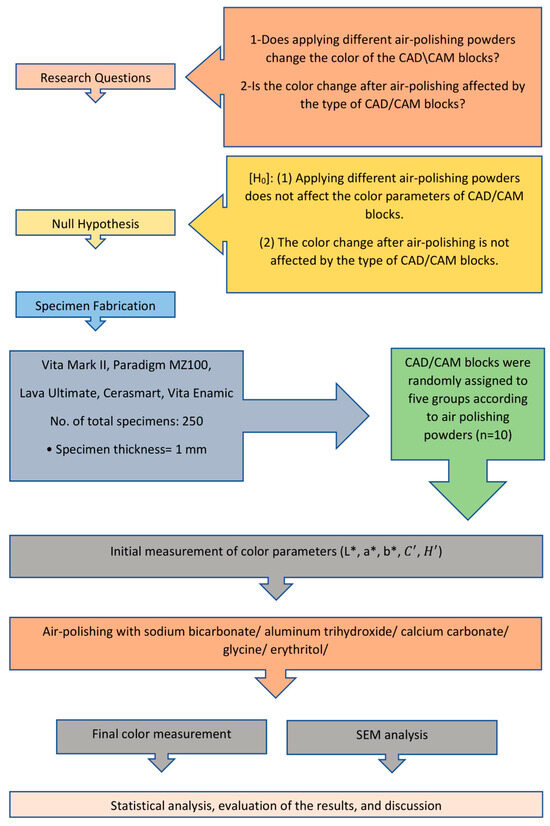
Figure 1.
Study design.
The sample size was calculated with power analysis using G*Power software (Version 3.1; University of Dusseldorf, Dusseldorf, Germany). The effect size (d) was taken as 0.606, and the standard deviation value for ΔE was taken as 1, so the number of samples, determined for power: 0.80 and α: 0.05, was calculated as minimum n = 6 for each group, related to a previous study [34]. Fifty samples of 1.1 mm thickness were prepared from each CAD-CAM block with diamond disks using a slow-speed precision cutter (Mecatome T180, PRESI, Eybens, France). All specimens were polished with a polishing machine (Mecatech 234 TCI-10, Presi, Eybens, France) and ground to the final thickness of 1 mm with waterproof abrasive papers from 320 to 2000 grits. Then, the samples were further polished for 180 s with the same machine, in accordance with the manufacturer’s instructions, at a constant speed of 150 rpm clockwise and 30 rpm counterclockwise, with a pressure of 3.00 daN, using a blue polishing felt (PRESI, Reflex Concept Pad Mag, Eybens, France) and a 0.50 mL drop of diamond polishing solution (PRESI, Preparations Diamentees Mecaprex, Eybence, France) every 10 s. The final thicknesses of the specimens were checked with a digital micrometer (C-master; Mitutoyo, Japan). Then, the specimens were cleaned in distilled water with an ultrasonic cleaner (Eurosonic Energy; Euronda SpA, Vicenza, Italy) for 10 min and dried with oil-free air for 30 s. After the CAD/CAM blocks were randomly divided into five groups (n = 10) for air-polishing with different APPs, the color measurement of each specimen was performed.
The color parameters, L* (lightness), a* (green-red coordinates), and b* (yellow-red coordinates) of each specimen were measured on a white background (L* = 98.20, a* = −3.52, b* = 4.65) using a spectrophotometer (CM-3600d, Konica Minolta, Tokyo, Japan), with 360–740 nm wavelength interval, specular component included, standard D65 (daylight) light source, and 2° observer [35], double-ray UV-100% visible reflection feature with 10 nm intervals, by an experienced operator who had been trained to use the spectrophotometer. Each specimen was measured three times; the mean value was the sample’s color. The device was calibrated before the measurement of each group, according to the manufacturer’s instructions, using computer software (Spectra-Magic NX, Version 3.61, Konica Minolta Sensing, Inc., Tokyo, Japan).
After initial color measurements, five different APPs, namely, sodium bicarbonate, aluminum trihydroxide, calcium carbonate, glycine, and erythritol, were applied to each group with an air-polishing device (Air-Flow S1, EMS, Nyon, Switzerland). The brands, manufacturers, lot numbers, particle sizes, Mohs hardness numbers, and densities of the tested APPs are presented in Table 2.
Table 2.
Air-polishing powders used in this study.
The APPs were applied on the surface of the specimens with a 60° angle and 4–5 mm distance at a medium setting (water pressure: 3 bar, air pressure: 5.75 bar) always by the same trained operator [24,36,37]. In addition, the powder chamber of the device was filled to the maximum level after each application to ensure constant powder flow. A previous study reported that the application period of APP during a recall appointment should be 1–2 s for a tooth’s anatomical crown, and more than 10 s would be harmful to the patient [38]. In a dental arch without any loss of interdental papilla and ideally aligned, an average upper central incisor’s surface area that can be air-polished is approximately 150 mm2 [39]. Therefore, if air polishing can be applied for 1 s to a 150 mm2 area in a recall appointment, then it should be applied for 1/150 s to an area of 1 mm2 in one recall appointment. Consequently, to simulate ten years of 3 monthly recall appointments [15,18,40,41,42], air-polishing should be applied for a period of 40 times longer, equivalent to 40/150 s for a 1 mm2 area [43]. Since the surface areas of the CAD/CAM blocks were different from each other, the duration of air-polishing was calculated for the area of each CAD/CAM block separately.
After surface treatment with the APPs, each specimen’s L*, a*, and b* values were measured in the same way. The quantitative color difference (ΔE00) values between the initial and final measurements of the specimens were calculated using the CIEDE2000 color difference formula [44].
In this formula, , , and indicate the differences in lightness, chroma, and hue, respectively; SL, SC, and SH are weighting functions that provide a better correspondence with visual evaluation; and RT is the rotation function. The parametric weighting factors kL, kC, and kH are correction terms for experimental conditions, and they were determined as 1 [45,46].
Microscopic morphologies and particle sizes of the APPs were analyzed under scanning electron microscopy (SEM) (Zeiss EVO LS 10, Oberkochen, Germany). The APPs were sputter-coated with gold/palladium (Quorum SC7620, West Sussex, England) and then analyzed at 10 kV, with 100×, 200×, and 15,000× magnifications. In addition, ten specimens from each CAD/CAM block were processed, with two samples for each APP. The CAD/CAM block was then split into two halves. One half was sprayed with APP, while the other half was covered and left untreated, serving as control. Then, these specimens were also sputter-coated with gold/palladium (Quorum SC7620, West Sussex, England), and the surface topography was analyzed under SEM with 10 kV at 200× magnification.
Mean ± standard deviation values were calculated according to the raw data in Table S1 and subjected to the Shapiro–Wilk test to analyze the convenience of quantitative data for a normal distribution; a one-way ANOVA analysis to compare three or more groups that showed a normal distribution; and the Tukey HSD test to determine which group made the difference. A two-way ANOVA analysis was used to evaluate the effect of both the APP and the restorative material type on quantitative variables. Significance was evaluated at p < 0.01 and p < 0.05 levels.
3. Results
The results of the colorimetric analysis revealed that applying APPs affects the final color of CAD/CAM restorative material (p < 0.05, p < 0.001). In accordance with the two-way ANOVA test results in Table 3, the type of CAD/CAM restorative material had a significant effect on the ΔL* (F: 21.121; p < 0.001), Δa* (F: 18.063; p < 0.001), Δb* (F: 49.923; p < 0.001), (F: 43.939; p < 0.001), (F: 19.243; p < 0.001), and ΔE00 (F: 6.205; p < 0.001) values. In addition, the type of APP had a significant effect on the Δa* (F: 28.589; p < 0.001), ΔH (F: 27.578; p < 0.001), and ΔE00 (F: 5.505; p < 0.001) values, but did not have a significant effect on ΔL* (F: 2.399; p: 0.051), Δb* (F: 1.805; p: 0.129), or (F: 2.062; p: 0.087) values. The interaction terms were not significant for the ΔL* (F: 1.095; p: 0.361), Δa* (F: 1.552; p: 0.084), Δb* (F: 0.854; p: 0.623), (F: 0.948; p: 0.515), (F: 1.669; p: 0.054), or ΔE00 (F: 0.996; p: 0.462) values.
Table 3.
Influence of CAD/CAM block type and air-polishing powder on the change in color parameters according to two-way ANOVA.
As illustrated in Table 4, Table 5, Table 6, Table 7 and Table 8, in the MZ groups, the samples’ ΔL*, Δa*, Δb*, and values decreased, while the values increased, regardless of the APP used. The APP of aluminum trihydroxide increased the Δb* and values of LU significantly compared with the APP of glycine. In addition, calcium carbonate increased the Δa* and values of LU significantly, compared with the other APPs. Furthermore, aluminum trihydroxide and calcium carbonate changed the Δa* and values of CS significantly, compared with the rest of the APPs.
Table 4.
Mean ± standard deviation values of the ΔL* value and post hoc analysis for pairwise comparisons.
Table 5.
Mean ± standard deviation values of the Δa* value and post hoc analysis for pairwise comparisons.
Table 6.
Mean ± standard deviation values of the Δb* value and post hoc analysis for pairwise comparisons.
Table 7.
Mean ± standard deviation values of the ΔC′ value and post hoc analysis for pairwise comparisons.
Table 8.
Mean ± standard deviation values of the ΔH’ value and post hoc analysis for pairwise comparisons.
As shown in Table 9, there were significant differences in terms of the ΔE00 values related to the type of APP only in the LU and CS groups (p: 0.004). According to the pairwise comparison, it was found that the ΔE00 values of the samples that were exposed to erythritol in the LU group and glycine in the CS group were significantly lower than those exposed to calcium carbonate (p: 0.012; p: 0.003); and aluminum trihydroxide (p: 0.047; p: 0.008), respectively. There was no significant difference in terms of the ΔE00 values in the VM, MZ, and VE groups according to different APPs (p > 0.05).
Table 9.
Mean ± standard deviation values of the ΔE00* value and post hoc analysis for pairwise comparisons.
When comparing the effect of the APPs on the ΔE00 values of the CAD/CAM blocks, as presented in Table 9, in the sodium bicarbonate, aluminum trihydroxide, calcium carbonate, or glycine-treated groups, there were significant differences in terms of the ΔE00 values according to the type of CAD/CAM restorative material (p: 0.005; p: 0.003; p: 0.005; and p: 0.002), respectively, whereas in the erythritol group, there was no significant difference (p > 0.05). According to the pairwise comparisons, it was found that among the sodium bicarbonate-treated groups, the ΔE00 values of the MZ samples were significantly higher than LU (p: 0.005) and VE (p: 0.027); among the aluminum trihydroxide-treated groups, the ΔE00 values of MZ were significantly higher than the LU (p: 0.026) and VE (p: 0.006) CAD/CAM blocks. Among the calcium carbonate-treated groups, the ΔE00 values of the VM samples were significantly lower than CS (p: 0.017) and MZ (p: 0.007); among the glycine-treated groups, the ΔE00 values of MZ samples were significantly higher than the VM (p: 0.005), LU (p: 0.007), CS (p: 0.013) and VE (p: 0.008) samples.
Figure 2 presents the SEM micrographs showing different microscopic morphologies and particle sizes of the APPs tested. As seen in Figure 2, with 100× and 200× magnifications, the particles of sodium bicarbonate and glycine are not evenly distributed; their sizes are in a wide range, with a large variety of fragment forms with sharp edges and corners. On the other hand, the particles of the calcium carbonate powder are in the form of large and small spherical clusters, and the aluminum trihydroxide particles are again spherical but in a more irregular form. Furthermore, erythritol particles have a homogenous distribution. They seem denser than the rest of the powders in terms of particle per unit area, and their particle sizes are similar to each other with an irregular microstructure. In addition, at 15,000× magnification, the particles of the powders are analyzed in detail, and their sizes are measured. It is observed that sodium bicarbonate powder resembles a rocky shape, whereas glycine powder seems to have smoother, rounder borders. In addition, aluminum trihydroxide powder seems to have interlockings with sharp edges; calcium carbonate powder appears as a cluster of small particles; and erythritol powder has an amorphous microstructure with an irregular surface. Furthermore, according to the SEM measurements of this study, particle sizes of sodium bicarbonate range between 10 and 258 µm; aluminum trihydroxide range between 110 and 227 µm; calcium carbonate range between 4 and 94 µm; glycine range between 7 and 211 µm; and erythritol range between 4 and 75 µm.
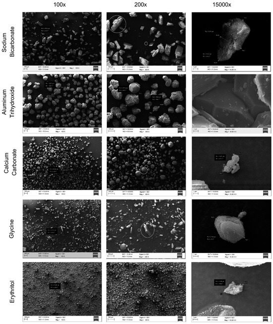
Figure 2.
SEM micrographs of the air polishing powders at 100×, 200×, 15,000× magnifications. In the 100× images, the size of a big particle is measured. In the 200× images, a big particle and a small particle to be measured are marked, and the small particle of aluminum trihydroxide is measured at this magnification since its particle size is too large to measure at 15,000× magnification. In the 15,000× images, the size of a small particle is measured.
The SEM micrographs in Figure 3, Figure 4, Figure 5, Figure 6 and Figure 7 show the differences between the air-polished and non-air-polished parts for each CAD/CAM restorative material and the APPs. As seen in these SEM images, calcium carbonate caused a significant roughness on all CAD/CAM restorative materials. In addition, aluminum trihydroxide caused a distinct roughness in CAD/CAM restorative materials, especially in MZ, LU, and CS blocks belonging to the resin with scattered fillers group (Figure 4, Figure 5 and Figure 6). No significant difference was observed between CAD/CAM blocks consisting of resin with scattered fillers air-polished with sodium bicarbonate, glycine, and erythritol (Figure 4, Figure 5 and Figure 6).
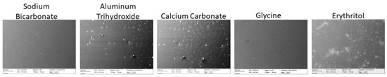
Figure 3.
SEM micrographs of Vita Mark II specimens with a half-air-polished, half-non-polished surface at 200×.
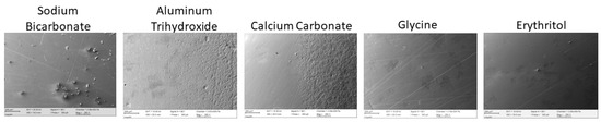
Figure 4.
SEM micrographs of Paradigm MZ 100 specimens with a half-air-polished, half-non-polished surface at 200×.
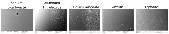
Figure 5.
SEM micrographs of Lava Ultimate specimens with a half-air-polished, half-non-polished surface at 200×.
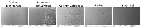
Figure 6.
SEM micrographs of Cerasmart specimens with a half-air-polished, half-non-polished surface at 200×.
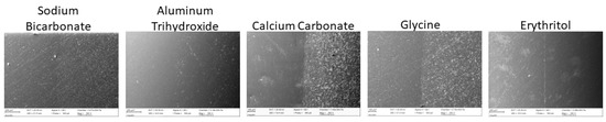
Figure 7.
SEM micrographs of Vita Enamic specimens with a half-air-polished, half-non-polished surface at 200×.
4. Discussion
In this in vitro study, the effect of five different APPs on the color change in five different CAD/CAM restorative materials was evaluated, and statistically significant differences were found (p < 0.05, p < 0.001). According to the results, both the first and second parts of the null hypothesis were rejected since the color of CAD/CAM restorative materials changed in different amounts after applying different APPs.
Based on the results, the ΔE00 values of the LU samples that were exposed to erythritol and the CS samples that were exposed to glycine were significantly lower than those exposed to calcium carbonate and aluminum trihydroxide. Considering the APPs, calcium carbonate has the highest density, and aluminum trihydroxide has the highest hardness value (Table 2) and the largest average particle size, as seen in the SEM images in Figure 2. On the other hand, erythritol has the lowest density, the lowest hardness value, and the smallest average particle size, and glycine follows subsequently. Previous studies reported that the APP becomes more abrasive as the mean particle size, hardness, and density increase [21,23,26]. In addition, as surface roughness increases, gloss decreases, affecting color perception [13,19,34,45,47]. Therefore, the color changes in the restorative materials can be attributed to the surface roughness that air-polishing causes, as can be seen in the SEM images of the CAD/CAM restorative materials (Figure 3, Figure 4, Figure 5, Figure 6 and Figure 7). For instance, the LU (Figure 5) and CS (Figure 6) samples treated with calcium carbonate and aluminum trihydroxide show apparent porosity in the SEM images compared with the other APPs, and this can explain the significant color change. Furthermore, Pelka et al. and Barnes et al. reported that in terms of abrasiveness, glycine < sodium bicarbonate < calcium carbonate < aluminum trihydroxide [24,29]. This result corresponds to the amount of color change caused by the APPs that are used in the present study.
In terms of color change according to the type of CAD/CAM restorative material, the ΔE00 values of the MZ samples were significantly higher than the LU and VE samples treated with sodium bicarbonate or aluminum trihydroxide. Previous research reported that air-polishing with sodium bicarbonate or aluminum trihydroxide increases the roughness of resin composites, and the higher ΔE00 values of the MZ samples could be partially attributed to this [25,32]. In addition, Guler et al. revealed that an increase in filler size and replacement of the fillers in restorative materials after air-polishing might increase roughness [31]. The filler size of LU is much smaller than MZ, which probably caused less roughness of LU and, thereby, less color change. Among the calcium carbonate-treated groups in this study, the ΔE00 values of the VM samples were found to be significantly lower than those of CS and MZ, which is parallel with the results of a recent study reporting that glass–ceramic materials have significantly better color stability than resin composites [48]. In addition, among the glycine-treated groups, the color change in the MZ samples was significantly higher than the other restorative materials, which is due to the differences in composition and microstructure of the restorative materials [13,48]. However, when the SEM images of the CAD/CAM blocks were analyzed, the glycine-treated VE samples (Figure 7) seemed more porous than the glycine-treated MZ samples (Figure 4). There might be two reasons why the MZ samples do not appear significantly rougher than the other blocks in SEM images (Figure 3, Figure 4, Figure 5, Figure 6 and Figure 7) but their ΔE00 values are significantly higher: 1—when the glycine powder is sprayed on VE, it might have roughened the surface and splashed, but may have partially penetrated into the MZ sample owing to MZ’s lower hardness value compared with the other blocks in this study, and glycine’s tiny size, thereby changed the color and 2—the SEM images’ magnification is 200×, and this might not be enough to show the exact surface topography that the small-sized APPs created [49].
In this study, to determine the color difference, the CIEDE2000 system, which is the most recent formula that CIE officially suggests, is used because the CIELAB system is not sufficient for determining especially small color differences [35,44]. The ΔE00 values of this study were interpreted according to the literature reports on visual thresholds, which provide guidance on the clinical relevance of the results [35]. According to the 50:50% CIEDE2000 acceptability thresholds for ΔE00 = 1.8 [44], = 2.92, = 2.52, and = 1.90 [35], all groups had acceptable color changes. However, according to the 50:50% perceptibility threshold (ΔE00 = 0.8) [44], the VM samples air-polished with aluminum trihydroxide or erythritol; the MZ samples treated with any of the APPs; the LU samples exposed to calcium carbonate; the CS samples air-polished with any of the APPs, except glycine; and the VE samples treated with calcium carbonate or erythritol demonstrated ΔE00 values above the perceptibility threshold. Regarding these results, glycine seems safe to use on restorative materials, except MZ, a resin composite. Cobb CM et al. also reported that glycine is the most efficient in removing biofilm from natural teeth and restorative materials, and it causes the least surface damage compared with the other powders [15]. Another study revealed that glycine and erythritol have a similar surface-damaging potential [22]. However, despite its small particle size, as seen in the SEM image (Figure 2), and its low density and hardness, though statistically insignificant, erythritol caused a sufficient color change to exceed the perceptibility threshold of all restorative materials except LU. This effect of erythritol might be because more particles may have struck the sample per unit of time owing to its small and uniform particle size (Figure 2) [37]. In addition, LU seems to be discolored the least among the other CAD/CAM blocks, which is probably due to its small filler size, which sustains better resistance to abrasion due to the quality of the interfacial bonding between the small fillers and the resin matrix [21]. It is reported that large and insufficiently embedded filler particles usually result in a greater loss of volume. In contrast, small filler particles demonstrate higher resistance to air-polishing abrasion and a more regular microstructure, which also leads to better chemical and optical properties [21]. Furthermore, LU is composed of aggregated nanoclusters, and owing to that, polishing causes drifting of the nanofillers instead of turning out of the whole cluster, which provides better resistance to wear [28]. Moreover, Barnes et al. revealed that sodium bicarbonate and glycine are safe for resin composites, whereas aluminum trihydroxide and calcium carbonate are not [24]. However, in this study, it was found that in terms of color stability, sodium bicarbonate and glycine are also not safe to use on MZ resin composite blocks. In addition, the ΔE00 values of the MZ samples were above the perceptibility threshold in all the APP groups, probably due to their organic components and relatively big filler particles [21].
In this in vitro study, it was observed that the APPs, which are used to remove stains, themselves cause discolorations on the restorative materials. Although they changed the color of the restorative materials within 50:50% acceptable limits, the majority of them exceeded the 50:50% perceptible limits, which means that 50% of the observers could notice the color difference [35]. Therefore, considering the findings of this study and the previous studies, regardless of the APP used, clinicians should consider avoiding or reducing the use of APPs on CAD/CAM restorative materials during supportive periodontal therapy, since they may have doubts about which restorative material they are facing intraorally. Another feasible method is to repolish the restorations after air-polishing, either with the finest polishing pastes and rubber cups or according to the manufacturer’s instructions, if the restorative material is known, to smoothen the surface as much as possible and thereby sustain color stability [13,28,31].
There are some limitations to this study. Primarily, in vitro studies do not demonstrate the intra-oral environment completely in terms of coloring beverages, the washing effect of saliva, habits like smoking, brushing the teeth, thermal alterations, etc., which may affect the color change. In addition, the samples were not subjected to artificial aging because this study aimed to evaluate the effect of APP, not aging. Secondly, intra-oral feldspathic ceramic restorations are glazed, but most restorative materials used in this study did not require glaze as the manufacturers instructed, and in order not to add another variable to this study and to standardize the study protocol, the specimens were not glazed, but they were all mechanically polished with a diamond polishing solution.
Further laboratory and clinical studies, without such limitations, should be executed to confirm the findings of this study. Although combining variables will not provide information on the effect of any single variable, aging and glaze can be added as variables to simulate the intraoral conditions better. In addition, this study analyzed only the color change and the SEM micrographs for surface analysis, but the surface roughness and gloss should also be investigated with a combination of repolishing procedures. Moreover, the influence of different APPs to CAD/CAM blocks stored in staining solutions such as coffee, coke, and black tea can also be researched in future studies in terms of optical and surface properties.
5. Conclusions
Within the limitations of this in vitro study, the following conclusions were drawn:
- Both the type of CAD/CAM restorative material type and the APP had a significant effect on color difference, but their interaction terms were not significant.
- The color changes in all groups were clinically acceptable, but most of the groups presented perceptible color changes.
- Among the APPs, glycine seems the safest to use on restorative materials, but even glycine caused a perceptible color change for MZ.
- Dentists who are uncertain about the specific restorative material should limit prolonged air-polishing or avoid it altogether.
Supplementary Materials
The following supporting information can be downloaded at: https://www.mdpi.com/article/10.3390/app132011573/s1, Table S1: Raw Data of Color Parameters.
Author Contributions
Conceptualization, R.T.O. and E.Y.; methodology, R.T.O. and E.Y; validation, R.T.O. and E.Y.; formal analysis, R.T.O. and E.Y; investigation, R.T.O.; resources, R.T.O.; data curation, R.T.O. and E.Y.; writing—original draft preparation, R.T.O.; writing—review and editing, R.T.O. and E.Y.; supervision, E.Y. All authors have read and agreed to the published version of the manuscript.
Funding
This research received no external funding.
Institutional Review Board Statement
Not applicable.
Informed Consent Statement
Not applicable.
Data Availability Statement
The raw data presented in this study are available in the Supplementary Material.
Acknowledgments
This study is based on Rana Turunç Oğuzman’s thesis, which was submitted to the Department of Prosthetic Dentistry of the Faculty of Dentistry at İstanbul Medipol University, as partial fulfillment of the requirements for specialization in dentistry.
Conflicts of Interest
The authors declare no conflict of interest.
References
- Zarone, F.; Russo, S.; Sorrentino, R. From porcelain-fused-to-metal to zirconia: Clinical and experimental considerations. Dent. Mater. 2011, 27, 83–96. [Google Scholar] [CrossRef]
- Bajraktarova-Valjakova, E.; Korunoska-Stevkovska, V.; Kapusevska, B.; Gigovski, N.; Bajraktarova-Misevska, C.; Grozdanov, A. Contemporary dental ceramic materials, a review: Chemical composition, physical and mechanical properties, indications for use. Open Access Maced. J. Med. Sci. 2018, 6, 1742–1755. [Google Scholar] [CrossRef]
- Park, J.S.; Lim, Y.J.; Kim, B.; Kim, M.J.; Kwon, H.B. Clinical evaluation of time efficiency and fit accuracy of lithium disilicate single crowns between conventional and digital impression. Materials 2020, 13, 5467. [Google Scholar] [CrossRef]
- Giordano, R. Materials for chairside CAD/CAM-produced restorations. J. Am. Dent. Assoc. 2006, 137, 14S–21S. [Google Scholar] [CrossRef] [PubMed]
- Ruse, N.D.; Sadoun, M.J. Resin-composite blocks for dental CAD/CAM applications. J. Dent. Res. 2014, 93, 1232–1234. [Google Scholar] [CrossRef] [PubMed]
- Awada, A.; Nathanson, D. Mechanical Properties of Resin-Ceramic CAD/CAM restorative materials. J. Prosthet. Dent. 2015, 114, 587–593. [Google Scholar] [CrossRef] [PubMed]
- Gracis, S.; Thompson, V.; Ferencz, J.; Silva, N.; Bonfante, E. A new classification system for all-ceramic and ceramic-like restorative materials. Int. J. Prosthodont. 2015, 28, 227–235. [Google Scholar] [CrossRef]
- Coldea, A.; Swain, M.V.; Thiel, N. Mechanical properties of polymer-infiltrated-ceramic-network materials. Dent. Mater. 2013, 29, 419–426. [Google Scholar] [CrossRef] [PubMed]
- Bergamo, E.T.P.; Yamaguchi, S.; Coelho, P.G.; Lopes, A.C.O.; Lee, C.; Bonfante, G.; Benalcázar Jalkh, E.B.; de Araujo-Júnior, E.N.S.; Bonfante, E.A. Survival of Implant-Supported Resin-Matrix Ceramic Crowns: In Silico and Fatigue Analyses. Dent. Mater. 2021, 37, 523–533. [Google Scholar] [CrossRef]
- Mainjot, A.K.; Dupont, N.M.; Oudkerk, J.C.; Dewael, T.Y.; Sadoun, M.J. From Artisanal to CAD-CAM Blocks: State of the Art of Indirect Composites. J. Dent. Res. 2016, 95, 487–495. [Google Scholar] [CrossRef]
- Spitznagel, F.A.; Boldt, J.; Gierthmuehlen, P.C. CAD/CAM Ceramic Restorative Materials for Natural Teeth. J. Dent. Res. 2018, 97, 1082–1091. [Google Scholar] [CrossRef]
- Günal-Abduljalil, B.; Ulusoy, M.M. The Effect of Resin Cement Shade and Restorative Material Type and Thickness on the Final Color of Resin-Matrix Ceramics. J. Prosthodont. Res. 2022, 66, 75–82. [Google Scholar] [CrossRef] [PubMed]
- Liebermann, A.; Spintzyk, S.; Reymus, M.; Schweizer, E.; Stawarczyk, B. Nine prophylactic polishing pastes: Impact on discoloration, gloss, and surface properties of a cad/cam resin composite. Clin. Oral Investig. 2019, 23, 327–335. [Google Scholar] [CrossRef]
- Sawai, M.A.; Bhardwaj, A.; Jafri, Z.; Sultan, N.; Daing, A. Tooth polishing: The current status. J. Indian Soc. Periodontol. 2015, 19, 375–380. [Google Scholar] [CrossRef] [PubMed]
- Cobb, C.M.; Daubert, D.M.; Davis, K.; Deming, J.; Flemmig, T.F.; Pattison, A.; Roulet, J.-F.; Stambaugh, R.V. Consensus conference findings on supragingival and subgingival air polishing. Compend. Contin. Educ. Dent. 2017, 38, e1–e4. [Google Scholar] [PubMed]
- Martins, O.; Costa, A.; Silva, D. The efficacy of air polishing devices in supportive periodontal therapy: Clinical, microbiological and patient-centred outcomes. A Systematic Review. Int. J. Dent. Hyg. 2023, 21, 41–58. [Google Scholar] [CrossRef]
- Manresa, C.; Ec, S.; Twigg, J.; Bravo, M. Supportive periodontal therapy (SPT) for maintaining the dentition in adults treated for periodontitis. Cochrane Database Syst. Rev. 2018, 1, 2–3. [Google Scholar] [CrossRef]
- Lu, H.; He, L.; Zhao, Y.; Meng, H. The effect of supragingival glycine air polishing on periodontitis during maintenance therapy: A randomized controlled trial. PeerJ 2018, 2018, e4371. [Google Scholar] [CrossRef]
- Colucci, V.; Dos Santos, C.D.; Do Amaral, F.L.B.; Corona, S.A.M.; Catirse, A.B.C.E.B. Influence of NaHCO3 powder on translucency of microfilled composite resin immersed in different mouthrinses. J. Esthet. Restor. Dent. 2009, 21, 242–248. [Google Scholar] [CrossRef]
- Graumann, S.J.; Sensat, M.L.; Stoltenberg, J.L. Air polishing: A review of current literature. J. Dent. Hyg. 2013, 87, 173–180. [Google Scholar]
- Pelka, M.A.; Altmaier, K.; Petschelt, A.; Lohbauer, U. The effect of air-polishing abrasives on wear of direct restoration materials and sealants. J. Am. Dent. Assoc. 2010, 141, 63–70. [Google Scholar] [CrossRef] [PubMed]
- Janaphan, K.; Hill, R.G.; Gilllam, D.G. In vitro evaluation of the abrasiveness of novel bioactive glass powders (Biominf®) on ivory dentine in air polishing procedures compared to selected reference powders. J. Dent. Maxillofac. Res. 2021, 4, 1–6. [Google Scholar] [CrossRef]
- Karmakar, S.; Kamath, D.G. Subgingival airpolishing: A simple and cost effective medical insurance. J. Pharm. Sci. Res. 2017, 9, 199–201. [Google Scholar]
- Barnes, C.M.; Covey, D.; Watanabe, H.; Simetich, B.; Schulte, J.R.; Chen, H. An in vitro comparison of the effects of various air polishing powders on enamel and selected esthetic restorative materials. J. Clin. Dent. 2014, 25, 76–87. [Google Scholar]
- Johnson, W.W.; Barnes, C.M.; Covey, D.A.; Walker, M.P.; Ross, J.A. The effects of a commercial aluminum airpolishing powder on dental restorative materials. J. Prosthodont. 2004, 13, 166–172. [Google Scholar] [CrossRef] [PubMed]
- Janiszewska-Olszowska, J.; Drozdzik, A.; Tandecka, K.; Grocholewicz, K. Effect of air-polishing on surface roughness of composite dental restorative material-comparison of three different air-polishing powders. BMC Oral Health 2020, 20, 30. [Google Scholar] [CrossRef]
- Barnes, C.M. An In-Depth Look at Air Polishing. Dimens Dent. Hyg. 2010, 8, 32–36. [Google Scholar]
- Németh, K.D.; Haluszka, D.; Seress, L.; Lovász, B.V.; Szalma, J.; Lempel, E. Effect of air-polishing and different post-polishing methods on surface roughness of nanofill and microhybrid resin composites. Polymers 2022, 14, 1643. [Google Scholar] [CrossRef]
- Pelka, M.; Trautmann, S.; Petschelt, A.; Lohbauer, U. Influence of air-polishing devices and abrasives on root dentin-an in vitro confocal laser scanning microscope study. Quintessence Int. 2010, 41, e141–e148. [Google Scholar]
- Ng, E.; Byun, R.; Spahr, A.; Divnic-Resnik, T. The efficacy of air polishing devices in supportive periodontal therapy: A systematic review and meta analysis. Quintessence Int. 2018, 49, 453–467. [Google Scholar]
- Güler, A.U.; Duran, I.; Yücel, A.Ç.; Özkan, P. Effects of air-polishing powders on color stability of composite resins. J. Appl. Oral Sci. 2011, 19, 505–510. [Google Scholar] [CrossRef]
- Mathias, P.; Cunha, T.M.d.S.; Rocha, I.A.R.; Vitória, L.A.; Mathias, C.; Cavalcanti, A.N. Effect of air-polishing on properties of nanocomposite submitted to coffee, red wine and cigarette smoke. Braz J. Oral Sci. 2018, 17, e18021. [Google Scholar] [CrossRef]
- Valian, A.; Ansari, Z.J.; Rezaie, M.M.; Askian, R. Composite surface roughness and color change following airflow usage. BMC Oral Health 2021, 21, 398. [Google Scholar] [CrossRef]
- Eldwakhly, E.; Ahmed, D.R.M.; Soliman, M.; Abbas, M.M.; Badrawy, W. Color and translucency stability of novel restorative CAD/CAM materials. Dent. Med. Probl. 2019, 56, 349–356. [Google Scholar] [CrossRef]
- Paravina, R.D.; Pérez, M.M.; Ghinea, R. Acceptability and perceptibility thresholds in dentistry: A comprehensive review of clinical and research applications. J. Esthet. Restor. Dent. 2019, 31, 103–112. [Google Scholar] [CrossRef] [PubMed]
- Babina, K.; Polyakova, M.; Sokhova, I.; Doroshina, V.; Arakelyan, M.; Zaytsev, A.; Novozhilova, N. The Effect of ultrasonic scaling and air-powder polishing on the roughness of the enamel, three different nanocomposites, and composite/enamel and composite/cementum interfaces. Nanomaterials 2021, 11, 3072. [Google Scholar] [CrossRef] [PubMed]
- Tada, K.; Kakuta, K.; Ogura, H.; Sato, S. Effect of particle diameter on air polishing of dentin surfaces. Odontology. 2010, 98, 31–36. [Google Scholar] [CrossRef]
- Rayman, A.S.; Dincer, E. Air Polishing. Hygiene 2013, 1, 7–12. [Google Scholar]
- Nelson, S.J.; Ash, M.M. Wheeler’s Dental Anatomy, Physiology and Occlusion, 9th ed.; Saunders/Elsevier: St. Louis, MO, USA, 2010; p. 41. [Google Scholar]
- Sanz, M.; Herrera, D.; Kebschull, M.; Chapple, I.; Jepsen, S.; Beglundh, T.; Sculean, A.; Tonetti, M.S.; Merete Aass, A.; Aimetti, M.; et al. Treatment of stage I–III Periodontitis—The EFP S3 level clinical practice guideline. J. Clin. Periodontol. 2020, 47, 4–60. [Google Scholar] [CrossRef]
- Rinke, S.; Bettenhäuser-Hartung, L.; Leha, A.; Rödiger, M.; Schmalz, G.; Ziebolz, D. Retrospective evaluation of extended glass-ceramic ceramic laminate veneers after a mean observational period of 10 years. J. Esthet. Restor. Dent. 2020, 32, 487–495. [Google Scholar] [CrossRef]
- Sampaio, F.B.W.R.; Özcan, M.; Gimenez, T.C.; Moreira, M.S.N.A.; Tedesco, T.K.; Morimoto, S. Effects of manufacturing methods on the survival rate of ceramic and indirect composite restorations : A systematic review and meta-analysis. J. Esthet. Restor. Dent. 2019, 31, 561–571. [Google Scholar] [CrossRef]
- Atkinson, D.R.; Cobb, C.M.; Killoy, W.J. The effect of an air-powder abrasive system on in vitro root surfaces. J. Periodontol. 1984, 55, 13–18. [Google Scholar] [CrossRef] [PubMed]
- Paravina, R.D.; Ghinea, R.; Herrera, L.J.; Bona, A.D.; Igiel, C.; Linninger, M.; Sakai, M.; Takahashi, H.; Tashkandi, E.; Del Mar Perez, M. Color difference thresholds in dentistry. J. Esthet. Restor. Dent. 2015, 27, S1–S9. [Google Scholar] [CrossRef] [PubMed]
- Perez, M.D.M.; Ghinea, R.; Herrera, L.J.; Ionescu, A.M.; Pomares, H.; Pulgar, R.; Paravina, R.D. Dental Ceramics: A CIEDE2000 acceptability thresholds for lightness, chroma and hue differences. J. Dent. 2011, 39, e37–e44. [Google Scholar] [CrossRef] [PubMed]
- Silva, J.; Engler, M.L.P.D.; Baumgardt Barbosa Lima, R.; Jesús Suarez, M.; Guy Oliver Salomon, J.P.; Maziero Volpato, C.A. Color stability of a resin nanoceramic after surface treatments, adhesive cementation, and thermal aging. J. Prosthet. Dent. 2022, 127, 498.e1–498.e8. [Google Scholar] [CrossRef]
- Chae, Y. Color appearance shifts depending on surface roughness, illuminants, and physical colors. Sci. Rep. 2022, 12, 1371. [Google Scholar] [CrossRef]
- Paolone, G.; Mandurino, M.; De Palma, F.; Mazzitelli, C.; Scotti, N.; Breschi, L.; Gherlone, E.; Cantatore, G.; Vichi, A. Color Stability of Polymer-Based Composite CAD/CAM Blocks: A Systematic Review. Polymers 2023, 15, 464. [Google Scholar] [CrossRef]
- Şen, N.; Tuncelli, B.; Göller, G. Surface Deterioration of Monolithic CAD/CAM Restorative Materials after Artificial Abrasive Toothbrushing. J. Adv. Prosthodont. 2018, 10, 271–278. [Google Scholar] [CrossRef]


Disclaimer/Publisher’s Note: The statements, opinions and data contained in all publications are solely those of the individual author(s) and contributor(s) and not of MDPI and/or the editor(s). MDPI and/or the editor(s) disclaim responsibility for any injury to people or property resulting from any ideas, methods, instructions or products referred to in the content. |
© 2023 by the authors. Licensee MDPI, Basel, Switzerland. This article is an open access article distributed under the terms and conditions of the Creative Commons Attribution (CC BY) license (https://creativecommons.org/licenses/by/4.0/).