Article

Variability in Phytochemical Contents and Biological Activities among Adenophora triphylla Genotypes

1
Department of Industrial Plant Science and Technology, College of Agriculture, Life and Environment Sciences, Chungbuk National University, Cheongju 28644, Republic of Korea
2
Chungbuk Agricultural Research and Extension Services, Cheongju 28130, Republic of Korea
3
LG Chem Ltd., Seoul 07336, Republic of Korea
4
Department of Food Science and Biotechnology, College of Agriculture, Life and Environment Sciences, Chungbuk National University, Cheongju 28644, Republic of Korea
*
Author to whom correspondence should be addressed.
These authors contributed equally to this work.
Appl. Sci. 2023, 13(20), 11184; https://doi.org/10.3390/app132011184
Submission received: 27 September 2023 / Revised: 9 October 2023 / Accepted: 10 October 2023 / Published: 11 October 2023
(This article belongs to the Special Issue Advances in Biological Activities of Natural Products)

Abstract

:
Genetic diversity can remarkably impact the quality of derived products. This study focused on Adenophora triphylla, an herbaceous perennial used in East Asian traditional medicine. It is known for its diverse therapeutic effects that are attributed to phytochemicals. However, despite its therapeutic potential, a comprehensive exploration of the influence of genetic diversity on the pharmaceutical properties of various A. triphylla genotypes remains unknown. To address this knowledge gap, we employed DNA barcoding regions (ITS2, matK, and psbA-trnH) to elucidate the genetic diversity among the collected genotypes. The combined application of these barcoding regions is a robust method to identify A. triphylla genotypes. In addition, leaf extracts consistently exhibited higher antioxidant potential than root extracts. Nitric oxide production was strongly inhibited by the leaf extracts of the BG genotype (IC50 = 124.52 ± 12.97 µg/mL), and the leaf extracts (200 µg/mL) of the BT genotype contained significant antimelanogenic properties (IC50 = 181.56 ± 16.17 µg/mL). Various genotypes displayed diverse levels of phytochemical content, and the total saponin content exhibited a strong correlation with various biological activities of A. triphylla, suggesting a genetic influence on their pharmaceutical attributes. These findings highlight the significance of genotype selection for optimizing health benefits and promoting the potential utilization of specific genotypes in various industries, such as cosmetics and pharmaceuticals.

1. Introduction

Over the last few decades, there has been a remarkable global surge in the utilization of phytochemicals or nutraceuticals, with numerous individuals adopting them as their primary healthcare option [1]. Generally, plant-based products encompass crude extracts derived from individual plants or a combination of various plants believed to exhibit synergistic effects [2,3]. Although it is well established that plant-derived bioactive compounds play a crucial role in promoting human health, it remains unclear whether these effects are uniform across all plant species. Particularly, the phytochemical content and plant quality can significantly impact the quality of derived products, which is influenced by environmental factors and genetic diversity [4]. For example, different metabolic compositions among various Perilla frutescens genotypes led to the variation in biological activity [5,6], indicating that understanding the impact of genetic diversity on pharmaceutical properties is crucial for ensuring the quality control of medicinal plant-based products. Nevertheless, a comprehensive comparative analysis of these activities among genotypes is lacking.
Adenophora triphylla, an herbaceous perennial plant from the Campanulaceae family, has been employed in traditional East Asian herbal medicine to treat a range of illnesses, including cough, phlegm, asthma, and airway inflammatory diseases [7]. The extract of A. triphylla has been reported to exhibit various beneficial effects, including antiobesity, antioxidant, antifungal, antimelanogenic, anti-inflammatory, and antitumor activities. These effects have been attributed to the presence of diverse phytochemicals within the extract, such as saponin, inulin, polysthicol, lupenone, β-sitosterol, triphyllol, and daucosterol [7,8,9,10,11]. Interestingly, A. triphylla demonstrates a broad range of morphological characteristics and genetic traits among individual plants, even within the same species [9], indicating its genetic diversity. Therefore, it can be hypothesized that the pharmaceutical properties differ among the A. triphylla genotypes.
In this study, we differentiated five A. triphylla genotypes using the DNA barcoding approach, which is an increasingly popular method for accurate species identification based on reliable DNA regions. Moreover, 70% of ethanol extracts from the five different genotypes of A. triphylla differed in their antioxidant, antimelanogenic, and anti-inflammatory effects, indicating that their biological activities are influenced by genetic differences. Furthermore, phytochemical and marker compound analysis revealed that variations in their biological activities were attributed to varying levels of phytochemicals. The results of this investigation will promote further research into the potential utilization of specific A. triphylla genotypes as valuable plant breeding and genetic resources. The enhancement of the phytochemical contents of A. triphylla genotypes will enable their increased application in diverse industries, including cosmetics, functional foods, and pharmaceuticals.

2. Materials and Methods

2.1. Plant Materials and Extract Preparation

To mitigate the influence of geographical and environmental factors on the composition and content of phytochemicals, we acquired the leaves and roots of five A. triphylla genotypes that were cultivated at research farms under the supervision of the Chungcheongbuk-do Agricultural Research and Extension Service (Cheongju-si, Chungcheongbuk-do, Republic of Korea). Following freeze-drying, the leaf- or root-ground materials were immersed in 70% (v/v) ethanol for a duration of 24 h. The resulting filtered extracts were then concentrated using a rotary vacuum evaporator and stored at −20 °C until needed.

2.2. DNA Extraction and Analysis of DNA Barcoding Regions

Genomic DNA (gDNA) was extracted from each sample according to the manufacturer’s instructions provided on the DNeasy® Plant Mini kits (Qiagen, Hilden, Germany) and measured with an Optizen Nano Q spectrophotometer (Mecasys, Deajeon, Republic of Korea). PCR amplification and sequence analysis of the IST2, matK, and psbA-trnH regions were performed as described previously [12]. For the phylogenetic analysis, the quality-trimmed Sanger sequences were aligned with ClustalW, and the maximum composite likelihood method was used to determine the evolutionary distances. The unweighted pair-group method using arithmetic averages (UPGMA) trees was created using MEGA version 7. The primer sequences are listed in Table S1.

2.3. Phytochemical Analysis

Total flavonoid content (TFC) and total phenolic content (TPC) were measured using the method described by Ju et al. [6]. To analyze TFC, 0.2 mL of the 70% ethyl alcohol extract and its fractions were combined with 0.1 mL of 10% aluminum nitrate (w/v), 0.1 mL of 1 M potassium acetate, and 4.6 mL of 80% ethanol. These reaction mixtures were allowed to stand at room temperature for 40 min, after which the absorbance was measured against a blank at 415 nm. For TPC analysis, 100 μL of each sample and 50 μL of Folin–Ciocalteu reagent (2 N) was mixed and incubated for 5 min. Then, 0.3 mL of 20% Na2CO3 was added to the mixture and incubated for 15 min. The resultant blue color was read at an absorbance of 725 nm. The TPC and TFC in each extract were quantified in terms of milligrams of gallic acid equivalents (µg GAE/mg of extract) and quercetin equivalents (µg QE/mg of extract), respectively. Furthermore, the total saponin content (TSC) was assessed using the vanillin–sulfuric acid assay, following the method outlined in a prior study [13]. Briefly, 10 mg of each extract were dissolved in 1 mL of ethyl acetate and mixed with 500 μL of A reagent [0.5% (v/v) p-anisaldehyde in ethyl acetate] and 500 μL of B reagent [50% (v/v) H2SO4 in ethyl acetate]. After incubation at 60 °C for 10 min, the absorbance was measured at 430 nm. TSC was expressed as diosgenin equivalents (µg DE/mg of extract).
The contents of lupenone and β-sitosterol were determined using high-performance liquid chromatography equipped with a diode array detector (Agilent Technologies, Waldbronn, Germany) and a Poroshell 120 EC-c18 column (4.6 × 150 mm, 4 μm), as described by Yoon et al. [14]. The contents of all these compounds were analyzed by comparing the retention times and the ultraviolet spectral data. The concentration of these compounds was calculated by comparing the peak areas of the samples with the calibration curve of each standard.

2.4. Analysis of the Antioxidant Activities

The 1,1-diphenyl-2-picrylhydrazyl (DPPH) free-radical scavenging activity of each extract obtained from leaves or roots of five genotypes was measured using DPPH free radicals, as described by Ju et al. [6]. With various concentrations (62.5, 125, 250, 500, and 1000 μg/mL) of each test sample, 90 μL of 0.4 mM DPPH solution was mixed. After incubation for 10 min at room temperature, the absorbance was measured at 520 nm using a spectrophotometer. The findings were presented as the concentration necessary to neutralize 50% of the initial DPPH free radicals (IC50).
Reducing power was measured by monitoring the ability of the extract to reduce Fe3+ to Fe2+ at 750 nm, as described by Ju et al. [6], with ascorbic acid (AsA) as a positive control. Briefly, each extract (200 µg/mL) was mixed with 0.2 mL of 200 mM sodium phosphate buffer (pH 6.6) and 0.2 mL of 1% potassium ferricyanide. After incubation at 50 °C for 20 min, the mixture was mixed with 1 mL of 10% trichloroacetic acid and centrifuged at 6500 rpm for 10 min. Then, 500 μL of aliquots of the supernatants were mixed with 500 μL of deionized water and 100 μL of 0.1% ferric chloride, and the absorbance was measured at 750 nm.
The oxygen radical antioxidant capacity (ORAC) assay was conducted as previously described [6], using Trolox as a standard. A total of 150 μL of 0.08 μM fluorescein, diluted in a phosphate buffer (75 mM, pH 7.0), was mixed with 25 μL of phosphate buffer (used as a blank), Trolox standard (ranging from 6.25 to 50 μM), or each extract (40 μg/mL), with each placed in separate wells of a microplate. Following incubation at 37 °C for 10 min in the dark, 25 μL of freshly prepared 2,2′-azobis(isobutyramidine) dihydrochloride solution (0.12 g/mL) was added. Fluorescence intensity was monitored with fluorescent filters (485 and 530 nm excitation and emission wavelengths, respectively). ORAC values were represented as µM Trolox equivalent concentration (µM TE).

2.5. Cell Culture

RAW264.7 macrophage cells and B16F10 melanoma cells were cultured in Dulbecco’s Modified Eagle medium containing 10% fetal bovine serum, 100 µg/mL of spectinomycin, and 100 U/mL of penicillin. The cells were incubated at 37 °C in a humidified incubator with 5% CO2.

2.6. Determination of Cell Viability, Nitric Oxide (NO), and Melanin Production

To analyze the NO level in the cultured medium, RAW264.7 cells were treated with or without lipopolysaccharide (LPS) in the presence of extracts (50, 100, 150, and 200 µg/mL). After 24 h, 100 μL of Griess reagent (Promega Co., Ltd., Madison, WI, USA) was mixed with an equal volume of cell supernatant and incubated at room temperature for 10 min. The optical density at 540 nm was measured, and NO concentrations were calculated using a nitrite standard curve. The IC50 value (the concentration of 50% inhibition) was calculated from the graph of inhibition percentage against different extract concentrations.
The antimelanogenic activity was determined by treating B16F10 cells with each extract (50, 100, 150, and 200 µg/mL) and 100 µM of 3-isobutyl-1-methylxanthine (IBMX). Cultured B16F10 cells were plated at a density of 1 × 10⁵ cells/mL in 6-well plates, followed by incubation at 37 °C for 24 h. Subsequently, the B16F10 cells were treated with each extract and IBMX and incubated at 37 °C for 48 h. Afterward, the cells were washed twice with ice-cold PBS and harvested by centrifugation at 4000 rpm for 10 min. The resulting cell pellets were solubilized in 1 N NaOH with 10% dimethyl sulfoxide at 65 °C for 1 h. The concentration of melanin was determined using a microplate reader at 490 nm. Data are expressed in terms of melanin-synthesis inhibitory activity compared to the mock control. IC50 value is the concentration of extract at which IBMX-induced melanin production is inhibited by 50%.
Using a previously described protocol, the cell viability was assessed using a 3-[4,5-dimethylthiazol-2-yl]-2,5 diphenyl tetrazolium bromide (MTT) solution, and the absorbance was measured at 520 nm [6]. RAW264.7 or B16F10 cells were treated in each concentration of leaf or root extracts and stimulated with LPS or IBMX. After 24 h (for RAW264.7 cells) or 48 h (for B16F10 cells), 20 μL of MTT (5 mg/mL in PBS) solution was added to each well. Incubation was continued for 4 h more. The formazan crystals were dissolved in DMSO, and the optical density was measured at 520 nm.

2.7. Statistical Analysis

All experiments were performed in triplicates, and the Duncan’s test was utilized to assess the significance of differences between the groups, with p ≤ 0.05 considered statistically significant.

3. Results and Discussion

3.1. Genetic Diversity of A. triphylla Genotypes

As mentioned above, A. triphylla has a wide range of morphological variations [15,16]. Additionally, A. triphylla is native to Japan and is classified into rheophytic and nonrheophytic types based on leaf morphology and anatomy [15]. The leaves of the genotypes NT, NG, and L are lanceolate or cuneate, whereas those of BT and BG are elliptic or ovate. This shows that genotypes NT, NG, and L are of the rheophytic type (Figure 1a). Furthermore, these genotypes have been classified based on the presence of trichomes on the stem or the wavy phenotype (Figure 1a). This morphological diversity in A. triphylla is likely due to the adaptation of plants to different natural environments [17].
DNA barcoding is a special tool for the rapid identification of species based on short and standard DNA fragments (400–800 bp) [18,19]. Although the mitochondrial cytochrome oxidase I gene has been widely used for the identification of animal species and phylogenetic analysis [20], the chloroplast gene sequence (matK), the gene sequence of the noncoding region of chloroplast RNA (psbA-trnH), and the nuclear gene internal transcribed spacer (ITS) sequence are used in plant phylogeny research and to determine genetic diversity in species [21,22,23]. To investigate the genetic relationships between the A. triphylla genotypes, we collected and amplified the selected barcode regions using the ITS2, matK, and psbA-trnH primers (Table S1, Supplementary Materials). The sequence lengths of ITS2, matK, and psbA-trnH were 257–259 bp, 601 bp, and 311–312 bp, respectively, as determined through amplification of template gDNA extracted from each sample using universal primers (Table 1). Based on the ITS2 region, the phylogenetic analysis indicated that genotypes BG, BT, NG, and NT were closely related species and shared close affinities with A. verticillata var. hirsuta, whereas genotype L exhibited a more distant relationship from the other genotypes (Figure S1a). For the matK region, all genotypes, except for the NG genotype, exhibited close similarity to A. triphylla (Figure S1b). Moreover, the psbA-trnH sequences of all genotypes showed high similarity to A. triphylla (Figure S1c). Based on the trees generated using the regions ITS2 + matK  +  psbA-trnH (Figure 1b), all genotypes were grouped within the same clade as A. triphylla (90% bootstrap), indicating their close genetic relationships. Taken together, these results suggested that all collected genotypes constitute varieties of A. triphylla exhibiting morphological variations.

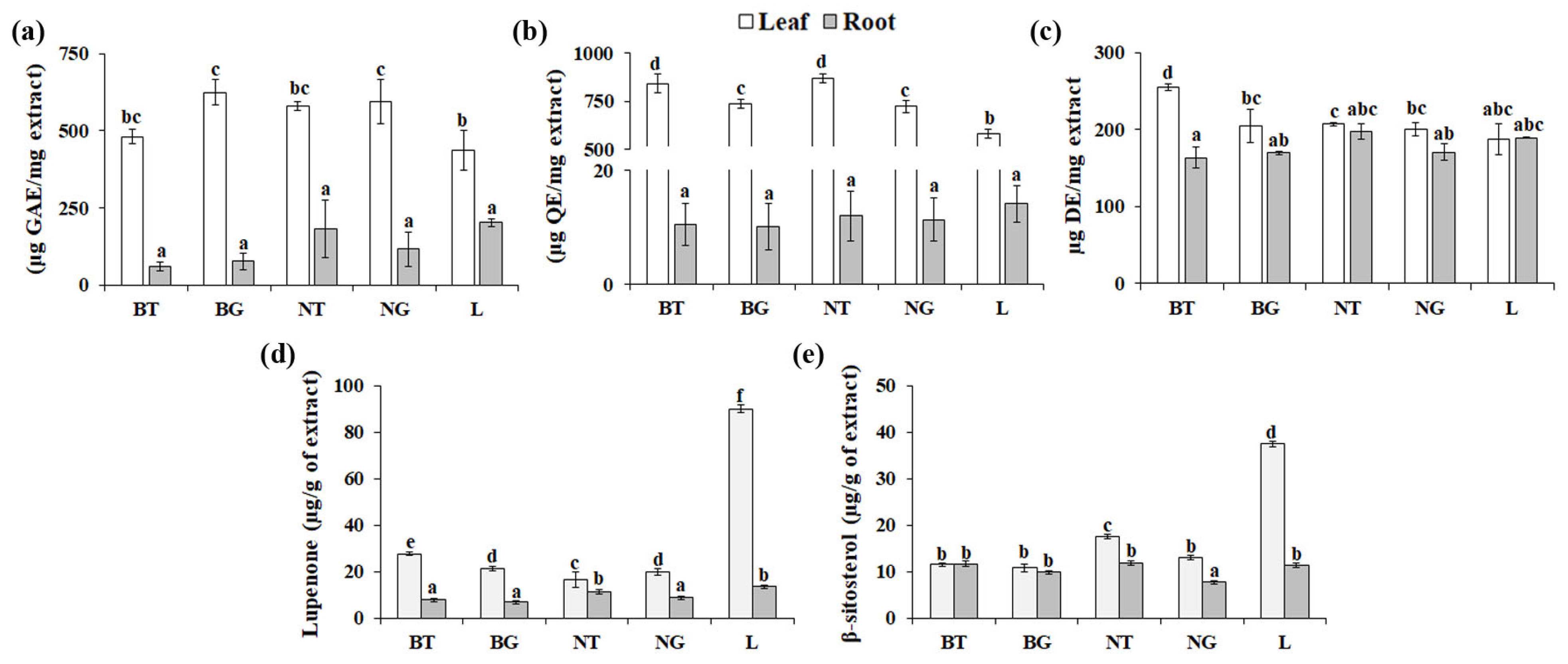
3.2. Variability in Phytochemical Contents among A. triphylla Genotypes

Genotype is a significant factor that can influence both the quality and yield of phytochemicals [24]. Consequently, this factor contributes to the diverse array of pharmacological effects witnessed in medicinal plants. To investigate the effect of genotypes on phytochemicals, five A. triphylla genotypes were grown and harvested under the same environmental conditions. As shown in Figure 2, the mean values of TPC, TFC, and TSC varied among the A. triphylla genotypes. In leaf extracts, TPC ranged from 435.89 to 624.03 µg GAE/mg (Figure 2a), and the TFC values could be ordered as follows: NT (869.40 µg QE/mg) > BT (840.91 µg QE/mg) > BG (734.64 µg QE/mg) > NG (723.48 µg QE/mg) > L (580.79 µg QE/mg) (Figure 2b). The root extracts had significantly lower levels of TPC and TFC that were approximately 2–11 times and 40–80 times lower, respectively, than those identified in the leaf extracts. Unlike TPC and TFC, the analysis of TSC revealed the richness of saponin compounds in both the root and leaf extracts (Figure 2c). Notably, the leaf extract of the BT phenotype exhibited the highest TSC (255 ± 4.35 µg DE/mg). Lupenone (lupane-type triterpenoid) and β-sitosterol (sterol) are marker compounds used to differentiate the Campanulaceae family and assess the quality of A. triphylla. [14,25,26]. As shown in Figure 2d,e, the leaf extracts of the collected genotypes contained lupenone and β-sitosterol within the range of 16.52–89.90 µg/g of extract and 10.82–37.49 µg/g of extract, respectively. The root extracts contained lupenone in the range of 6.81–11.32 µg/g of extract, accompanied by β-sitosterol in the range of 7.67–11.80 µg/g of extract. The highest contents of lupenone and β-sitosterol were found in leaf extracts of the L genotype. Rheophytic types, like the L genotype, possess thick cuticles on the leaf surface, which have been associated with adaptations for avoiding or resisting thermal damage in open environments [15]. In various plants, the accumulation of steroids and triterpenoids in cuticular waxes may be necessary to enhance the mechanical strength of the fruit cuticle by acting as nanofillers [27]. The highest levels of lupenone and β-sitosterol in the L genotype might be necessary for the development of cuticles and suggest that the genotype has a substantial impact on the phytochemical contents of A. triphylla.

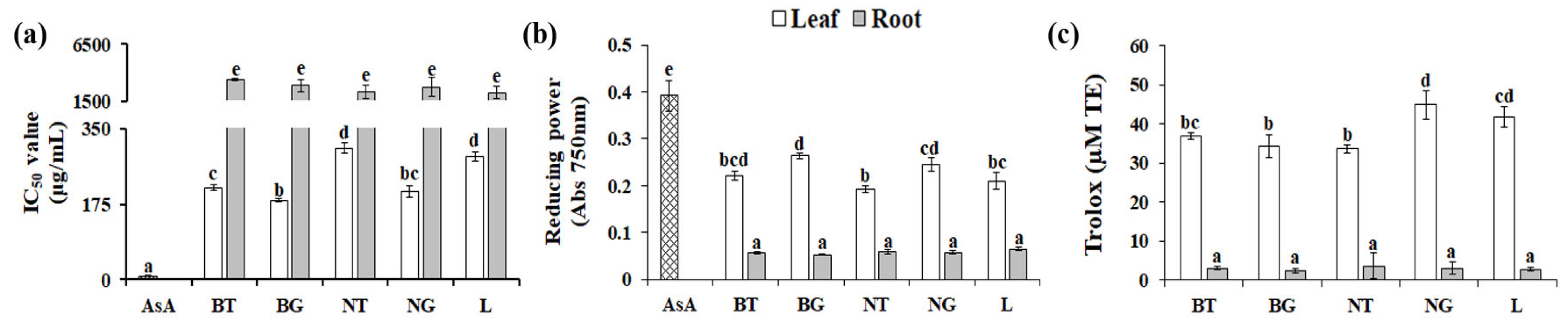
3.3. Effect of Genotype on Antioxidant Activities

The DPPH, reducing power, and ORAC assays were used to reveal the effect of genotype on antioxidant activity. As shown in Figure 3a, all tested root extracts exhibited similar IC50 values of ≥3500 μg/mL, which were approximately 270–400 times higher than those associated with AsA. This indicates that the root extracts had very weak free-radical scavenging activities. The leaf extracts demonstrated substantial DPPH-radical scavenging capabilities (7–16 times higher than those of the root extracts), although their IC50 values (183.69 to 305.24 μg/mL) were higher than AsA (IC50 value = 8.32 ± 0.74 µg/mL) (Figure 3a). The leaf extract of the BG genotype exhibited the highest DPPH-radical scavenging activities among the leaf extracts, owing to its low inhibitory concentration value (IC50 = 183.69 ± 3.95 μg/mL), followed by the NG, BT, L, and NT genotypes.
In addition, we assessed the capability of each extract to facilitate the Fe3+–Fe2+ transformation utilizing the reducing power assay based on a single electron transfer reaction (SET) [6]. The reducing power activities of root extracts (200 µg/mL) from all five genotypes were low, and no significant differences (p ≤ 0.05) were observed between the various genotypes. When compared with root extracts, leaf extracts demonstrated higher reducing power activities, with the BG leaf extract displaying the most pronounced activity, followed by NG, BT, L, and NT (Figure 3b). Moreover, the antioxidant potentials of these extracts were evaluated through the ORAC assay, a method based on hydrogen atom transfer (HAT) [6]. As shown in Figure 3c, 40 µg/mL of NG displayed the most elevated ORAC value at 44.96 ± 3.06 µM TE. Conversely, the antioxidant activities of the root extracts were relatively modest and displayed no significant differences (p ≤ 0.05) across the five phenotypes. We conducted Pearson’s correlation analysis to assess the relationship between antioxidant activities and phytochemical contents. The most significant negative correlation was identified between TFC and DPPH-radical scavenging activity (R2 = −0.883 **; ** p < 0.01), followed by the correlations between TPC and DPPH-radical scavenging activity (R2 = −0.743 **; ** p < 0.01), and TSC and DPPH-radical scavenging activity (R2 = −0.536 **; ** p < 0.01). The tested phytochemical contents exhibited no correlation with the results of other antioxidant-activity tests. Consistent with our results, the leaf extract from 2-month-old A. triphylla displayed more potent antioxidant activities when compared with the root extract [14]. In higher plants, polyphenolic compounds are synthesized in a light-dependent manner and serve to protect the photosynthetic apparatus from photoinhibition induced by excessive light [28]. In addition, the distribution and types of saponins largely depend on species and tissues [29], indicating that spatial accumulation of polyphenolic compounds and saponins can cause variations in the DPPH-radical scavenging activity of A. triphylla leaves and roots. Overall, these findings suggest that the leaf extracts of A. triphylla can be a potential source of antioxidants. However, it is important to note that the genotype influences the antioxidant mechanism, which can neutralize radicals through either HAT or SET.

3.4. Effect of Genotype on the Anti-Inflammatory and Antimelanogenic Activities

The skin is the primary line of defense, and, thus, it is constantly exposed to diverse external factors that can set off a chain of inflammatory responses [30], driven by the excessive production of NO caused by the activation of inducible nitric oxide synthase [31]. Therefore, inhibiting NO production in the presence of inflammatory triggers is a significant goal while searching for potential anti-inflammatory agents. Previous studies have highlighted the anti-inflammatory activities of the ethyl acetate fraction of A. triphylla root extract on macrophage cells [32]. Nonetheless, to the best of our knowledge, no studies have investigated the influence of genetic variation on the anti-inflammatory activities of A. triphylla. The effect of genotype on NO production was determined in LPS-stimulated RAW264.7 cells. As shown in Figure 4a, the cells treated with LPS alone exhibited a marked increase in NO production (10.65 μM nitrite), whereas all tested leaf extracts (150 µg/mL) exhibited an inhibitory effect on LPS-induced NO production without cytotoxic activity (Figure 4b). NO production was strongly inhibited by the leaf extracts of BG (124.52 ± 12.97 µg/mL), NT (133.97 ± 0.56 µg/mL), and NG (115.61 ± 1.31 µg/mL), with comparable IC50 values of LPS-induced NO production (Figure 4c). On the other hand, the root extracts of the five genotypes exhibited no inhibitory effect on LPS-induced NO production (Figure 4a), although the ethyl acetate fraction of A. triphylla root extract has been reported to possess anti-inflammatory properties [32]. This variation is likely a result of concentrating the target components through solvent fractionation of the crude extract. Similar to DPPH-radical scavenging activity, the most significant negative correlation was identified between TFC and anti-inflammatory activity (R2 = −0.805 **; ** p < 0.01), followed by the correlations between TPC and anti-inflammatory activity (R2 = −0.774 **; ** p < 0.01), and TSC and anti-inflammatory activity (R2 = −0.478 **; ** p < 0.01). Taken together, these findings suggest that the leaf extract of A. triphylla exhibits anti-inflammatory activity that is both promising and demonstrates genotype-specific variability.
Melanin production is a crucial physiological process that protects the skin against ultraviolet light damage. However, excessive melanin accumulation can result in hyperpigmentation-related conditions, including melanocytic nevus, seborrheic keratosis, solar lentigo, and melanoma [6]. Yoo et al. [33], showed that A. triphylla sprout extract had an inhibitory effect on the production of melanin in B16F10 melanoma cells, suggesting the potential of A. triphylla as a skin whitening agent; however, the variability in the antimelanogenic properties among different genotypes remains unknown. To explore the antimelanogenic potential of the A. triphylla genotypes, we examined the suppressive impact of each extract on IBMX-induced melanin accumulation in B16F10 cells. Similar to the results of the antioxidant and anti-inflammatory assays, treatment with the root extracts (200 µg/mL) did not display any inhibitory influence on IBMX-induced B16F10 cells (Figure 4d). However, the leaf extracts (200 µg/mL) of BT, NT, and NG contained significant antimelanogenic properties without inducing cytotoxicity in B16F10 melanoma cells (Figure 4d,e). The antimelanogenic properties of the leaf extract obtained from BT (IC50 = 181.56 ± 16.17 µg/mL), NT (IC50 = 185.81 ± 5.24 µg/mL), and NG (IC50 = 195.32 ± 5.62 µg/mL) were significantly greater than those of other genotypes (Figure 4f). Correlations were identified between TSC and antimelanogenic activity (R2 = −0.511 *; * p ≤ 0.05). This indicates that the leaf of A. triphylla has the potential for development as a whitening agent. Nevertheless, the antimelanogenic activities of A. triphylla exhibit diversity among various genotypes. In addition, we discovered a strong correlation between various biological activities of A. triphylla, such as DPPH-radical scavenging activity, anti-inflammatory activity, and antimelanogenic activity, and its TSC. Triterpenoid saponin from A. triphylla suppressed the growth of human gastric cancer cells via inhibition of the extracellular (ERK) signal-regulated kinase signaling pathway [34], which is also known to play crucial roles in melanogenesis and inflammation [35,36]. As a result, it is reasonable to hypothesize that the anti-inflammatory and antimelanogenic activities of A. triphylla leaf extract (Figure 4) may be attributed to the inhibition of the ERK signaling pathway, which requires further investigation.

4. Conclusions

In conclusion, our study highlights the crucial role of genetic diversity in shaping the pharmaceutical attributes of A. triphylla. Various genotypes displayed diverse levels of phytochemical content and antioxidant, antimelanogenic, and anti-inflammatory activities, suggesting that genetic factors substantially contribute to the modulation of phytochemical concentrations and biological activities. Notably, within the genotypes examined, the leaf extract of NG generally demonstrated high biological activities, whereas the leaf extract of L exhibited relatively low biological activities. This indicates the critical importance of judicious genotype selection to optimize the acquisition of health benefits.

Supplementary Materials

The following supporting information can be downloaded at: https://www.mdpi.com/article/10.3390/app132011184/s1, Figure S1: Phylogenetic analyses of A. triphylla genotypes based on the nucleotide sequences of ITS2 region (a), matK region (b), and psbA-trnH region (c); Table S1: Primer sequences used in this study.

Author Contributions

Conceptualization, J.-W.C. and T.K.H.; investigation, O.E.A., S.Y.Y., S.M., J.H.K. and K.H.K.; resources, J.H.K. and K.H.K.; writing—original draft preparation, O.E.A., K.-I.J. and T.K.H.; writing—review and editing, K.-I.J. and T.K.H. All authors have read and agreed to the published version of the manuscript.

Funding

This work was carried out with the support of the “Cooperative Research Program for Agriculture Science and Technology Development (Project No. RS-2022-RD010345)” Rural Development Administration, Republic of Korea.

Institutional Review Board Statement

Not applicable.

Informed Consent Statement

Not applicable.

Data Availability Statement

The data presented in this study are available on request from the corresponding author. The data are not publicly available due to reasons of privacy.

Conflicts of Interest

The authors declare no conflict of interest.

References

  1. Nasim, N.; Sandeep, I.S.; Mohanty, S. Plant-Derived Natural Products for Drug Discovery: Current Approaches and Prospects. Nucleus 2022, 65, 399–411. [Google Scholar] [CrossRef] [PubMed]
  2. Choudhury, A. Potential role of bioactive phytochemicals in combination therapies against antimicrobial activity. J. Pharmacopunct. 2022, 25, 79–87. [Google Scholar] [CrossRef] [PubMed]
  3. Pandey, P.; Khan, F.; Alshammari, N.; Saeed, A.; Aqil, F.; Saeed, M. Updates on the anticancer potential of garlic organosulfur compounds and their nanoformulations: Plant therapeutics in cancer management. Front. Pharmacol. 2023, 14, 1154034. [Google Scholar] [CrossRef]
  4. Kusano, M.; Yang, Z.; Okazaki, Y.; Nakabayashi, R.; Fukushima, A.; Saito, K. Using metabolomic approaches to explore chemical diversity in rice. Mol. Plant 2015, 8, 58–67. [Google Scholar] [CrossRef] [PubMed]
  5. Ju, H.J.; Bang, J.-H.; Chung, J.-W.; Hyun, T.K. Variation in essential oil composition and antimicrobial activity among different genotypes of Perilla frutescens var. crispa. J. Appl. Biol. Chem. 2021, 64, 127–131. [Google Scholar] [CrossRef]
  6. Ju, H.J.; Kim, K.C.; Kim, H.; Kim, J.-S.; Hyun, T.K. Variability of polyphenolic compounds and biological activities among Perilla frutescens var. crispa genotypes. Horticulturae 2021, 7, 404. [Google Scholar] [CrossRef]
  7. Kim, D.; Kim, K.Y. Adenophora triphylla var. japonica inhibits candida biofilm formation, increases susceptibility to antifungal agents and reduces infection. Int. J. Mol. Sci. 2021, 22, 12523. [Google Scholar] [CrossRef] [PubMed]
  8. Lee, S.E.; Lee, E.H.; Lee, T.J.; Kim, S.W.; Kim, B.H. Anti-obesity effect and action mechanism of Adenophora triphylla root ethanol extract in C57BL/6 obese mice fed a high-fat diet. Biosci. Biotechnol. Biochem. 2013, 77, 544–550. [Google Scholar] [CrossRef]
  9. Park, K.; Kim, Y.; Hwangbo, K.; Gil, J.; Chung, H.; Park, S.; Hong, C.P.; Lee, Y. Development of simple sequence repeat markers from Adenophora triphylla var. japonica (Regel) H. Hara using next generation sequencing. Korean J. Med. Crop Sci. 2017, 25, 411–417. [Google Scholar] [CrossRef]
  10. Park, H.J.; Park, S.H. Hexane fraction of Adenophora triphylla var. japonica root extract induces apoptosis of human lung cancer cells by inactivating Src/STAT3 pathway. Nat. Prod. Res. 2023, 37, 2924–2928. [Google Scholar] [CrossRef]
  11. Lee, H.R.; Kim, H.M.; Jeong, H.W.; Kim, G.G.; Na, C.I.; Oh, M.M.; Hwang, S.J. Growth Characteristics of Adenophora triphylla var. japonica Hara Seedlings as Affected by Growing Medium. Plants 2019, 8, 466. [Google Scholar] [CrossRef]
  12. Han, S.; Sebastin, R.; Wang, X.; Lee, K.J.; Cho, G.-T.; Hyun, D.Y.; Chung, J.-W. Identification of Vicia species native to South Korea using molecular and morphological characteristics. Front. Plant Sci. 2021, 12, 608559. [Google Scholar] [CrossRef] [PubMed]
  13. Le, A.V.; Parks, S.E.; Nguyen, M.H.; Roach, P.D. Improving the vanillin-sulphuric acid method for quantifying total saponins. Technologies 2018, 6, 84. [Google Scholar] [CrossRef]
  14. Yoon, S.Y.; Kim, K.H.; Hyun, T.K. Antioxidant and anticancer activities of Adenophora triphylla leaf and root extracts. J. Plant Biotechnol. 2023, 50, 137–141. [Google Scholar] [CrossRef]
  15. Ohga, K.; Muroi, M.; Hayakawa, H.; Yokoyama, J.; Ito, K.; Tebayashi, S.-I.; Arakawa, R.; Fukuda, T. Morphological and anatomical analyses of the serpentine ecotype of Adenophora triphylla var. japonica (Campanulaceae). J. Plant Stud. 2012, 1, 180. [Google Scholar] [CrossRef]
  16. Moon, B.C.; Kim, W.J.; Han, K.S.; Yang, S.; Kang, Y.; Park, I.; Piao, R. Differentiating authentic Adenophorae radix from its adulterants in commercially-processed samples using multiplexed ITS sequence-based SCAR markers. Appl. Sci. 2017, 7, 660. [Google Scholar] [CrossRef]
  17. Ohga, K.; Muroi, M.; Hayakawa, H.; Yokoyama, J.; Ito, K.; Tebayashi, S.-I.; Arakawa, R.; Fukuda, T. Coastal adaptation of Adenophora triphylla var. japonica (Campanulaceae). Am. J. Plant Sci. 2013, 4, 596–601. [Google Scholar] [CrossRef]
  18. Feng, S.; Jiang, Y.; Wang, S.; Jiang, M.; Chen, Z.; Ying, Q.; Wang, H. Molecular identification of Dendrobium Species (Orchidaceae) based on the DNA barcode ITS2 region and Its application for phylogenetic study. Int. J. Mol. Sci. 2015, 16, 21975–21988. [Google Scholar] [CrossRef]
  19. De Boer, H.J.; Ghorbani, A.; Manzanilla, V.; Raclariu, A.-C.; Kreziou, A.; Ounjai, S.; Osathanunkul, M.; Gravendeel, B. DNA metabarcoding of orchid-derived products reveals widespread illegal orchid trade. Proc. Biol. Sci. 2017, 284, 20171182. [Google Scholar] [CrossRef]
  20. Hebert, P.D.; Ratnasingham, S.; de Waard, J.R. Barcoding animal life: Cytochrome C oxidase subunit 1 divergences among closely related species. Proc. Biol. Sci. 2003, 270, S96–S99. [Google Scholar] [CrossRef]
  21. Li, H.; Xiao, W.; Tong, T.; Li, Y.; Zhang, M.; Lin, X.; Zou, X.; Wu, Q.; Guo, X. The specific DNA barcodes based on chloroplast genes for species identification of orchidaceae plants. Sci. Rep. 2021, 11, 1424. [Google Scholar] [CrossRef]
  22. Acharya, G.C.; Mohanty, S.; Dasgupta, M.; Sahu, S.; Singh, S.; Koundinya, A.V.V.; Kumari, M.; Naresh, P.; Sahoo, M.R. Molecular phylogeny, DNA barcoding, and ITS2 secondary structure predictions in the medicinally important Eryngium Genotypes of east coast region of India. Genes 2022, 13, 1678. [Google Scholar] [CrossRef] [PubMed]
  23. Yong, W.; Chunbo, X.; Laga, T.; Xiaoming, Z.; Mingjiu, W. Genetic evaluation and germplasm identification analysis on ITS2, trnL-F, and psbA-trnH of alfalfa varieties germplasm resources. Open Life Sci. 2023, 18, 20220582. [Google Scholar] [CrossRef] [PubMed]
  24. Mannino, G.; Gentile, C.; Maffei, M.E. Chemical partitioning and DNA fingerprinting of some pistachio (Pistacia vera L.) varieties of different geographical origin. Phytochemistry 2019, 160, 40–47. [Google Scholar] [CrossRef] [PubMed]
  25. Kim, W.I.; Zhao, B.T.; Lee, J.H.; Lee, D.-U.; Kim, Y.S.; Min, B.S.; Son, J.K.; Woo, M.H. Quantitative and classification analyses of lupenone and β-Sitosterol by GC-FID in Adenophora triphylla var. japonica Hara and Codonopsis lanceolata. Nat. Prod. Sci. 2014, 20, 243–250. [Google Scholar]
  26. Kim, H.-J.; Son, D.C.; Kim, H.-J.; Choi, K.; Oh, S.-H.; Kang, S.-H. The chemotaxonomic classification of Korean campanulaceae based on triterpene, sterol, and polyacetylene contents. Biochem. Syst. Ecol. 2017, 74, 11–18. [Google Scholar] [CrossRef]
  27. Dashbaldan, S.; Pączkowski, C.; Szakiel, A. Variations in triterpenoid deposition in cuticular waxes during development and maturation of selected fruits of Rosaceae family. Int. J. Mol. Sci. 2020, 21, 9762. [Google Scholar] [CrossRef] [PubMed]
  28. Vrábl, D.; Nezval, J.; Pech, R.; Volná, A.; Mašková, P.; Pleva, J.; Kuzniciusová, N.; Provazová, M.; Štroch, M.; Špunda, V. Light drives and temperature modulates: Variation of phenolic compounds profile in relation to photosynthesis in spring barley. Int. J. Mol. Sci. 2023, 24, 2427. [Google Scholar] [CrossRef]
  29. Chen, X.; Wang, Y.; Zhao, H.; Fu, X.; Fang, S. Localization and dynamic change of saponins in Cyclocarya paliurus (Batal.) Iljinskaja. PLoS ONE 2019, 14, e0223421. [Google Scholar] [CrossRef]
  30. Kabashima, K.; Honda, T.; Ginhoux, F.; Egawa, G. The immunological anatomy of the skin. Nat. Rev. Immunol. 2019, 19, 19–30. [Google Scholar] [CrossRef]
  31. Bogdan, C. Nitric oxide and the immune response. Nat. Immunol. 2001, 2, 907–916. [Google Scholar] [CrossRef] [PubMed]
  32. Park, S.-H. Ethyl Acetate Fraction of Adenophora triphylla var. japonica Inhibits Migration of Lewis Lung Carcinoma Cells by Suppressing Macrophage Polarization toward an M2 Phenotype. J. Pharmacopunct. 2019, 22, 253–259. [Google Scholar] [CrossRef] [PubMed]
  33. Yoo, S.K.; Park, S.K.; Kang, J.Y.; Kim, J.M.; Park, S.H.; Kwon, B.S.; Lee, C.J.; Kang, J.E.; Park, S.B.; Lee, U.; et al. Skin Whitening effect of ethyl acetate fraction of Adenophora triphylla var. japonica Sprout. Korean J. Plant Res. 2017, 30, 352–363. [Google Scholar]
  34. Chun, J.; Kang, M.; Kim, Y.S. A triterpenoid saponin from Adenophora triphylla var. japonica suppresses the growth of human gastric cancer cells via regulation of apoptosis and autophagy. Tumor Biol. 2014, 35, 12021–12030. [Google Scholar] [CrossRef] [PubMed]
  35. Pillaiyar, T.; Manickam, M.; Jung, S.H. Downregulation of melanogenesis: Drug discovery and therapeutic options. Drug Discov. Today 2017, 22, 282–298. [Google Scholar] [CrossRef]
  36. Manzoor, Z.; Koh, Y.-S. Mitogen-activated protein kinases in inflammation. J. Bacteriol. Virol. 2012, 42, 189–195. [Google Scholar] [CrossRef]
Figure 1. Phenotype (a) and phylogenetic analyses (b) of A. triphylla genotypes based on the nucleotide sequences of the combined ITS2 + matK + psbA-trnH regions.
Applsci 13 11184 g001
Figure 2. Effects of genotype on phytochemical concentrations. (a) Total phenolic (TPC), (b) total flavonoid (TFC), (c) total saponin (TSC), (d) lupenone, and (e) β-sitosterol contents of the five A. triphylla genotypes. Different letters indicate significant differences at p ≤ 0.05 (means ± SE from three independent replicate experiments).
Applsci 13 11184 g002
Figure 3. The antioxidant activities of five A. triphylla genotypes were assessed through DPPH free-radical scavenging (a), reducing power (b), and ORAC assays (c). Different letters indicate significant differences at p ≤ 0.05 (means ± SE from three independent replicate experiments).
Applsci 13 11184 g003
Figure 4. Comparison of anti-inflammatory and antimelanogenic activities among the five A. triphylla genotypes. Effects of each genotype (150 µg/mL) on NO production (a) and cell viability (b) in LPS-stimulated RAW264.7 cells. (c) IC50 values of the LPS-induced NO production of each extract. The effect of extracts (200 µg/mL) obtained from each genotype on melanin production (d) and cell viability (e) was analyzed in IBMX-treated B16F10 cells. (f) IC50 values of the IBMX-induced melanin production of each extract. Different letters indicate significant differences at p ≤ 0.05 (means ± SE from three independent replicate experiments).
Applsci 13 11184 g004
Table 1. Characteristics and variation of the ITS2, matK, and psbA-trnH regions.
GenotypesSize (bp)/Accession Number (NABIC)
ITS2matKpsbA-trnH
NG259 bp/NU-1869601 bp/NU-1875312 bp/NU-1882
BT258 bp/NU-1870601 bp/NU-1878311 bp/NU-1880
BG257 bp/NU-1871601 bp/NU-1877311 bp/NU-1879
L257 bp/NU-1872601 bp/NU-1874311 bp/NU-1881
NT259 bp/NU-1873601 bp/NU-1876311 bp/NU-1883
Disclaimer/Publisher’s Note: The statements, opinions and data contained in all publications are solely those of the individual author(s) and contributor(s) and not of MDPI and/or the editor(s). MDPI and/or the editor(s) disclaim responsibility for any injury to people or property resulting from any ideas, methods, instructions or products referred to in the content.

