Article

Implant–Prosthetic Rehabilitation of Maxillary Lateral Incisor Agenesis with Narrow Diameter Implants and Metal–Ceramic vs. All-Ceramic Single Crowns: A 16-Year Prospective Clinical Study

1
Department of Neurosciences, University “Federico II” of Naples, Reproductive and Odontostomatological Science, 80131 Naples, Italy
2
Department of Medical and Surgical Specialties and Dentistry, University of Campania “Luigi Vanvitelli”, 80138 Naples, Italy
*
Authors to whom correspondence should be addressed.
These authors contributed equally to this work.
Appl. Sci. 2023, 13(2), 964; https://doi.org/10.3390/app13020964
Submission received: 19 December 2022 / Revised: 4 January 2023 / Accepted: 9 January 2023 / Published: 11 January 2023
(This article belongs to the Special Issue Advances in Reconstruction Dentistry)

Abstract

:
The main objectives of the present prospective clinical study were to evaluate the marginal bone resorption and the peri-implant soft tissue conditions around narrow diameter implants, as well as the clinical performances of metal–ceramic and all-ceramic single crowns in the implant–prosthetic treatment of maxillary lateral incisors agenesis. Seventy-two patients affected by monolateral or bilateral agenesis of the maxillary lateral incisors were included in the study, and a total of 105 narrow diameter implants were inserted. The final single crowns were fabricated 4 months after the surgical procedures by means of either titanium abutments with Auro-Galvan Crowns (AGC) veneered with feldspathic ceramics or zirconia abutments with all-ceramic (alumina or zirconia) crowns layered with dedicated veneering ceramics. Both marginal bone resorption and soft tissue quality were evaluated after a maximum period of 16 years. Two independent survival curves for patients wearing AGC or all-ceramic crowns were calculated by means of Kaplan–Meier analysis, and a log-rank test was performed in order to compare these curves (p < 0.05). After 16 years of clinical function, a mean marginal bone resorption of 1.2 ± 0.61 mm and good response of peri-implant soft tissues were observed. Descriptive statistics resulted in 80% and 87.1% cumulative success rates for patients provided with AGC prostheses onto titanium abutments and all-ceramic crowns on-to zirconia abutments, respectively. The cumulative survival rate calculated for AGC group was 93.3%, whereas all-ceramic group showed a cumulative survival rate of 95.7%. No statistically significant differences were found with the log-rank tests. The overall success and survival rates calculated were 85% and 95% for all the restorations, respectively. Mucositis, abutment unscrewing, loss of retention, chipping, core fracture, and esthetic failure were the major complications reported. The implant–prosthetic approach can be considered an effective and viable solution to treat cases of maxillary lateral incisor agenesis in the long term.

1. Introduction

Dental agenesis was defined as the absence or failure of formation of a tooth [1]. The prevalence of tooth agenesis was reported to range between 1–3% among different population groups [2,3]. Missing permanent maxillary lateral incisors represents about 20% of all cases of agenesis; this anomaly was noticed to be more frequently bilateral [3]. Different prevalence percentages were reported in the literature for maxillary lateral incisor agenesis on the basis of race and sex: its prevalence varied across population between 0.8% to 4.5% [4], although studies on North-Western European populations showed a lower congenital prevalence, from 1% to 2% [5].
Missing teeth were often associated with tooth ectopias and/or other abnormal dental conditions, such as smaller or peg-shaped teeth on the contralateral side. In such cases, the canines are often mesial or lingual positioned, and the midline is displaced towards the side affected by agenesis. Deciduous maxillary lateral incisors often remain in the dental arch for a time longer than its physiologic falling [6,7].
The lack of maxillary lateral incisors was reported to cause remarkable esthetic concerns by patients [8].
Several treatment options have been proposed for the rehabilitation of maxillary lateral incisor agenesis, as follows:
  • Conservative approach: this approach should be considered a short-term solution and consists only in an esthetic reshaping of the deciduous lateral incisor with composite resins [9].
  • Orthodontic approach: intercepting or orthodontic treatment is used to move the canines into the place of the lateral incisors and the space of the missing tooth is closed by moving the adjacent premolars and molars mesially [10].
  • Prosthetic approach: the missing maxillary lateral incisor is replaced by means of an inlay bridge (i.e., Maryland bridge) or a fixed partial denture (metal–ceramic or all-ceramic) sustained by the upper canine and central incisor [11,12]. The proper position of the canine is a paramount factor, since an adequate space in occlusion is indispensable to guarantee the correct thickness of an inlay bridge. Furthermore, a sufficient space between the upper central incisor and the upper canine is necessary to realize a prosthetic restoration of appropriate size.
  • Implant approach: osseointegrated implant-supported restoration replaces the missing tooth. Occasionally, a previous orthodontic adjustment of the prosthetic space is indispensable [13,14].
According to the literature, patients usually prefer the orthodontic approach to close the edentulous space or the implant treatment, since no tooth preparation is required. As reported by several authors, the implant–prosthetic rehabilitation of maxillary lateral incisor agenesis or the orthodontic closure of the edentulous space can be considered the most conservative and widespread treatments [15,16].
Scientific investigations confirmed that the crestal bone resorption of submerged and non-submerged implants are comparable [17].
Recent investigations found that clinical outcome with narrow-diameter implants was comparable to standard-diameter implants in the anterior zone [18,19].
Particularly, the cumulative success rate reported was satisfactory, ranging from 84.2% to 100% (mean of 95.2%). Prosthetic (fracture of the abutment, loosening of the abutment screw, chipping of the veneering ceramic) and biologic (mucositis, peri-implantitis, fistulae, food impaction) complications were the major drawbacks reported [20].
A 10-year study on submerged implant-supported prostheses showed that prosthetic complications represent 18.8% of the drawbacks in the implant–prosthetic treatment of missing teeth [21].
The present prospective clinical study aimed at evaluating the marginal bone resorption and the peri-implant soft tissue conditions around Narrow-Neck ITI implants in the implant–prosthetic rehabilitation of the maxillary lateral incisor agenesis. Cumulative survival and success rates of the implant–prosthetic systems were also calculated after 16 years of clinical function.

2. Materials and Methods

The present prospective study included 72 patients (49 females and 37 males) affected by maxillary lateral incisor agenesis and rehabilitated by means of implant–prosthetic restorations. The age of the patients ranged from 19 to 46 years. In 39 patients, the anomaly was unilateral (54.16%), while in 33 subjects it was bilateral (45.8%). All patients were in good general health; 15 were smokers (20.83%), and 6 showed occlusal parafunctional habits (8.3%). Patients were provided with a consent form and a written explanation regarding alternative techniques, the nature of the treatment, and associated procedures and risks. The patients were also provided with a consent form to participate in the study.
The study was performed by expert prosthodontists of the Department of Fixed Prosthodontics of the University “Federico II” of Naples (Italy); an ethical review was stated by the university.
The recruitment of patients was carried out between September 2003 and July 2004 according to the common criteria established by the literature for implant-supported prostheses. The following inclusion criteria were adopted [22,23]:
  • single tooth space(s) with adjacent teeth intact or restored with functionally and aesthetically good restorations;
  • the presence of prostheses precluding the addition of the missing tooth;
  • patient reluctance of preparation of adjacent teeth;
  • patient refusal to wear removable partial dentures.
Patients with a history of substance abuse, psychoses, unrealistic aesthetic expectations, inability to undergo minor oral surgical procedures, comprised health of the local site, insufficient bone quality or mouth opening precluding the placement of the implant, inadequate vertical space, or incomplete facial growth and tooth eruption were excluded from the study.
Before surgery, the quantity and quality of bone tissue were analyzed by means of clinical examination with Rinn standardized periapical radiographs according to the long cone technique with parallel rays and CT Dental Scan. All patients showed an average bone thickness from the buccal to the lingual side of 6 ± 1 mm, an average bone height of ±131 mm (from the margin of the alveolar ridge to the floor of the nose), and an edentulous space (measured from the distal side of the central incisor to the mesial aspect of the canine) of ±60.5 mm.
According to the bone crest height, 105 ITI-SLA Narrow Neck implants (Institut Straumann AG, Waldenburg, Switzerland) were used, as reported below:
  • Sixty-one Narrow-Neck implants 3.3 mm × 10 mm;
  • Twenty-one Narrow-Neck implants 3.3 mm × 12 mm;
  • Twenty-three Narrow-Neck implants 3.3 mm × 14 mm.
The clinician performed a crestal incision located slightly on the palatal aspect and extended the incision through the sulcus of the adjacent teeth to both the buccal and palatal aspect of the alveolar ridge to surgically insert implants. Buccal relieving incisions were only performed for 37 patients.
A fine tissue elevator was used to elevate the buccal and palatal muco-periostal flaps to ensure low-trauma soft tissue handling. Narrow-Neck protective copings with an internal occlusal screw were employed to cover the implants after the surgery.
During the healing time, 48 patients did not wear any temporary restoration, whereas the other 24 patients wore a resin denture tooth supported by a fake orthodontic appliance.
Four months after the surgical procedure, the osseointegrated implants were loaded. In order to take a fixture-level impression, a thin layer of an adhesive specific for polyethers (Polyether Adhesive, 3M ESPE) was spread on custom autopolimerizing acrylic resin trays (SR-Ivolen, Ivoclar, Schaan, Liechtenstein), and impression transfers and polyether materials (Permadyne Penta H,Permadyne Penta L, 3M ESPE, Seefeld, Germany) were used.
A dental technician prepared 105 titanium standard hollow abutments (height of 9 mm) and 105 screwed acrylic temporary crowns.
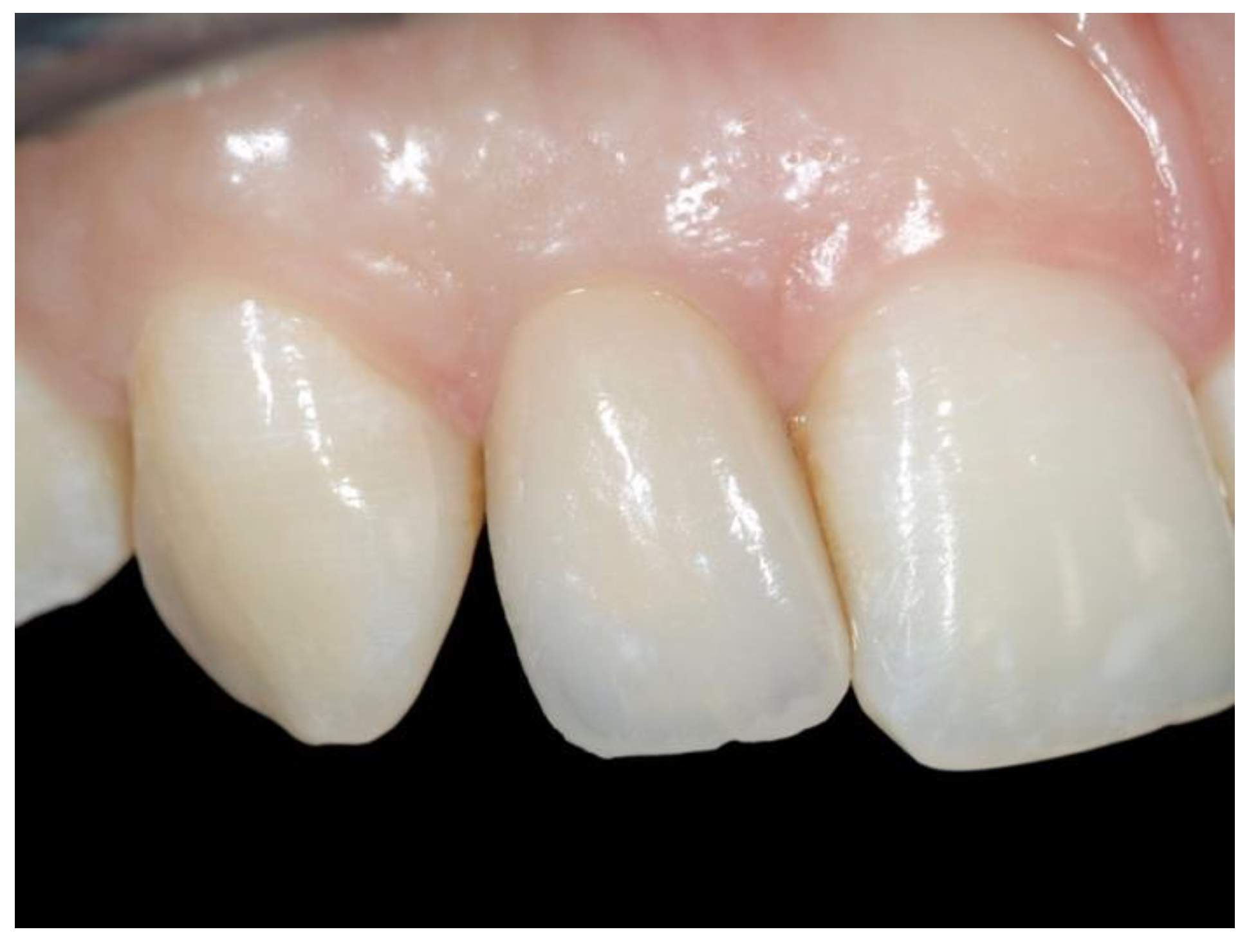
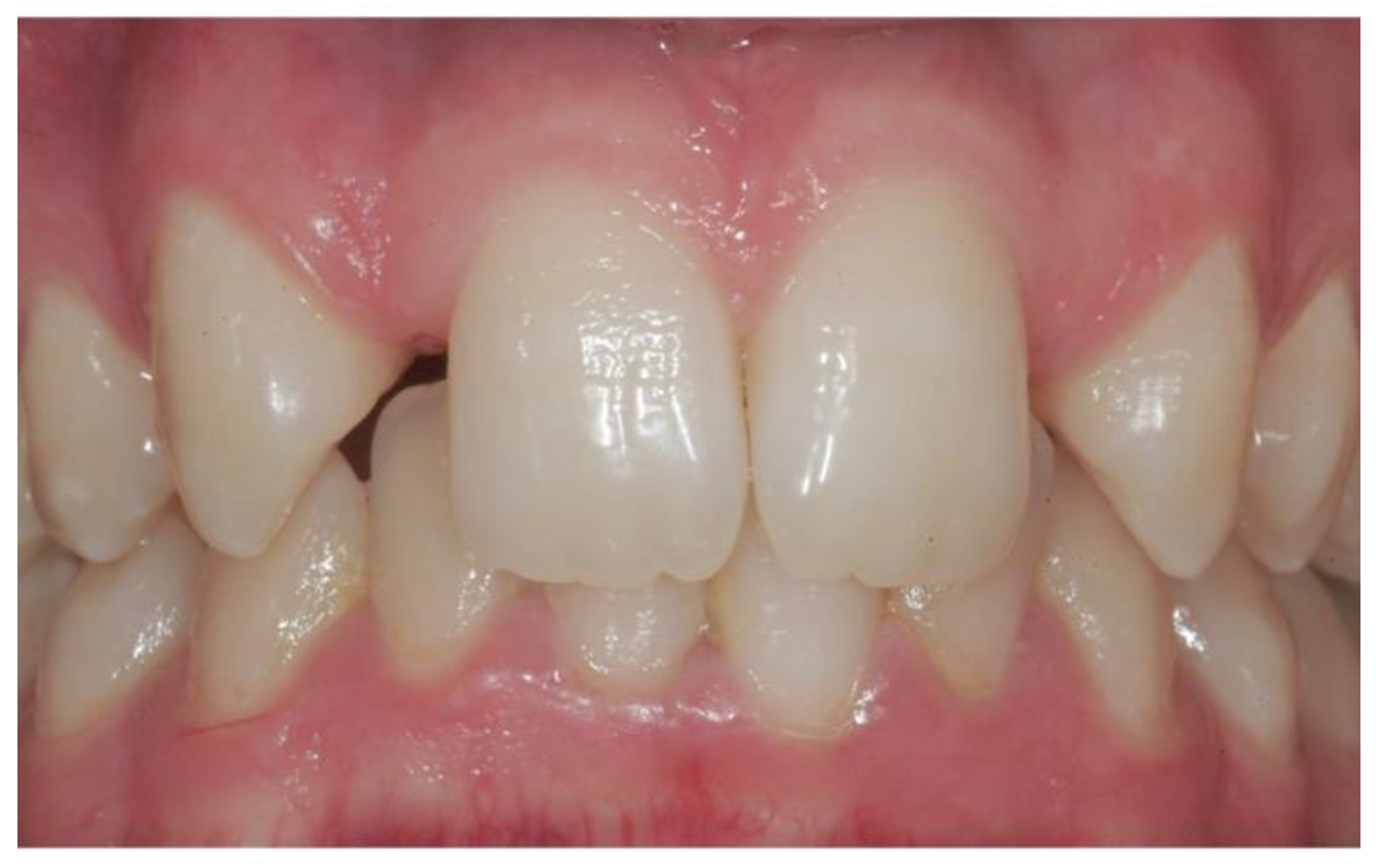
After 25 days of function with temporary prostheses, the final restorations were tried and delivered, using 33 Auro Galvan Crowns (AGC) on titanium abutments (Figure 1, Figure 2, Figure 3 and Figure 4) and 72 all-ceramic (alumina or zirconia) on zirconia abutments (Figure 5, Figure 6, Figure 7 and Figure 8). The patients were not randomly allocated to the study groups but were divided according to mucosal biotype and biomechanical variables for each single case. Patients with thin biotypes were considered more suitable to receive all-ceramic crowns onto zirconia abutments in order to obtain the best esthetic outcome. Differently, patients with thicker buccal peri-implant tissues and/or unfavorable implant–prosthetic lever arms were provided with conventional titanium abutments and AGC crowns.
The final AGC crowns were cemented with a zinc–phosphate luting agent, whereas all ceramic crowns were cemented with a glass ionomer cement. Excess cement was removed by means of Teflon curettes in order to avoid potential peri-implant soft tissues inflammation (i.e., mucositis and/or peri-implantitis).
Follow-up controls were performed 1 month after loading, 6 months after loading, and then every year to a maximum period of 16 years.
The clinical success of the implant–prosthetic rehabilitation of the missing maxillary lateral incisors was evaluated by means of the following parameters:
  • standardized periapical radiographs;
  • four-point peri-implant probing (i.e., mesial, buccal, distal, and palatal), determined after removing the crowns;
  • peri-implant soft tissues conditions;
  • mobility and pain of osseointegrated implants.
The peri-implant tissue conditions were established by means of the following periodontal parameters: Plaque Index (PI), Gingival Index (GI), Bleeding On Probing (BOP), and Papilla Index Score (PIS) [24,25].
More particularly, PIS describes the quantity of the papilla by means of five levels:
  • PIS 0: no papilla;
  • PIS 1: less than half papilla in the vertical direction compared with the other teeth;
  • PIS 2: half or more than half papilla in the vertical direction compared with the other teeth but not in harmony with the contiguous papillae;
  • PIS 3: papilla filling the interdental space and in harmony with the contiguous papillae;
  • PIS 4: hyperplasic papilla covering the restoration.
Marginal bone resorption was evaluated clinically by means of 4-point peri-implant probing and measured radiographically with standardized periapical radiographs. A customized silicon check bite was added to the centering device in order to make periapical radiographs reproducible for each patient, allowing clinicians to measure the marginal bone resorption by means of digital software.
The marginal bone resorption and four-point peri-implant probing were evaluated after 16 years of clinical function. Potential differences among the different aspects of each implant were analyzed by means of One-way ANOVA and Tukey’s post hoc test for comparisons. Cumulative survival and success curves of both AGC and all-ceramic crowns were calculated after a maximum period of 16 years of clinical function by means of Kaplan–Meier analysis, and the log-rank tests were performed in order to compare these curves.
Furthermore, the global survival and success rates of all the implant–prosthetic restorations were reported. The success rate was defined as the number of implant–prosthetic units that did not report any complication; all the complications were considered as events for Kaplan–Meier analysis. The level of significance was set at p < 0.05 for all the statistical tests. Statistical analysis was performed using the software SPSS 22.0 (SPSS Inc., Chicago, IL, USA).

3. Results

Osseointegration was achieved for all 105 narrow diameter implants. Five patients (three provided with AGC and two provided with all-ceramic crowns) did not reach the 16-year follow-up after the load application, because three patients relocated to another city and two patients chose to rely on other dental clinics. As for the other fixtures, standardized periapical radiographs revealed a marginal bone resorption ranging from 0.46 to 1.32 mm 1 year after prosthetic loading and from 0.62 to 1.81 mm (mean of 1.2 ± 0.61 mm) at a 16-year follow-up. Correspondingly, four-point peri-implant examination revealed the following probing measures:
  • a mean mesial probing 3.4 ± 0.2 mm;
  • a mean buccal probing of 2.6 ± 0.1 mm;
  • a mean distal probing of 3.2 ± 0.1 mm;
  • a mean palatal probing of 2.3 ± 0.2 mm.
The one-way ANOVA and Tukey’s post hoc tests did not show any significant difference between both the buccal and palatal aspects (p > 0.05) and between the mesial and distal sides (p > 0.05) of the implants.
Conversely, statistically significant differences emerged between buccal vs. mesial (p < 0.05), buccal vs. distal (p < 0.05), palatal vs. mesial (p < 0.05), and palatal vs. distal aspects (p < 0.05) of the implants (Table 1).
As regards the peri-implant tissue conditions (Table 2), at the 16-year follow-up, 95 restorations showed a GI = 0 (95%).
Ninety-four prostheses revealed a PI = 0, whereas the other 6 restorations showed a PI = 1. BOP was negative for 97 implants.
Furthermore, a PIS = 1 score was found in 5 crowns, PIS = 2 score in 6 crowns, and PIS = 3 score in 86 crowns.
After 16 years of clinical function, descriptive analysis resulted in 80% and 87.1% cumulative success rates for patients provided with AGC prostheses onto titanium abutments and all-ceramic crowns onto zirconia abutments, respectively, as reported in the Kaplan–Meier graph considering complications as events (Figure 9). No statistically significant difference was found with the log-rank test (p > 0.05).
The cumulative survival rate calculated for AGC group was 93.3%, whereas the all-ceramic group showed a cumulative survival rate of 95.7% (Figure 10). No statistically significant difference was found with the log-rank test (p > 0.05). The overall success and survival rates calculated for all the restorations were 85% and 95%, respectively.
During the entire period of observation, in the AGC group the following complications were reported: 1 aesthetic failure after 8 years of clinical function (3.33%), at the 11-year recall 1 loss of retention (3.33%), 2 mucositis after 12 years and 1 mucositis at the 15-year follow-up (10%). At the 16-year recall, 1 case of severe peri-implantitis was observed in the AGC group, causing loss of the implant (3.33%).
On the other hand, in the all-ceramic crowns group, 1 chipping of the veneering ceramic was observed after 6 years of function (1.42%), at the 8-year recall 1 decementation was reported (1.42%), 3 cases of mucositis were observed at the 8-year, 10-year, and 14-year recalls (4.28%), and 1 irreversible core fracture was observed after 9 years of clinical function (1.42%). Two cases of mild peri-implantitis were observed 10 years after the prosthetic load, showing grade-2 implant mobility and impairing function (2.86%). After 14 years of clinical function, 1 abutment unscrewing was detected (1.42%).

4. Discussion

The treatment of maxillary lateral incisor agenesis requires a multidisciplinary approach.
Previous orthodontic therapy is frequently a mandatory requirement to achieve a correct surgical space to place the implant. The implant–prosthetic treatment often offers the best cost/benefit ratio and is considered a biological conservative approach for maxillary lateral incisor agenesis. Several authors suggested placing the implant into the bone at the end of the cranium-facial growth, at about 21 for males and 17 for females [26,27].
After the surgical placement of the implant and the recovery screw, a period of healing of about 4–6 months is needed to obtain the osseointegration [23]. It is well-known that a good aesthetic result cannot be achieved without respecting the peri-implant soft tissues and the papillae.
In the present prospective study, both AGC on titanium abutment and all-ceramic crowns onto zirconia abutment have shown good marginal bone level stability and peri-implant soft tissues response in the long-term, since 86% of the restorations showed satisfactory morphology of the papillae after 16 years of function.
The presence of either titanium or zirconia abutment seems not to influence peri-implant soft tissue stability, confirming the data previously published by other authors [28]. It can be assumed that other factors, such as proper 3D implant positioning, a conservative design of the surgical flaps, and the correct application of temporary acrylic resin restorations, may have played an important role in obtaining this result.
Similar investigations highlighted the stability of the marginal bone level and soft tissues around the dental implants in the medium-term [29,30].
In the present prospective study, healthy and stable peri-implant soft tissues were evidenced in the long-term, probably thanks to the appropriate design of the final restorations and the maintenance of good oral hygiene. All the periodontal parameters considered to evaluate the peri-implant soft tissues conditions confirmed these data.
Several studies recognize smoking, together with a history of periodontitis, as a great risk factor to develop peri-implantitis [31,32]. The long-term health of peri-implant tissues evidenced in the present investigation may also be explained with the high percentage of non-smoker patients (79.2%) included in the study.
In the present study, after 16 years of prosthetic loading, high global survival (95%) and success rates (85%) were found, and no statistically significant differences in functional or aesthetic outcomes emerged between AGC crowns and all-ceramic crowns (p > 0.05).
High cumulative success (87.1%) and survival (95.7%) rates emerged in the all-ceramic crowns group.
The few minor complications were abutment unscrewing (1.42%), chipping of the veneering ceramic (1.42%), 1 decementation (1.42%), and mucositis (4.28%). In none of these cases was an invasive surgical treatment or replacement of the prosthetic restoration necessary.
After 10 years of serviceability, a patient with bilateral agenesis returned to the clinic, complaining about sensitivity and mobility of his implants. After peri-implantitis was clinically and radiographically assessed by the clinician, a guided bone regeneration (GBR) procedure was required to restore function and maintain the implant–prosthetic restorations. A high number of cigarettes (15–20 per day) and low daily standards of hygiene of the patient may explain this case.
Another patient wearing a zirconia crown required a new restoration after 9 years of function, due to a catastrophic core fracture which occurred while he was eating.
Similarly, in the AGC group, satisfactory cumulative success (80%) and survival (93.3%) rates were reported in the present investigation.
The minor complications reported were mucositis (10%) and loss of retention (3.33%). Few major complications were detected: 1 aesthetic failure after 8 years requiring a new restoration (3.33%) and 1 implant loss due to severe peri-implantitis (3.33%) after 16 years of serviceability.
The data collected in the present study have confirmed evidence pointed out by precedent investigations [14,27,33,34], conscious that a larger sample and randomization of the patients should be advisable for future studies. Aware of the limits of the present prospective study, very good stability and satisfactory long-term aesthetic and functional results for implant–prosthetic systems used to restore maxillary lateral incisors agenesis emerged.

5. Conclusions

The present 16-year prospective clinical study has pointed out satisfactory long-term results of Narrow-Neck ITI implants restored with both Auro-Galvan on titanium abutment and all-ceramic (alumina and zirconia) crowns on zirconia abutment. The implant–prosthetic treatment of monolateral or bilateral maxillary lateral incisor agenesis has proved to be a reliable and predictable option from both an aesthetic and functional point of view, showing stability of marginal bone and good conditions of peri-implant tissue in the long term.

Author Contributions

Conceptualization, R.S. and F.Z.; Methodology M.I.D.M.; Software M.I.D.M.; Validation, R.L., G.R. and M.A.; Formal Analysis, F.Z.; Investigation, R.S. and M.I.D.M.; Data Curation, R.L.; Writing—Original Draft Preparation, M.I.D.M. and G.R.; Writing—Review & Editing, R.S. and F.Z.; Supervision, M.A., R.S. and M.I.D.M. contributed equally to this work. All authors have read and agreed to the published version of the manuscript.

Funding

This research received no external funding.

Institutional Review Board Statement

The study was conducted in accordance with the Declaration of Helsinki, and approved by the Institutional Review Board of the University “Federico II” of Naples.

Informed Consent Statement

Informed consent was obtained from all subjects involved in the study.

Data Availability Statement

Data available on request due to privacy and ethical restrictions.

Conflicts of Interest

The authors declare no conflict of interest.

References

  1. The Glossary of Prosthodontic Terms: Ninth Edition. J. Prosthet. Dent. 2017, 117, e1–e105. [CrossRef] [Green Version]
  2. Yemitan, T.A.; Adediran, V.E.; Ogunbanjo, B.O. Pattern of agenesis and morphologic variation of the maxillary lateral incisors in nigerian orthodontic patients. J. West Afr. Coll. Surg. 2017, 7, 71–91. [Google Scholar] [PubMed]
  3. Stamatiou, J.; Symons, A.L. Agenesis of the permanent lateral incisor: Distribution, number and sites. J. Clin. Pediatr. Dent. 1991, 15, 244–246. [Google Scholar] [PubMed]
  4. Pandey, P.; Ansari, A.A.; Choudhary, K.; Saxena, A. Familial aggregation of maxillary lateral incisor agenesis (MLIA). BMJ Case Rep. 2013, 2013, bcr2012007846. [Google Scholar] [CrossRef] [PubMed] [Green Version]
  5. Robertsson, S.; Mohlin, B. The congenitally missing upper lateral incisor. A retrospective study of orthodontic space closure versus restorative treatment. Eur. J. Orthod. 2000, 22, 697–710. [Google Scholar] [CrossRef] [Green Version]
  6. Hua, F.; He, H.; Ngan, P.; Bouzid, W. Prevalence of peg-shaped maxillary permanent lateral incisors: A meta-analysis. Am. J. Orthod. Dentofac. Orthop. 2013, 144, 97–109. [Google Scholar] [CrossRef]
  7. Lo Muzio, L.; Bucci, P.; Carile, F.; Riccitiello, F.; Scotti, C.; Coccia, E.; Rappelli, G. Prosthetic rehabilitation of a child affected from anhydrotic ectodermal dysplasia: A case report. J. Contemp. Dent. Pract. 2005, 6, 120–126. [Google Scholar] [CrossRef] [PubMed]
  8. Rosa, M.; Olimpo, A.; Fastuca, R.; Caprioglio, A. Perceptions of dental professionals and laypeople to altered dental esthetics in cases with congenitally missing maxillary lateral incisors. Prog. Orthod. 2013, 14, 34. [Google Scholar] [CrossRef] [Green Version]
  9. Laverty, D.P.; Thomas, M.B. The restorative management of microdontia. Br. Dent. J. 2016, 221, 160–166. [Google Scholar] [CrossRef]
  10. Pithon, M.M.; Vargas, E.O.A.; da Silva Coqueiro, R.; Lacerda-Santos, R.; Tanaka, O.M.; Maia, L.C. Impact of oral-health-related quality of life and self-esteem on patients with missing maxillary lateral incisor after orthodontic space closure: A single-blinded, randomized, controlled trial. Eur. J. Orthod. 2021, 43, 208–214. [Google Scholar] [CrossRef]
  11. Hebel, K.; Gajjar, R.; Hofstede, T. Single-tooth replacement: Bridge vs. implant-supported restoration. J. Can. Dent. Assoc. 2000, 66, 435–438. [Google Scholar] [PubMed]
  12. Kinzer, G.A.; Kokich, V.O., Jr. Managing congenitally missing lateral incisors. Part II: Tooth-supported restorations. J. Esthet. Restor. Dent. 2005, 17, 76–84. [Google Scholar] [CrossRef] [PubMed]
  13. Kinzer, G.A.; Kokich, V.O., Jr. Managing congenitally missing lateral incisors. Part III: Single-tooth implants. J. Esthet. Restor. Dent. 2005, 17, 202–210. [Google Scholar] [CrossRef] [PubMed]
  14. Priest, G. The treatment dilemma of missing maxillary lateral incisors-Part II: Implant restoration. J. Esthet. Restor. Dent. 2019, 31, 319–326. [Google Scholar] [CrossRef]
  15. Šikšnelytė, J.; Guntulytė, R.; Lopatienė, K. Orthodontic canine substitution vs. implant-supported prosthetic replacement for maxillary permanent lateral incisor agenesis: A systematic review. Stomatologija 2021, 23, 106–113. [Google Scholar]
  16. Kiliaridis, S.; Sidira, M.; Kirmanidou, Y.; Michalakis, K. Treatment options for congenitally missing lateral incisors. Eur. J. Oral Implantol. 2016, 9 (Suppl. S1), S5–S24. [Google Scholar]
  17. Al Amri, M.D. Crestal bone loss around submerged and nonsubmerged dental implants: A systematic review. J. Prosthet. Dent. 2016, 115, 564–570.e1. [Google Scholar] [CrossRef]
  18. Momberger, N.; Mukaddam, K.; Zitzmann, N.U.; Bornstein, M.A.; Filippi, A.; Kühl, S. Esthetic and functional outcomes of narrow-diameter implants compared in a cohort study to standard diameter implants in the anterior zone of the maxilla. Quintessence Int. 2022, 53, 502–509. [Google Scholar] [CrossRef]
  19. Telles, L.H.; Portella, F.F.; Rivaldo, E.G. Longevity and marginal bone loss of narrow-diameter implants supporting single crowns: A systematic review. PLoS ONE 2019, 14, e0225046. [Google Scholar] [CrossRef]
  20. Parize, H.N.; Bohner, L.O.L.; Gama, L.T.; Porporatti, A.L.; Mezzomo, L.A.M.; Martin, W.C.; Gonçalves, T.M.S.V. Narrow-diameter implants in the anterior region: A meta-analysis. Int. J. Oral Maxillofac. Implant. 2019, 34, 1347–1358. [Google Scholar] [CrossRef]
  21. Priest, G. Single-tooth implants and their role in preserving remaining teeth: A 10-year survival study. Int. J. Oral Maxillofac. Implant. 1999, 14, 181–188. [Google Scholar]
  22. Schmitt, A.; Zarb, G.A. The longitudinal clinical effectiveness of osseointegrated dental implants for single-tooth replacement. Int. J. Prosthodont. 1993, 6, 197–202. [Google Scholar] [PubMed]
  23. Avivi-Arber, L.; Zarb, G.A. Clinical effectiveness of implant-supported single-tooth replacement: The Toronto Study. Int. J. Oral Maxillofac. Implant. 1996, 11, 311–321. [Google Scholar]
  24. Löe, H. The Gingival Index, the Plaque Index and the Retention Index Systems. J. Periodontol. 1967, 38, S610–S616. [Google Scholar] [CrossRef]
  25. Jemt, T. Regeneration of gingival papillae after single-implant treatment. Int. J. Periodontics Restor. Dent. 1997, 17, 326–333. [Google Scholar]
  26. Krassnig, M.; Fickl, S. Congenitally missing lateral incisors—A comparison between restorative, implant, and orthodontic approaches. Dent. Clin. N. Am. 2011, 55, 283–299, viii. [Google Scholar] [CrossRef] [PubMed]
  27. Lacarbonara, M.; Cazzolla, A.P.; Lacarbonara, V.; Lo Muzio, L.; Ciavarella, D.; Testa, N.F.; Crincoli, V.; Di Venere, D.; De Franco, A.; Tripodi, D.; et al. Prosthetic rehabilitation of maxillary lateral incisors agenesis using dental mini-implants: A multicenter 10-year follow-up. Clin. Oral Investig. 2022, 26, 1963–1974. [Google Scholar] [CrossRef] [PubMed]
  28. Linkevicius, T.; Vaitelis, J. The effect of zirconia or titanium as abutment material on soft peri-implant tissues: A systematic review and meta-analysis. Clin. Oral Implant. Res. 2015, 26 (Suppl. S11), 139–147. [Google Scholar] [CrossRef] [PubMed]
  29. King, P.; Maiorana, C.; Luthardt, R.G.; Sondell, K.; Øland, J.; Galindo-Moreno, P.; Nilsson, P. Clinical and radiographic evaluation of a small-diameter dental implant used for the restoration of patients with permanent tooth agenesis (hypodontia) in the maxillary lateral incisor and mandibular incisor regions: A 36-month follow-up. Int. J. Prosthodont. 2016, 29, 147–153. [Google Scholar] [CrossRef] [Green Version]
  30. Zarone, F.; Sorrentino, R.; Vaccaro, F.; Russo, S. Prosthetic treatment of maxillary lateral incisor agenesis with osseointegrated implants: A 24–39-month prospective clinical study. Clin. Oral Implant. Res. 2006, 17, 94–101. [Google Scholar] [CrossRef]
  31. Smeets, R.; Henningsen, A.; Jung, O.; Heiland, M.; Hammächer, C.; Stein, J.M. Definition, etiology, prevention and treatment of peri-implantitis—A review. Head Face Med. 2014, 10, 34. [Google Scholar] [CrossRef] [Green Version]
  32. Kormas, I.; Pedercini, C.; Pedercini, A.; Raptopoulos, M.; Alassy, H.; Wolff, L.F. Peri-implant diseases: Diagnosis, clinical, histological, microbiological characteristics and treatment strategies. A narrative review. Antibiotics 2020, 9, 835. [Google Scholar] [CrossRef] [PubMed]
  33. Degidi, M.; Nardi, D.; Piattelli, A. Immediate versus one-stage restoration of small-diameter implants for a single missing maxillary lateral incisor: A 3-year randomized clinical trial. J. Periodontol. 2009, 80, 1393–1398. [Google Scholar] [CrossRef] [PubMed]
  34. Scarano, A.; Conte, E.; Mastrangelo, F.; Greco Lucchina, A.; Lorusso, F. Narrow single tooth implants for congenitally missing maxillary lateral incisors: A 5-year follow-up. J. Biol. Regul. Homeost. Agents 2019, 33 (Suppl. S2), 69–76. [Google Scholar] [PubMed]
Figure 1. Monolateral agenesis of maxillary right lateral incisor.
Applsci 13 00964 g001
Figure 2. Detail of the titanium abutment after peri-implant soft tissues conditioning.
Applsci 13 00964 g002
Figure 3. Cement-retained Auro-Galvan Crown (AGC).
Applsci 13 00964 g003
Figure 4. Post-operative view 3 weeks after the cementation of the implant-supported AGC onto the titanium abutment in position 12.
Applsci 13 00964 g004
Figure 5. Bilateral agenesis of the maxillary lateral incisors.
Applsci 13 00964 g005
Figure 6. Detail of the zirconia abutment after peri-implant soft tissues conditioning in position 12.
Applsci 13 00964 g006
Figure 7. Details of the zirconia abutments and crowns.
Applsci 13 00964 g007
Figure 8. Post-operative front view 3 weeks after the cementation of the implant-supported zirconia single crowns onto the zirconia abutments in position 12 and 22.
Applsci 13 00964 g008
Figure 9. Kaplan–Meier graph showing cumulative success rates in relation to time. Two different curves were reported for patients wearing AGC and all-ceramic crowns, respectively.
Applsci 13 00964 g009
Figure 10. Kaplan–Meier graph showing cumulative survival rates in relation to time. Two different curves as reported for patients wearing AGC and all-ceramic crowns, respectively.
Applsci 13 00964 g010
Table 1. Results of the one-way analysis of variance (ANOVA) and Tukey’s post hoc test for the interproximal marginal bone resorption and the four-point peri-implant probing at 16-year follow-up.
TimeAspectMean Difference (mm)p-Value
16-year follow upB vs. P
B vs. M
B vs. D
P vs. M
P vs. D
M vs. D
0.31
1.2
1.08
1.07
1.08
0.38
>0.05
<0.05
<0.05
<0.05
<0.05
>0.05
Table 2. Gingival Index (GI) score, Plaque Index (PI) score, and Papilla Index Score (PIS) at 16-year follow-up.
Periodontal
Parameters
16-Year Follow-Up
n = 100
GI 095
GI 12
GI 23
GI 30
PI 094
PI 16
PI 20
PI 30
PIS 03
PIS 15
PIS 26
PIS 386
PIS 40
Disclaimer/Publisher’s Note: The statements, opinions and data contained in all publications are solely those of the individual author(s) and contributor(s) and not of MDPI and/or the editor(s). MDPI and/or the editor(s) disclaim responsibility for any injury to people or property resulting from any ideas, methods, instructions or products referred to in the content.