Systematic Review

Effect of Adjunctive Use of Probiotics in the Treatment of Peri-Implant Mucositis: A Systematic Review and Meta-Analysis

by
Kelly Rocio Vargas Villafuerte
1,*,
Cristhiam de Jesus Hernandez Martinez
2 and
Karina Oliveira Santos
3
1
Faculty of Health Sciences, School of Psychology, Universidad César Vallejo, Lima Campus, Callao 07001, Peru
2
Department of Oral & Maxillofacial Surgery and Periodontology, School of Dentistry of Ribeirão Preto, University of São Paulo (USP), Ribeirão Preto 14040-904, Brazil
3
Department of Biological Sciences, Bauru School of Dentistry, University of São Paulo (USP), Bauru 17012-901, Brazil
*
Author to whom correspondence should be addressed.
Appl. Sci. 2023, 13(19), 10940; https://doi.org/10.3390/app131910940
Submission received: 6 April 2023 / Revised: 30 June 2023 / Accepted: 4 July 2023 / Published: 3 October 2023
(This article belongs to the Section Applied Dentistry and Oral Sciences)

Abstract

:
This review was performed to analyze evidence from the scientific literature of the adjunctive effect of probiotics in the treatment of peri-implant mucositis (PiM). Only randomized clinical trials evaluating the effect of probiotics as an adjunct to mechanical debridement in PiM were included. A search was performed in PubMed/MEDLINE, LILACS, ScienceDirect, and Web of Science to identify articles published in English up to January 2023. The quality of the studies was evaluated using the JADAD scale, and the risk of bias was assessed with the Cochrane Collaboration assessment tool. Of the 159 potentially relevant studies, only 9 studies were included. The most commonly used strain was Lactobacillus reuteri, and the meta-analysis was conducted on studies with a follow-up period of 3 to 6 months, observing an overall effect on the reduction of bleeding on probing (BOP) at 3 and 6 months (WMD = −1.13, 95% CI = −1.95 to −0.30, p = 0.007; and WMD = −1.32, 95% CI = −2.15 to −0.48, p = 0.002), plaque index (PI) at 3 months (WMD = −1.22, 95% CI = −2.25 to −0.19, p = 0.02), and probing pocket depth (PPD) at 3 and 6 months, which was statistically significant in favor of the probiotic group (WMD = −1.34, 95% CI = −2.42 to −0.25, p = 0.02 and WMD = −1.36, 95% CI = −2.61 to −0.11, p = 0.03). On the other hand, there were no significant changes in the subgingival microflora around the implants with the use of probiotics. Probiotic therapy, as an adjunct to mechanical debridement, promotes a greater reduction in BOP, PPD, and PI, in relation to the control group without probiotics.

1. Introduction

Peri-implant diseases are defined as inflammatory lesions of the surrounding peri-implant tissues and include mucositis and peri-implantitis [1]. Peri-implant mucositis (PiM) is a reversible inflammatory lesion that affects the soft tissues around a dental im-plant, and the main clinical feature is bleeding on probing (BOP) [2]; radiographically, bone loss is absent [1]. An increase in probing depth is often observed due to swelling or decreased resistance to probing [2]. Mucositis can cause discomfort due to bleeding and implant failure if left untreated [1].
The therapy for peri-implant mucositis involves mechanical debridement of the implant surface using curettes, ultrasonic devices, and air abrasives [3]. However, when mechanical debridement fails to control inflammatory changes [3], adjunctive treatments such as antibiotics and local antiseptics [1] have been used, and their efficacy has been demonstrated for mucositis, showing an improvement in clinical parameters, especially bleeding on probing [4]. However, there is a growing interest in exploring alternative treatments that can reduce inflammation and improve immune function [5].
The World Health Organization (WHO) describes probiotics as living microorganisms that can provide health benefits when consumed in adequate amounts. They have been extensively studied for their potential to improve intestinal health and stimulate the immune system [6]. However, there is a growing body of evidence [7,8,9,10] suggesting that probiotics may also be beneficial in the treatment of peri-implant mucositis. Probiotics containing Lactobacillus spp. have been shown to reduce inflammation and bleeding in patients with peri-implant mucositis [11]. Additionally, it has been found that probiotic lozenges containing Lactobacillus reuteri prevent the development of peri-implant mucositis and mucositis related to chemotherapy and radiation therapy [12].
In this context, systematic reviews have emerged to evaluate the efficacy of different non-surgical protocols for the treatment of peri-implant mucositis [13], and at the same time, other systematic reviews have evaluated the effectiveness of probiotics in the treatment of peri-implant diseases [14,15]. However, these reviews included patients with and without mechanical instrumentation and also included patients who had been diagnosed not only with PiM and peri-implantitis, or they included patients treated with other adjunctive therapies besides probiotic therapy.
This systematic review evaluated the effects of adjunctive probiotic therapy on mechanical debridement in peri-implant mucositis. This article aims to inform healthcare professionals and patients about the benefits and limitations of probiotics for peri-implant mucositis through the PICOS question addressed: “What are the clinical and microbiological effects of adjunctive probiotic use in the treatment of peri-implant mucositis?”.

2. Materials and Methods

This systematic review was structured according to the Preferred Reporting Items for Systematic Reviews and Meta-Analyses (PRISMA) guidelines.
The review was registered on the Open Science Framework (OSF) and is publicly accessible in the database https://osf.io/, accessed on 4 May 2023 (registration number osf.io/v874h).

2.1. Inclusion Criteria

Types of studies to be included: Randomized controlled trials (RCTs) and studies published in English were considered eligible for inclusion.
Participants/population: Healthy adult patients diagnosed with peri-implant mucositis (Pi-M) in at least one dental implant.
Intervention(s)/exposure(s): Use of probiotic therapy as an adjunct to submarginal mechanical instrumentation (mechanical prophylaxis and/or debridement (ultrasonic devices, curettes, and polishing) with probiotic therapy (MD + P).
Comparator(s)/control: Only submarginal mechanical instrumentation therapy (me-chanical prophylaxis and/or debridement without probiotic therapy (MD)) or in combination with placebo (MD + placebo).
Outcome measures/primary outcome(s): Changes in bleeding on probing (BOP), plaque index, and probing pocket depth (PPD). Changes in subgingival plaque bacterial population.
Secondary outcome(s): Adverse effects as secondary outcomes.
Exclusion criteria: (1) Studies without mechanical debridement or professional plaque removal; studies that only used probiotics; (2) studies in which patients received other adjunctive therapies (e.g., laser/probiotics or use of antibiotics/nonsteroidal anti-inflammatory drugs) or any type of periodontal decontamination or treatment within the last 3 months; (3) patients with systemic disorders such as diabetes mellitus, liver or kidney diseases, or cardiovascular or autoimmune diseases that may influence treatment outcomes; (4) heavy smokers (≥10 cigarettes per day); (5) pregnant or lactating women; (6) studies including individuals presenting peri-implantitis; (7) studies with letters, case reports, and short communications; and (8) animal model and in vitro studies.
Search strategy: The search included all articles indexed in PubMed/MEDLINE, LI-LACS, ScienceDirect, and Web of Science. Articles published in the English language up to January 2023 were searched. We used the following search descriptors: “mucositis” OR “peri-implant mucositis”, each combined with Boolean operators (OR, AND)—“probiotic” OR “prebiotics” OR “lactobacillus” OR “bacillus” OR “enterococcus” OR “bifidobacterium” AND “mechanical curettage” OR “scaling” OR “periodontal debridement” OR “mechanical debridement” OR “plaque removal” OR “nonsurgical mechanical debridement”.
After electronic searches, manual searches were conducted on the reference lists of selected articles.

2.2. Data Extraction (Selection and Coding)

The study selection process followed PRISMA guidelines. Two independent reviewers (K.R.V.V. and C.d.J.H.M.) selected titles and abstracts. When a disagreement was reached, a third reviewer was consulted (K.O.S). Studies with insufficient information in the title and abstract to make a decision were selected for full-text evaluation, which was also independently conducted by the same two reviewers to determine study eligibility. Studies that met the inclusion criteria were subjected to validation and data extraction.

2.3. Extracted Data

The relevant data extracted from each study were the study and publication year; country; study design; sample size; participant characteristics; periodontal diagnosis; definition of PiM; the number of implants/implants per patient; smoking habits; intervention and follow-up; clinical parameters (PPD, BOP, PI); type of probiotic strain (oral cavity and gut strains); protocol for administration of probiotics, intervals, and frequency (dose); sample collection; microbiological techniques; collection time; evaluated bacteria; clinical and microbiological outcome measures of interest; and source of funding.
To obtain data that were missing in the reports, if necessary, the authors of the included studies were contacted. The GetData Graph Digitizer 2.26 was also used (http://getdata-graph-digitizer.com, software downloaded on 23 December 2022) to read the data that were only illustrated in the figures [16].

2.4. Quality Assessment and Risk of Bias

The assessment of methodological quality and risk of bias of the studies was conducted by two researchers (K.R.V.V and C.d.J.H.M.), according to the Jadad scale, where a score ≥3 points is considered high methodological quality, and a score ≤2 points is considered low methodological quality.
The risk of bias in each study was assessed using the Cochrane Collaboration tool [17], which evaluates bias risk in six domains (selection bias, performance bias, detection bias, attrition bias, reporting bias, and other biases). Each domain is assessed for low risk of bias, high risk of bias, or unclear risk of bias.
Bias risks were classified as adequate (+), inadequate (−), or unclear (?). Randomization and allocation methods (selection bias); blinding of study patients (performance bias); operators and examiners (detection bias); completeness of follow-up period/incomplete outcome data (attrition bias); selective reporting (reporting bias); and others based on these domains. The overall risk of bias was categorized as follows: (1) low risk of bias if all criteria were met, (2) unclear risk of bias if one or more criteria were partially met, and (3) high risk of bias if one or more criteria were not met.

2.5. Statistical Analysis

A meta-analysis was separately performed for bleeding on probing reduction (BOP), plaque index (PI), and reduction in probing pocket depth (PPD) to identify the weighted mean difference (WMD) of the probiotic effect as an adjunct to mechanical debridement (MD + P) compared to the control group without probiotic use.
For heterogeneity between the included studies, the I2 formula was taken as a measure between the studies, with values of I2 = 25%, I2 = 50%, and I2 = 70% indicating low, moderate, and high heterogeneity, respectively.
If heterogeneity was statistically significant (p < 0.05), the random-effects model was used, and the fixed-effects model was used if heterogeneity was not significant. The alpha level was maintained at p ≤ 0.05 to determine statistically significant differences. Forest plots were produced to illustrate the effects in the meta-analysis, reporting WMD differences and a 95% confidence interval (CI). The analyses were performed using the RevMan software (Review Manager, version 5.3.5; the Cochrane Collaboration, Copenhagen, Denmark).

3. Results

3.1. Literature Search

A total of 159 published articles were identified by the search strategy in the database, of which 28 were excluded after the removal of duplicates. In total, 131 studies were selected; of these, 119 were excluded after the title or abstract review. The full texts of the remaining 12 publications were reviewed, and of these, 9 studies [8,9,11,18,19,20,21,22,23] were included, and the other 3 studies [24,25,26] were excluded for different reasons (1 article included photodynamic therapy adjunct to probiotics [25],1 article included patients with periimplantitis [26], and 1 article was in Russian [24]). A flowchart according to PRISMA is shown in Figure 1.

3.2. Characteristics of Included Studies

The characteristics of the included studies are shown in Supplementary Tables S1–S3. All RCTs were published between 2015 and 2022, of which eight studies [8,9,19,20,21,22,23] were parallel designs, and one study [11] was a crossover design. Three studies were from Spain [11,21,22], two from Saudi Arabia [19,20], one from Brazil [18], one from Iran [9], one from Italy [8], and one from Sweden [23].
Regarding the follow-up time, it varied among the studies. One study [9] had a follow-up of 4 weeks, three studies [8,22] had a follow-up of 3 months, one study [21] had a follow-up of 135 days, and five studies [11,18,19,20,23] had a follow-up of 6 months. In total, 364 patients were initially enrolled, of which 361 completed the follow-up period, giving a percentage of 99%.
Of the RCTs, five studies [8,10,11,19,21] included periodontally healthy patients, one study [18] included edentulous patients, two studies [9,23] did not report any previous periodontal diagnostic information, and one study [22] included patients with a history of chronic periodontitis.
Regarding smoking habits, seven studies presented exclusion criteria for smokers, and two studies [21,23] included smokers (≤10 cigarettes per day).

3.3. Definition of Peri-Implant Mucositis (PiM)

The RCTs included in this RS defined peri-implant mucositis (PiM) as an inflamed mucosa with bleeding on probing (BOP) [8,9,11,19,20,21,22] and/or suppuration [18,23], with no evidence of radiographic bone loss [8,9,11,20,21,22] of ≥2 mm and/or ≥3 implant threads [8,20,22], or in the absence of a 3 mm [18] or more distant bone crest from the most coronal portion of the intraosseous part of the implant. In addition, two studies [20,23] included PPD ≥ 4 mm combined with BOP in the definition of PiM.

3.4. Clinical Parameters

In all studies, after completing the medical history, clinical parameters were collected. In each patient, the PPD [8,9,11,18,19,20,21,22,23], BOP [8,9,18,19,20,21,22,23], and PI [8,11,19,20,21,22,23] were collected in four sites [8,9,11,23] (buccal, lingual, mesial, and distal) or six implant sites [18,19,20,21,22] (three buccal sites and three lingual sites), using a manual probe [8,9,11,18,19,20,21,22,23] (PCP-UNC15 [8,19,20,21,22]; UNC colorvue [18]; and/or plastic probe, Perioprobe, Kerr [11]). Three studies specified the probing force, which ranged from 0.2 [11,23] to 0.3 N [22]. Two studies mentioned that a single dichotomous value was assigned to the presence or absence of plaque [8] or bleeding on probing [8,18] at the implant level.
Flichy et al. (2015) [11] and Santana et al. (2022) [18] used the modified gingival index (mGI) [11] and the modified plaque index (mPI) [18], according to scores (0, 1, 2, and 3), following Mombelli et al. 1987 [27]. Furthermore, three studies [19,20,21] used digital interproximal radiographs taken at baseline to verify the extent of CBL (linear distance from the mesial and distal surfaces of the implant pillar interface to the most crestal location of the alveolar bone).

3.5. Submucosal Instrumentation and Administration of Probiotics

All RCTs [8,9,11,18,19,20,21,22,23] performed submucosal instrumentation, which was performed by prophylaxis and/or mechanical debridement before probiotic therapy. Five studies [8,20,21,22,23] performed mechanical debridement by using an ultrasonic device [8,20,21,22,23], and some authors mentioned that they were using titanium [8,21] and/or carbon tips [22] (Ph1, Ph2L, and Ph2R), with water irrigation and a mode set at medium power [8,20,21,23].
Two studies [8,23], in addition to using an ultrasonic device, performed manual instrumentation. Four studies performed MD, using curettes [8,18,19,23] (titanium or Teflon-coated) and polishing with a rubber cup and polishing paste. Sargolzaei et al. 2022 [9] and Flichy et al. 2015 [11] did not specify how the performed the mechanical debridement from implants with PiM.
After submucosal instrumentation, patients received oral hygiene instructions [8,9,11,18,19,20,21,22,23] and were instructed on how to use probiotics.

3.6. Administration of Probiotics

Seven studies administered probiotics in tablet form [8,11,19,20,21,22,23], and two studies in capsule form [9,18]. In three studies [8,11,21], patients were instructed to take one probiotic tablet after brushing their teeth once a day for 30 days [8,11,21]. In one study [19], patients were instructed to take one probiotic tablet after brushing their teeth twice a day for 21 days. In another three studies [20,22,23], patients were instructed to dissolve the tablets in their mouths without chewing, with a frequency of once [22] (at night) and/or twice a day (morning and evening) [20,23].
Finally, two studies [9,18] administered probiotic therapy in capsule form: one study [9] instructed patients to dissolve each capsule in 2 tablespoons of warm water and rinse their mouth daily for 14 days (used as a mouthwash); and the other study [18] instructed patients to dissolve its contents in water (20 mL), to rinse with it, and then swallow twice a day for 12 weeks [18], after brushing their teeth. In addition, in two studies [18,23], probiotic strains were administered in topical oil form and applied around the implants, in the submucosal and submucosal areas after mechanical debridement.

3.7. Probiotic Strains

Out of nine studies, seven [8,11,19,20,21,22,23] used Lactobacillus spp. (L. reuteri) bacteria for probiotic purposes, in concentrations ranging from 1 × 10⁸ to 2 × 10⁸ colony-forming units (CFUs); one study [18] combined strains of B. lactis, L. rhamnosus, and L. paracasei at a dosage of 1 × 10⁹ CFUs; and another study [9] did not mention the name of the strain used, and the duration of treatment varied from 2 weeks to 3 months (Supplementary Table S3).
In three studies [11,22,23], the test and placebo products were generously provided by BioGaia AB, Lund, Sweden.

3.8. Clinical Parameter Results

Clinical parameter results are presented in Supplementary Table S2. The 9 studies reported clinical criteria data.
Regarding changes in the form of the average reduction of BOP at 3 months, for the intervention group (MD + P), it ranged from 0.12 [21] to 33.30 [8], while the reduction of BOP for the control group ranged from 0.15 [21] to 35.18 [8]. Regarding 6 months, the average reduction of BOP ranged from 1.30 [11] to 33.56 [19] for the intervention group, while for the control group, the reduction of BOP at 6 months ranged from 1.65 [11] to 44.29 [19]
At 3 months, there was an average reduction of PI for the intervention group, ranging from 0.24 [21] to 42.04 [8], while for the control group, it varied from 0.26 [21] to 39.88 [8]. Regarding 6 months, the intervention group had a reduction of 0.96 [11] to 27.3 [20], and the control group had a reduction of 1.09 [11] to 36.22 [19].
The average reduction of PPD at 3 months for the intervention group ranged from 0.56 [19] to 3.7 mm [23], and the reduction for the control group ranged from 1.55 [18] to 3.66 [22]. At 6 months, the average reduction of PPD for the intervention group ranged from 1.36 [19] to 2.6 [20], while for the control group, the reduction ranged from 1.62 [18] to 3.5 [23].
In one study [18], to obtain the missing data in the reports, the first author was contacted, and the data were sent. In another study [19], the data of the clinical parameters were presented in figures, and the GetData Graph Digitizer was used to read the data (http://getdata-graph-digitizer.com).

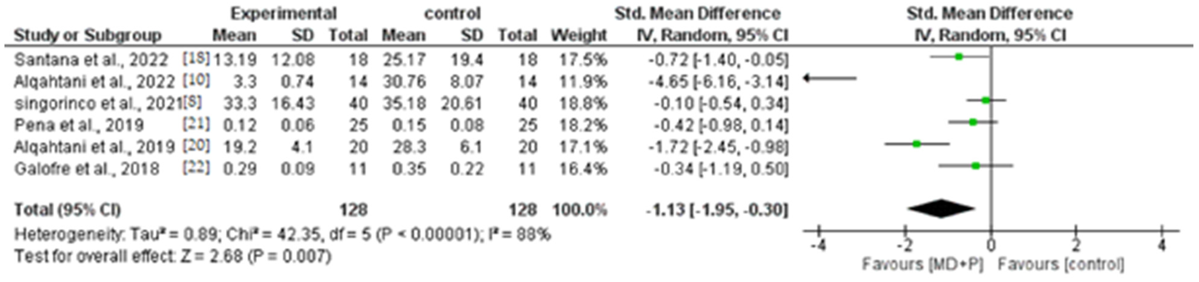
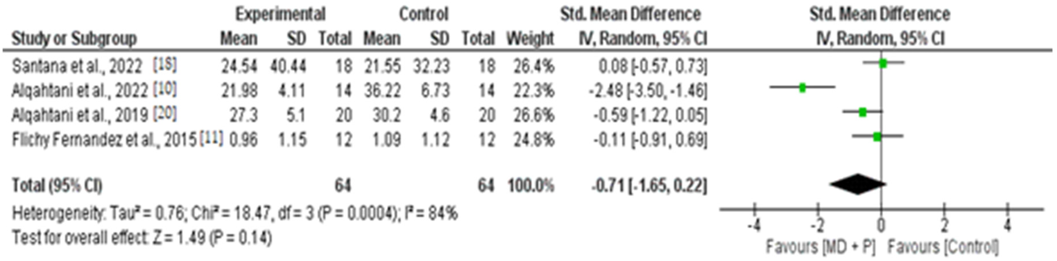
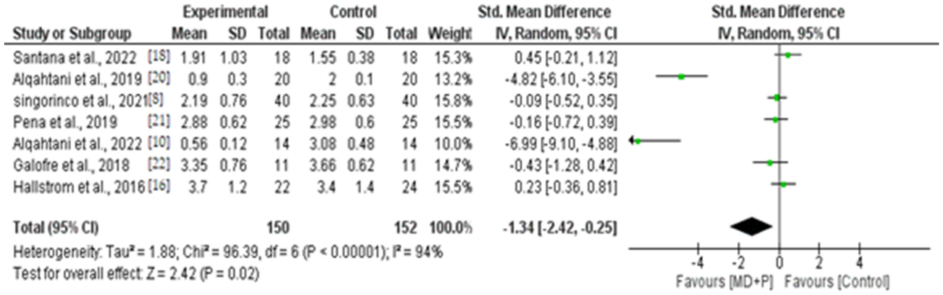
3.9. Quantitative Results of BOP, PI, and PPD

Eight studies [8,11,18,19,20,21,22,23] were included in the quantitative evaluation, considering the effects of probiotic therapy on the mean reduction of BOP, PI, and PPD at 3 and 6 months. The overall effect for both was calculated using the weighted mean difference (WMD).
The random effects model was used for PPD, BOP, and PI analysis, as heterogeneity was statistically significant at 3 and 6 months, respectively. For BOP (Chi2 = 42.35, p < 0.00001, I2 = 88% and Chi2 = 13.22, p < 0.004, I2 = 77%), PI (Chi2 = 63.95, p = 0.02, I2 = 92% and Chi2 = 18.47, p < 0.0004, I2 = 84%), and for PPD (Chi2 = 50.15, p < 0.00001, I2 = 92% and Chi2 = 96.39, p < 0.00001, I2 = 94%.
The overall effect for the reduction of BOP at 3 and 6 months was statistically significant in favor of the probiotic group, (WMD = −1.13, 95% CI = −1.95 to −0.30, p = 0.007; Figure 2) and (WMD = −1.32, 95% CI = −2.15 to −0.48, p = 0.002; Figure 3), respectively. Similarly, the overall effect on the reduction of PI at 3 months (WMD = −1.22, 95% CI = −2.25 to −0.19, p = 0.02; Figure 4) was statistically significant in favor of the probiotic group. However, at 6 months, there was no statistically significant difference between the groups (WMD = −0.71, 95% CI = −1.65 to 0.22, p = 0.14; Figure 5). Additionally, there was a statistically significant effect in reducing PPD at 3 and 6 months in favor of the probiotic group, (WMD = −1.34, 95% CI = −2.42 to −0.25, p = 0.02; Figure 6) and (WMD = −1.36, 95% CI = −2.61 to −0.11, p = 0.03; Figure 7).

3.10. Microbiological Parameters

Three studies [11,22,23] evaluated microbiological parameters in patients with PiM. Subgingival plaque samples were collected from the deepest peri-implant pockets [21,22,23]. Before sample collection, the supragingival plaque was removed using a sterile cotton ball, without penetrating the peri-implant mucosa [21,22,23]. The area of interest was isolated, and the sample was collected using sterile paper and left there for 10–20 s (10 s [22], 15 s [21,22], and the 20 s [23]). After collection, the paper tips were placed in Eppendorf tubes, with patient data, and sent to the laboratory for microbiological testing. The follow-up time of the included studies ranged from 90 days to 194 days (26 weeks).

3.11. Microbiological Techniques

Regarding microbiological techniques, two studies [21,22] used quantitative polymerase chain reaction (qPCR), and another study [23] used the checkerboard DNA-DNA hybridization technique to quantify subgingival species. In all three studies [21,22,23], the evaluated microorganisms were Aggregatibacter actinomycetemcomitans (Aa), Tannerella forsythia (Tf), Porphyromonas gingivalis (Pg), Treponema denticola (Td), Prevotella intermedia (Pi), Peptostreptococcus micros (Pm), Fusobacterium nucleatum (Fn), Campylobacter rectus (Cr), and Eikenella corrodens (Ec). Furthermore, a study evaluated Prevotella nigrescens (Pn), Parvimonas micra, P. endodontis (Pe). F. alocis (Fa), and P. Tannerae (Pt).

3.12. Results of Microbiological Parameters

The results of the microbiological parameters are reported in Supplementary Table S3. Only one study showed statistically significant results in favor of the probiotic group in reducing levels of Pg between baseline and 90 days. The other two studies showed no significant changes over time and no differences between groups.
It was not possible to perform a meta-analysis of the microbiological parameters due to the limited number of studies and the heterogeneity of the microbiological techniques used. Additionally, the data were presented in different outcome measures.

3.13. Adverse Effects

The nine studies [8,9,11,18,19,20,21,22,23] included in this review did not report any adverse effects or reactions.

3.14. Quality Evaluation

The evaluation of the methodological quality and risk of bias of the studies was consistent between the two examiners. According to the JADAD scale, all nine studies included in this review had high methodological quality (Supplementary Table S4).
To evaluate the risk of bias, the Cochrane tool was employed, which revealed that five studies [8,11,18,21,22] had a low risk of bias, one study [23] had an unclear risk of bias, and three studies [9,19,20] had a high risk of bias.
The study with an unclear risk of bias was classified as such because it did not indicate how the sample size was calculated [23]. Two studies [19,20] had a high risk of bias because the blinding of the participants and examiners was not verified, and the other study [9] had partially completed results data and did not indicate the type of strain used.
In all studies, appropriate statistical analysis was performed, and dropouts were described [23].

4. Discussion

This text is a systematic review that aims to evaluate the clinical and microbiological effects of probiotics as an adjunct to mechanical debridement alone through RCTs. Nine studies were included, and all of them reported clinical parameters’ results, while three reported microbiological results. Therefore, it was possible to perform a meta-analysis of the clinical parameters, but not the microbiological parameters, due to the limited number of studies and the heterogeneity of microbiological techniques for identifying pathogenic bacteria.
The meta-analysis was conducted on studies with follow-ups at 3 and 6 months. It demonstrated that adjunctive probiotic therapy to scaling and root planing (SRP) was more effective in reducing clinical parameters (BOP, PI, and PPD). One of the mechanisms of action of probiotics is the modulation of the immune system, helping to control inflammation and prevent infection [28,29]. The main characteristic of mucositis is inflammation and bleeding on probing [2]. Thus, the studies included in the meta-analysis have shown that probiotics can attenuate inflammation by reducing bleeding upon probing.
Three studies monitored the immune response by measuring levels of im-munomarkers in the gingival crevicular fluid. Two studies [11,18] showed a greater reduction in the inflammatory markers interleukin (IL)-1β, IL-6, IL-8, and tumor necrosis factor-alpha (TNF-α) in favor of the probiotic group compared to the control group without the use of probiotics after 6 months. One study indicated [23] a significant reduction in pro-inflammatory markers IL-1β, IL-8, and CCL5 in the probiotic group, but without statistical differences between the groups after 26 weeks. Studies indicate [30,31] that probiotics promote a potential immunomodulatory effect by reducing pro-inflammatory cytokines associated with the destruction of peri-implant tissues.
In this systematic review, seven studies [8,11,19,20,21,22,23] used the probiotic bacterium Lactobacillus spp. (L. reuteri) at concentrations ranging from 1 × 10⁸ to 2 × 10⁸ colony-forming units (CFU), and one study [18] combined strains of B. lactis, L. rhamnosus, and L. paracasei at a dosage of 1 × 10⁹ CFU, and the treatment duration ranged from 2 weeks to 3 months. The Lactobacillus species were the most commonly used strain in the studies. Bacteria of the genus Lactobacillus spp. comprise a large group of facultatively anaerobic, Gram-positive, non-spore-forming bacteria. Among these, the most studied strain is L. reuteri (Lr), and there is evidence that this strain is capable of producing lactic acid, which can help maintain a lower pH in the mouth, making it less favorable for the growth of pathogenic bacteria [32]. In addition, a systematic review [33] showed that especially the strain Lactobacillus reuteri, without combination with other strains, offered a greater reduction of pathogenic bacteria and deep periodontal pockets. Another study indicates that Lactobacillus reuteri (intestinal strain/species) is capable of producing various antimicrobial substances, such as reuterin, reuteran, and reutericyclin, which can inhibit the growth of pathogenic bacteria [34]. This explains why the authors of the included studies preferred to use the Lactobacillus reuteri strain. It was also observed that probiotic strains were used in adequate amounts greater than 1 × 10⁷, which is important since inadequate dosages can influence results [33]. However, there was heterogeneity in the duration of probiotic treatment, requiring more studies with more homogeneous times.
Regarding the microbiological results, two studies [21,23] showed no major changes in the total bacterial load of pathogenic species, and there was no difference in time and between groups. Meanwhile, one study [22] showed statistically significant results in favor of the probiotic group in reducing the count of Pg species between baseline and 90 days. The three studies used the same bacterial strain (L. reuteri); however, one study used intestinal strains [21], and two studies [22,23] used strains from the oral cavity. Although the mechanism of action of probiotics in the oral cavity is analogous to that of the intestinal microbiota [35], the strains may have adhered differently to periodontal tissues. Another factor to highlight was the periodontal diagnosis before treatment. Galofre et al. 2018 [22] indicated that they included patients with a history of periodontitis, one study [23] did not report, and another study [21], indicated that periodontally healthy and/or periodontitis patients included in a maintenance program were included. There is evidence that diseased periodontal and peri-implant sites and their healthy counterparts have distinct microbiological profiles [36]. One study [37] showed that patients with implants and periodontal health have a significantly higher relative abundance of Actinomyces and Streptococcus genera, while patients with a history of periodontitis with implants have a higher number of Por-phyromonas gingivallis and Prevotella spp. in subgingival plaque samples and, thus, a higher risk for PiM. This suggests the importance of periodontal diagnosis before PiM treatment, as patients with a history of periodontitis have an increase in periodontal pathogens. Therefore, this could be a reason why the studies obtained different results.
In addition, the long-term use of probiotics as an adjuvant to conventional treatment for peri-implant mucositis can assist in reducing inflammation and controlling disease progression [18], because probiotics can modulate the inflammatory response. However, the effectiveness of the treatment will also depend on the level of oral hygiene, since good oral hygiene is essential for maintaining oral health [33].
Regarding adverse effects, none of the included studies reported any associated adverse events; therefore, probiotics can be considered a safe intervention. Currently, “safety”, “individualized treatment”, and “internal interaction within the flora” are requirements of potential next-generation probiotics (NGPs) [38]. Furthermore, in the complex ecosystem of humans and microbes, it is challenging to identify the relationship between specific strains, specific flora, and hosts to warrant a therapeutic intervention in the case of disease [38].
Despite some limitations of the study, the meta-analysis showed that probiotic therapy as an adjunct to scaling and root planing (SRP) has a greater effect in reducing clinical parameters such as BOP, PI, and PD more effectively than the control group without probiotics. However, with regard to microbiological parameters, probiotics did not show greater reductions in pathogenic bacteria associated with both periodontal and peri-implant diseases.

5. Conclusions

Probiotic therapy as an adjunct to mechanical debridement is more effective in reducing clinical parameters (BOP, PPD, and PI) compared to the control group without probiotics. However, probiotics did not affect the subgingival microflora around implants. Therefore, more studies with high methodological quality and RCTs are needed.

Supplementary Materials

The following supporting information can be downloaded at https://www.mdpi.com/article/10.3390/app131910940/s1, Table S1: Characteristics of the studies; Table S2: Periodontal diagnosis, PiM definition, number of implants/implants per patient, smoking habits, interventions and follow-ups, clinical parameters (PPD, BOP, and PI), and outcome measures of clinical parameters; Table S3: Probiotic strain, type of strains/species isolated from, the protocol of probiotics administration, samples collection location, microbial techniques, collection time, bacteria evaluated, and main results of microbiology; Table S4: Summary of assessment of quality and risk of bias (low +/high −/? unclear) in selected studies.

Author Contributions

K.R.V.V. contributed to the study conceptualization and methodology, validation, data statistics and interpretation of the data, and writing—original draft preparation; C.d.J.H.M. contributed to the study design and methodology, validation, and critical review of the manuscript; K.O.S. contributed to the study design and helped with the data analysis and the critical review of the manuscript. All authors have read and agreed to the published version of the manuscript.

Funding

This research received no external funding.

Institutional Review Board Statement

Not applicable.

Informed Consent Statement

Not applicable.

Data Availability Statement

Data sharing is not applicable to this article. All raw data supporting this systematic review are from previous literature studies, which were cited in the text/figures.

Conflicts of Interest

The authors declare no conflict of interest.

References

  1. Figuero, E.; Graziani, F.; Sanz, I.; Herrera, D.; Sanz, M. Management of peri-implant mucositis and peri-implantitis. Periodontology 2000 2014, 66, 255–273. [Google Scholar] [CrossRef] [PubMed]
  2. Berglundh, T.; Armitage, G.; Araujo, M.G.; Avila-Ortiz, G.; Blanco, J.; Camargo, P.M.; Chen, S.; Cochran, D.; Derks, J.; Figuero, E.; et al. Peri-implant diseases and conditions: Consensus report of workgroup 4 of the 2017 World Workshop on the Classification of Periodontal and Peri-Implant Diseases and Conditions. J. Periodontol. 2018, 89 (Suppl. S1), S313–S318. [Google Scholar] [CrossRef]
  3. Verket, A.; Koldsland, O.C.; Bunaes, D.; Lie, S.A.; Romandini, M. Non-surgical therapy of peri-implant mucositis-Mechanical/physical approaches: A systematic review. J. Clin. Periodontol. 2023, 50, 135–145. [Google Scholar] [CrossRef] [PubMed]
  4. Ramanauskaite, A.; Fretwurst, T.; Schwarz, F. Efficacy of alternative or adjunctive measures to conventional non-surgical and surgical treatment of peri-implant mucositis and peri-implantitis: A systematic review and meta-analysis. Int. J. Implant Dent. 2021, 7, 112. [Google Scholar] [CrossRef] [PubMed]
  5. Saiz, P.; Taveira, N.; Alves, R. Probiotics in Oral Health and Disease: A Systematic Review. Appl. Sci. 2021, 11, 8070. [Google Scholar] [CrossRef]
  6. Reid, G. Probiotics: Definition, scope and mechanisms of action. Best practice & research. Clin. Gastroenterol. 2016, 30, 17–25. [Google Scholar] [CrossRef]
  7. Yu, S.C.; Zhang, Y.C.; Zhu, C.H.; Zhou, H.; Liu, J.; Sun, J.Y.; Li, A.; Pei, D.D. Adjunctive Diode Laser Therapy and Probiotic Lactobacillus Therapy in the Treatment of Periodontitis and Peri-Implant Disease. Jove-J. Vis. Exp. 2022, 183, e63893. [Google Scholar] [CrossRef]
  8. Signorinco, F.; Zouri, N.N.; Allocca, G.; Maiorana, C.; Poli, P.P. Effectiveness of orally administered probiotic Lactobacillus reuteri in patients with peri-implant mucositis: A prospective clinical study. J. Osseointegration 2021, 13, 144–149. [Google Scholar] [CrossRef]
  9. Sargolzaei, N.; Arab, H.; Gerayeli, M.; Ivani, F. Evaluation of the Topical Effect of Probiotic Mouthwash in the Treatment of Patients with Peri-Implant Mucositis. J. Long Term Eff. Med. Implant 2022, 32, 85–91. [Google Scholar] [CrossRef]
  10. Alqahtani, F.; Alshaikh, M.; Mehmood, A.; Alqhtani, N.; Alkhtani, F.; Alenazi, A. Role of Probiotics for the Treatment of Peri-Implant Mucositis in Patients with and without Type 2 Diabetes Mellitus. J. Oral Implantol. 2022, 48, 37–42. [Google Scholar] [CrossRef]
  11. Flichy-Fernandez, A.J.; Ata-Ali, J.; Alegre-Domingo, T.; Candel-Marti, E.; Ata-Ali, F.; Palacio, J.R.; Penarrocha-Diago, M. The effect of orally administered probiotic Lactobacillus reuteri-containing tablets in peri-implant mucositis: A double-blind randomized controlled trial. J. Periodontal Res. 2015, 50, 775–785. [Google Scholar] [CrossRef]
  12. Cereda, E.; Caraccia, M.; Caccialanza, R. Probiotics and mucositis. Curr. Opin. Clin. Nutr. Metab. Care 2018, 21, 399–404. [Google Scholar] [CrossRef] [PubMed]
  13. Barootchi, S.; Ravidà, A.; Tavelli, L.; Wang, H.L. Nonsurgical treatment for peri-implant mucositis: A systematic review and meta-analysis. Int. J. Oral Implantol. 2020, 13, 123–139. [Google Scholar]
  14. Arbildo-Vega, H.I.; Panda, S.; Bal, A.; Mohanty, R.; Rendón-Alvarado, A.; Das, A.C.; Cruzado-Oliva, F.H.; Infantes-Ruíz, E.D.; Manfredi, B.; Vásquez-Rodrigo, H.; et al. Clinical effectiveness of Lactobacillus reuteri in the treatment of peri-implant diseases: A systematic review and meta-analysis. J. Biol. Regul. Homeost. Agents 2021, 35, 79–88. [Google Scholar] [CrossRef] [PubMed]
  15. Sayardoust, S.; Johansson, A.; Jönsson, D. Do Probiotics Cause a Shift in the Microbiota of Dental Implants—A Systematic Review and Meta-Analysis. Front. Cell. Infect. Microbiol. 2022, 12, 823985. [Google Scholar] [CrossRef]
  16. Li, S.M.; Ji, Y.Z.; Wu, S.S.; Zhan, S.Y.; Wang, B.; Liu, L.R.; Li, S.Y.; Wang, N.L.; Wang, J.J. Multifocal versus single vision lenses intervention to slow progression of myopia in school-age children: A meta-analysis. Surv. Ophthalmol. 2011, 56, 451–460. [Google Scholar] [CrossRef] [PubMed]
  17. Higgins, J.P.; Altman, D.G.; Gotzsche, P.C.; Juni, P.; Moher, D.; Oxman, A.D.; Savovic, J.; Schulz, K.F.; Weeks, L.; Sterne, J.A. The Cochrane Collaboration’s tool for assessing risk of bias in randomised trials. BMJ Clin. Res. Ed. 2011, 343, d5928. [Google Scholar] [CrossRef]
  18. Santana, S.I.; Silva, P.H.F.; Salvador, S.L.; Casarin, R.C.V.; Furlaneto, F.A.C.; Messora, M.R. Adjuvant use of multispecies probiotic in the treatment of peri-implant mucositis: A randomized controlled trial. J. Clin. Periodontol. 2022, 49, 828–839. [Google Scholar] [CrossRef]
  19. Alqahtani, F.; Alshaikh, M.; Mehmood, A.; Alqhtani, N.; Alkhtani, F.; Alenazi, A. Efficacy of Antibiotic Versus Probiotics as Adjuncts to Mechanical Debridement for the Treatment of Peri-Implant Mucositis. J. Oral Implantol. 2022, 48, 99–104. [Google Scholar] [CrossRef]
  20. Alqahtani, F.; Alqahtani, M.; Shafqat, S.S.; Akram, Z.; Al-Kheraif, A.A.; Javed, F. Efficacy of mechanical debridement with adjunctive probiotic therapy in the treatment of peri-implant mucositis in cigarette-smokers and never-smokers. Clin. Implant Dent. Relat. Res. 2019, 21, 734–740. [Google Scholar] [CrossRef]
  21. Pena, M.; Barallat, L.; Vilarrasa, J.; Vicario, M.; Violant, D.; Nart, J. Evaluation of the effect of probiotics in the treatment of peri-implant mucositis: A triple-blind randomized clinical trial. Clin. Oral Investig. 2019, 23, 1673–1683. [Google Scholar] [CrossRef] [PubMed]
  22. Galofre, M.; Palao, D.; Vicario, M.; Nart, J.; Violant, D. Clinical and microbiological evaluation of the effect of Lactobacillus reuteri in the treatment of mucositis and peri-implantitis: A triple-blind randomized clinical trial. J. Periodontal Res. 2018, 53, 378–390. [Google Scholar] [CrossRef] [PubMed]
  23. Hallstrom, H.; Lindgren, S.; Widen, C.; Renvert, S.; Twetman, S. Probiotic supplements and debridement of peri-implant mucositis: A randomized controlled trial. Acta Odontol. Scand. 2016, 74, 60–66. [Google Scholar] [CrossRef] [PubMed]
  24. Ahmedbeyli, D.R.; Seyidbekov, O.S.; Dirikan, I.S.; Mamedov, F.Y.; Ahmedbeyli, R.M. Efficacy of probiotic application in the treatment and prevention of peri-implant mucositis. Stomatologiia 2019, 98, 20–24. [Google Scholar] [CrossRef]
  25. Mongardini, C.; Pilloni, A.; Farina, R.; Di Tanna, G.; Zeza, B. Adjunctive efficacy of probiotics in the treatment of experimental peri-implant mucositis with mechanical and photodynamic therapy: A randomized, cross-over clinical trial. J. Clin. Periodontol. 2017, 44, 410–417. [Google Scholar] [CrossRef]
  26. Lauritano, D.; Carinci, F.; Palmieri, A.; Cura, F.; Caruso, S.; Candotto, V. Reuterinos((R)) as adjuvant for peri-implant treatment: A pilot study. Int. J. Immunopathol. Pharmacol. 2019, 33, 2058738419827745. [Google Scholar] [CrossRef]
  27. Mombelli, A.; van Oosten, M.A.; Schurch, E., Jr.; Land, N.P. The microbiota associated with successful or failing osseointegrated titanium implants. Oral Microbiol. Immunol. 1987, 2, 145–151. [Google Scholar] [CrossRef]
  28. Zhou, K.; Xie, J.; Su, Y.; Fang, J. Lactobacillus reuteri for chronic periodontitis: Focus on underlying mechanisms and future perspectives. Biotechnol. Genet. Eng. Rev. 2023, 39, 1–28. [Google Scholar] [CrossRef]
  29. Staab, B.; Eick, S.; Knöfler, G.; Jentsch, H. The influence of a probiotic milk drink on the development of gingivitis: A pilot study. J. Clin. Periodontol. 2009, 36, 850–856. [Google Scholar] [CrossRef]
  30. Oliveira, L.F.; Salvador, S.L.; Silva, P.H.; Furlaneto, F.A.; Figueiredo, L.; Casarin, R.; Ervolino, E.; Palioto, D.B.; Souza, S.L.; Taba, M., Jr.; et al. Benefits of Bifidobacterium animalis subsp. lactis Probiotic in Experimental Periodontitis. J. Periodontol. 2017, 88, 197–208. [Google Scholar] [CrossRef]
  31. Cardoso, R.S.; Messora, M.R.; Silva, P.H.F.; Oliveira, L.F.; Leite-Panissi, C.; Salvador, S.; Casarin, R.; Novaes, A.B., Jr.; Palioto, D.B.; Furlaneto, F.A.C. Effects of Bifidobacterium animalis subsp. lactis HN019 on ligature-induced periodontitis in rats with experimental rheumatoid arthritis. Benef. Microbes 2020, 11, 33–46. [Google Scholar] [CrossRef] [PubMed]
  32. Ali, M.S.; Lee, E.B.; Lim, S.K.; Suk, K.; Park, S.C. Isolation and Identification of Limosilactobacillus reuteri PSC102 and Evaluation of Its Potential Probiotic, Antioxidant, and Antibacterial Properties. Antioxidants 2023, 12, 238. [Google Scholar] [CrossRef] [PubMed]
  33. Villafuerte, K.R.V.; Martinez, C.J.H.; Nobre, A.V.V.; Maia, L.P.; Tirapelli, C. What are microbiological effects of the adjunctive use of probiotics in the treatment of periodontal diseases? A systematic review. Benef. Microbes 2021, 12, 307–319. [Google Scholar] [CrossRef]
  34. Yang, K.M.; Kim, J.S.; Kim, H.S.; Kim, Y.Y.; Oh, J.K.; Jung, H.W.; Park, D.S.; Bae, K.H. Lactobacillus reuteri AN417 cell-free culture supernatant as a novel antibacterial agent targeting oral pathogenic bacteria. Sci. Rep. 2021, 11, 1631. [Google Scholar] [CrossRef] [PubMed]
  35. Deandra, F.A.; Ketherin, K.; Rachmasari, R.; Sulijaya, B.; Takahashi, N. Probiotics and metabolites regulate the oral and gut microbiome composition as host modulation agents in periodontitis: A narrative review. Heliyon 2023, 9, e13475. [Google Scholar] [CrossRef] [PubMed]
  36. Barbagallo, G.; Santagati, M.; Guni, A.; Torrisi, P.; Spitale, A.; Stefani, S.; Ferlito, S.; Nibali, L. Microbiome differences in periodontal, peri-implant, and healthy sites: A cross-Sectional pilot study. Clin. Oral Investig. 2022, 26, 2771–2781. [Google Scholar] [CrossRef]
  37. Zhou, N.; Huang, H.; Liu, H.; Li, Q.; Yang, G.; Zhang, Y.; Ding, M.; Dong, H.; Mou, Y. Microbiota analysis of peri-implant mucositis in patients with periodontitis history. Clin. Oral Investig. 2022, 26, 6223–6233. [Google Scholar] [CrossRef]
  38. Zhang, H.; Duan, Y.; Cai, F.; Cao, D.; Wang, L.; Qiao, Z.; Hong, Q.; Li, N.; Zheng, Y.; Su, M.; et al. Next-Generation Probiotics: Microflora Intervention to Human Diseases. BioMed Res. Int. 2022, 2022, 5633403. [Google Scholar] [CrossRef]
Figure 1. Flowchart of the selection strategy according to PRISMA guidelines.
Applsci 13 10940 g001
Figure 2. Forest plot of BOP reduction at 3 months.
Applsci 13 10940 g002
Figure 3. Forest plot of BOP reduction at 6 months.
Applsci 13 10940 g003
Figure 4. Forest plot of PI reduction at 3 months.
Applsci 13 10940 g004
Figure 5. Forest plot of PI reduction at 6 months.
Applsci 13 10940 g005
Figure 6. Forest plot of PPD reduction at 3 months.
Applsci 13 10940 g006
Figure 7. Forest plot of PPD reduction at 6 months.
Applsci 13 10940 g007
Disclaimer/Publisher’s Note: The statements, opinions and data contained in all publications are solely those of the individual author(s) and contributor(s) and not of MDPI and/or the editor(s). MDPI and/or the editor(s) disclaim responsibility for any injury to people or property resulting from any ideas, methods, instructions or products referred to in the content.

