Article

The Role of Ultrasound Guidance in Mini-Invasive Musculoskeletal Surgery—A Pictorial Essay

1
Department of Orthopaedics, Orvit Clinic, Citomed Healthcare Center, 87-100 Torun, Poland
2
Department of Orthopaedics and Traumatology, Faculty of Medicine, Collegium Medicum in Bydgoszcz, Nicolaus Copernicus University in Toruń, 85-092 Bydgoszcz, Poland
3
Department of Orthopaedics and Traumatology, Faculty of Medicine, Jan Kochanowski University in Kielce, 25-001 Kielce, Poland
4
Orthopaedics and Traumatology Department, Hacettepe Universitesi, Ankara 06230, Turkey
*
Author to whom correspondence should be addressed.
Appl. Sci. 2023, 13(19), 10900; https://doi.org/10.3390/app131910900
Submission received: 2 July 2023 / Revised: 24 September 2023 / Accepted: 28 September 2023 / Published: 30 September 2023
(This article belongs to the Special Issue Novel Technologies in Diagnostic and Interventional Radiology)

Abstract

:
In this article, the authors review the role of ultrasound guidance in MSK mini-invasive surgery. Ultrasound imaging has become an important tool in the field of musculoskeletal examination and has advantages over the X-ray guidance traditionally used by orthopaedic surgeons. Ultrasound provides the 3D localization of the area where the instruments are being used, does not require X-ray’s certified personnel, and poses less of a health risk to the patient and the medical staff. This article explores the development and application of ultrasound navigation in mini-invasive surgery, including the use of ultrasound during different stages of arthroscopic procedures or for the entire operation. Ultrasounds can assist in setting access points around the joint, localize the pathology and assure its complete resection, identify vessels and nerves, establish access to and constant control of difficult operating areas with a high potential for neurovascular complications. In this paper, the authors also acknowledge that there are some disadvantages, including the need for additional equipment and personnel, a long learning curve, and the potential elongation of the procedure. To identify all of the essential studies that report relevant information and data concerning the ultrasounds navigation in mini-invasive MSK surgery, an extensive search of the major and significant electronic databases was performed by two authors. An investigation was conducted in January 2023 using the following key terms: ultrasounds navigation, ultrasounds in arthroscopy, ultrasounds in MSK, with no limits regarding the year of publication. The authors focused both on the advantages and disadvantages of ultrasound navigation in MSK mini-invasive surgery, and also on particular techniques in mini-invasive and arthroscopic surgeries. The described techniques are the application of ultrasound in arthroscopy (knee, hip, and shoulder), hallux rigidus surgery, mini-invasive Achilles tendon surgery, gastrocnemius recession, carpal tunnel release, and hematoma evacuation.

1. Introduction

The ultrasound imaging technique has developed significantly during last decade, especially in the field of musculoskeletal examination, and nowadays, it is a well-established tool [1]. Sonography has some advantages over the X-ray guidance traditionally used by orthopaedic surgeons in diagnostics and intervention [1]. It provides the three-dimensional localization of the area where the instruments are being used, depending only on the sonographic probe position. The operator has full control over the probe position and can visualise both instruments and tissues according to the visualisation plane. Such multiplanar three-dimensional orientation is impossible with a single X-ray C-arm unit. There are biplane C-arm units available, but the use of such systems during orthopaedic surgery makes wound sterile draping more complicated. On the other hand, changing the position of the single plane C-arm during the procedure elongates the procedure and puts the sterile environment at risk. Unlike during pure diagnostic procedures (i.e., standard two plane X-ray of the extremity), the exact dose of ionizing radiation (IR) during C-arm guided surgery is unpredictable. There is always a risk of prolonged exposition due to complications or intraoperative difficulties. It should be noted that prolonged exposition to IR places not only the patient at biological risk, but also the medical staff. Low-dose IR used during radiography leads to the production of reactive oxygen species (ROS) that can lead to oxidative stress, DNA mutations, mitochondrial dysfunction and other biological side effects. It has been proved that it slightly increases the risk of cancer. Ultrasound imaging uses non-ionizing radiation (ultrasound wave), so it does not have the same risks as X-rays do on cells at the molecular level. It should be noted that ultrasound energy also has the potential to produce some biological effects. The prolonged use of ultrasound can heat tissues. In some cases, it can also produce cavitation. The long-term consequences of these effects are unknown. To summarise, the physical nature of ultrasound provides the possibility to continuously visualize during whole procedure with no proven hazard to human tissues, depending on use time; however, X-ray beams do cause harm. Moreover, sonographic equipment is less expensive than an X-ray C-arm unit is, and it does not require X-ray’s certified personnel. That is why ultrasound guidance has increased popularity in many aspects of modern endoscopic surgery, especially in musculoskeletal, mini-invasive surgery. We are able to perform not only guiding injections, but complex surgical procedures, such as simultaneous, hybrid, arthro-sonographic surgical operations.

2. Materials and Methods

To identify all of the essential studies that reported relevant information and data concerning the ultrasounds navigation in mini-invasive MSK surgery, an extensive search of the major and significant electronic databases (PubMed, Cochrane Central, ScienceDirect, Web of Science, and Embase) was performed by two authors.
An investigation was conducted in January 2023, using the following key terms: ultrasounds navigation, ultrasounds in arthroscopy, and ultrasounds in MSK, with no limits regarding the year of publication. The authors focused both on the advantages and disadvantages of the ultrasounds navigation in MSK mini-invasive surgery, and also on particular techniques in mini-invasive and arthroscopic surgery.

3. Technique Description

In 2006, sonography was used as a part of more complex CT-based navigation system in phantom shoulder arthroscopy to augment the standard procedure [2]. However, nowadays, this imaging modality is commonly used in numerous branches of mini-invasive surgery. Owing to the popularity of modern, high-resolution transducers and the familiarisation of orthopaedic surgeons with this imaging technique, the possible applications are growing in number. Sonography can be performed during different stages of an arthroscopic procedure or for the entire operation. It is useful in the setting of access points around the joint and provides optimal portal placement. Moreover, ultrasound facilitates the localization of the pathology and assures its complete resection. The simultaneous use of arthroscopy and ultrasound in the operative theatre allows surgeons to intra-operatively identify the vessels and nerves, especially during establishing of the arthroscopic portals. Furthermore, ultrasound navigation during orthopaedic surgery can be used to establish the access to and constant control of difficult operating areas, with a high potential for neurovascular complications, such as the sural nerve and popliteal neurovascular bundle [3]. Furthermore, an arthroscopic shaver can be placed exactly where the pathology is located (i.e., calcium deposits and osteophytes in B-mode or neovascularity in power Doppler mode) and seen via US probe navigation.
Hattori et al., in 2004, described a special intra-articular US probe used for the quantitative assessment of articular cartilage during knee arthroscopy [4]. The idea was subsequently developed by Huang et al., which resulted in the creation of a special probe to assess the cartilage degeneration during arthroscopic procedures [5,6]. It should be emphasized, that in this case, the purpose of the ultrasound device was not an intra-operative guidance tool. These papers proved that US can be used as a quantitative measurements tool for cartilage and subchondral bones [7] Kaleva et al. used an intra-articular probe of a higher frequency (40 MHz) to evaluate the cartilage in knee joints [8]. However, the authors emphasized that intra-operative ultrasound provided quantitative information about the cartilage integrity and thickness, which are not available when using conventional arthroscopy. Another author described the feasibility of the quantitative evaluation of the shoulder cartilage with a clinical intravascular (40 MHz) ultrasound imaging system [9]. However, Joukainen et al. showed that USs, on the one hand, provide essential information on the integrity of the articular cartilage, and on the other hand, are a useful for monitoring arthroscopy-assisted procedures, such as retrograde drilling and bone transplantations. Thus, the quantitative assessment of the cartilage does not exclude intraoperative guidance [10].
In 2006, Bethune et al. first described the use of a laparoscopic US probe for the augmentation of the arthroscopic removal of the calcium deposits from the rotator cuff [11] (Figure 1). This technique was modified by Sabeti et al., and they recommended the use of linear transducers [12,13]. Nevertheless, this procedure is extremely difficult to accomplish without double visualisation, as calcifications are often hidden intra-tendinous from the arthroscopic spectrum of view, while being easy localized using US due to their characteristic echogenicity.
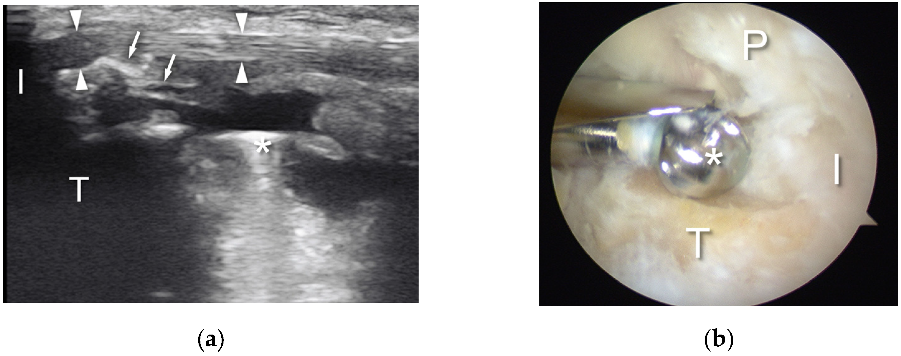
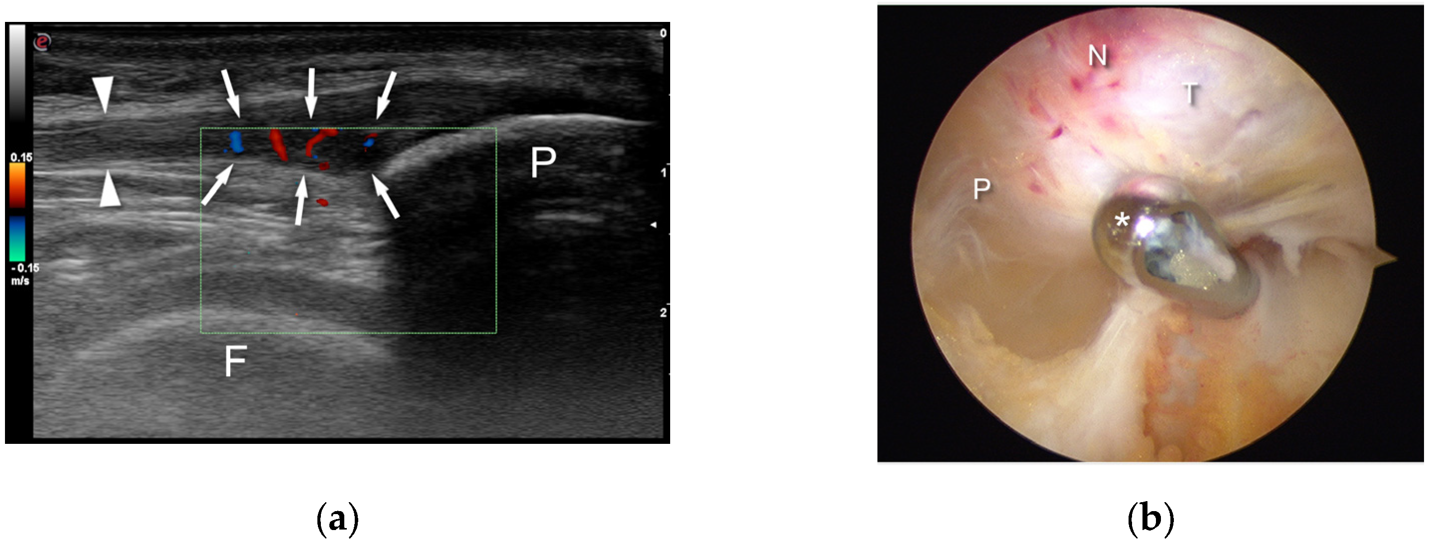
De Lucia et al. (2007) reported the use of double visualisation (arthroscopy and ultrasound) during three knee surgeries [14]. They confirmed the important role of ultrasounds and good correlation between the sonographic and arthroscopic views. However, the authors also emphasized some disadvantages: the ultrasound device was consumed a lot of-space, there was a need of more personnel movement during the surgery, the high cost of the additional equipment, there was a long learning curve of the sonography, and the potential elongation of the procedure. In another paper, Paresce et al. described the advantages of synovectomy performed under double visualisation (arthroscopy and ultrasound). Owing to this method, it was possible to more clearly to identify the hypertrophic synovium. Ultrasound assistance allowed them to resect the septum dividing the knee joint. In another case, this modality was used to navigate the resection of the excessive synovium from a Baker cyst [15]. Akatsu et al. described the original technique of the localisation of menisco-capsular separation during knee arthroscopy [16]. According to their findings, ultrasounds could be helpful in both the diagnosis and treatment of this kind of meniscal lesions. Another indication of ultrasound augmentation in mini-invasive surgery is during tibia bone resection and near patellar tendon insertion due to deformation caused by Osgood–Schlatter disease [17] (Figure 2). Sunding et al. analysed the results 3–5 years after the double visualization (US/arthroscopic) shaving of the involved part of the patellar tendon in tendinopathy (Figure 3). They proved that there is a correlation between the low score of pain during activity, a high patient satisfaction rate, and poor local blood flow. In comparison with sclerosing injections, both the treatment methods rendered good and equal clinical results at the mean 46-month follow-up, but US/ColorDoppler-guided arthroscopic shaving revealed a significant decrease in the anteroposterior thickness of the proximal patellar tendon [18].
Ultrasound guidance was also found to be effective in establishing portals for hip arthroscopy. It was described by Hua et al. (2009) as a method which allows surgeons to avoid the fluoroscopy [19]. Weinrauch et al. revealed that sonographic guidance can be used for both peripheral and central compartment procedures in hip arthroscopy in prone and lateral patient positions [20]. Keough et al. showed the limitations of this technique. It should not be taken into consideration in patients with morbid obesity and in cases of extensive heterotopic ossifications [21]. Moreover, Ali et al., in their 3D models of FAI deformity, showed that ultrasound guidance can be used when confirming joint distraction and establishing portals for hip arthroscopy [22].
The mini-invasive, arthroscopic treatment of hallux rigidus can also be augmented using ultrasound assistance (Figure 4) [23]. The arthroscopy of the first metatarsophalangeal joint was first described by Bartlett in 1988 during an osteochondritis dissecans treatment [24]. However, this procedure has gained popularity in recent years and has been used to treat osteochondritis dissecans, hallux valgus, and gout, as well as the most common indication, hallux rigidus. Arthroscopic debridement cannot definitively remove the entire bony eminence. Even with extended synovectomy, a collapsing capsule, and often, overgrowth synovium may deceive the surgeon. Intraoperative sonographic navigation during first metatarsophalangeal joint arthroscopy allows a constant view of osteophyte removal, ensuring complete resection. Furthermore, the first metatarsal joint is relatively small, and navigation over the upper surface of the metatarsal head under both the capsule and the extensor hallucis longus tendon requires excellent orientation. Moreover, US imaging allows surgeons to omit performing an open procedure in numerous cases. Double visualization makes the procedure safer, faster, and more reliable.
Paczesny et al., in their study, suggest sterile draping for the transducer, and additionally, gel was added frequently during visualisation [23]. After the sterile draping of the ultrasound transducer (linear probe: 7.5 cm; frequency: 10–12 MHz), the joint was visualised using the ultrasound scanner by placing the probe longitudinally over the upper surface of the first metatarso-phalangeal joint. The authors revealed that all three patients were satisfied with the procedure and the functioning of the treated feet. The average surgery time was 81 min, with no complications having occurred.
Yamamoto et al. used sonographic navigation to avoid complications during arthroscopic ganglion resection. The authors emphasized the role of double visualisation and the decreased risk of inadvertent injury to the arteries or nerves surrounding the joint during arthroscopy. Using sonography, the vessels, nerves, and tendons around the ganglion can be easily visualized. In addition, the position of the tip of the shaver can be controlled throughout the all procedures [25].
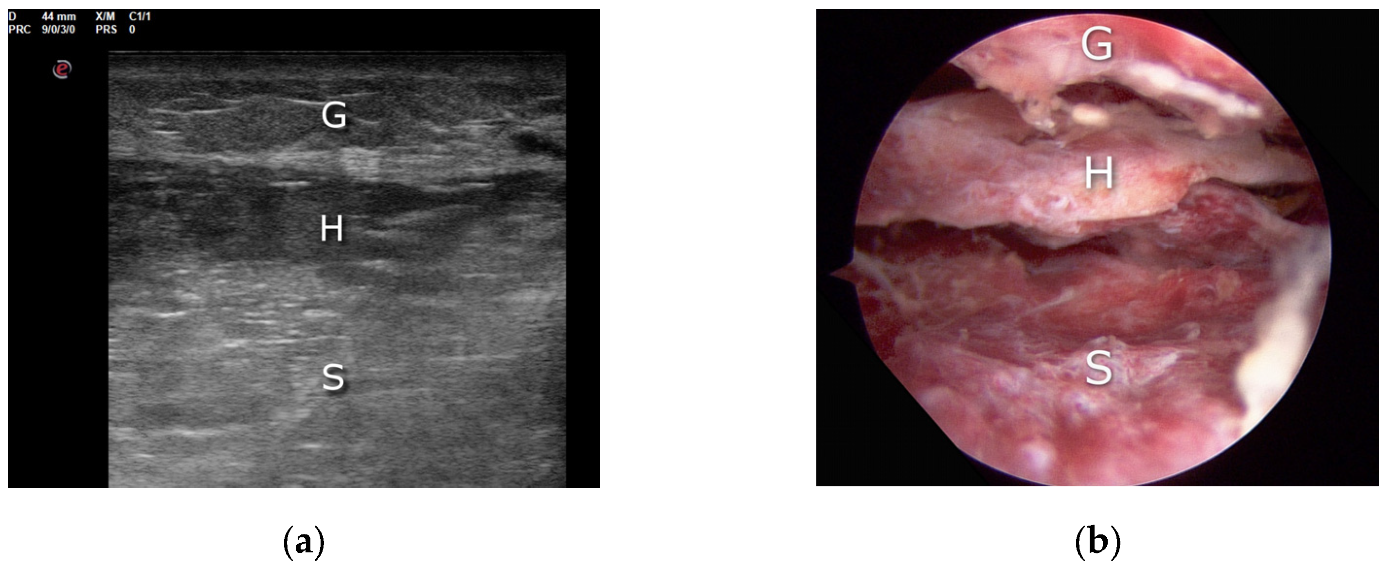
Hematoma of the calf is a rare complication following anterior cruciate ligament reconstruction, and the golden standard method for its treatment is puncture and aspiration (Figure 5). However, prolonged hematomas may demand more advance procedures, such as surgical evacuation. Michalski et al. presented a case study where an open surgery failed, and reoperation using a hybrid–US/endoscopic procedure resolved the clinical problem [26]. The authors concluded that the endoscopic and ultrasound-assisted evacuation of the prolonged calf hematoma is an effective, minimally invasive and targeted procedure, even in old hematomas with evident scarring of the soft tissues.
Lastly, ultrasound navigation can also be used in mini-invasive, percutaneous Achilles tendon repair. Ultrasounds are an adequate tool for assessing the pathological alteration of the Achilles tendon, effectively represents histopathological changes, and moreover, in Europe, it is regarded as a primary imaging method [27]. Achilles tendinopathy with the degeneration of tendinous tissue occurs in 5.9% of seniors and 50% of elite athletes [28]. There are multiple methods of treatment for completely torn Achilles tendons, such as an operative treatment (open, percutaneous, or mini-invasive repair) and a non-operative treatment (cast, brace, or orthosis). Still, no consensus has been reached regarding a universal treatment option. Surgical treatment always carries a risk of infection, wound healing problems, adhesions of sutures to the tendon, and iatrogenic nerve injury, i.e., a sural nerve [29].
Both open and percutaneous surgeries always entail the risk of damaging the nerves and causing wound-related complications [30]. However, the development of mini-invasive surgery on the Achilles tendon allows surgeons to reduce the wound complications rate, but various percutaneous techniques increase the risk of sural nerve entrapment (1.7–27%) [31]. On the one hand, high-resolution ultrasounds used intra-operatively in Achilles tendon suturing can minimize the risk of the a sural nerve injury during repair; on the other hand, it can improve the accuracy of needle penetration of the tendon’s stump [32]. Paczesny et al., in their study, examined 57 patients who underwent acute Achilles tendon ruptures with percutaneous sutures between 2005 and 2015. In the non-ultrasound-guided procedure group, three of fifteen patients had symptoms of sural nerve entrapment, whereas in the ultrasounds navigated group, none suffered from this. The authors revealed also that the real-time intra-operative ultrasound imaging of the sural nerve requires experience and proper skills. Giannetti et al. combined ultrasound and percutaneous Achilles surgery in their study and also reported excellent results with no sural nerve complications [33]. Severyns et al. described the ultrasonographic visualization of the ruptured tendon for sural nerve visualization before endoscopic portal placement during endoscopic Achilles tendon repair [34]. It was designed to minimize the risk of sural nerve injury during Achilles repair, and the authors supported endoscopy during repair. The double-assisted surgery (both ultrasonographically and endoscopically guided) reduces the complication rates.
Another important method is ultrasound-guided gastrocnemius recession as a mini-invasive technique in MSK surgery [34]. An isolated gastrocnemius contracture is thought to lead to numerous foot and ankle pathologic conditions, and as the authors mentioned, the resulting equinus deformity alters the foot biomechanics and increases forefoot pressure, aggravating or predisposing the patients to conditions such as Achilles tendinosis, flatfoot, lower back pain or strain, diabetic foot ulcer, knee hyperextension (genu recurvatum), metatarsalgia, plantar fasciitis, midfoot pain or arthritis, lateral foot pain, and nerve entrapment [35]. Gastrocnemius recession can be performed as a traditional open surgery or as an endoscopic, mini-invasive technique. The complications after open techniques have been described, such as poor cosmetic outcomes, neurovascular compromises, and wound complications. Endoscopic gastrocnemius recession is less invasive and has advantages over traditional procedures: a shorter operative time, fewer complications, and reduced morbidity rate [36]. Villanueva et al. observed the complete release of the gastrocnemius tendon in all cases in the cadaveric study. No damage to the sural nerve or vessels occurred, and there was a minimal damage to the underlying muscle fibers. The authors observed that the degree of ankle dorsiflexion increased for every patient in the study, with a mean preoperative VAS score of 7, which improved to 0 postoperatively. The AOFAS Ankle-Hindfoot Score improved from a mean of 30 to 93 at 6 months with no major complications.
Another important modality in MSK mini-invasive surgery is ultrasound application in carpal tunnel surgery [37]. Carpal tunnel syndrome is the most common peripheral entrapment neuropathies, occurring in approximately 3–5% of the general population. The surgical treatment is based on transection of the transverse carpal ligament using either an open or endoscopic technique to reduce the compression of the median nerve within the carpal tunnel [37]. To decrease postoperative pain and disability, new techniques such as ultrasound-guided techniques for carpal tunnel release have been introduced. These techniques are designed to allow patients to regain to hand function earlier. Kamel et al., in their study, performed 61 ultrasound-guided carpal tunnel release procedures performed on 46 patients (15 bilateral procedures) with clinically diagnosed carpal tunnel syndrome [37]. The authors performed these procedures with a single-use transection device and local anaesthesia at an outpatient office. Over 90% of the patients were satisfied or very satisfied with the long-term outcomes, and no urgent postoperative complications occurred. Only two patients needed surgical intervention in the few days after surgery; one was for an infection after injury and one was for post-traumatic compartment syndrome.
The last modality in MSK mini-invasive surgery for ultrasounds is chronic exertional compartment syndrome treatment. Chronic exertional compartment syndrome is an exertional pain syndrome that typically affects the lower legs of participants involved in high-intensity running or marching activities [38].
The treatment of chronic exertional compartment syndrome begins with conservative methods, including activity modification, physiotherapy, gait re-training, orthotics, and analgesics [38]. Moreover, various fasciotomy techniques have been described, including single and multiple incisions and endoscopic techniques for the release of the fascia. Traditional, open fasciotomy is the standard treatment for recalcitrant cases of chronic exertional compartment syndrome. Alternatively, other authors described a mini-invasive fasciotomy technique, with reduced rates of procedural complications and expedite recovery [38]. Sommerfeldt et al. found that an ultrasound-guided, minimally invasive looped thread fasciotomy procedure is feasible and is not associated with injury to the neurovascular structures [38]. Finnoff et al. also revealed in their study a novel, mini-invasive technique with ultrasound application; the procedure required a 3 mm incision, was performed in the office under local anaesthesia, and allowed the athlete to resume running within 1 week of the procedure. Although the preliminary results of this study were promising, further translational research is required before widespread clinical application can be achieved [39]. Moreover, Balius et al. showed a good clinical outcomes of ultrasound-navigated mini-invasive fasciotomy in patients with chronic exertional compartment syndrome. All the studied patients had a decrease in their level of pain, and all except one returned to presymptomatic exercise levels, with a median return to play of 35 days.

4. Possible Future Applications

The technology of ultrasonic transducers is growing very rapidly, following development in other medical fields [40,41]. The gradual increase in the frequency of transducer use and the evolution of ultrasound software has led to gradual improvement of ultrasound images. Nowadays, progressive miniaturization with the use of capacitive or piezoelectric micromachined ultrasonic transducers provides the possibility of the integration of ultrasonic probes with endoscopic instruments [41]. Such integration could allow the direct, high-resolution ultrasonographic assessment of tissues (i.e., menisci or rotator cuff) during arthroscopy.

5. Conclusions

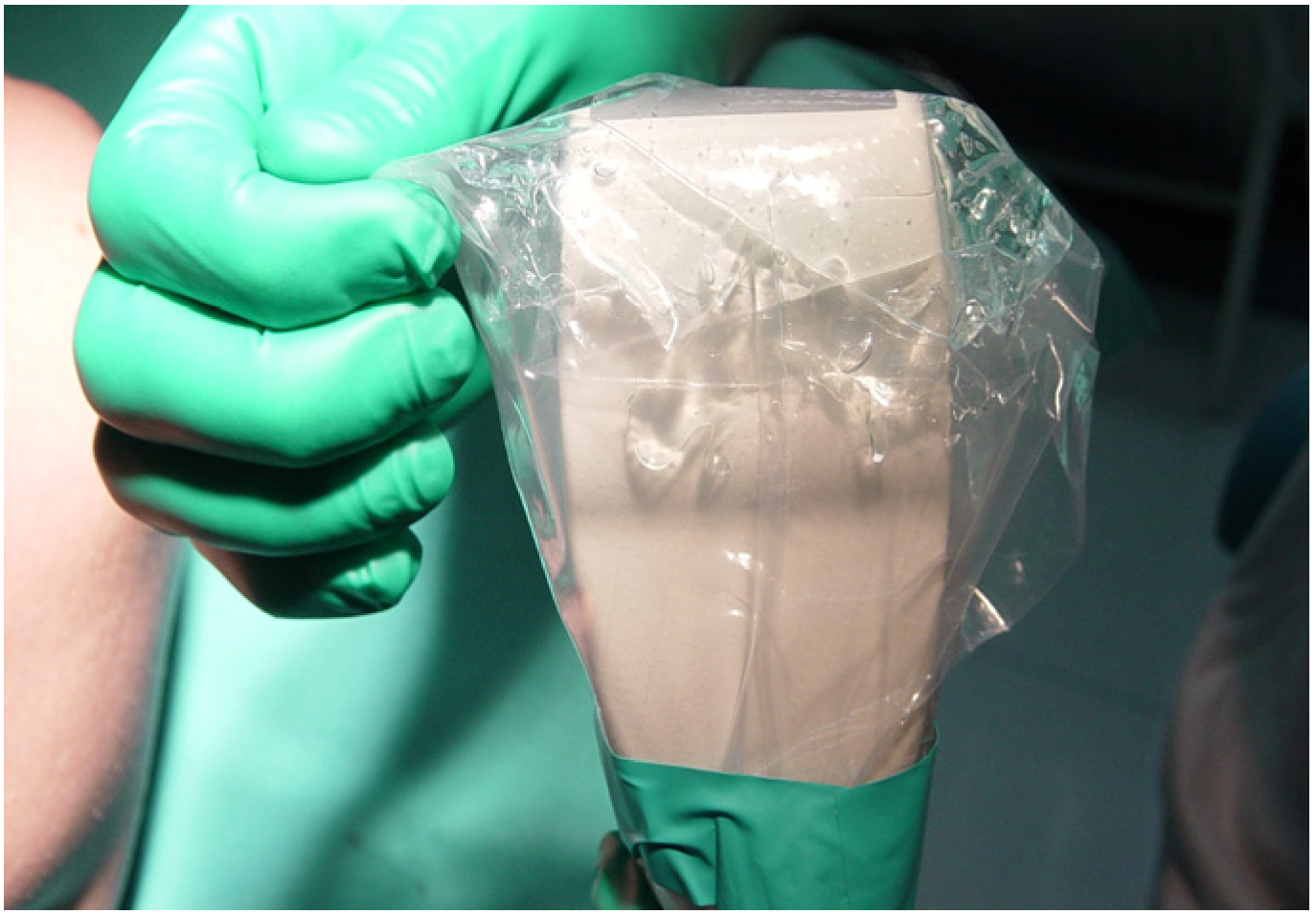
The main disadvantage of ultrasound guidance during orthopaedic surgery is the need for the extra equipment in the operating room and the additional cost of the needle, ultrasound gel, and draping (Figure 6).
It should also be noted that basic ultrasound skills are mandatory for such procedures. The other disadvantages are the potential risk of the procedure connected with the misinterpretation of the ultrasound image by the surgeon and possible infections due to wound contamination via an insufficiently draped transducer. We recommend ultrasounds for secondary visualization in the described arthroscopic procedures to surgeons who are well experienced in diagnostic musculoskeletal ultrasounds. Real-time intra-operative ultrasound imaging requires experience in diagnostics ultrasounds and proper skills.

Author Contributions

Conceptualization, Ł.P. and J.Z.; methodology, Ł.P. and J.Z.; software, M.L.; validation, Ł.P. and J.Z.; formal analysis, J.Z.; investigation, M.L.; resources, T.P.; data curation Ł.P. and R.W.; writing—original draft preparation, Ł.P., J.Z., T.P. and R.W.; writing—Ł.P., J.Z., T.P. and R.W.; visualization, Ł.P. supervision, G.H.; project administration, J.Z.; funding acquisition, Ł.P. All authors have read and agreed to the published version of the manuscript.

Funding

This research received no external funding.

Institutional Review Board Statement

Not applicable.

Informed Consent Statement

Not applicable.

Data Availability Statement

No data applicable.

Conflicts of Interest

The authors declare no conflict of interest.

References

  1. Hattori, S.; Onishi, K. Ultrasound-Guided Surgery in Musculoskeletal Medicine. J. Med. Ultrason. 2022, 49, 513–515. [Google Scholar] [CrossRef] [PubMed]
  2. Tyryshkin, K.; Mousavi, P.; Pichora, D.R.; Abolmaesumi, P. Identification of Anatomical Landmarks for Registration of CT and Ultrasound Images in Computer-Assisted Shoulder Arthroscopy. In Proceedings of the 2006 International Conference of the IEEE Engineering in Medicine and Biology Society, New York, NY, USA, 30 August–3 September 2006; IEEE: New York, NY, USA, 2006; pp. 416–419. [Google Scholar]
  3. Ho, T.-F.; Lee, P.-Y.; Lan, H.H.-C.; Ku, M.-C. Establishing a Popliteal Portal Using the METRx System Under Ultrasound Guidance. Arthrosc. J. Arthrosc. Relat. Surg. 2006, 22, 1363.e1–1363.e4. [Google Scholar] [CrossRef] [PubMed]
  4. Hattori, K.; Takakura, Y.; Ishimura, M.; Habata, T.; Uematsu, K.; Ikeuch, K. Quantitative Arthroscopic Ultrasound Evaluation of Living Human Cartilage. Clin. Biomech. 2004, 19, 213–216. [Google Scholar] [CrossRef] [PubMed]
  5. Huang, Y.-P.; Zheng, Y.-P. Intravascular Ultrasound (IVUS): A Potential Arthroscopic Tool for Quantitative Assessment of Articular Cartilage. TOBEJ 2009, 3, 13–20. [Google Scholar] [CrossRef]
  6. Huang, Y.P.; Zheng, Y.P. Development of an Arthroscopic Ultrasound Probe for Assessment of Articular Cartilage Degeneration. In Proceedings of the 2013 35th Annual International Conference of the IEEE Engineering in Medicine and Biology Society (EMBC), Osaka, Japan, 3–7 July 2013; IEEE: Osaka, Japan, 2013; pp. 144–147. [Google Scholar]
  7. Liukkonen, J.; Hirvasniemi, J.; Joukainen, A.; Penttilä, P.; Virén, T.; Saarakkala, S.; Kröger, H.; Jurvelin, J.S.; Töyräs, J. Arthroscopic Ultrasound Technique for Simultaneous Quantitative Assessment of Articular Cartilage and Subchondral Bone: An In Vitro and In Vivo Feasibility Study. Ultrasound Med. Biol. 2013, 39, 1460–1468. [Google Scholar] [CrossRef]
  8. Kaleva, E.; Virén, T.; Saarakkala, S.; Sahlman, J.; Sirola, J.; Puhakka, J.; Paatela, T.; Kröger, H.; Kiviranta, I.; Jurvelin, J.S.; et al. Arthroscopic Ultrasound Assessment of Articular Cartilage in the Human Knee Joint: A Potential Diagnostic Method. Cartilage 2011, 2, 246–253. [Google Scholar] [CrossRef]
  9. Puhakka, J.; Afara, I.O.; Paatela, T.; Sormaala, M.J.; Timonen, M.A.; Virén, T.; Jurvelin, J.S.; Töyräs, J.; Kiviranta, I. In Vivo Evaluation of the Potential of High-Frequency Ultrasound for Arthroscopic Examination of the Shoulder Joint. Cartilage 2016, 7, 248–255. [Google Scholar] [CrossRef]
  10. Joukainen, A.; Virén, T.; Penttilä, P.; Liukkonen, J.; Puhakka, P.H.; Kröger, H.; Töyräs, J. Ultrasound Arthroscopy of Hip in Treatment of Osteochondritis Dissecans. Arthrosc. Tech. 2017, 6, e1063–e1068. [Google Scholar] [CrossRef]
  11. Bethune, R.; Bull, A.M.J.; Dickinson, R.J.; Emery, R.J.H. Removal of Calcific Deposits of the Rotator Cuff Tendon Using an Intra-Articular Ultrasound Probe. Knee Surg. Sports Traumatol. Arthrosc. 2007, 15, 289–291. [Google Scholar] [CrossRef]
  12. Sabeti, M.; Schmidt, M.; Ziai, P.; Graf, A.; Nemecek, E.; Schueller-Weidekamm, C. The Intraoperative Use of Ultrasound Facilitates Significantly the Arthroscopic Debridement of Calcific Rotator Cuff Tendinitis. Arch. Orthop. Trauma. Surg. 2014, 134, 651–656. [Google Scholar] [CrossRef]
  13. Sabeti-Aschraf, M.; Gonano, C.; Nemecek, E.; Cichocki, L.; Schueller-Weidekamm, C. Intra-Operative Ultrasound Facilitates the Localization of the Calcific Deposit during Arthroscopic Treatment of Calcifying Tendinitis. Knee Surg. Sports Traumatol. Arthrosc. 2010, 18, 1792–1794. [Google Scholar] [CrossRef]
  14. De Lucia, O.; Paresce, E.; Murgo, A.; Epis, O.; Pisoni, L.; Schito, E.; Valcamonica, E.; Piana, C.; Fantini, F. Simultaneous Ultrasonography and Arthroscopy for the Study of the Joint Environment: Indications and Limits. Reumatismo 2011, 59, 146–152. [Google Scholar] [CrossRef] [PubMed]
  15. Paresce, E.; De Lucia, O.; Bruschi, E.; Giacomelli, L.; Epis, O.M. Use of Ultrasound-Assisted Arthroscopy in Rheumatology: An Experience in 11 Patients with Different Rheumatic Diseases. Expert. Opin. Med. Diagn. 2013, 7, 309–312. [Google Scholar] [CrossRef]
  16. Akatsu, Y.; Akagi, R.; Fukawa, T.; Yamaguchi, S.; Sasho, T. Ultrasound for Treating Meniscocapsular Separation Together With Arthroscopy. Arthrosc. Tech. 2016, 5, e1457–e1460. [Google Scholar] [CrossRef]
  17. Willberg, L.; Sunding, K.; Forssblad, M.; Alfredson, H. Ultrasound- and Doppler-Guided Arthroscopic Shaving to Treat Jumper’s Knee: A Technical Note. Knee Surg. Sports Traumatol. Arthrosc. 2007, 15, 1400–1403. [Google Scholar] [CrossRef]
  18. Sunding, K.; Willberg, L.; Werner, S.; Alfredson, H.; Forssblad, M.; Fahlström, M. Sclerosing Injections and Ultrasound-Guided Arthroscopic Shaving for Patellar Tendinopathy: Good Clinical Results and Decreased Tendon Thickness after Surgery—A Medium-Term Follow-up Study. Knee Surg. Sports Traumatol. Arthrosc. 2015, 23, 2259–2268. [Google Scholar] [CrossRef] [PubMed]
  19. Hua, Y.; Yang, Y.; Chen, S.; Wang, Y.; Li, Y.; Chen, J.; Li, H. Ultrasound-Guided Establishment of Hip Arthroscopy Portals. Arthrosc. J. Arthrosc. Relat. Surg. 2009, 25, 1491–1495. [Google Scholar] [CrossRef]
  20. Weinrauch, P.; Kermeci, S. Ultrasound-Assisted Hip Arthroscopy. Arthrosc. Tech. 2014, 3, e255–e259. [Google Scholar] [CrossRef]
  21. Keough, T.; Wilson, D.; Wong, I. Ultrasound-Guided Portal Placement for Hip Arthroscopy. Arthrosc. Tech. 2016, 5, e851–e856. [Google Scholar] [CrossRef]
  22. Ali, M.I.; Ravipati, A.P.T.; Wong, I. Hip Arthroscopy for Femoral Acetabular Impingement: A Bird’s-Eye/En Face Perspective With Ultrasound Guidance and 3-Dimensional Hip Printing. Arthrosc. Tech. 2019, 8, e1301–e1307. [Google Scholar] [CrossRef]
  23. Paczesny, L.; Kruczyński, J. Ultrasound-Guided Arthroscopic Management of Hallux Rigidus. Videosurgery Other Miniinvasive Tech. 2016, 3, 144–148. [Google Scholar] [CrossRef]
  24. Bartlett, D.H. Arthroscopic Management of Osteochondritis Dissecans of the First Metatarsal Head. Arthrosc. J. Arthrosc. Relat. Surg. 1988, 4, 51–54. [Google Scholar] [CrossRef]
  25. Yamamoto, Y.; Yamaguchi, S.; Sasho, T.; Fukawa, T.; Akatsu, Y.; Akagi, R.; Yamaguchi, T.; Takahashi, K.; Nagashima, K.; Takahashi, K. Quantitative US Elastography Can Be Used to Quantify Mechanical and Histologic Tendon Healing in a Rabbit Model of Achilles Tendon Transection. Radiology 2017, 283, 408–417. [Google Scholar] [CrossRef]
  26. Michalski, Ł.; Paczesny, Ł.; Zabrzyński, J.; Kruczyński, J. Ultrasound-Assisted Endoscopic Evacuation of Recurrent Calf Hematoma Following Anterior Cruciate Ligament Reconstruction. Case Study. Ortop. Traumatol. Rehabil. 2019, 21, 359–368. [Google Scholar] [CrossRef] [PubMed]
  27. Longo, U.G.; Ronga, M.; Maffulli, N. Achilles Tendinopathy. Sports Med. Arthrosc. Rev. 2009, 17, 112–126. [Google Scholar] [CrossRef] [PubMed]
  28. Kaux, J.-F.; Forthomme, B.; Goff, C.L.; Crielaard, J.-M.; Croisier, J.-L. Current Opinions on Tendinopathy. J. Sports Sci. Med. 2011, 10, 238–253. [Google Scholar]
  29. Carmont, M.R.; Rossi, R.; Scheffler, S.; Mei-Dan, O.; Beaufils, P. Percutaneous & Mini Invasive Achilles Tendon Repair. BMC Sports Sci. Med. Rehabil. 2011, 3, 28. [Google Scholar] [CrossRef]
  30. Park, S.-H.; Lee, H.S.; Young, K.W.; Seo, S.G. Treatment of Acute Achilles Tendon Rupture. Clin. Orthop. Surg. 2020, 12, 1. [Google Scholar] [CrossRef]
  31. Maffulli, N. Minimally Invasive Surgery for Achilles Tendon Pathologies. Open Access J. Sports Med. 2010, 1, 95–103. [Google Scholar] [CrossRef] [PubMed]
  32. Paczesny, Ł.; Zabrzyński, J.; Domżalski, M.; Gagat, M.; Termanowski, M.; Szwedowski, D.; Łapaj, Ł.; Kruczyński, J. Mini-Invasive, Ultrasound Guided Repair of the Achilles Tendon Rupture—A Pilot Study. J. Clin. Med. 2021, 10, 2370. [Google Scholar] [CrossRef]
  33. Giannetti, S.; Patricola, A.A.; Stancati, A.; Santucci, A. Intraoperative Ultrasound Assistance for Percutaneous Repair of the Acute Achilles Tendon Rupture. Orthopedics 2014, 37, 820–824. [Google Scholar] [CrossRef]
  34. Severyns, M.; Andriamananaivo, T.; Rollet, M.-E.; Kajetanek, C.; Lopes, R.; Renard, G.; Noailles, T.; Odri, G.A.; Rouvillain, J.-L. Acute Achilles Tendon Rupture: Ultrasonography and Endoscopy-Assisted Percutaneous Repair. Arthrosc. Tech. 2019, 8, e489–e493. [Google Scholar] [CrossRef]
  35. Villanueva, M.; Iborra, Á.; Rodríguez, G.; Sanz-Ruiz, P. Ultrasound-Guided Gastrocnemius Recession: A New Ultra–Minimally Invasive Surgical Technique. BMC Musculoskelet. Disord. 2016, 17, 409. [Google Scholar] [CrossRef] [PubMed]
  36. Schroeder, S.M. Uniportal Endoscopic Gastrocnemius Recession for Treatment of Gastrocnemius Equinus with a Dedicated EGR System with Retractable Blade. J. Foot Ankle Surg. 2012, 51, 714–719. [Google Scholar] [CrossRef]
  37. Kamel, S.I.; Freid, B.; Pomeranz, C.; Halpern, E.J.; Nazarian, L.N. Minimally Invasive Ultrasound-Guided Carpal Tunnel Release Improves Long-Term Clinical Outcomes in Carpal Tunnel Syndrome. Am. J. Roentgenol. 2021, 217, 460–468. [Google Scholar] [CrossRef]
  38. Sommerfeldt, M.; Jack, E.; Playfair, L.; Satkunam, L.; Loh, E.; Rambaransingh, B.; Burnham, R. Ultrasound-Guided, Minimally Invasive Looped Thread Fasciotomy for Chronic Exertional Compartment Syndrome of the Lower Leg: A Cadaveric Feasibility Study. Interv. Pain. Med. 2022, 1, 100074. [Google Scholar] [CrossRef]
  39. Finnoff, J.T.; Fowler, S.P.; Lai, J.K.; Santrach, P.J.; Willis, E.A.; Sayeed, Y.A.; Smith, J. Treatment of Chronic Tendinopathy with Ultrasound-Guided Needle Tenotomy and Platelet-Rich Plasma Injection. PM&R 2011, 3, 900–911. [Google Scholar] [CrossRef]
  40. Roy, K.; Lee, J.E.-Y.; Lee, C. Thin-Film PMUTs: A Review of over 40 Years of Research. Microsyst. Nanoeng. 2023, 9, 95. [Google Scholar] [CrossRef] [PubMed]
  41. Joseph, J.; Ma, B.; Khuri-Yakub, B.T. Applications of Capacitive Micromachined Ultrasonic Transducers: A Comprehensive Review. IEEE Trans. Ultrason. Ferroelectr. Freq. Control. 2022, 69, 456–467. [Google Scholar] [CrossRef] [PubMed]
Figure 1. (a) Calcium deposit in intraoperative sonography. The probe is in axial plane, showing deltoid muscle (D) superficially in relation to supraspinatus tendon (arrows). Acromion border is seen on the right (A). The calcium deposit (arrowheads) is evident in the centre of supraspinatus fibres. (b) Multiple calcium deposits (arrows) during arthroscopic removal, temporarily disseminated above the upper surface of supraspinatus tendon (arrowheads). The tendon is palpated under double visualisation using arthroscopic hook (H).
Applsci 13 10900 g001
Figure 2. (a) Sonographic visualisation of the arthroscopic removal of the tibia (T) deformity (arrows) with the use of motorised shaver (asterisk) of tibia (T) bony deformation caused by Osgood–Schlatter disease. The resection is carried out distally to the insertion (I) of the patellar tendon (arrowheads). (b) Arthroscopic removal with motorised shaver (asterisk) of tibia (T) bony deformation after Osgood–Schlatter disease. The resection is carried out distally to the insertion (I) of the patellar tendon (P).
Applsci 13 10900 g002
Figure 3. (a) Intraoperative sonography of the patellar tendon (arrowheads) tendinopathy. The transducer is placed in sagittal plane parallel to the axis of the femur (F). Doppler modality allows the precise localisation of neovascularised pathologic tissue (arrows) of the patellar tendon near the inferior pole of the patella (P). (b) Arthroscopic shaving (motorised shaver; asterisk) of the neovascularised pathologic tissue (N) of the bottom part of the patellar tendon (T) near inferior pole of the patella (P).
Applsci 13 10900 g003
Figure 4. (a) Sonographic visualisation of the arthroscopic cannula introduced the first metatarsal joint between first metatarsal (M) and phalanx (P). The aim of the procedure is the removal of the bony osteophyte (arrowhead). Note hyperechoic artefacts (A) caused by steel cannula (arrow) (b) Resection of the first metatarsal (M) head osteophyte using the motorised shaver (S).
Applsci 13 10900 g004
Figure 5. (a) Organised hematoma (H) is seen in intraoperative sonography as hypoechoic heterogenic echo between the soleus (S) and gastrocnemius (G) muscles. (b) Fibrotic organised hematoma evolving into scar (H) was evacuated using arthroscopic shaver. The pathologic tissue was localised intraoperatively via sonography between the soleus (S) and gastrocnemius (G) muscles.
Applsci 13 10900 g005
Figure 6. Ultrasound transducer is draped in a sterile translucent tunnel. Note: ultrasonic gel put into draping to cover the working side of the transducer. To achieve proper visualisation, sterile gel has also to be put on the skin during sonography.
Applsci 13 10900 g006
Disclaimer/Publisher’s Note: The statements, opinions and data contained in all publications are solely those of the individual author(s) and contributor(s) and not of MDPI and/or the editor(s). MDPI and/or the editor(s) disclaim responsibility for any injury to people or property resulting from any ideas, methods, instructions or products referred to in the content.